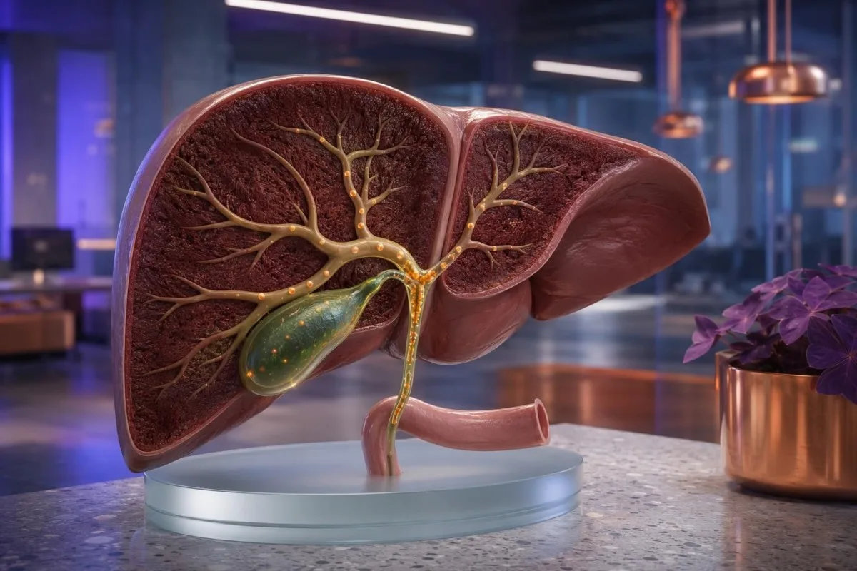
A cholestatic liver pattern usually means ALP and GGT rise more than ALT and AST, sometimes followed by direct bilirubin elevation. New jaundice, dark urine with pale stools, fever, or severe right-upper-abdominal pain needs same-day medical assessment rather than a wait-and-see approach.


- Cholestatic pattern usually means alkaline phosphatase is disproportionately high, with an R ratio below 2.
- ALP plus GGT elevation supports a liver or bile-duct source; isolated ALP with normal GGT is often bone-related.
- Direct bilirubin above 5 µmol/L or 0.3 mg/dL can rise when conjugated bilirubin cannot drain normally into the bowel.
- Adult ALP commonly falls around 30-120 IU/L, but the laboratory upper limit matters more than one universal cutoff.
- GGT above 60 IU/L in many adult male reference intervals, or above 40 IU/L in many female intervals, merits context when ALP is also high.
- Fever, jaundice and abdominal pain together can indicate biliary infection and require emergency assessment.
- Pale or clay-coloured stool with dark urine suggests reduced bile reaching the gut and should be assessed urgently.
- Itching without a rash can precede visible jaundice in cholestasis, especially during pregnancy or autoimmune bile-duct disease.
How a Cholestatic Pattern Appears on Liver Blood Tests
A cholestatic liver pattern occurs when alkaline phosphatase and usually GGT rise out of proportion to ALT and AST, suggesting impaired bile formation or bile flow. As of July 21, 2026, the most useful first calculation is the R ratio: (ALT ÷ ALT upper limit of normal) divided by (ALP ÷ ALP upper limit of normal); below 2 is cholestatic, above 5 is hepatocellular, and 2-5 is mixed.
A cholestatic result does not automatically mean a blocked duct. It can reflect gallstones, medication effects, pregnancy-related cholestasis, primary biliary cholangitis, primary sclerosing cholangitis, or infiltrative liver conditions; the pattern tells us where to look first, not the final diagnosis.
In my clinical work, I pay more attention to the proportionate change than to a single flagged number. An ALT of 90 IU/L can look alarming on a portal, yet an ALP of 360 IU/L with an upper limit of 120 IU/L shifts the clinical question toward bile ducts, ultrasound, medicines, and symptoms.
Kantesti is an AI blood test analyzer used by more than 2 million people across 127 countries, and it reads ALP, GGT, bilirubin, ALT, AST, albumin, and platelet trends together rather than treating one flagged value as a diagnosis. Our organisational and clinical background is available at About Kantesti.
The European Association for the Study of the Liver advises confirming the pattern before extensive testing, because ALP can come from liver, bone, placenta, and intestine (EASL, 2009). Thomas Klein, MD, has seen more anxiety caused by an unexplained isolated ALP flag than by a clearly interpreted cluster of abnormal liver tests.
Why Alkaline Phosphatase Is the Starting Point
Alkaline phosphatase (ALP) often rises first in cholestasis because bile-duct lining cells release more of the enzyme when bile flow is impaired. Typical adult reference intervals are roughly 30-120 IU/L, although age, pregnancy, method, and local laboratory limits can move that range substantially.
An ALP result above 1.5 times the laboratory upper limit that persists on repeat testing deserves an explanation, particularly when GGT is also elevated. A result of 180 IU/L may be modest in a lab with an upper limit of 130 IU/L but is more meaningful in a lab whose upper limit is 104 IU/L.
The subtle trap is that ALP is not liver-specific. Healing fractures, vitamin D deficiency with increased bone turnover, hyperparathyroidism, adolescence, and late pregnancy can elevate ALP without obstructed bile flow; this is why an ALP result should never be interpreted in isolation.
When the source remains uncertain, clinicians may request GGT, ALP isoenzymes, or 5′-nucleotidase. A normal GGT does not absolutely exclude liver disease, but it makes a bone source more plausible and can prevent an unnecessary sequence of liver scans.
Before assuming an ALP flag is a bile-duct problem, compare the other components of what a liver panel contains. The pattern across tests is usually more reliable than the red or amber colour assigned by a lab portal.
What High ALP and GGT Together Usually Mean
High alkaline phosphatase and GGT together usually point toward a hepatobiliary source rather than bone, especially when ALP is more than 1.5 times the upper limit of normal. GGT is sensitive but not specific: alcohol exposure, fatty liver, diabetes, obesity, and several medicines can raise it without a physical blockage.
Many laboratories use GGT upper limits near 40 IU/L for women and 60 IU/L for men, but some European laboratories use lower limits. A GGT of 92 IU/L with ALP 105 IU/L may fit metabolic or medication-related enzyme induction; GGT 92 IU/L with ALP 310 IU/L is more convincingly cholestatic.
GGT can rise within days of enzyme-inducing drugs such as phenytoin, carbamazepine, phenobarbital, and occasionally antibiotics. It can also stay elevated for weeks after alcohol intake falls, so a one-off value cannot reliably calculate alcohol consumption or prove alcohol-related disease.
I often explain GGT as a corroborating witness, not the verdict. It supports the liver origin of ALP, but it does not distinguish a gallstone from primary biliary cholangitis, and it does not measure liver function.
If ALP is elevated but GGT is normal, read our focused guide to ALP with normal GGT before assuming cholestasis. The distinction matters because bone investigations and liver investigations are very different clinical paths.
Direct Bilirubin Elevation and the Meaning of Dark Urine
Direct bilirubin elevation occurs when conjugated bilirubin enters the circulation instead of reaching the intestine through bile. Total bilirubin is commonly about 3-21 µmol/L or 0.2-1.2 mg/dL in adults, while direct bilirubin is usually below 5 µmol/L or 0.3 mg/dL.
Conjugated bilirubin is water-soluble, so it can pass into urine and make it tea- or cola-coloured. By contrast, unconjugated bilirubin from Gilbert syndrome or haemolysis does not appear in urine; this is a useful bedside clue, though never a replacement for fractionated bilirubin testing.
Pale stools develop when too little bilirubin reaches the gut to be converted into stercobilin, the pigment that normally makes stool brown. One unusually light stool after diarrhoea is not diagnostic, but repeated pale, grey, or putty-coloured stools with dark urine warrant urgent contact with a clinician.
Kantesti AI interprets bilirubin fractions alongside ALP, GGT, urine findings, and prior results because a total bilirubin of 45 µmol/L means something very different when the direct fraction is 38 µmol/L than when it is 3 µmol/L. This is also why fasting-related bilirubin rises should not be mistaken for duct obstruction.
For a deeper explanation of the fractions, see our fractionated bilirubin guide. In adults, visible jaundice often becomes noticeable around total bilirubin of 35-50 µmol/L, although skin tone and lighting make this variable.
Cholestatic Versus Hepatocellular Injury: The Practical Difference
Hepatocellular injury primarily raises ALT and AST, while cholestasis primarily raises ALP and GGT. An R ratio above 5 supports hepatocellular injury, below 2 supports cholestasis, and a 2-5 ratio requires a mixed-pattern work-up.
ALT is more liver-specific than AST, but neither test measures bile drainage. ALT above 1,000 IU/L usually triggers a different urgent differential—acute viral hepatitis, ischaemia, or medication toxicity—than ALP 400 IU/L with ALT 85 IU/L, which more often prompts biliary imaging.
Mixed patterns matter because drug-induced liver injury can begin hepatocellular and evolve into cholestatic injury over 2-8 weeks. The date a medicine, herbal product, anabolic agent, or antibiotic began is sometimes more revealing than the peak enzyme number.
The American College of Gastroenterology guideline recommends confirming an ALP elevation with GGT and reviewing prescribed and non-prescribed agents before expanding the work-up (Kwo, Cohen, and Lim, 2017). That sounds straightforward, but patients frequently forget topical products, teas, powders, and recently stopped medication.
A raised ALT can coexist with cholestasis, particularly with a passing gallstone or fatty liver disease. Our ALT pattern guide explains why a hepatocellular-looking result still needs context from ALP, bilirubin, symptoms, and timing.
When Itching, Jaundice or Pale Stools Need Urgent Care
Jaundice with fever, shaking chills, confusion, low blood pressure, or significant right-upper-abdominal pain is an emergency. These symptoms can indicate infected obstructed bile ducts, and assessment should happen in an emergency department rather than after a routine repeat panel.
Itching from cholestasis is often intense on the palms and soles, worse in the evening, and has no primary rash. Its severity does not reliably match bilirubin or ALP values; I have treated patients with miserable pruritus and bilirubin below 30 µmol/L, so symptoms deserve attention even when the panel looks only moderately abnormal.
New yellow eyes or skin without fever still merits medical review within 24 hours, particularly when urine is dark or stools are pale. Persistent vomiting, inability to keep fluids down, new drowsiness, or easy bruising lowers the threshold for same-day emergency evaluation.
Thomas Klein, MD, advises patients not to self-treat unexplained itching with high-dose supplements or imported herbal blends while awaiting assessment. Some products complicate the liver pattern, and antihistamines may sedate without addressing cholestatic itch.
The colour of stool is a surprisingly useful clue when it is persistent, not occasional. Our pale stool guide explains the difference between transient light stool and a repeated loss of bile pigment.
Common Causes of a Cholestatic Liver Enzyme Pattern
Gallstones, medication effects, fatty liver with cholestatic features, autoimmune bile-duct disease, pregnancy, and pancreatic or biliary narrowing are common causes of cholestasis. The first divide is extrahepatic obstruction outside the liver versus intrahepatic cholestasis within liver tissue or small ducts.
A stone in the common bile duct often produces a rapid rise in bilirubin and ALP, but enzymes may lag behind pain by 24-48 hours. A stone can also pass before an ultrasound is performed, leaving a falling bilirubin with a still-elevated ALP and GGT.
Primary biliary cholangitis is more common in middle-aged women and typically presents with persistent ALP elevation, fatigue, and itch; antimitochondrial antibody is positive in roughly 90-95% of established cases. The AASLD practice guidance supports testing for this condition when cholestatic tests persist without an obvious obstruction (Lindor et al., 2021).
Primary sclerosing cholangitis often requires MRCP imaging because the larger ducts can show characteristic irregular narrowing even when ultrasound is unrevealing. It has a meaningful association with inflammatory bowel disease, but bowel symptoms are not required for diagnosis.
Kantesti is an AI lab test interpretation service that prompts a structured review of medicines, supplements, symptom timing, and repeat values when a cholestatic cluster appears. Our doctors set the clinical escalation principles through the medical advisory board, but urgent symptoms always need real-time medical care.
What Clinicians Usually Check After a Cholestatic Result
The first investigation for new cholestasis is usually abdominal ultrasound, combined with a repeat liver panel and medication review. Ultrasound can identify gallstones, bile-duct dilation, gallbladder abnormalities, and many structural liver changes without radiation.
A typical repeat panel includes ALP, GGT, ALT, AST, total and direct bilirubin, albumin, and INR or prothrombin time. Clinicians may add full blood count, C-reactive protein, lipase, hepatitis tests, antimitochondrial antibody, immunoglobulins, and serum IgG4 depending on the clinical story.
A normal ultrasound does not close the case when ALP remains high. MRCP can visualise the biliary tree more clearly, while endoscopic ultrasound or ERCP may be considered when obstruction remains likely; ERCP is now usually reserved for a situation where treatment such as stone removal or stenting may be needed.
The urgency is determined by symptoms and deterioration, not imaging availability alone. Bilirubin rising from 28 to 96 µmol/L over several days, particularly with fever or pain, is much more concerning than a stable ALP of 145 IU/L over 6 months.
Our AI workflow is designed to organise this timeline, not replace examination or imaging. The safeguards behind that approach are described in our technology guide, including why a laboratory pattern is a follow-up trigger rather than a definitive diagnosis.
Can Cholestasis Exist With Normal Bilirubin?
Yes—cholestasis can be present with normal bilirubin, especially early in the course or in small-duct disease. ALP and GGT often rise before bilirubin because partial obstruction or impaired microscopic bile transport may still allow enough bilirubin to reach the bowel.
A normal bilirubin is reassuring against severe complete obstruction at that moment, but it does not rule out a bile-duct disorder. This is one reason persistent ALP of 220 IU/L with GGT 180 IU/L should not be dismissed merely because bilirubin remains 12 µmol/L.
ALP has a relatively slow decline because its circulating half-life is about 7 days. After a transient obstruction resolves, bilirubin can normalise first while ALP and GGT remain elevated for several weeks—an awkward but common pattern that can confuse a single repeat test.
Isolated GGT elevation is different. GGT 75 IU/L with normal ALP, bilirubin, ALT, and AST rarely indicates obstructive cholestasis on its own, although it may justify a review of alcohol, metabolic risk, and medication exposure.
Reference limits vary enough that the lab report should anchor interpretation. Our GGT range guide explains sex-specific intervals and why a change from 18 to 55 IU/L can matter even when both results sit near local cutoffs.
Why the Direction of ALP, GGT and Bilirubin Changes Matters
A rising bilirubin or ALP trend is generally more informative than a single mildly abnormal value. Comparing results drawn 1-4 weeks apart can distinguish recovery after a passing obstruction from a progressive process that needs accelerated imaging or specialist review.
A patient whose bilirubin falls from 74 to 22 µmol/L in 5 days while pain settles may have passed a stone, but follow-up is still sensible if ALP remains 280 IU/L. Conversely, bilirubin rising from 14 to 48 µmol/L alongside worsening itch should not wait for a routine three-month retest.
GGT often moves more slowly than symptoms. In my experience, patients understandably expect it to normalise within days after stopping alcohol or a suspected medication, but it can take 4-8 weeks and occasionally longer depending on the cause and baseline liver health.
Kantesti AI can compare past panels and identify whether the same pattern appeared after a prior illness, medication course, or lifestyle change. A trend is not proof of cause, though; correlation is useful for questions to ask, not a licence to restart a suspected medicine.
For a practical sense of expected recovery timing, review liver markers after alcohol reduction. Repeating tests too soon can create noise, while waiting too long in someone with jaundice can be unsafe.
Pregnancy, Children and Bone Conditions That Alter ALP
Pregnancy and growth can increase ALP without liver disease, but itch or jaundice during pregnancy requires prompt bile-acid assessment. Placental ALP rises most in the third trimester, whereas GGT is often normal or lower than non-pregnant reference values.
Intrahepatic cholestasis of pregnancy commonly presents after 20 weeks with itch, often on palms and soles, and no primary rash. Non-fasting bile acids of 19 µmol/L or higher support the diagnosis in many contemporary pathways, while levels of 100 µmol/L or higher are associated with substantially higher fetal risk and require specialist obstetric management.
Children and adolescents can have ALP values two to four times adult levels during rapid bone growth. A 13-year-old with ALP 350 IU/L may be entirely within an age-specific interval, which is why adult reference ranges should never be copied into paediatric interpretation.
Bone disease is particularly worth considering when ALP is high, GGT and bilirubin are normal, and there is bone pain, recent fracture, low vitamin D, or abnormal calcium and phosphate. ALP isoenzymes can sometimes clarify the source, though clinicians often begin with the simpler GGT-plus-history approach.
Pregnancy-related itch is not something to troubleshoot from a general liver panel alone. Our guide to same-day pregnancy red flags explains when maternity triage should take over.
What ALP, GGT and Bilirubin Do Not Tell You About Liver Function
ALP and GGT are injury or cholestasis markers, not direct measures of how well the liver is functioning. Albumin, INR, platelet count, glucose, mental status, and the clinical examination provide a different layer of information about liver synthetic capacity and severity.
Albumin usually changes slowly because its half-life is around 20 days, so a normal albumin cannot rule out an acute biliary event. INR can change more quickly because clotting factor VII has a short half-life of roughly 4-6 hours; an elevated INR with jaundice deserves urgent clinician attention.
Low platelets can suggest portal hypertension in chronic liver disease, but they also fall for many non-liver reasons. A platelet count of 110 × 10⁹/L alongside splenomegaly and chronic cholestatic tests carries a different implication from 110 × 10⁹/L during a viral illness.
Kantesti is an AI biomarker interpretation platform that separates enzyme signals from synthetic-function clues, so a patient does not mistake a normal AST for proof that everything is fine. It also highlights albumin and protein context, which you can explore in our serum proteins guide.
A patient can have very high ALP and preserved liver function, or modest enzymes with serious functional decline. That mismatch is exactly why clinicians assess the whole picture rather than ranking liver disease by one enzyme.
Common Cholestasis Test Misconceptions and Avoidable Errors
A mildly abnormal liver result is not always disease, but it should not be explained away by fasting, exercise, or alcohol without checking the pattern. Fasting commonly raises unconjugated bilirubin in susceptible people; it does not typically create a convincing ALP-and-GGT cholestatic pattern.
Gilbert syndrome can raise total bilirubin to roughly 20-80 µmol/L during fasting, illness, menstruation, or dehydration, but the direct fraction stays low and ALP and GGT remain normal. That profile is biologically different from direct bilirubin 32 µmol/L with ALP 300 IU/L.
Strenuous exercise can elevate AST and sometimes ALT through muscle release, particularly after endurance events or resistance training, but it does not usually explain a marked ALP-GGT combination. Checking creatine kinase is helpful when muscle injury is genuinely suspected.
Another avoidable error is repeating the same panel without recording timing. Write down recent antibiotics, new prescriptions, weight-loss products, supplements, viral symptoms, alcohol changes, pregnancy status, abdominal pain, and whether the sample was fasting; these details markedly improve the next consultation.
If fasting was involved, our explanation of bilirubin during fasting can help you distinguish a familiar benign pattern from new conjugated hyperbilirubinaemia. Do not assume the same explanation applies to a changed pattern.
How to Prepare for a Medical Review of Cholestatic Results
Bring a dated list of symptoms, medicines, supplements, and prior liver tests to a cholestasis review. The most useful question is often not “How high is my ALP?” but “When did ALP, GGT, bilirubin, pain, itch, and medication exposure first change?”
Ask whether your pattern is cholestatic, hepatocellular, or mixed, and ask for the actual upper limits used by that laboratory. Bring old reports if available: an ALP of 150 IU/L may be a new 50% rise from your baseline of 100 IU/L even if it is only mildly flagged.
Seek urgent care now rather than waiting for a scheduled appointment if you have fever, chills, worsening jaundice, significant abdominal pain, repeated pale stools, vomiting, confusion, or rapid deterioration. Blood tests alone cannot safely exclude an obstructed or infected biliary system.
Thomas Klein, MD, recommends treating an AI interpretation as a clear briefing document for your clinician, not as a prescription or a diagnosis. Kantesti can help organise a PDF or photo of your results in about 60 seconds, but it cannot perform an examination, arrange urgent imaging, or replace emergency care.
For transparency around model oversight and clinical boundaries, review our clinical validation standards. The best outcome is a timely, well-informed conversation with the clinician who can examine you and act on the result.
Frequently Asked Questions
What blood test pattern suggests cholestasis?
A cholestatic blood test pattern shows alkaline phosphatase and usually GGT elevated more than ALT and AST, with an R ratio below 2. The R ratio is calculated as ALT divided by its upper limit of normal, then divided by ALP divided by its upper limit of normal. Direct bilirubin may remain normal early on, but a rising direct bilirubin above about 5 µmol/L or 0.3 mg/dL strengthens concern about impaired bile flow. The pattern needs clinical review because gallstones, medicines, pregnancy, autoimmune disease, and other conditions can look similar initially.
What does high alkaline phosphatase and GGT mean?
High alkaline phosphatase and GGT together usually indicate that the ALP is coming from the liver or bile ducts rather than bone. An ALP value above 1.5 times the laboratory upper limit with elevated GGT commonly leads to medication review, repeat tests, and abdominal ultrasound. This pattern can occur with gallstones, fatty liver disease, alcohol exposure, medication effects, and autoimmune bile-duct disease. GGT is supportive but not diagnostic because it can rise from several non-obstructive causes.
Can GGT be high without a blocked bile duct?
Yes, GGT can be elevated without a blocked bile duct. Alcohol exposure, metabolic dysfunction-associated steatotic liver disease, diabetes, obesity, and enzyme-inducing medicines can raise GGT into the 60-150 IU/L range while bilirubin and ALP remain normal. An isolated high GGT is less specific for cholestasis than a combined ALP-and-GGT rise. Persistent results still deserve review in the context of symptoms, medicines, and prior liver panels.
When is direct bilirubin elevation concerning?
Direct bilirubin elevation is concerning when it rises with jaundice, dark urine, pale stools, high ALP, high GGT, fever, or abdominal pain. Direct bilirubin is commonly below 5 µmol/L or 0.3 mg/dL, although each laboratory sets its own reference interval. A rapid rise in total bilirubin from 20 to 80 µmol/L over days needs prompt medical assessment, especially if the direct fraction is predominant. Fever or shaking chills with jaundice is an emergency because infected bile-duct obstruction can become dangerous quickly.
Can cholestasis cause itching without a rash?
Yes, cholestasis can cause severe itching without a visible primary rash, often affecting the palms and soles and worsening at night. The intensity of itch does not reliably correlate with ALP, GGT, or bilirubin, so even bilirubin below 30 µmol/L can be associated with substantial symptoms. In pregnancy, new itch after 20 weeks should prompt urgent maternity assessment and bile-acid testing; non-fasting bile acids of 19 µmol/L or higher are used in many current diagnostic pathways. Itching with jaundice, dark urine, or pale stools should not be treated as a simple skin problem.
Are pale stools an emergency with abnormal liver tests?
Repeated pale, grey, or clay-coloured stools with dark urine and abnormal cholestatic liver tests need urgent medical assessment, ideally the same day. These symptoms can mean too little bile pigment is reaching the intestine, which may occur with significant bile-flow obstruction. One lighter stool after diarrhoea or dietary change is not enough to diagnose cholestasis, but persistence beyond 24-48 hours is clinically meaningful. Add fever, chills, jaundice, or right-upper-abdominal pain, and emergency department assessment is appropriate.
Can gallstones cause liver test results to fluctuate?
Yes. A small gallstone may temporarily block and then pass through a bile duct, causing bilirubin, ALP, GGT, ALT, or AST to change over days. Recurrent pain, jaundice, fever, dark urine, or pale stools needs prompt medical assessment even if symptoms later ease.
Get AI-Powered Blood Test Analysis Today
Join over 2 million users worldwide who trust Kantesti for instant, accurate lab test analysis. Upload your blood test results and receive comprehensive interpretation of 15,000+ biomarkers in seconds.
📚 Referenced Research Publications
Klein, T., Mitchell, S., & Weber, H. (2026). C3 C4 Complement Blood Test & ANA Titer Guide. Kantesti AI Medical Research.
Klein, T., Mitchell, S., & Weber, H. (2026). Nipah Virus Blood Test: Early Detection & Diagnosis Guide 2026. Kantesti AI Medical Research.
📖 External Medical References
Lindor KD et al. (2021). AASLD practice guidance on primary biliary cholangitis. Hepatology.
⚕️ Medical Disclaimer
This article is for educational purposes only and does not constitute medical advice. Always consult a qualified healthcare provider for diagnosis and treatment decisions.
E-E-A-T Trust Signals
Experience
Physician-led clinical review of lab interpretation workflows.
Expertise
Laboratory medicine focus on how biomarkers behave in clinical context.
Authoritativeness
Written by Dr. Thomas Klein with review by Dr. Sarah Mitchell and Prof. Dr. Hans Weber.
Trustworthiness
Evidence-based interpretation with clear follow-up pathways to reduce alarm.