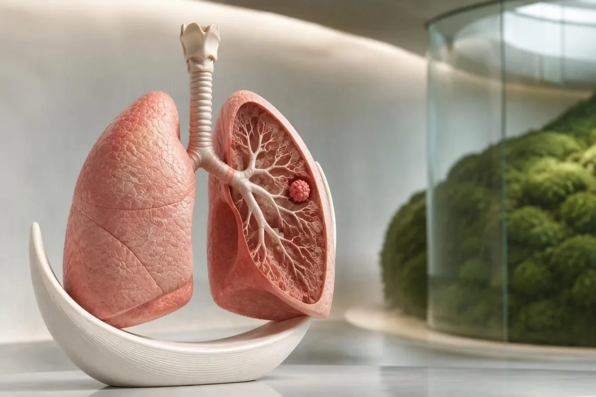
A cough deserves attention when it persists, changes, or comes with warning signs—not because its sound can identify cancer. Normal blood results and screening eligibility do not decide whether symptoms need investigation.

Key takeaways
- Persistent cough: Arrange assessment for a new cough lasting more than 3 weeks, or sooner if it worsens or accompanies other warning signs.
- Changing cough: A familiar smoker's cough that becomes more frequent, different, or associated with breathlessness warrants evaluation without waiting another 3 weeks.
- Coughing up blood: Even a few unexplained spots or streaks need urgent medical advice; more than a few spots, or blood with breathing difficulty or chest pain, warrants emergency care.
- Silent early disease: A small lung cancer can cause no symptoms if it does not obstruct an airway or affect pain-sensitive structures.
- Unintentional weight loss: Losing more than 5% of body weight over 6–12 months deserves investigation, although many noncancer conditions cause this.
- Diagnostic imaging: Symptoms lead to a diagnostic pathway, often starting with chest X-ray; persistent concern can require CT despite a normal X-ray.
- Low-dose CT screening: The 2021 USPSTF recommendation covers annual screening for eligible adults aged 50–80 with at least 20 pack-years who currently smoke or quit within 15 years.
- Blood test limits: Normal CBC, CRP, calcium, or sodium results do not exclude lung cancer, and abnormal results do not confirm it.
When should a new or changing cough be checked?
Lung cancer symptoms can include a cough lasting more than 3 weeks, a changing usual cough, coughing up blood, breathlessness, chest pain, or unexplained weight loss. Arrange assessment for a persistent or changing cough; seek help sooner for warning signs. Symptoms do not confirm cancer, and early lung cancer can cause no symptoms.

A 3-week cough threshold is a practical prompt to seek assessment, not a biological point at which cancer becomes likely. A cough that is steadily improving after a respiratory illness differs from one that grows more frequent, interrupts sleep, or appears alongside reduced exercise tolerance.
Changing symptoms matter even when a cough has existed for years. For example, a hypothetical 61-year-old with a longstanding morning cough should not wait another 3 weeks if the cough becomes continuous and new breathlessness develops; the change, rather than the original duration, needs explanation.
NICE guideline NG12 recommends an urgent chest X-ray within 2 weeks for people aged 40 or older with 2 specified unexplained symptoms, or 1 if they have ever smoked; the symptoms include cough, fatigue, breathlessness, chest pain, weight loss, and appetite loss (NICE, 2015). These criteria guide UK referral decisions rather than diagnose cancer.
Kantesti is an AI blood test analyzer that helps explain laboratory findings alongside symptoms, but a normal CBC cannot clear a persistent cough. Our organization and service scope explain Kantesti Ltd's role; readers with trigger-related wheeze can also review patterns that suggest asthma without assuming asthma explains every new change.
Does a lung cancer cough sound or feel different?
A lung cancer cough has no reliable identifying sound: it may be dry, produce mucus, or resemble an ordinary chronic cough. Duration beyond 3 weeks, progression, accompanying symptoms, and personal risk provide more useful clues than whether the cough sounds harsh or occurs at night.
Cough recordings and symptom descriptions cannot reliably distinguish lung cancer from reflux, asthma, or postinfectious cough. Two people with the same dry cough may need different investigations because one is recovering normally while the other has unexplained weight loss and an abnormal chest examination.
Night-time coughing is not specific to cancer. Reflux, nasal drainage, and asthma commonly worsen after lying down, so a 4-week cough associated with sour taste or throat clearing may prompt assessment for reflux-related symptom patterns as well as consideration of chest imaging when indicated.
Coughing fits with vomiting or a whoop can suggest pertussis, but adults do not always produce the characteristic sound. Our explanation of whooping cough test timing shows why a negative test collected several weeks into illness may not settle the cause of a prolonged cough.
A brief symptom record is more useful than trying to label the cough yourself. Over 7 days, note frequency, mucus, sleep disruption, breathlessness during ordinary activities, and any visible blood; improvement after a cough suppressant does not establish the cause or remove the need to investigate warning signs.
Why can early lung cancer cause no symptoms?
Early lung cancer can be asymptomatic because a small growth may not block an airway, disturb breathing, or reach pain-sensitive structures. A peripheral growth measuring 1–2 cm can sometimes be discovered incidentally, while a smaller centrally located growth may cause coughing or recurrent airway obstruction.
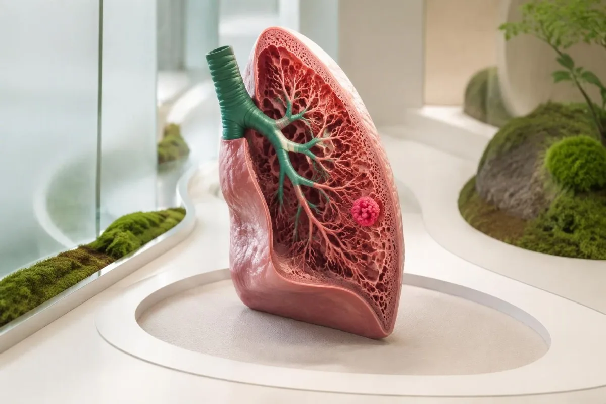
Location can influence symptoms as much as size. A hypothetical 15 mm peripheral growth might leave airflow unchanged, whereas a growth beside a major bronchus can irritate the airway or obstruct mucus drainage; neither example allows its stage to be determined without imaging and further assessment.
Lung tissue itself is not uniformly sensitive to pain. Discomfort becomes more likely when an affected area involves the pleura or chest wall, so the absence of pain—even over 6 months—cannot establish that the lungs are free of cancer.
A normal oxygen saturation does not rule out a localized growth. Someone with a small affected area may still have a resting saturation of 97% because the remaining lung exchanges oxygen normally; pulse oximetry measures oxygenation, not whether an abnormal growth exists.
Symptoms and screening answer different questions: one investigates a change, while the other searches for disease before symptoms appear in selected risk groups. Our medical content standards distinguish established guideline recommendations from illustrative examples; a 3-week cough rule should never be interpreted as proof that shorter coughs are harmless.
Which other lung cancer symptoms deserve attention?
Other lung cancer symptoms include unexplained breathlessness, recurrent chest infections, persistent chest or shoulder pain, hoarseness, appetite loss, fatigue, and weight loss. Losing more than 5% of body weight over 6–12 months is a commonly used trigger for investigation, not a cancer-specific finding.
Unintentional weight loss deserves a numerical check rather than an impression alone. A hypothetical decline from 80 kg to 75 kg is a 6.25% loss; clinicians consider appetite, swallowing, gastrointestinal symptoms, thyroid disease, mood, medications, and other explanations alongside possible cancer.
Repeated pneumonia in the same lung region can suggest an underlying drainage or airway problem. Two documented episodes affecting the same lobe may lead clinicians to review previous images for obstruction, whereas infections in different locations may raise other questions about aspiration, immunity, or chronic lung disease.
Persistent hoarseness can arise from reflux or voice strain, but it can also reflect a problem affecting the nerve that supplies the voice box. Hoarseness persisting beyond approximately 3 weeks, especially with cough or swallowing difficulty, deserves assessment rather than repeated treatment without an explanation.
Breathlessness and fatigue are particularly nonspecific because anemia, heart disease, and airway disorders can produce both. Our breathlessness laboratory results guide explains supporting tests; an oxygen saturation of 98% or normal hemoglobin does not replace investigation when a person's walking tolerance has clearly deteriorated.
How urgently should coughing up blood be assessed?
Coughing up blood requires urgent medical advice, even when only a few unexplained spots or streaks appear. Seek emergency care for more than a few spots or streaks, ongoing or increasing blood, or blood accompanied by breathlessness, chest pain, faintness, or a very fast heartbeat; do not wait to measure the amount precisely.
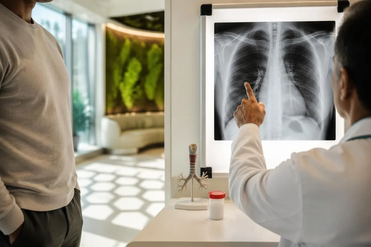
Hemoptysis means blood arising from the lower respiratory tract, but a nosebleed or gum bleeding can contaminate saliva. A single episode still needs clarification: tell the clinician whether blood followed coughing, throat clearing, or vomiting, because these lead to different examination and testing pathways.
There is no dependable home volume cutoff that makes coughing up blood safe. A hypothetical teaspoon is about 5 mL, but visual estimates are unreliable and the danger depends on whether blood affects breathing, how quickly it accumulates, and the person's underlying lung reserve.
Anticoagulants can increase bleeding, but they do not explain why bleeding started. If blood appears while taking a medicine such as warfarin or apixaban, seek prompt advice rather than stopping treatment independently; our clotting test interpretation guide explains why normal routine tests cannot exclude every bleeding-related problem.
NICE NG12 recommends a suspected cancer pathway referral for unexplained hemoptysis in people aged 40 or older (NICE, 2015). Younger people also need assessment, because infection, pulmonary embolism, and other respiratory conditions can cause hemoptysis; the referral threshold is not a permission to ignore symptoms below age 40.
Which breathing or chest symptoms are emergencies?
Severe or sudden breathlessness, major coughing up of blood, chest pressure, collapse, or new neurological deficits require emergency assessment. A newly low oxygen reading around 92% or below warrants urgent advice, but severe symptoms require help even with a normal reading; people with prescribed oxygen targets need individualized guidance.
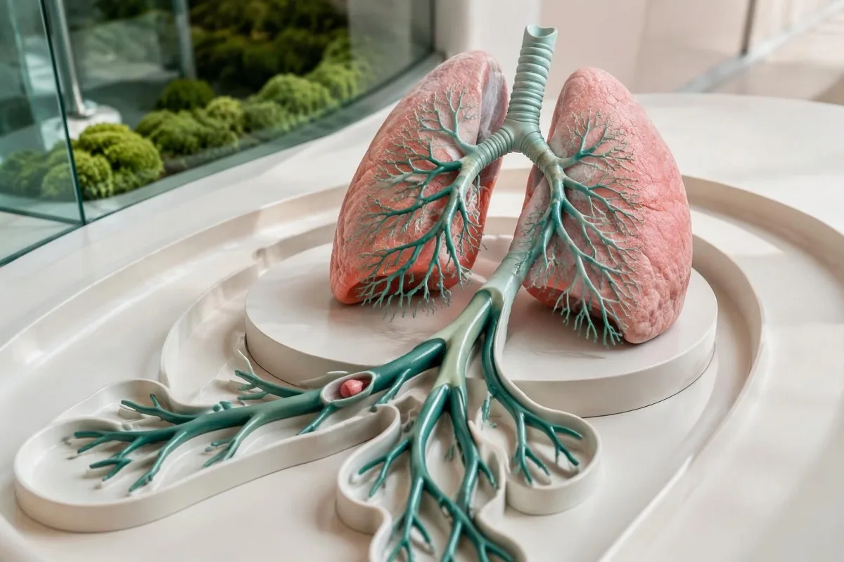
Sudden breathlessness with pain on breathing can indicate pulmonary embolism, particularly when one leg is newly swollen. The combination deserves urgent assessment even if the cough began only 1 day earlier; our urgent leg swelling signs explain why clot symptoms should not be managed as a routine persistent cough.
Chest pressure with sweating, nausea, or pain spreading to the arm or jaw may indicate a heart emergency. Waiting 3 weeks to see whether a cough improves is inappropriate in this situation, because symptom-duration advice for uncomplicated cough does not apply to potentially time-critical chest symptoms.
Sudden weakness, facial drooping, speech difficulty, or a new seizure require emergency care rather than an outpatient cancer appointment. Our stroke emergency warning signs describe immediate action; a deficit lasting even a few minutes can still need urgent evaluation.
New face or neck swelling accompanied by breathlessness, difficulty swallowing, or a severe headache can reflect obstruction of a major chest vein or another urgent problem. Do not wait for a second episode, and do not use a consumer oximeter as the deciding test; readings can be inaccurate and do not detect every emergency.
What are lung cancer symptoms in nonsmokers?
Lung cancer symptoms in nonsmokers are generally the same as in smokers: persistent cough, breathlessness, coughing up blood, chest discomfort, or unexplained weight loss. Never-smoking lowers risk but does not eliminate it, and a cough lasting more than 3 weeks still deserves assessment when it persists or changes.

Never-smoker usually means a person has smoked fewer than 100 cigarettes over their lifetime in epidemiological definitions. That classification does not describe secondhand smoke, radon, occupational exposures, or air pollution, so clinicians ask about these separately rather than treating smoking history as a complete risk assessment.
Radon exposure is assessed in buildings, not diagnosed from a cough or routine blood panel. The US EPA recommends fixing homes at 4 pCi/L, approximately 148 Bq/m³, or higher; a result above that level supports exposure reduction but cannot tell whether an individual has lung cancer.
Nicotine exposure tests are not lung cancer tests. As our cotinine result interpretation guide explains, cotinine may reflect nicotine replacement or vaping as well as tobacco exposure; a negative result cannot erase a smoking history accumulated over 20 or 30 years.
A hypothetical 46-year-old never-smoker with a 5-week cough and new hoarseness needs symptom assessment even though they would not meet standard USPSTF smoking-based screening criteria. Routine low-dose CT screening is not established for all never-smokers, but diagnostic imaging can still be appropriate when symptoms or examination findings warrant it.
What else commonly causes a persistent cough?
Common alternatives to cancer include postinfectious cough, asthma, upper-airway cough syndrome, reflux, COPD, and medication effects. Postinfectious cough often occupies the 3–8 week interval after an illness; improvement and the absence of warning signs support that explanation, but duration alone cannot establish the cause.
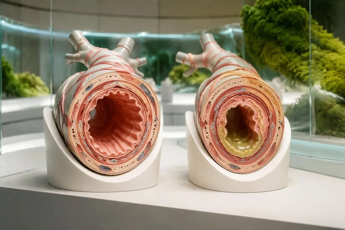
A cough lasting more than 8 weeks is usually classified as chronic in adults. That definition organizes investigation; it does not mean clinicians should postpone assessment until week 8 when a cough is worsening, a person is coughing up blood, or unexplained weight loss has appeared.
Respiratory viruses can leave sensitive airways after the initial fever and congestion resolve. Our adult RSV symptom guide discusses illness patterns, but a 4-week cough should not automatically be attributed to a previous positive viral test if new focal chest symptoms develop.
An ACE inhibitor such as ramipril can cause a dry cough even after the medicine has been taken for some time. When a medication is suspected, clinicians may arrange a supervised alternative and reassessment over the following weeks; do not stop prescribed treatment yourself or assume the medicine explains every accompanying symptom.
Tuberculosis becomes more relevant with prolonged cough, fever, night sweats, weight loss, and a compatible exposure history. Our IGRA test limitations guide explains why 1 negative immune-response test does not exclude active pulmonary tuberculosis; chest imaging and respiratory samples may still be needed.
Which imaging tests investigate lung cancer symptoms?
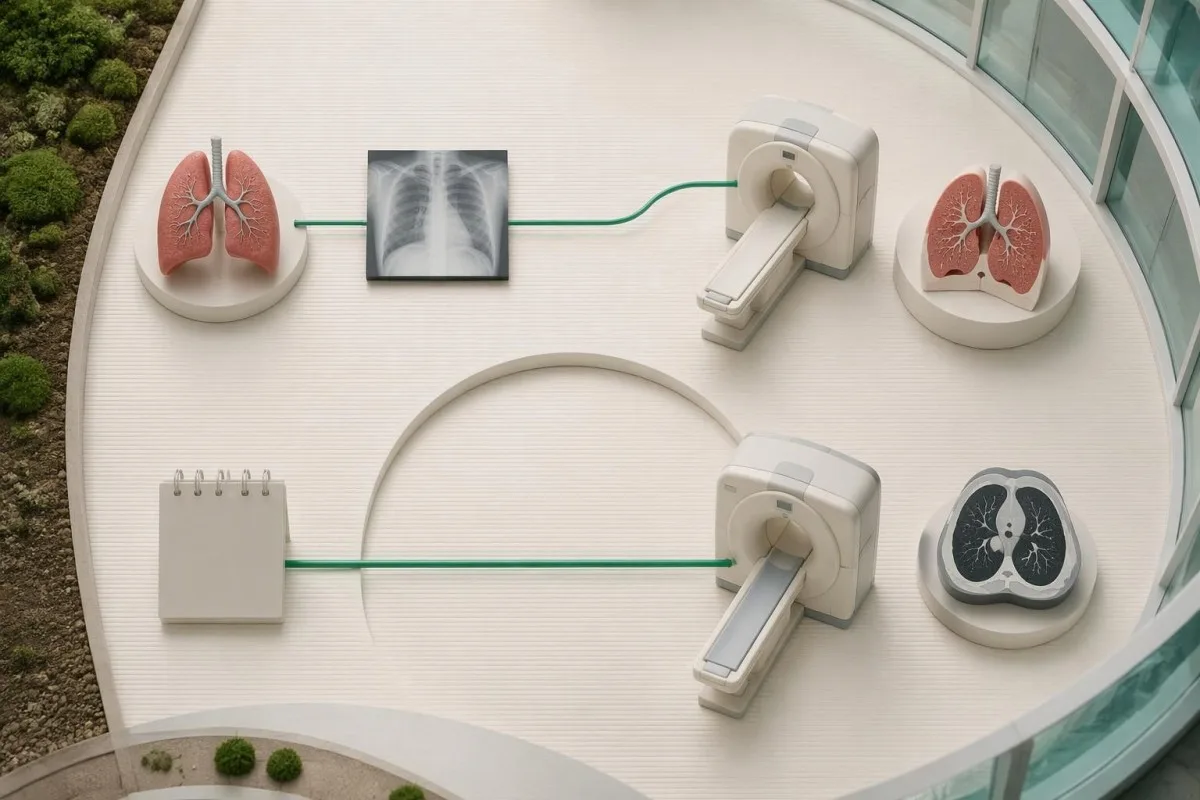
Symptom-driven imaging investigates a current clinical concern, often with a chest X-ray followed by CT when findings or ongoing suspicion justify it. NICE's 2-week urgent X-ray pathway is a diagnostic referral standard, not a screening schedule, and a normal X-ray cannot exclude every lung cancer.
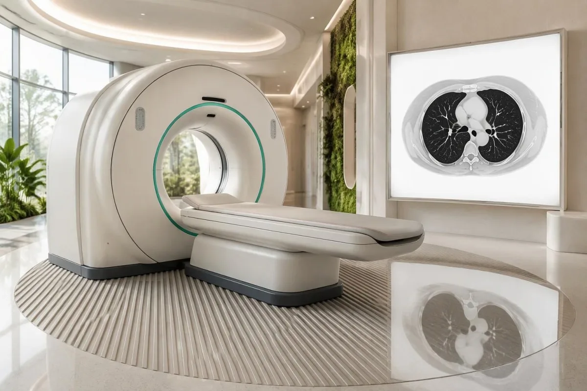
Chest X-rays compress 3-dimensional anatomy into a projection, allowing ribs, the heart, or other structures to obscure an affected area. CT produces cross-sectional images, so clinicians may request it after a reassuring X-ray when persistent hemoptysis, focal signs, or a concerning symptom combination remains unexplained.
Diagnostic CT protocols vary with the clinical question. Contrast-enhanced CT may help assess chest structures and suspected cancer, whereas CT pulmonary angiography uses a different contrast-timing protocol for suspected embolism; these are not interchangeable simply because both involve a CT scanner.
An abnormal scan does not equal cancer. Granulomatous conditions, scars, and inflammatory changes can produce nodules or enlarged chest lymph nodes; our sarcoidosis ACE test guide explains why 1 supporting laboratory marker cannot reliably distinguish these possibilities.
A screening CT performed 6 months earlier does not remove the need to assess new symptoms. Bring the previous report and, if possible, the images: comparison can identify change, but clinicians still choose a diagnostic test suited to the current problem rather than automatically repeating the previous screening protocol.
How does low-dose CT screening differ from diagnosis?
Low-dose CT screening is intended for eligible people without symptoms requiring investigation. The 2021 USPSTF recommends annual screening for adults aged 50–80 with at least 20 pack-years who currently smoke or quit within the past 15 years; symptoms require diagnostic assessment regardless of screening eligibility.
Pack-years equal packs smoked per day multiplied by years smoked, using 20 cigarettes as 1 pack. Smoking 10 cigarettes daily for 40 years equals 20 pack-years; brief periods of heavier smoking should be included in the calculation rather than estimating exposure from the current daily amount alone.
Screening evidence concerns repeated assessment of selected higher-risk populations, not a CT for every cough. In the National Lung Screening Trial, 3 annual low-dose CT rounds reduced lung-cancer mortality by approximately 20% relative to chest X-ray screening among participants aged 55–74 with substantial smoking histories (National Lung Screening Trial Research Team, 2011).
That 20% figure is a relative mortality reduction, not a guarantee of protection or a 20-percentage-point reduction in an individual's risk. Screening can find benign nodules, cause anxiety, lead to additional procedures, and sometimes identify cancers that would not have caused harm; eligibility therefore includes discussion of benefits, harms, and fitness for treatment.
International systems separate symptom referral from screening selection in different ways. UK NICE symptom criteria begin at age 40 for specified presentations, whereas the cited US screening recommendation begins at 50; neither routine annual blood work nor being below a screening age substitutes for evaluating a concerning cough.
What happens if an X-ray or CT shows an abnormality?
An abnormal lung image usually requires clarification, not an immediate cancer label: clinicians assess its appearance, size, change over time, and the person's risk. An 8 mm nodule and a large airway-obstructing mass lead to different decisions, and incidental-nodule follow-up rules may not apply unchanged to someone with concerning symptoms.
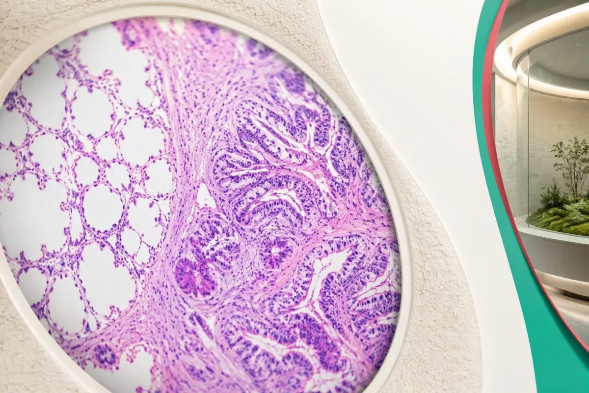
CT may identify a nodule, enlarged lymph nodes, fluid around a lung, or airway narrowing. Comparison with imaging from 12 or 24 months earlier can help, but the significance of stability depends on the finding's type; some slow-growing or subsolid nodules need longer observation than solid nodules.
A biopsy is usually used to establish the cancer type when feasible and clinically appropriate. Bronchoscopy samples accessible airways, endobronchial ultrasound can sample certain lymph nodes, and image-guided sampling can reach peripheral areas; clinicians choose 1 route that balances diagnostic yield, staging information, and procedural risk.
PET-CT can help evaluate activity and possible spread, but increased uptake is not specific to cancer. Infection and inflammatory disease can also be active, while small or certain slow-growing abnormalities may be less conspicuous; an imaging result therefore needs interpretation alongside tissue findings and the broader clinical picture.
Rare neurological presentations can prompt cancer investigation before a respiratory complaint dominates. Our anti-Hu antibody follow-up guide explains one such association, especially with small-cell lung cancer; 1 positive antibody result still requires specialist confirmation and does not establish the diagnosis by itself.
Can blood tests detect or rule out lung cancer?
Routine blood tests cannot reliably detect or rule out lung cancer. A CBC, kidney panel, calcium, and liver tests can reveal anemia, complications, or alternative explanations; a sodium concentration of 140 mmol/L or normal white-cell count does not exclude a growth seen only through imaging and further assessment.
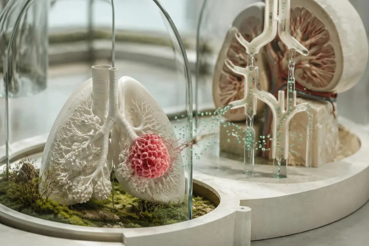
Low sodium can occur with SIADH, including some cases associated with small-cell lung cancer, but medications and noncancer illnesses are common causes. Sodium below 135 mmol/L is generally classified as hyponatremia; our SIADH laboratory pattern guide explains why paired serum and urine measurements matter more than sodium alone.
High calcium can accompany malignancy, but mild elevations often have other explanations, including primary hyperparathyroidism or supplements. A hypothetical calcium of 2.65 mmol/L should be interpreted against the laboratory range and albumin or confirmed with ionized calcium when appropriate; a single result cannot identify whether a lung abnormality is present.
Kantesti is an AI blood test interpretation platform that helps readers understand CBC, electrolyte, and chemistry results while preserving the distinction between laboratory clues and diagnosis. Our AI interpretation technology guide describes the workflow; a normal CRP result cannot cancel a 5-week worsening cough or replace a clinician's imaging decision.
Kantesti AI does not treat an elevated CEA or a neutrophil-to-lymphocyte ratio as proof of lung cancer. Our CEA result limitations guide explains why, for example, a CEA of 7 ng/mL may have several explanations; standard lung screening is not based on CEA, CBC ratios, or a general tumor-marker panel.
What should you bring to a cough appointment?
Bring a symptom timeline, medication list, smoking and exposure history, weight measurements, and previous imaging or laboratory reports. A 7-day record can show whether symptoms are improving or progressing; the appointment should also establish who will review results and what happens if the first investigation does not explain the cough.

A hypothetical 58-year-old has a 5-week cough, no fever, and weight falling from 72 kg to 68 kg over 4 months. The 5.6% weight loss plus persistent cough merits prompt assessment; a normal white-cell count would not justify simply waiting for an annual screening appointment.
A different hypothetical patient develops cough during a viral illness and is steadily improving by week 4, without blood, weight loss, or breathlessness. Clinicians may choose treatment for a likely alternative cause with planned reassessment, but our WBC and CRP comparison guide explains why 2 normal inflammatory tests do not independently settle the diagnosis.
Nutrition-related laboratory findings need context rather than a cancer interpretation. Albumin of 32 g/L, for example, can reflect inflammation, liver disease, kidney loss, or other illness—not simply inadequate protein intake; our serum protein interpretation guide explains why weight change and related tests must be considered together.
Ask for a specific follow-up plan rather than an open-ended instruction to return if worried. If a chest X-ray is requested within 2 weeks, confirm how results will reach you and whom to contact if they do not arrive; persistent or worsening symptoms after a normal result may require a fresh examination, CT, or specialist advice.
Follow-up, AI limits and supporting research publications
Follow-up should track unresolved symptoms, not just reassuring test results. A cough continuing beyond 3 weeks needs an explanation and a safety-net plan; new blood, worsening breathlessness, or neurological symptoms require escalation even while routine appointments or laboratory interpretations are pending.
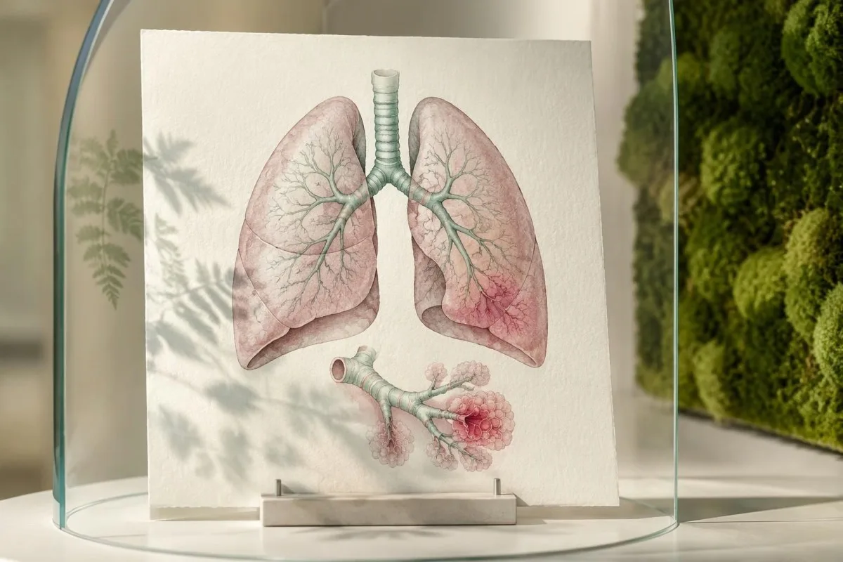
Kantesti is an AI-powered blood test analysis tool that can help organize laboratory questions before follow-up, but it does not diagnose lung cancer or interpret a cough as a cancer probability. Our responsible AI principles explain why 1 reassuring panel cannot replace examination, imaging, or urgent care.
Keeping reports together helps clinicians connect changes without overreading them. Record dates, laboratory units, medication changes, and symptom onset over the preceding 4–8 weeks; a change in sodium, hemoglobin, or albumin is useful clinical context, but none of these trends is a validated stand-alone lung cancer screen.
The 2 supporting publications listed below concern coagulation testing and serum proteins. They are background laboratory resources, not lung cancer diagnostic studies, screening trials, or evidence that AI can rule out cancer; the external references separately support the symptom-referral and low-dose CT statements in this article.
Kantesti's laboratory explanations should support, rather than delay, the next clinical step. For a persistent cough, agree on 3 practical points: the likely explanation, the next investigation or treatment, and the trigger for reassessment; for emergency warning signs, seek emergency help immediately instead of waiting for an online interpretation.
Frequently asked questions
How long does a cough last before you should worry about lung cancer?
A new cough lasting more than 3 weeks should be assessed, especially if it is not improving or has changed from a familiar pattern. The 3-week threshold prompts evaluation; it does not mean the cough is cancer. Seek help sooner for coughing up blood, worsening breathlessness, chest pain, or unexplained weight loss. A clinician uses symptoms, examination, risk history, and imaging when indicated—not duration alone—to investigate the cause.
What does a lung cancer cough sound like?
A lung cancer cough has no distinctive sound that reliably separates it from asthma, reflux, or infection. It may be dry or mucus-producing and can occur during the day or at night. A cough persisting beyond 3 weeks, becoming more frequent, or accompanying other warning signs provides more useful information than its sound. Neither a recording nor temporary improvement with cough medicine can confirm or exclude lung cancer.
Can you have lung cancer without any symptoms?
Lung cancer can cause no symptoms when a growth does not obstruct an airway or affect pain-sensitive structures. A small peripheral growth of approximately 1–2 cm can sometimes be found incidentally, although symptom development depends on location and other factors rather than size alone. Normal oxygen saturation and routine blood results do not exclude lung cancer. Low-dose CT screening seeks disease before symptoms in eligible higher-risk people, while new symptoms require diagnostic assessment.
What are the early signs of lung cancer in nonsmokers?
Early signs of lung cancer in nonsmokers can include a persistent or changing cough, unexplained breathlessness, blood in sputum, chest discomfort, or weight loss, but early disease may also be silent. Never-smoking lowers risk without eliminating it. A cough lasting more than 3 weeks still deserves assessment when it persists or changes, regardless of smoking history. Standard smoking-based screening eligibility does not decide whether a nonsmoker needs diagnostic imaging for symptoms.
Can a normal chest X-ray or blood test rule out lung cancer?
Neither a normal chest X-ray nor normal routine blood tests can reliably rule out lung cancer. Chest X-rays can miss abnormalities hidden by overlapping structures, and CBC or CRP tests do not detect lung growths directly. A 5-week worsening cough or unexplained coughing up of blood may justify further assessment even after reassuring initial results. A clinician decides whether CT or specialist evaluation is needed based on the continuing clinical concern.
Should I request low-dose CT screening if I already have a cough?
A cough that requires investigation belongs in a diagnostic pathway rather than being managed only through screening. The 2021 USPSTF recommends annual low-dose CT screening for eligible adults aged 50–80 with at least 20 pack-years who currently smoke or quit within 15 years. Those criteria apply to preventive screening, not to deciding whether symptoms deserve imaging. Tell the clinician or screening service about new symptoms so they can choose the appropriate assessment and CT protocol if needed.
Is a small amount of blood when coughing an emergency?
Even a few unexplained spots or streaks of blood when coughing require urgent medical advice. More than a few spots or streaks, ongoing or increasing blood, or blood with breathing difficulty, chest pain, faintness, or a fast heartbeat warrants emergency care. A teaspoon is approximately 5 mL, but home volume estimates are unreliable and should not determine whether help is needed. Coughing up blood can result from several conditions and does not, by itself, confirm cancer.
Get your blood test results explained
Upload your lab report and Kantesti's AI explains each biomarker in plain language within seconds. Used by more than 2 million people worldwide.
References
- Klein, T., Mitchell, S., & Weber, H. (2026). aPTT Normal Range: D-Dimer, Protein C Blood Clotting Guide. Kantesti AI Medical Research. DOI
- Klein, T., Mitchell, S., & Weber, H. (2026). Serum Proteins Guide: Globulins, Albumin & A/G Ratio Blood Test. Kantesti AI Medical Research. DOI
- National Institute for Health and Care Excellence (2015). Suspected cancer: recognition and referral. NICE guideline NG12.
- US Preventive Services Task Force (2021). Screening for Lung Cancer: US Preventive Services Task Force Recommendation Statement. JAMA. DOI
- National Lung Screening Trial Research Team (2011). Reduced Lung-Cancer Mortality with Low-Dose Computed Tomographic Screening. New England Journal of Medicine. DOI PubMed
- 2M+Tests analysed
- 127+Countries
- 100+Languages
About the medical team


E-E-A-T Trust Signals
Experience
Physician-led clinical review of lab interpretation workflows.
Expertise
Laboratory medicine focus on how biomarkers behave in clinical context.
Authoritativeness
Medically reviewed by Dr. Thomas Klein, with contributions from Dr. Sarah Mitchell and Prof. Dr. Hans Weber.
Trustworthiness
Evidence-based interpretation with clear follow-up pathways to reduce alarm.





Medical disclaimer
This article is for educational purposes only and does not constitute medical advice. Always consult a qualified healthcare provider for diagnosis and treatment decisions.
Published · Contact the editorial team
Published by Kantesti Ltd, registered in England & Wales, company no. 17090423, London, United Kingdom.
