
New, unexplained swelling in one leg needs same-day assessment, even if the pain is mild. Sudden breathlessness, chest pain, coughing up blood or fainting requires emergency care—not an online test.

Key takeaways
- Same-day assessment is needed for new, unexplained swelling in 1 leg, especially with pain, warmth, discoloration or a recent clot risk.
- Emergency symptoms include sudden unexplained breathlessness, chest pain, coughing up blood or fainting; call 999, 112, 911 or your local emergency number.
- Calf measurements differ by at least 3 cm in one Wells-score criterion, measured 10 cm below the tibial tuberosity; smaller differences do not exclude DVT.
- Wells scoring classifies DVT as likely at 2 points or more and unlikely at 1 point or less, but requires a clinician's examination and judgment.
- D-dimer commonly uses a cutoff of 500 ng/mL FEU; a positive result does not diagnose a clot, and a negative result is useful only within an appropriate diagnostic pathway.
- Ultrasound timing under NICE guidance is within 4 hours when DVT is likely; if unavailable, clinicians may prescribe interim anticoagulation and arrange imaging within 24 hours.
- Repeat ultrasound may be needed after 6–8 days when the initial proximal scan is negative but D-dimer is positive.
- Confirmed proximal DVT generally requires at least 3 months of anticoagulation; calf-only clots sometimes follow a different treatment or surveillance pathway.
When does one-sided leg swelling need same-day care?
Deep vein thrombosis symptoms include new swelling in one leg, calf or thigh pain, warmth and discoloration. Seek same-day assessment for unexplained one-sided swelling; call emergency services for sudden breathlessness, chest pain, coughing up blood or fainting. Symptoms alone cannot confirm or exclude DVT.

A DVT is a clot in a deep vein, usually in the leg; the immediate concern is extension or movement to the lungs, not simply the amount of swelling. A person with 1 newly swollen leg should not wait for redness, severe pain or a second symptom before seeking advice, particularly after surgery, immobilization or a previous clot.
For example, a hypothetical 52-year-old develops a tight shoe and a larger left calf after a long journey, but rates discomfort only 2 out of 10. Mild pain does not make that pattern safe to monitor for several days; the useful details are when the asymmetry began, whether it is progressing, and what recent risks accompanied it.
Kantesti is an AI blood test analyzer that can help explain laboratory reports, but it cannot diagnose DVT from a photograph or replace urgent ultrasound. Our guide to nerve-related leg pain explains a different pattern—shooting pain with tingling—yet nerve symptoms do not explain away new swelling in 1 leg.
What do DVT symptoms in the calf feel like?
DVT symptoms in the calf may feel like cramping, a persistent ache, tenderness or tightness, with or without visible swelling. No pain pattern reliably separates a clot from a muscle injury, and some DVTs produce few noticeable symptoms.
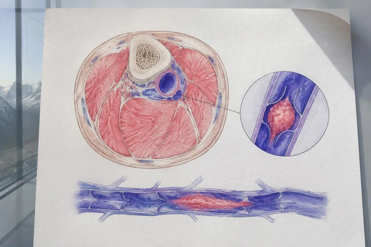
A calf at least 3 cm larger than the other contributes 1 point to the clinical Wells score when measured 10 cm below the tibial tuberosity. That threshold standardizes an examination; it is not a home diagnostic boundary, because a smaller clot, early presentation or naturally unequal calves can make circumference misleading.
Pain on pulling the toes upward—the historical Homan's sign—cannot confirm or exclude DVT. Repeatedly squeezing the calf or trying this movement adds no reliable diagnostic information; a person who can walk normally or perform a heel raise can still need assessment for new symptoms in 1 leg.
Exercise-related calf discomfort that repeatedly starts after a similar walking distance and settles with rest suggests a different mechanism, such as reduced arterial circulation. Our explanation of arterial circulation symptoms describes that distinction, but sudden swelling should not be attributed to an established walking problem without examination.
Proximal DVT can cause thigh discomfort or swelling of the whole leg, rather than a focal calf ache. If symptoms extend into the groin or the entire limb becomes larger, tell the assessing clinician explicitly; an investigation limited to the calf may not answer the question of a more central obstruction.
How should warmth and leg discoloration be interpreted?
Warmth and discoloration can accompany DVT, but they also occur with skin infection, superficial vein inflammation and injury. The combination of new asymmetry, warmth and swelling matters more than any particular shade of red.
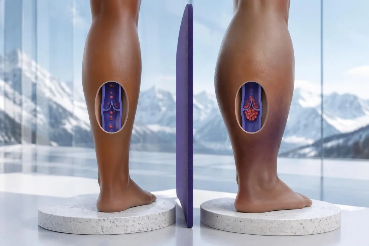
Redness may be less obvious on brown or dark skin, where a change can appear dusky, purple, gray-brown or simply darker than the opposite leg. Compare both legs in the same light, looking at size and texture as well as color; absence of visible redness is not evidence against a deep clot.
A tender, firm cord close to the surface suggests superficial venous thrombosis, but that condition can coexist with DVT or extend toward the deep system. A line of localized redness is therefore not automatically less urgent, especially if there is accompanying calf swelling or the affected area approaches the groin or back of the knee.
Fever and spreading redness raise concern for cellulitis, although neither fever nor a raised inflammatory marker proves that infection is the only explanation. Our discussion of WBC and CRP disagreement explains why laboratory inflammation signals cannot distinguish all causes of a warm, swollen leg.
A rapidly swelling leg that becomes markedly blue or purple, extremely painful, numb or weak needs emergency assessment, even without chest symptoms. A cold, pale foot is also an emergency: that pattern can indicate arterial compromise rather than uncomplicated DVT, and waiting for a routine appointment risks missing a limb-threatening circulation problem.
Which breathing and chest symptoms require emergency care?
Sudden unexplained breathlessness, chest pain, coughing up blood, fainting or collapse require emergency care because they can indicate pulmonary embolism. These symptoms may occur without leg pain or swelling, and a normal home oxygen reading does not exclude a lung clot.
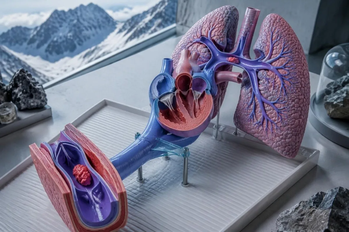
Call 999, 112, 911 or your local emergency number for new symptoms suggesting pulmonary embolism; do not drive yourself if breathless, faint or experiencing chest pain. Tell the dispatcher about a swollen leg, recent surgery, pregnancy, a previous clot or anticoagulant medication, but do not spend time collecting every detail before calling.
Pulmonary embolism pain often worsens with a deep breath, yet the absence of that feature is not reassuring. NICE's venous thromboembolism guidance treats suspected PE as a separate diagnostic pathway from suspected DVT; a leg ultrasound alone cannot exclude PE when chest or breathing symptoms are present (NICE, 2020, updated 2023).
A hypothetical person has sudden breathlessness, a pulse of 118 beats/minute and a home oxygen saturation of 97%. The oxygen result does not cancel the other warning signs, and our breathlessness testing guide is background reading—not a reason to wait for outpatient laboratory results.
Chest pain also has urgent alternatives, including heart attack and other cardiovascular conditions. Our explanation of urgent chest-pain tests distinguishes their purposes, but no single normal troponin, ECG or D-dimer result should be interpreted outside the clinician's assessment and the timing of symptoms.
Which clot risks make mild leg symptoms more concerning?
Recent surgery, reduced mobility, previous DVT, active cancer, pregnancy and estrogen-containing medication increase concern when one leg changes. A risk factor changes the probability of DVT; its absence does not make new one-sided swelling safe to ignore.

The Wells score assigns 1 point for bed rest lasting at least 3 days or major surgery within the previous 12 weeks requiring general or regional anesthesia. A plaster cast, paralysis or significant leg weakness can also matter; tell the clinician about mobility restrictions even if you were never admitted to hospital.
Long journeys—often discussed from around 4 hours onward—can contribute to venous stasis, especially when combined with other risks, but journey duration is not itself a standard DVT Wells-score item. Report the actual travel and movement pattern rather than assuming that a flight below a particular duration rules out a clot.
Inherited thrombophilia affects background susceptibility rather than confirming today's symptoms. A positive Factor V Leiden result does not establish that a swollen leg contains a clot, and a negative result does not remove risks from surgery, cancer, immobility or estrogen exposure.
Testing clotting proteins during an acute event can be difficult to interpret because illness and treatment may change the results. Our guide to low antithrombin activity explains why a single low value may require later reassessment; acute ultrasound and treatment decisions should not wait for a broad inherited-risk panel.
How does a clinician estimate DVT probability?
The two-level DVT Wells score classifies DVT as likely at 2 points or more and unlikely at 1 point or less. It guides the sequence of D-dimer testing and ultrasound; it does not diagnose a clot and should not be used as a self-clearance checklist.
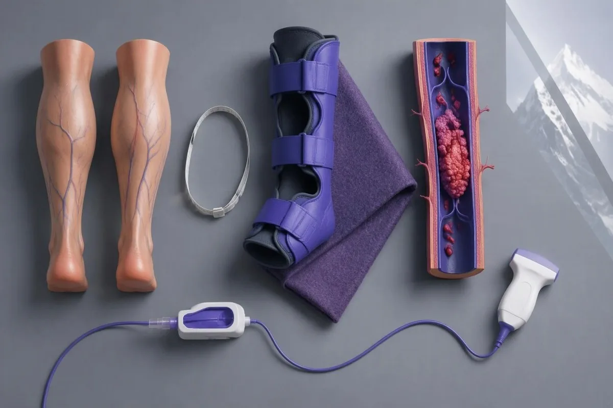
The score contains 9 findings worth 1 point each, including previous documented DVT, active cancer, relevant immobilization, deep-vein tenderness, whole-leg swelling, calf asymmetry, unilateral pitting edema and nonvaricose collateral veins. An alternative diagnosis at least as likely as DVT subtracts 2 points, which requires clinical judgment rather than simply identifying another possible cause.
For a clearly hypothetical calculation, previous DVT adds 1 point, unilateral pitting edema adds 1, and calf enlargement meeting the measurement criterion adds 1. With no equally likely alternative, the total is 3 and DVT is likely; if an alternative is judged at least as likely, subtracting 2 changes the total to 1 and the testing sequence changes.
An 'unlikely' score does not mean 'no clot': it means a suitable negative D-dimer may safely end testing within the validated pathway. Lim and colleagues' 2018 ASH diagnostic guideline similarly organizes investigation around pretest probability, because the meaning of a negative test depends on the probability before that test was performed.
At Kantesti, we distinguish explaining a reported score from performing the bedside assessment needed to generate it. Our technical validation information describes our interpretation methodology and its limits; it should not be mistaken for validation of an AI-only pathway that clears new swelling in 1 leg without clinical examination.
Can a D-dimer blood test confirm or exclude DVT?
A positive D-dimer cannot confirm DVT. A negative high-sensitivity D-dimer can exclude DVT in selected patients with low clinical probability, but patients with likely DVT usually need ultrasound regardless of whether a laboratory number looks reassuring.
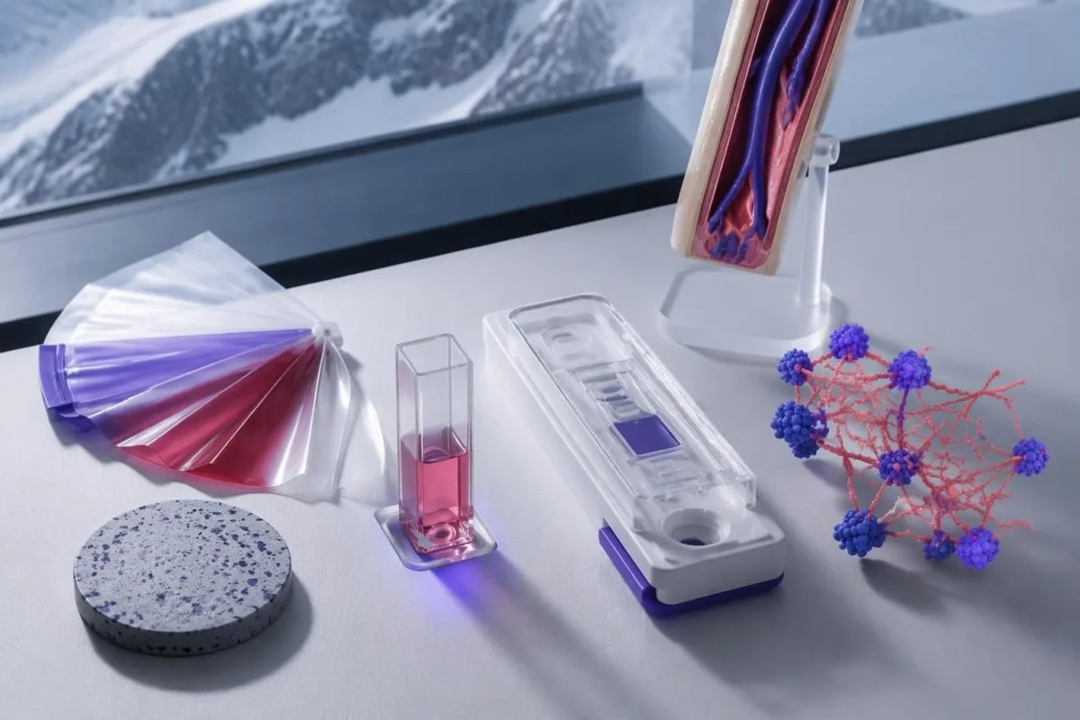
A common laboratory cutoff is 500 ng/mL FEU, equivalent to 0.5 mg/L FEU, although assays and reporting conventions differ. D-dimer measures products of cross-linked fibrin breakdown; surgery, pregnancy, cancer, infection and increasing age can raise it without an acute DVT, which explains why a positive result usually leads to imaging rather than an automatic diagnosis.
NICE recommends considering an age-adjusted threshold above age 50; a common validated convention is age multiplied by 10 ng/mL FEU. For a hypothetical 72-year-old, that gives 720 ng/mL FEU, but the threshold must match the assay and local diagnostic pathway—not be applied retrospectively to every elevated report.
FEU and D-dimer units, or DDU, are not interchangeable: FEU values are approximately twice DDU values, and a cutoff around 250 ng/mL DDU may correspond to 500 ng/mL FEU. Kantesti is an AI blood test interpretation platform that can help explain reported units; our D-dimer and clotting guide provides supporting laboratory context.
Anticoagulation started before sampling, later presentation and some smaller clots can complicate a negative result; a normal PT, INR or aPTT does not exclude DVT either. Our guide to normal coagulation test limits explains the distinction, consistent with ASH's warning against diagnosing DVT from a positive D-dimer alone (Lim et al., 2018).
When is ultrasound needed, and why repeat a negative scan?
Compression ultrasound is the main imaging test for suspected leg DVT. NICE recommends a proximal leg-vein scan within 4 hours when DVT is likely; when that is unavailable, the pathway may include clinician-prescribed interim anticoagulation and imaging within 24 hours.
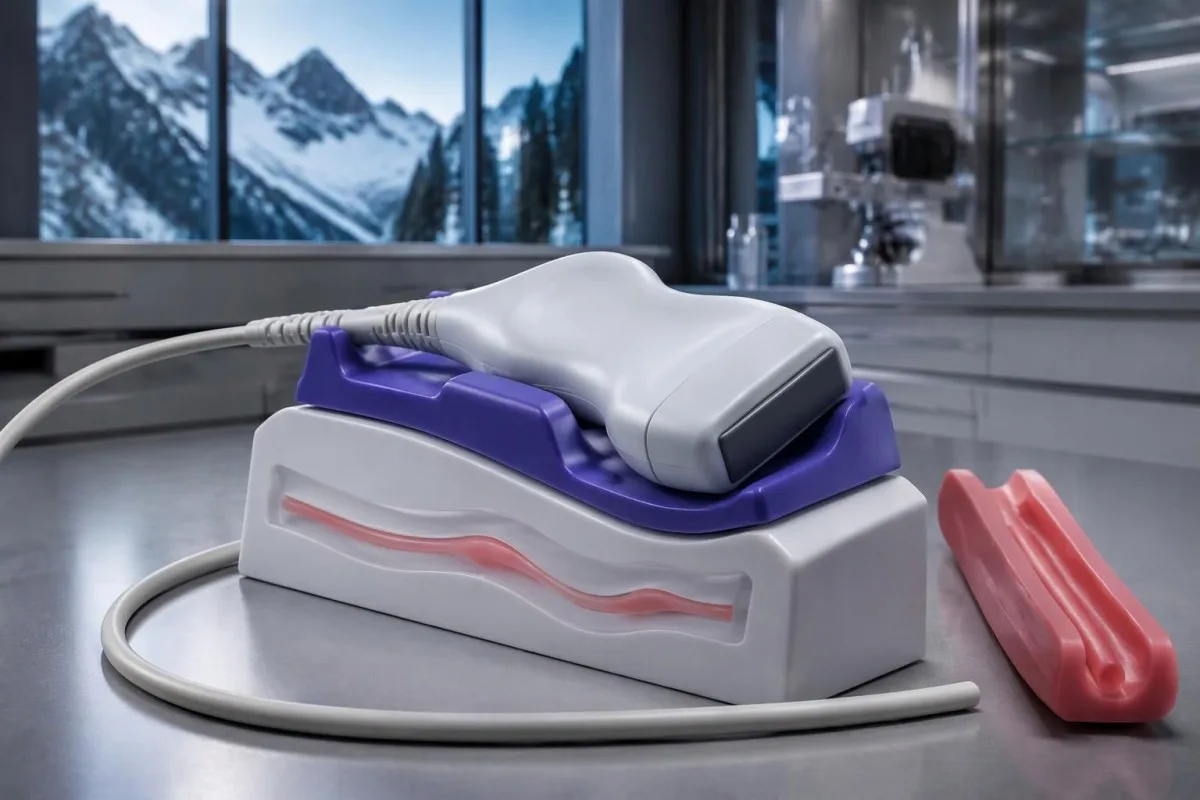
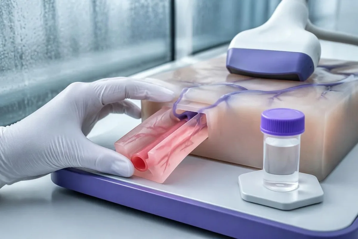
The ultrasound operator examines whether a vein compresses normally and may assess flow; an obstructed segment may fail to compress. Proximal ultrasound focuses on larger veins above the calf, while whole-leg ultrasound also evaluates distal veins, so a report saying 'no proximal DVT' does not necessarily mean every calf vein was assessed.
Under the NICE pathway, a negative proximal scan with a positive D-dimer generally leads to a repeat proximal scan after 6–8 days. The repeat checks for extension of an initially undetected distal clot; a technically adequate negative whole-leg scan can lead to a different follow-up plan depending on probability and local practice.
Persistent whole-leg swelling or groin symptoms after an unrevealing standard scan may need further consideration of pelvic or iliac-vein obstruction. The practical question is not simply 'Was the scan negative?' but 'Which veins were examined, was the study technically adequate, and what explains the symptoms now?'—especially if symptoms worsen before a planned repeat appointment.
Routine CBC, kidney tests and liver tests support treatment planning but do not replace imaging; the laboratory marker guide explains their separate roles. If an appointment is offered outside the recommended window, ask the assessing service whether interim treatment or another imaging route is needed rather than deciding to take an anticoagulant yourself.
What else can cause one leg to become swollen and painful?
A muscle injury, ruptured Baker's cyst, cellulitis, superficial venous thrombosis, venous insufficiency or lymphedema can mimic DVT. Several conditions can coexist, so identifying a plausible alternative does not automatically remove the need for a clot assessment.
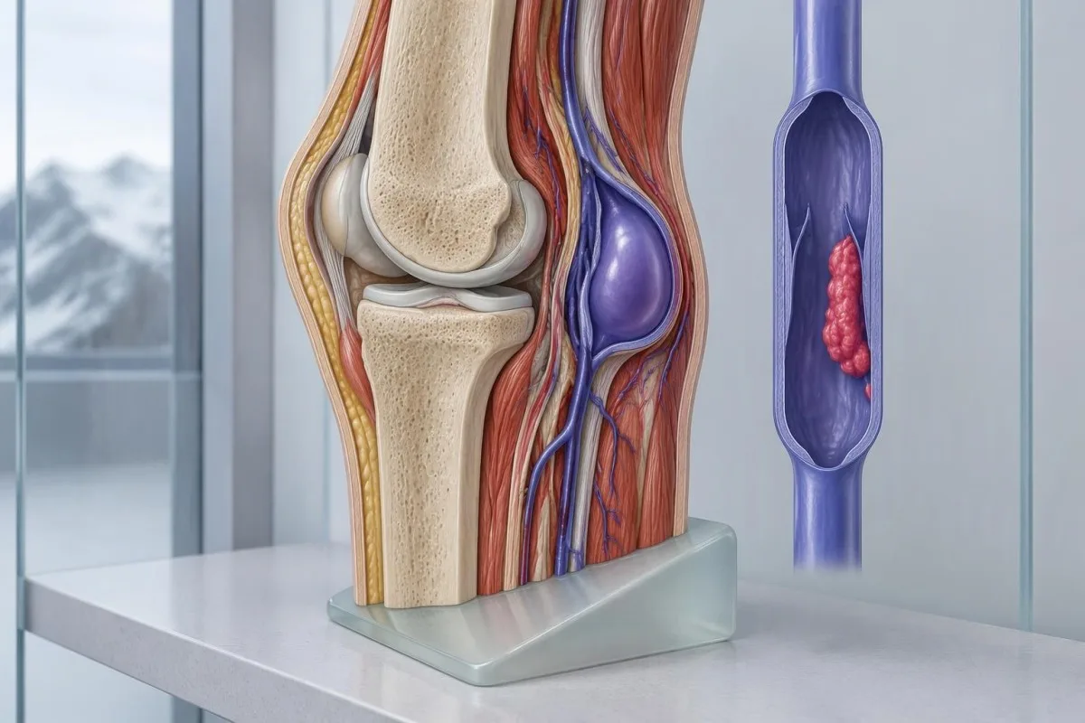
A ruptured Baker's cyst can cause sudden calf pain and swelling as fluid tracks down from behind the knee, sometimes with bruising near the ankle. Ultrasound may help distinguish it from DVT, but a history of knee arthritis alone is insufficient; the clinician still has to explain the new change in 1 leg.
Chronic venous insufficiency usually produces a longer-standing pattern of ankle swelling, heaviness and skin changes, while lymphedema may involve the foot and toes. A sudden change over 24–48 hours deserves reassessment even in someone with an established swelling disorder, because an old diagnosis does not prevent a new clot or skin infection.
Tingling, numbness or electric pain may indicate nerve involvement, but nerve disease alone generally does not account for marked new pitting edema. Our explanation of neuropathy and numb feet describes that diagnostic distinction; if 1 leg becomes swollen as well, report both patterns rather than assuming they share one cause.
Swelling in both legs more often prompts consideration of medication effects or heart, kidney, liver and protein-related causes, although bilateral swelling does not make DVT impossible. Our albumin and protein guide explains one systemic mechanism; bilateral swelling accompanied by sudden breathlessness still requires emergency assessment.
How do pregnancy, postpartum recovery and estrogen change assessment?
New one-sided swelling during pregnancy or postpartum recovery needs urgent same-day assessment. Pregnancy-related changes make ordinary D-dimer thresholds and the standard DVT Wells pathway less straightforward, so clinicians use pregnancy-appropriate assessment rather than simply applying the general adult algorithm.
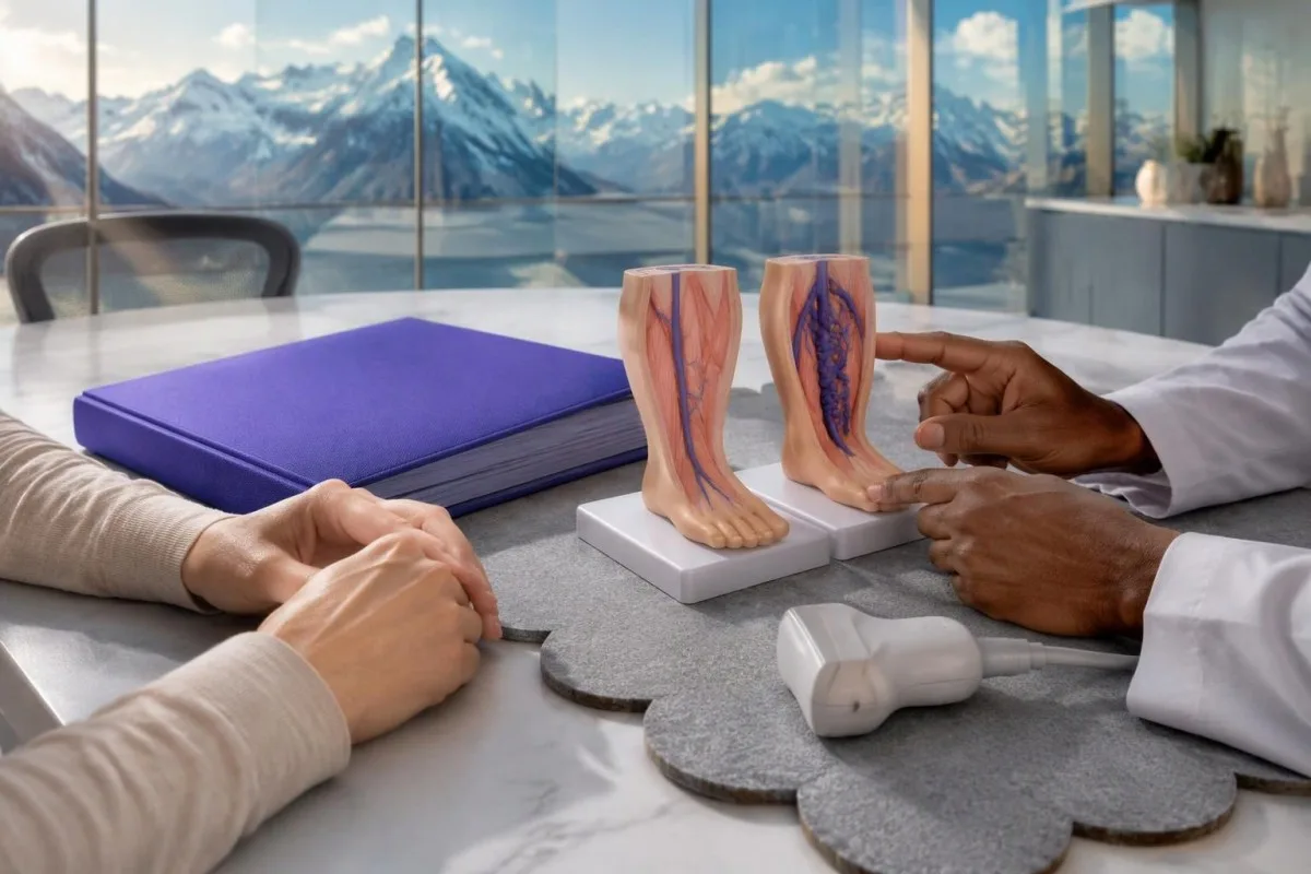
Venous thromboembolism risk is particularly increased during the first 6 weeks after birth, including after an apparently uncomplicated delivery. Normal postpartum fluid shifts tend to affect both legs; a newly enlarged, tender or warm single leg should not be dismissed as routine recovery, especially after cesarean birth or reduced mobility.
D-dimer rises physiologically during pregnancy, which reduces the usefulness of a conventional 500 ng/mL FEU threshold when used in isolation. Some validated pregnancy pathways address suspected PE, but those should not be casually transferred to suspected leg DVT; ultrasound remains central, with repeat or additional imaging when concern persists.
A negative scan does not settle every pregnancy-associated presentation, particularly with whole-leg swelling, groin discomfort or suspected iliac-vein involvement. Tell the maternity or emergency team about the distribution of symptoms and any chest complaints; pregnancy changes the diagnostic approach, not the need to investigate possible pulmonary embolism urgently.
Protein S activity can fall during pregnancy, so a low result is not automatic proof of inherited deficiency. Our explanation of temporary low protein S helps separate background laboratory interpretation from acute diagnosis; estrogen-containing contraception and some hormone therapies also deserve mention when discussing new symptoms in 1 leg.
What should you do while waiting for a DVT assessment?
Arrange assessment promptly, avoid deep calf massage and do not self-start aspirin or anticoagulants. Gentle necessary movement is usually reasonable if you feel well, but strenuous exercise, a long journey or trying to 'walk off' the swelling should wait for clinical advice.
Write down the symptom onset, which leg changed, relevant operations or journeys, and every medication with the time of the last dose. If you already take an anticoagulant, do not double the dose: breakthrough symptoms need assessment because missed doses, interactions, inappropriate dosing or another diagnosis may change the management.
Aspirin is not an adequate substitute for treatment of suspected acute DVT, and adding it to an anticoagulant can increase bleeding risk. Avoid starting leftover medication or borrowed compression garments; a treatment intended for 3 months after a confirmed clot is very different from an unsupervised dose taken before diagnosis.
When imaging is delayed, a clinician may prescribe interim therapeutic anticoagulation, after considering contraindications, kidney function and bleeding risk. NICE recommends baseline CBC, renal and liver function, PT and aPTT, but says not to delay needed interim treatment while waiting for those results; our baseline test preparation guide provides nonurgent preparation context.
Deep massage has no proven role in diagnosing or treating DVT; avoiding it is a precaution, not a claim that every touch dislodges a clot. Keep emergency symptoms in view while waiting: new breathlessness, chest pain, coughing up blood or fainting should trigger an immediate call to 999, 112, 911 or the local emergency number, not the next scheduled appointment.
What happens if a leg clot is confirmed?
Confirmed proximal DVT usually requires anticoagulation for at least 3 months. Anticoagulants prevent extension and new clot formation while the body gradually remodels the existing clot; they do not instantly remove it or guarantee that swelling disappears within days.
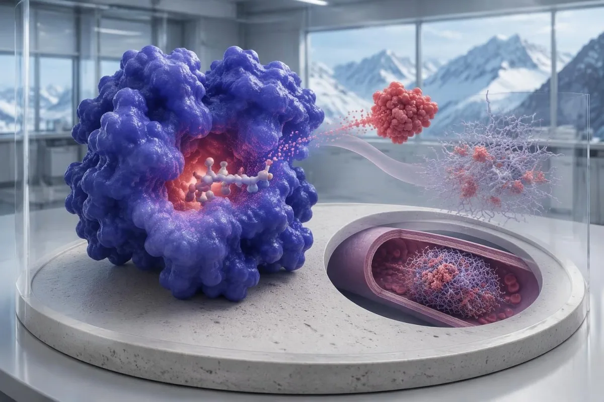
Treatment depends on clot location, pregnancy status, kidney function, cancer, drug interactions and bleeding risk—not just how painful the leg feels. NICE recommends at least 3 months for confirmed proximal DVT; the CHEST guideline distinguishes isolated distal calf DVT, where selected lower-risk patients may undergo serial ultrasound instead of immediate anticoagulation (Stevens et al., 2021).
For selected isolated distal DVT without severe symptoms or risk factors for extension, CHEST suggests serial imaging over 2 weeks; significant symptoms or extension risk can favor anticoagulation. This is a clinician-directed decision with a reliable follow-up plan, not permission to watch a suspected clot at home before its location is established.
Warfarin treatment commonly uses a target INR of 2.0–3.0 for venous thromboembolism, whereas INR does not measure the treatment effect of apixaban or rivaroxaban reliably. Our INR interpretation guide explains why a 'normal INR' means something different depending on the medication.
Anticoagulation can worsen menstrual bleeding, and medication changes require clinical advice rather than skipped doses. Our guide to heavy menstrual bleeding warnings explains when to seek help; vomiting blood, black stools, collapse, a severe new headache or a significant head injury while anticoagulated warrants urgent assessment.
Why can swelling persist after treatment, and what needs rechecking?
Swelling can persist after DVT because venous obstruction and valve damage may outlast the acute clot. New or worsening asymmetry still needs reassessment; persistent symptoms alone do not prove treatment failure, and a repeat D-dimer is not a universal treatment-monitoring test.
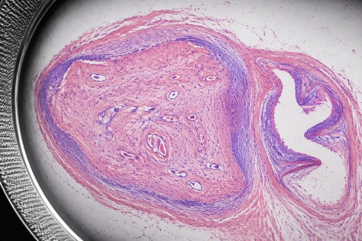
Post-thrombotic symptoms include heaviness, swelling and discomfort that may worsen after standing and improve with elevation. A useful follow-up record compares function and symptom progression over weeks, not repeated hourly calf measurements; abrupt new pain, increased swelling or chest symptoms should be treated as a new clinical problem rather than assumed to be routine recovery.
Compression stockings may help selected patients' symptoms, but guidelines do not recommend them routinely solely to prevent post-thrombotic syndrome after every DVT. The decision depends on fit, tolerance and arterial circulation; a pressure such as 20–30 mmHg is not automatically suitable for someone with significant peripheral arterial disease or an unexplained newly swollen leg.
Kantesti is an AI-powered blood test analysis tool that can help organize follow-up CBC, kidney and liver results after treatment begins. Our medication-safety trend guide explains the workflow, but laboratory trends cannot establish that a clot has resolved or determine whether anticoagulation should stop after 3 months.
Kantesti AI helps explain uploaded reports and their units; clinical decisions still require the medication, dose, symptoms and imaging context. Our AI interpretation technology describes that process, and a reassuring AI explanation must never override worsening symptoms, a prescribed repeat scan or a clinician's anticoagulation plan.
Research publications: which sources support DVT advice?
Clinical DVT guidance should come from directly relevant diagnostic and treatment evidence. This article's clinical pathways draw on NICE NG158, the 2018 ASH diagnostic guideline and the 2021 CHEST treatment guideline; the two additional DOI publications listed below are not DVT evidence.
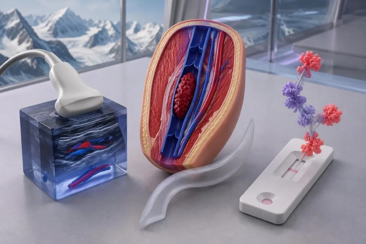
NICE NG158 was published in 2020 and updated in 2023; its general adult DVT pathway supplies the 4-hour imaging target, 24-hour alternative pathway and 6–8-day repeat-scan recommendation. These are service and diagnostic targets, not promises that every country uses an identical pathway or that symptoms become harmless once a certain number of hours passes.
Lim and colleagues' ASH guideline addresses diagnosis, while Stevens and colleagues' CHEST guideline addresses antithrombotic management. Keeping those questions separate prevents a common error: applying a surveillance option for an already-confirmed distal clot to someone whose new one-sided swelling has not yet been investigated.
Kantesti's two repository publications below concern Nipah testing and hematology topics; their DOI identifiers establish retrievable publication records, not peer review or validation of DVT triage. Our medical content standards explain our approach to sourcing, and those unrelated publications should not be used to justify a D-dimer cutoff or an ultrasound decision.
Frequently asked questions
Can you have a DVT without pain or redness?
DVT can occur without obvious pain or redness, and some clots cause few noticeable symptoms. New unexplained swelling in 1 leg still needs same-day assessment, particularly after surgery, immobility or a previous clot. A calf difference below 3 cm does not exclude DVT; that measurement is one clinical scoring criterion rather than a diagnostic boundary. Sudden breathlessness or chest pain requires emergency care even if both legs look normal.
How can I tell whether calf pain is a clot or a pulled muscle?
Symptoms alone cannot reliably distinguish a calf DVT from a muscle strain. New swelling in 1 leg, warmth, deep tenderness or a recent clot risk should prompt same-day assessment even if exercise preceded the pain. A clinician combines history and examination with a probability score, D-dimer when appropriate, and ultrasound. Squeezing the calf, walking normally or pulling the toes upward cannot safely rule out a clot.
Does a normal D-dimer rule out a blood clot in the leg?
A negative high-sensitivity D-dimer can exclude DVT in selected patients with low clinical probability when used within a validated pathway. The common threshold is 500 ng/mL FEU, but age adjustment, assay units and local protocols can change interpretation. Likely DVT generally requires ultrasound, and anticoagulation before sampling or other clinical circumstances may complicate a negative result. A positive D-dimer cannot diagnose DVT on its own.
Can an ultrasound miss a DVT?
An ultrasound can miss a clot if the affected segment is outside the examination area or the study is technically limited. A proximal scan does not necessarily assess every distal calf vein, which is why NICE recommends another proximal scan after 6–8 days when the first is negative and D-dimer is positive. A technically adequate whole-leg scan may lead to a different follow-up plan. Worsening symptoms or whole-leg swelling should be reported before the scheduled repeat scan.
Should I go to the emergency department for one swollen leg?
New unexplained swelling in 1 leg needs same-day assessment, which may be through an urgent clinic or emergency department depending on local access. Sudden breathlessness, chest pain, coughing up blood, fainting or collapse requires an emergency call to 999, 112, 911 or the local emergency number. A rapidly enlarging blue or purple leg, severe pain, numbness, weakness or a cold foot also needs emergency assessment. Do not delay care while waiting for an online interpretation or a routine blood test.
Is one swollen leg normal during pregnancy or after birth?
New one-sided leg swelling during pregnancy or after birth should not be assumed to be normal fluid retention. Clot risk is particularly increased during the first 6 weeks postpartum, and ultrasound is central to investigating suspected leg DVT. The standard adult Wells score and an isolated 500 ng/mL FEU D-dimer threshold should not be applied casually in pregnancy. Chest pain, sudden breathlessness or fainting requires emergency care regardless of whether leg symptoms are present.
Can I take aspirin or massage my calf if I think I have a DVT?
Do not self-treat suspected DVT with aspirin, borrowed anticoagulants or deep calf massage. Aspirin is not an adequate substitute for acute DVT treatment, and combining medicines can increase bleeding risk. When imaging is delayed, a clinician may prescribe interim anticoagulation and arrange ultrasound within 24 hours under an appropriate pathway. If you already take an anticoagulant, do not double a dose; new symptoms still require assessment.
Get your blood test results explained
Upload your lab report and Kantesti's AI explains each biomarker in plain language within seconds. Used by more than 2 million people worldwide.
References
- Klein, T., Mitchell, S., & Weber, H. (2026). Nipah Virus Blood Test: Early Detection & Diagnosis Guide 2026. Kantesti AI Medical Research. DOI
- Klein, T., Mitchell, S., & Weber, H. (2026). B Negative Blood Type, LDH Blood Test & Reticulocyte Count Guide. Kantesti AI Medical Research. DOI
- National Institute for Health and Care Excellence (2020). Venous thromboembolic diseases: diagnosis, management and thrombophilia testing (NG158). NICE guideline.
- Lim W et al. (2018). American Society of Hematology 2018 guidelines for management of venous thromboembolism: diagnosis of venous thromboembolism. Blood Advances. DOI
- Stevens SM et al. (2021). Antithrombotic Therapy for VTE Disease: Second Update of the CHEST Guideline and Expert Panel Report. Chest. DOI
- 2M+Tests analysed
- 127+Countries
- 100+Languages
About the medical team


E-E-A-T Trust Signals
Experience
Physician-led clinical review of lab interpretation workflows.
Expertise
Laboratory medicine focus on how biomarkers behave in clinical context.
Authoritativeness
Medically reviewed by Dr. Thomas Klein, with contributions from Dr. Sarah Mitchell and Prof. Dr. Hans Weber.
Trustworthiness
Evidence-based interpretation with clear follow-up pathways to reduce alarm.





Medical disclaimer
This article is for educational purposes only and does not constitute medical advice. Always consult a qualified healthcare provider for diagnosis and treatment decisions.
Published · Contact the editorial team
Published by Kantesti Ltd, registered in England & Wales, company no. 17090423, London, United Kingdom.
