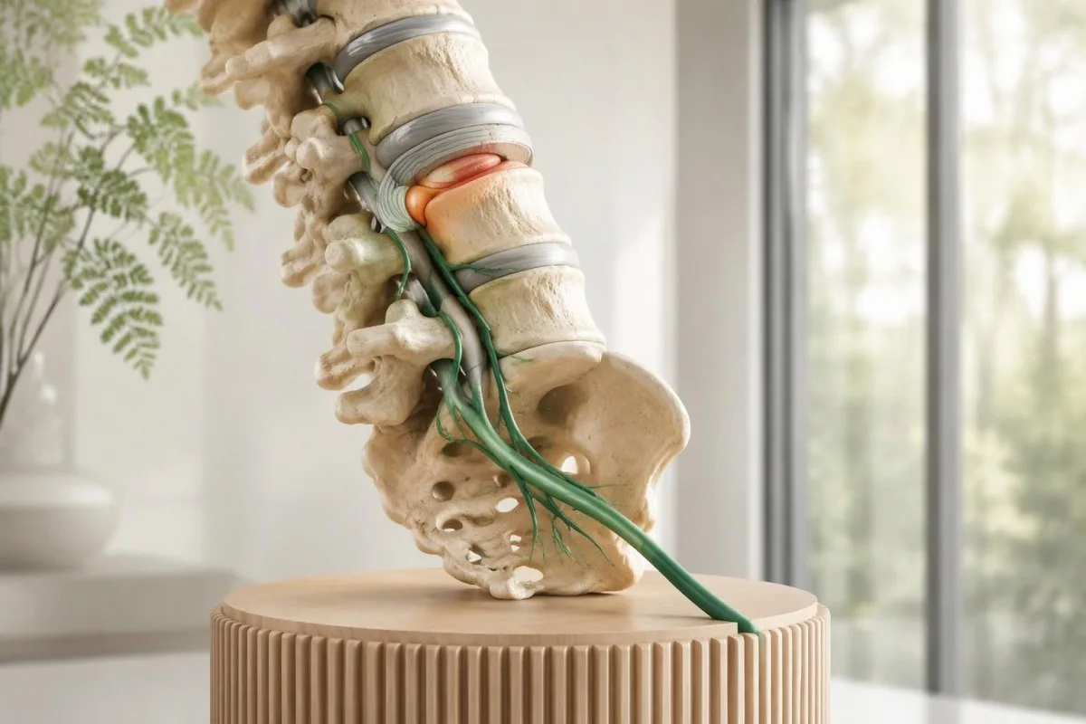
Sciatica usually starts with irritation of a lower-back nerve root, often from a herniated disc or spinal narrowing. Diagnosis depends on the pain pattern and neurological examination—not routine bloodwork—with MRI reserved for warning signs or decisions that could change treatment.

Key takeaways
- Sciatica causes most often involve a lumbar disc herniation or spinal narrowing irritating a nerve root; L5 and S1 roots are commonly involved.
- One-leg pain usually reflects irritation on one side of the spine, but symptoms do not always follow a perfect nerve-territory map.
- Neurological examination assesses strength on a 0–5 scale, sensation, reflexes and walking; worsening objective weakness changes the urgency.
- MRI timing depends on the clinical question: around 6 weeks of persistent symptoms may justify imaging if an injection or operation is being considered.
- Incidental MRI findings are common: a systematic review estimated disc bulges in 30% of asymptomatic 20-year-olds and 84% of asymptomatic 80-year-olds.
- Bloodwork limits mean a complete blood count, CRP, vitamin B12 or HbA1c cannot confirm sciatica; selected tests investigate alternative diagnoses.
- Emergency symptoms include new difficulty starting urination, loss of bladder sensation, saddle numbness or rapidly progressive weakness—even when only one leg hurts.
- Recovery monitoring should track function as well as pain: new foot dragging or loss of toe-standing ability needs reassessment rather than waiting for a scheduled scan.
What causes sciatica at the nerve root?
Sciatica causes usually involve irritation of a lower-back nerve root by a herniated disc, spinal narrowing or both. The irritated root can produce pain down one leg, often through L5 or S1 pathways; diagnosis starts with symptoms and neurological examination, not bloodwork.
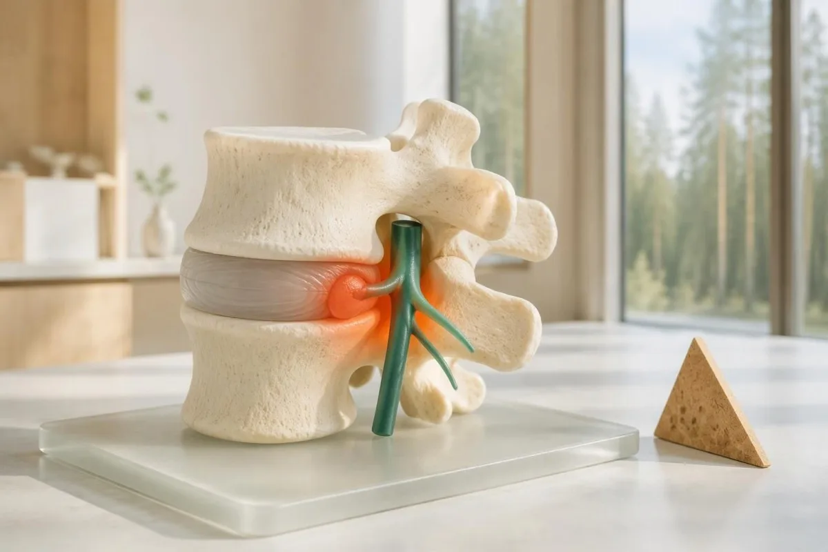
Radicular pain comes from abnormal signalling in a spinal nerve root, whereas radiculopathy means measurable nerve dysfunction, such as weakness, sensory loss or a reduced reflex. A person can have severe radiating pain with normal strength on a 0–5 examination scale; the two terms are not interchangeable.
Mechanical pressure is only part of the explanation: substances released around disc material can sensitise a nearby root without a large visible compression. Consequently, a 3 mm protrusion is not automatically less painful than a 6 mm protrusion, and scan size alone cannot determine symptom severity or treatment.
The sciatic nerve forms from contributions spanning L4 to S3, but most familiar lower-back-related sciatica begins before those contributions join into the peripheral nerve. This distinction explains why clinicians usually examine the lumbar spine first rather than ordering an ultrasound of the sciatic nerve in the thigh.
Kantesti is an AI blood test analyzer that can explain accompanying laboratory results, but no routine laboratory value identifies an irritated L5 or S1 root. Our organization and clinical scope should be understood separately from the hands-on assessment needed for radiating leg pain.
Why does sciatica pain affect one leg?
Sciatica pain in one leg usually occurs because a disc or narrowed nerve exit affects a root on one side of the spine. L5-related symptoms often reach the outer lower leg or top of the foot, while S1 symptoms often reach the calf and outer foot.
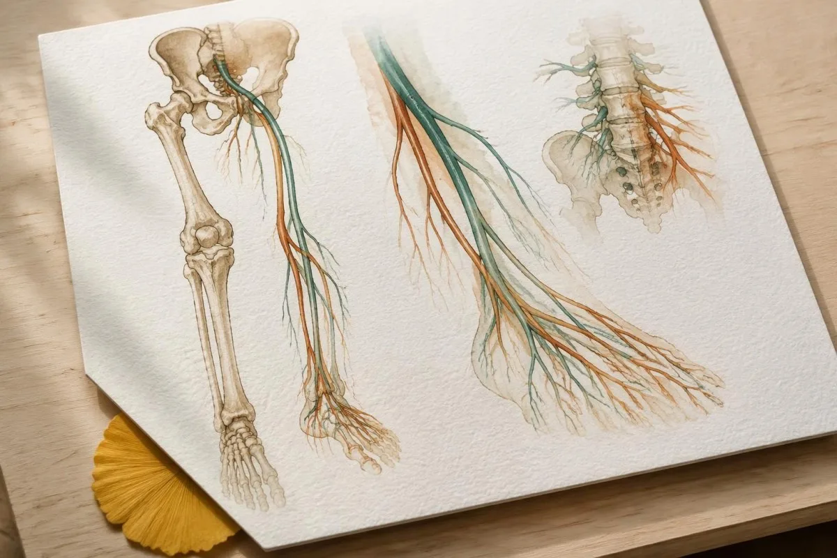
Pain location suggests a starting point, not a definitive diagnosis: L5 irritation can affect great-toe lifting, while S1 irritation can affect pushing down through the foot. These functions matter more than matching every centimetre of pain to a diagram, because adjacent roots share muscle and sensory territories.
A hypothetical 44-year-old with buttock-to-foot pain and difficulty lifting the great toe has a different pattern from someone with isolated outer-thigh numbness. The latter may have meralgia paraesthetica, involving a sensory nerve near the hip, which should not directly weaken the ankle or change the Achilles reflex.
Radiating nerve pain may feel electric, burning, stabbing or deeply aching; back pain can be mild or absent. During the first 2 weeks, noting whether symptoms reach below the knee and whether coughing reproduces familiar leg pain can help the examination, although neither feature proves sciatica alone.
Nerve distribution also helps distinguish a spinal problem from a peripheral nerve problem, much as finger-specific nerve symptoms help localise hand complaints. Bilateral leg symptoms deserve careful assessment, but even a one-sided presentation can accompany an urgent central spinal problem.
How does a herniated disc cause leg pain?
A herniated lumbar disc can cause sciatica when displaced disc material irritates a nearby nerve root. A typical posterolateral L4–L5 herniation affects the traversing L5 root; an L5–S1 herniation commonly affects S1, although far-lateral herniations follow a different anatomical pattern.
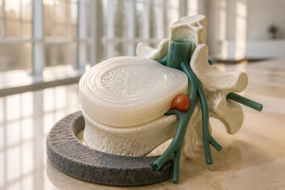
Disc names and root names do not always match, which makes MRI wording confusing. For example, an L4–L5 far-lateral herniation can affect the exiting L4 root rather than L5; an explanation that simply equates every L4–L5 disc finding with L5 symptoms can miss the actual anatomy.
Lifting, twisting or prolonged sitting may trigger an episode, but an identifiable injury is not required. In a hypothetical 38-year-old whose symptoms began after lifting a suitcase, the event suggests timing rather than proof that one movement created the entire disc change; degeneration may have developed beforehand.
Pain can improve while the disc remains abnormal on MRI, because nerve sensitivity and local tissue response change over time. Repeating a scan after 2 weeks solely to check whether a protrusion has disappeared usually does not answer a useful treatment question if strength and function are improving.
New back pain after a fall raises a different concern, particularly in someone aged 70 or taking long-term corticosteroids. A vertebral fracture can coexist with leg symptoms, so bone-loss investigations address fracture susceptibility—not whether a nerve root is currently irritated.
When does spinal narrowing cause sciatica?
Lumbar spinal stenosis can cause radiating leg symptoms when narrowing reduces space around nerve roots. Central narrowing often causes walking-related discomfort in one or both legs, while foraminal narrowing may affect a single exiting root; symptoms can improve with sitting or bending forward.
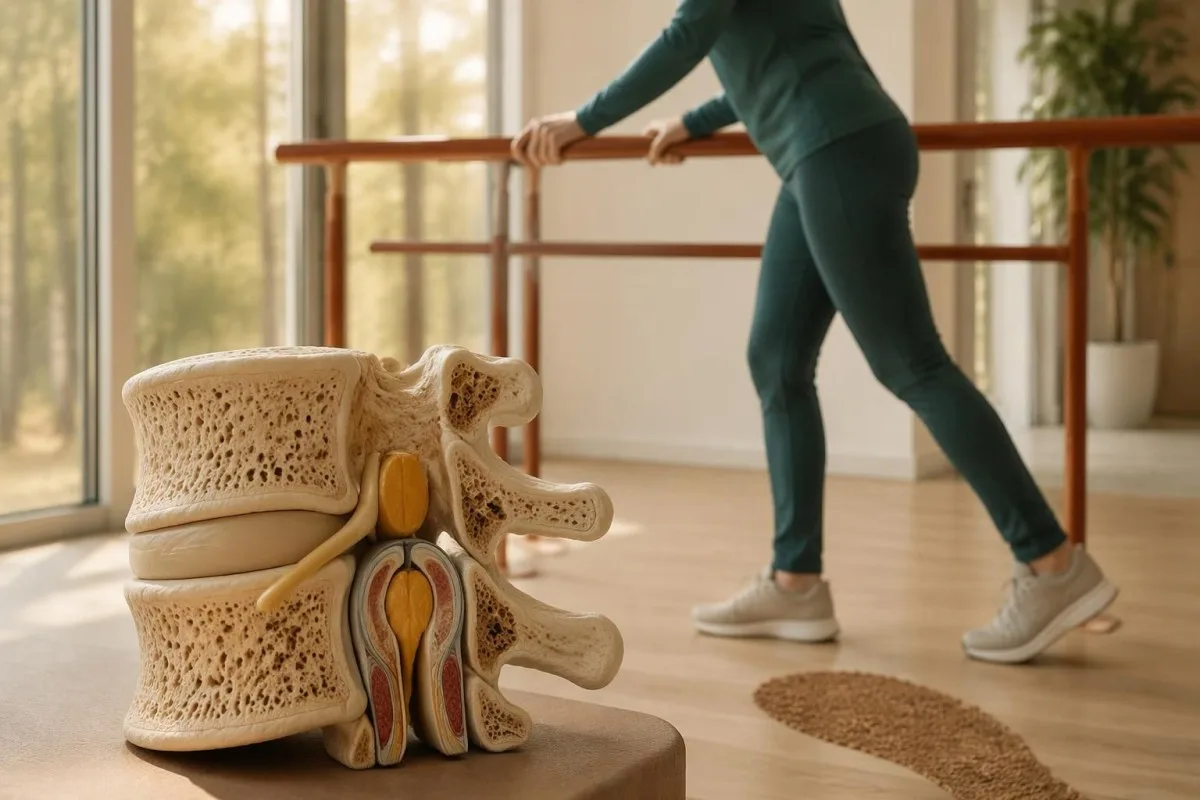
The characteristic walking pattern is called neurogenic claudication, but there is no single walking-distance cutoff that establishes it. A hypothetical person who develops leg heaviness after 150 metres yet cycles comfortably provides a useful clue: spinal position may matter more than the cardiovascular effort alone.
Standing upright can reduce available space in some narrowed spinal segments, whereas forward bending may increase it. That helps explain the shopping-trolley pattern, but improvement while leaning forward is not a stand-alone diagnostic test; hip conditions and general conditioning can also influence walking tolerance.
Anatomical stenosis on MRI does not always cause symptoms, especially in older adults with several degenerative findings. A scan reporting 2 narrowed levels should be interpreted alongside the side of symptoms, walking limitation, neurological findings and whether treatment directed at either level would plausibly improve function.
Vascular claudication is an alternative explanation when exertional calf pain improves simply by stopping, without needing to sit or bend. Our guide to leg circulation warning signs explains why foot pulses and, when appropriate, an ankle–brachial index around or below 0.90 help investigate arterial disease rather than sciatica.
Which conditions commonly mimic sciatica?
Hip joint disease, lateral-hip tendon pain, peripheral nerve entrapment and vascular problems can mimic sciatica. A careful assessment separates pain referred from muscles or joints from root-related pain; no single symptom, including pain below the knee, reliably distinguishes every alternative.
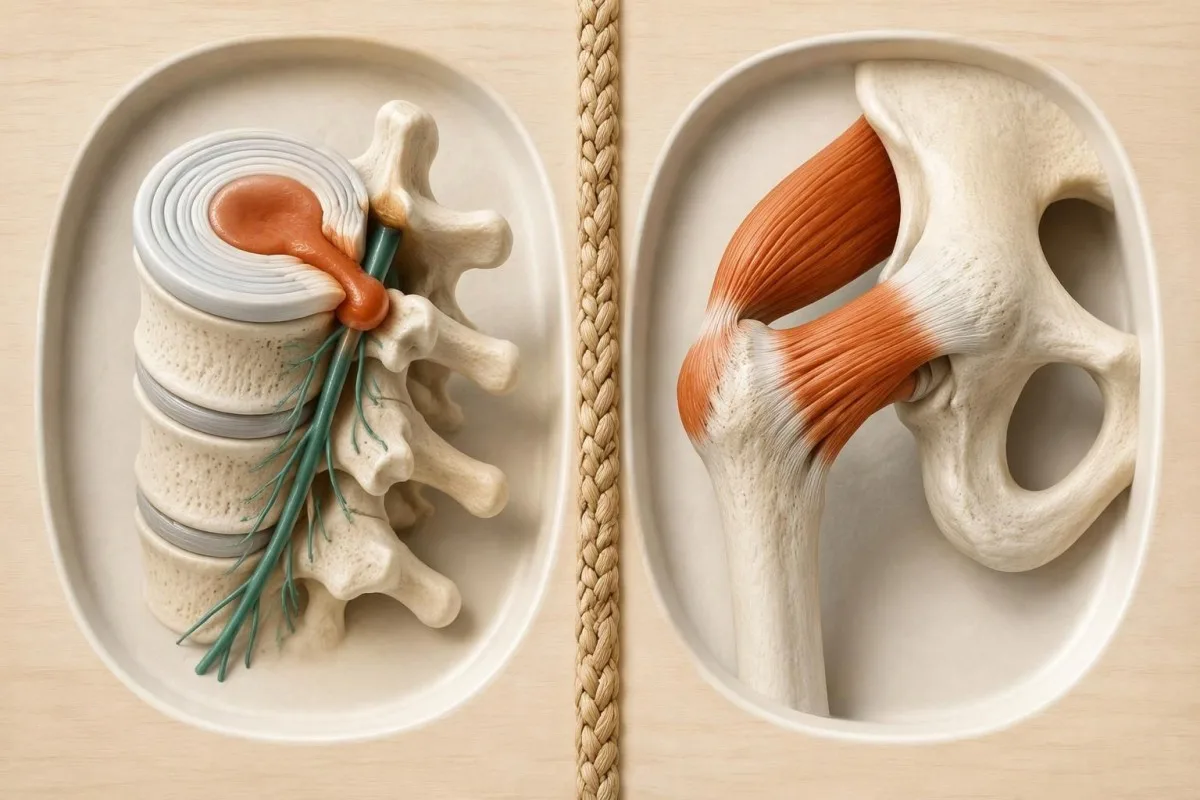
Hip osteoarthritis often produces groin pain, reduced hip rotation and sometimes pain reaching the knee. Lateral-hip tendon pain commonly worsens when lying on that side or climbing stairs; these 2 patterns direct attention toward the hip examination rather than automatically treating every buttock-to-thigh symptom as sciatica.
Deep gluteal syndrome describes sciatic nerve irritation outside the spine, sometimes associated with structures near the piriformis muscle. The evidence for individual provocative tests is mixed, and buttock tenderness alone does not establish the diagnosis; a hypothetical 35-year-old with sitting-related symptoms still needs spinal and peripheral nerve assessment.
Burning pain can precede a shingles rash by several days, making an early episode resemble nerve-root pain. Symptoms before a shingles rash become particularly relevant when light touch is unusually uncomfortable across one band of skin, but absence of a rash on day 1 does not confirm either diagnosis.
Medication-related muscle symptoms usually differ from a single-root pattern, often involving broader aching or proximal weakness. Someone taking a statin who develops pain in both thighs should discuss medication timing and examination findings; statin-related muscle risk cannot be translated into a diagnosis of L5 or S1 irritation.
How is sciatica diagnosed on examination?
Sciatica is usually diagnosed clinically through the symptom history and a focused neurological examination. Clinicians compare both legs for muscle strength, sensation, reflexes and walking; strength is commonly documented on a 0–5 scale, where 5 represents normal power and 0 means no visible contraction.
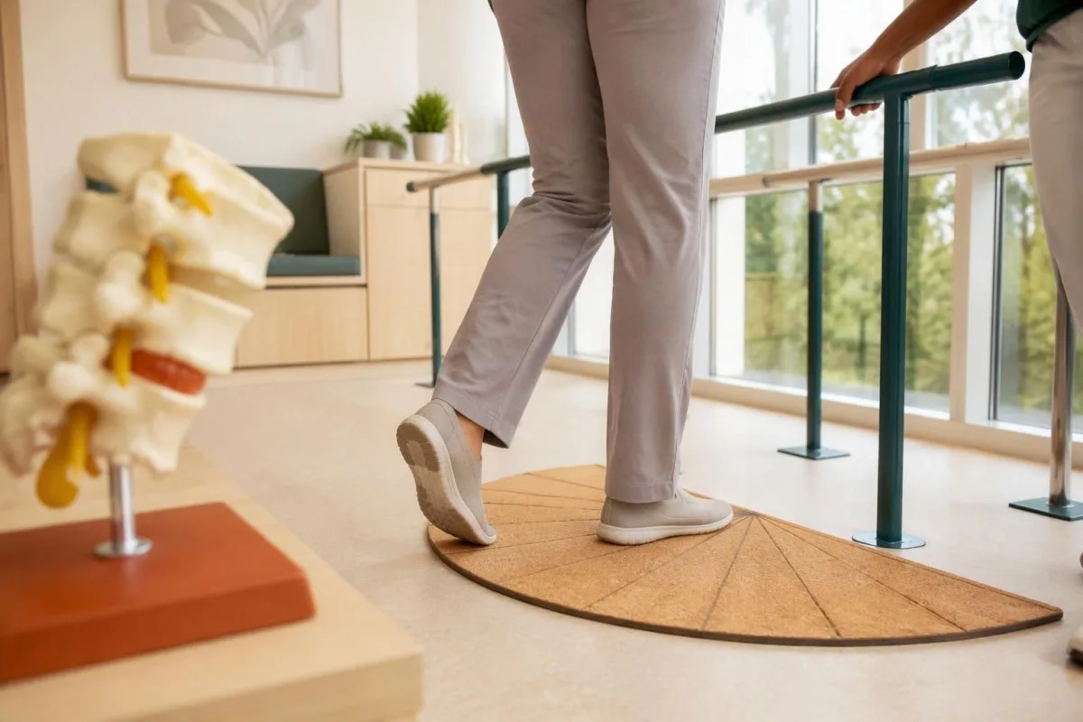
A root-focused examination checks functions rather than relying on pain intensity: knee extension can provide information about L3–L4, great-toe lifting about L5 and ankle push-off about S1. The patellar reflex mainly assesses L3–L4 pathways, while the Achilles reflex mainly assesses S1; overlap prevents perfect one-test localisation.
A grade of 4/5 means movement against resistance is reduced, while 3/5 generally means movement against gravity but not added resistance. Pain, fear and effort can affect the result, so a clinician interprets the grade alongside repeated testing and other findings rather than treating one uncertain measurement as permanent nerve damage.
For example, a hypothetical 56-year-old with unchanged pain but new foot dragging has a more concerning change than someone whose pain rose from 4/10 to 7/10 while walking and ankle strength stayed normal. Progressive objective weakness can require urgent assessment even when the pain itself seems tolerable.
Weakness spreading across both legs over 1–3 days, particularly with reduced reflexes or upper-body symptoms, is not a typical uncomplicated single-root episode. Rapidly developing weakness signs explain another urgent neurological pathway; a normal routine laboratory panel should never delay that assessment.
What do straight-leg raising and EMG add?
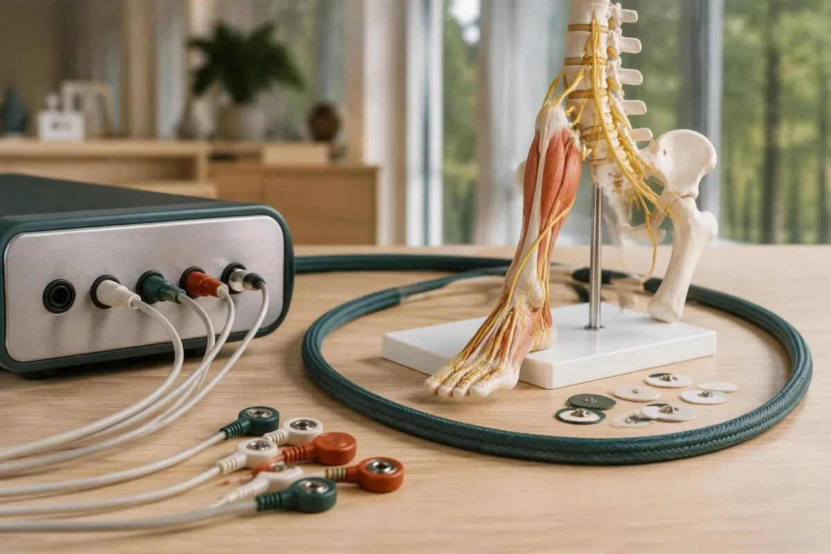
The straight-leg raise helps assess lower lumbar nerve-root irritation when it reproduces the person's familiar radiating leg pain, often between approximately 30° and 70° of elevation. EMG and nerve-conduction studies can help distinguish root dysfunction from peripheral neuropathy when the diagnosis remains uncertain.
A stretching sensation behind the thigh is not automatically a positive straight-leg raise. Familiar pain travelling below the knee carries a different meaning from isolated hamstring tightness, and pain produced only near 90° can be less characteristic; clinicians may modify ankle position to assess whether neural tension contributes.
A crossed straight-leg raise occurs when lifting the less symptomatic leg reproduces pain in the opposite leg. It can strengthen suspicion of disc-related root irritation, but neither this test nor the standard version identifies the exact disc level without the rest of the assessment.
EMG evaluates electrical activity in muscles, while nerve-conduction studies assess peripheral nerve responses. For suspected axonal radiculopathy, testing after roughly 3–4 weeks may reveal findings not yet apparent very early, although useful timing varies by muscle and question; urgent weakness must not wait for an optimal testing window.
Normal EMG does not exclude pain-only root irritation, because the test is better suited to detecting motor axon dysfunction than pain generation. Persistent symmetric numb feet may instead prompt evaluation for antibody-associated peripheral neuropathy, especially when examination and nerve-conduction findings do not fit a single L5 or S1 root.
When is an MRI useful for sciatica?
Lumbar MRI is useful when its result could change management, or when symptoms suggest an urgent spinal condition. Uncomplicated new sciatica usually does not need immediate imaging; persistent symptoms after about 6 weeks may justify MRI if an injection, operation or another targeted intervention is being considered.
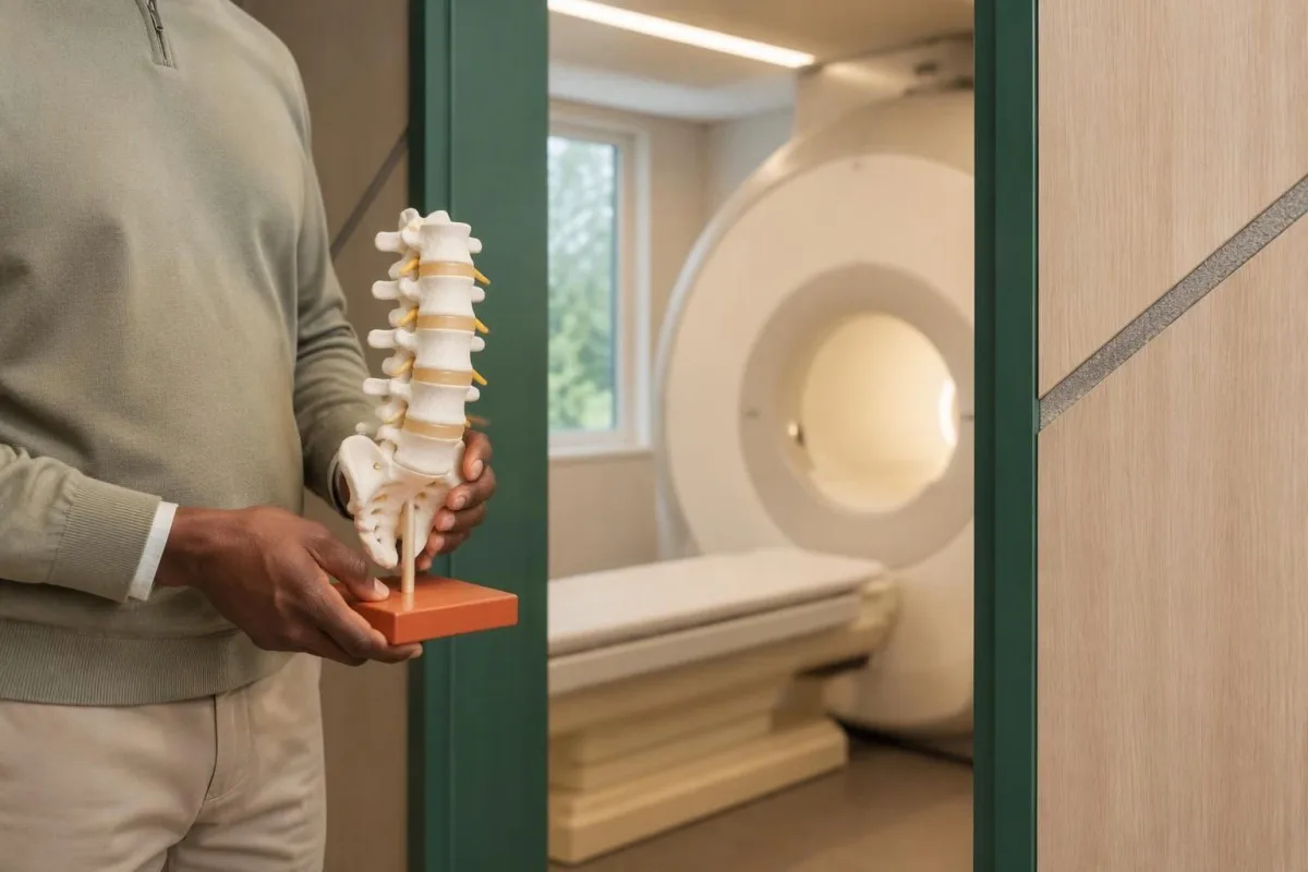
NICE guideline NG59 advises against routinely offering imaging in non-specialist settings and recommends specialist imaging only when the result is likely to change management (NICE, 2016; amended 2020). The rule is about clinical usefulness, not a blanket requirement to wait 6 weeks when warning signs are present.
The ACR Appropriateness Criteria: Low Back Pain—2021 Update supports lumbar MRI for persistent or progressive symptoms after about 6 weeks of appropriate management in intervention candidates. The same guideline treats suspected cauda equina syndrome differently: urgent imaging is appropriate rather than completing a routine conservative-care period.
Most straightforward disc-related investigations use MRI without contrast, whereas suspected spinal immune response, malignancy or certain postoperative questions may require a different protocol. An ordinary X-ray shows bones and alignment but cannot directly establish soft-tissue nerve-root irritation, and CT may be considered when MRI is unsuitable.
Kantesti's methodology and validation scope concerns laboratory interpretation, not lumbar MRI diagnosis or neurological examination. A hypothetical person with 2 days of worsening foot weakness should obtain urgent clinical assessment rather than seeking reassurance from either an AI laboratory explanation or an already scheduled outpatient scan.
Why can an abnormal MRI fail to explain pain?
An abnormal lumbar MRI does not automatically explain sciatica, because age-related disc findings are common in people without pain. The strongest explanation matches 3 elements: the symptomatic side, a plausible nerve-root level and examination findings compatible with that root.
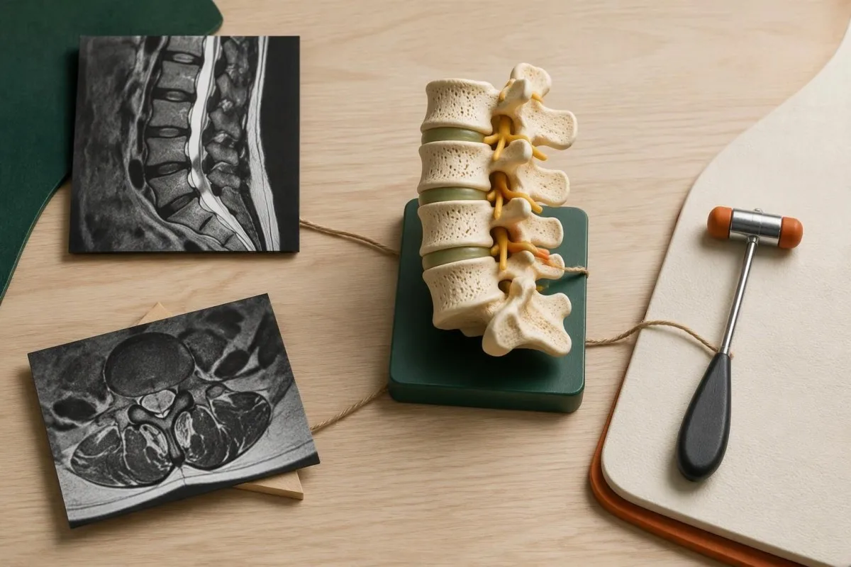
Brinjikji et al. estimated disc bulges in 30% of asymptomatic 20-year-olds and 84% of asymptomatic 80-year-olds in their 2015 AJNR systematic review. Disc protrusions were estimated in 29% and 43%, respectively; these were pooled age-specific estimates, not the probability that one individual's finding is harmless.
A hypothetical MRI showing a right-sided L5–S1 protrusion does not neatly explain left-sided foot symptoms. The mismatch should prompt reconsideration of the images, examination and alternatives rather than assuming any abnormal disc is responsible; anatomical variations and multiple abnormalities can make the answer less straightforward.
Words such as contact, displacement and compression describe different appearances, but reporting conventions vary between radiologists. A report mentioning contact at 2 levels needs clinical interpretation; neither the number of findings nor the strongest-sounding word substitutes for evidence that the corresponding root is actually causing symptoms.
When reading an AI-assisted report explanation, keep the original wording and distinguish observations from inferred diagnoses. Asking which finding matches the affected leg, and which examination result supports that match, is usually more useful than asking whether a 4 mm protrusion is universally dangerous.
Why can't routine bloodwork diagnose sciatica?
Routine bloodwork cannot diagnose or exclude sciatica because it does not measure local nerve-root irritation. A complete blood count, CRP, vitamin B12 and HbA1c can investigate selected alternative or accompanying conditions, but none confirms that an L5 or S1 root is responsible for leg pain.

A normal inflammatory marker does not demonstrate that a nerve root is functioning normally. Many laboratories use a CRP reference limit near 5 mg/L, but an individual can have disabling disc-related sciatica below that value; conversely, CRP of 12 mg/L may reflect another process entirely.
Kantesti is an AI blood test interpretation platform that explains measured results without treating a normal panel as proof against sciatica. If symptoms involve both feet or balance problems, borderline vitamin B12 interpretation may help investigate an alternative; the laboratory question differs from identifying a single irritated root.
Raised white-cell counts and CRP can support an investigation for systemic or spinal infection when the history makes it plausible, but normal results do not reliably exclude every serious cause. Our explanation of WBC and CRP disagreement shows why 2 apparently conflicting markers require context rather than a simple positive-or-negative answer.
HLA-B27 testing addresses suspected inflammatory spinal disease, not mechanical sciatica. Persistent back stiffness improving with movement over more than 3 months, especially with uveitis or other suggestive features, creates a different question; HLA-B27 and back pain explains why even a positive result is not a diagnosis by itself.
Which blood tests help investigate alternative causes?
Targeted laboratory tests help when the history suggests a problem beyond a single nerve root, such as diabetes, vitamin B12 deficiency, systemic inflammation or a protein-related neuropathy. Test selection follows the examination; HbA1c of 6.5%—48 mmol/mol—is a diabetes threshold, not a sciatica threshold.
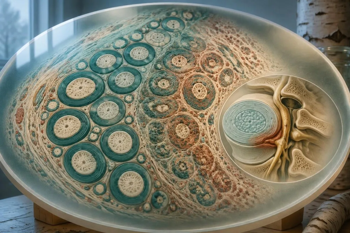
An HbA1c at or above 6.5% generally needs confirmation when there is no unequivocal hyperglycaemia, and it does not establish the cause of leg symptoms. High-glucose symptom patterns help separate diabetes-related clues from a root pattern; diabetes can coexist with disc disease or cause a different painful asymmetric neuropathy.
A vitamin B12 result below approximately 200 pg/mL, equivalent to 148 pmol/L, raises concern for deficiency, although assays and diagnostic thresholds differ. Borderline results may lead to methylmalonic acid testing, but impaired kidney function can elevate that marker independently; normal haemoglobin does not reliably exclude neurological B12 deficiency.
Protein studies are considered selectively for otherwise unexplained peripheral neuropathy rather than ordered for every person with 1 painful leg. Immunofixation result interpretation explains why a monoclonal finding needs its own assessment and does not automatically prove that a detected protein caused the neurological symptoms.
Kantesti AI can explain kidney markers accompanying medication review, but an eGFR below 30 mL/min/1.73 m² changes analgesic safety considerations rather than identifying a compressed root. Our kidney function ratio guide addresses renal interpretation; no BUN-to-creatinine ratio diagnoses sciatica.
Which sciatica warning signs need urgent care?
New bladder dysfunction, saddle numbness or rapidly progressive leg weakness requires emergency assessment when it occurs with back or radiating leg symptoms. These findings can indicate cauda equina compression; do not wait for symptoms in both legs, complete incontinence or a routine 6-week review.
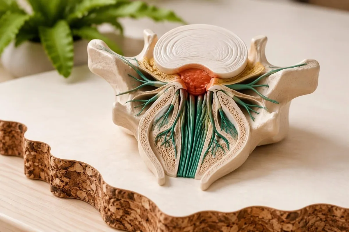
Bladder warning signs include new difficulty starting urination, inability to empty, reduced awareness of bladder filling or new leakage associated with retention. Saddle numbness means altered sensation around the inner thighs, genital region or anus; even 1 new symptom in a concerning clinical pattern warrants urgent assessment rather than a home diagnostic checklist.
A bladder scan may help the emergency assessment, but no single residual-volume cutoff safely rules out cauda equina syndrome. New urinary dysfunction accompanying leg symptoms must not be dismissed as a urinary infection solely from a dipstick; our urinalysis interpretation guide concerns urine findings, not exclusion of spinal compression.
Rapidly worsening foot drop, inability to walk safely, fever with significant spinal pain, or neurological symptoms after substantial trauma also need urgent evaluation. A history of cancer, immunosuppression or recent spinal procedures lowers the threshold for assessment; isolated night pain or an age above 50 is less specific than a concerning combination.
A suddenly cold, pale or severely painful leg suggests impaired arterial supply rather than routine sciatica, while a swollen warm calf needs assessment for a clot. New leg weakness with face or arm weakness, speech difficulty or imbalance follows the stroke emergency warning pathway—even if symptoms began only 10 minutes ago.
What helps recovery while symptoms are monitored?
Staying active within tolerable limits and monitoring neurological function are usually more useful than prolonged bed rest for uncomplicated sciatica. The activity plan should adapt to symptoms and ability; lack of improvement around 6 weeks warrants reassessment, while new weakness or bladder symptoms requires earlier action.
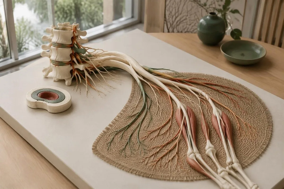
Short, repeatable activity periods can be easier to tolerate than a single long session—for example, 5 minutes of walking several times daily in a hypothetical starting plan. There is no universal exercise dose, and a sustained increase in radiating symptoms or new weakness should prompt adjustment and clinical advice rather than pushing through.
Pain relief and neurological recovery are different outcomes: a lower pain score does not establish restored strength. Recording walking tolerance, foot catching and ability to manage stairs over 7 days can reveal a useful trend, but repeated strenuous heel or toe tests are not a substitute for reassessment when function deteriorates.
NICE NG59 advises against offering gabapentinoids, other antiepileptics, oral corticosteroids or benzodiazepines for sciatica because overall benefit is lacking and harms exist; it also advises against opioids for chronic sciatica. NSAIDs require caution and individual risk assessment, with the lowest effective dose for the shortest suitable period rather than a fixed 14-day course for everyone.
Kantesti does not recommend supplements as a way to decompress a nerve root, and high-dose vitamin B6 can itself cause neuropathy. Our B-complex safety guidance explains that risk; responsible AI boundaries also mean no automated laboratory explanation should override new L5 or S1 motor weakness.
A safe testing plan and related research publications
The safest testing plan begins with urgency assessment, then clinical examination, then tests chosen to answer a specific question. For uncomplicated symptoms, a planned review over the next 2–6 weeks can track progress; suspected cauda equina compression or progressive weakness bypasses that routine timeline.
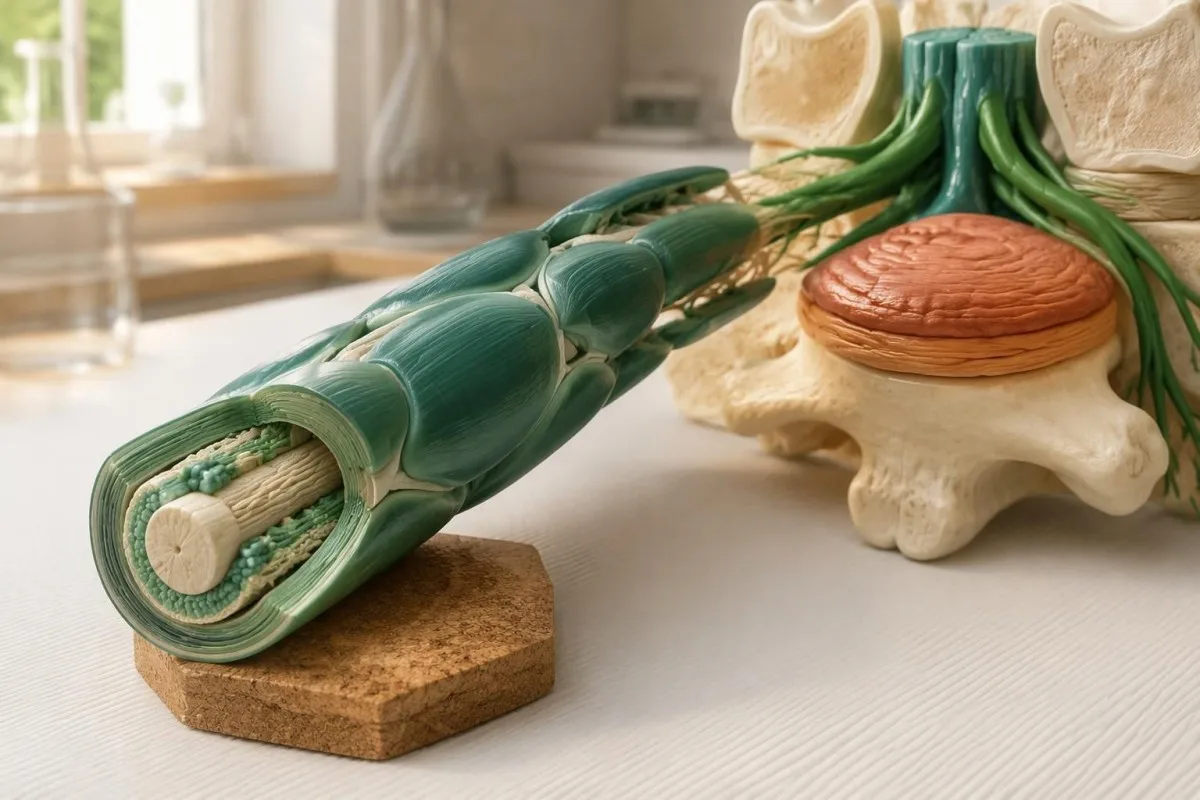
Bring a short symptom map to the appointment: affected side, where pain travels, any numbness, changes in strength and new urinary or bowel symptoms. If an MRI shows 3 abnormalities, ask which one matches the examination and what decision would change because of the finding; an extra scan is useful only when it answers something actionable.
Kantesti is an AI-powered blood test analysis tool for interpreting laboratory reports, not a replacement for assessment of one-leg neurological symptoms. Our medical content standards support keeping these boundaries explicit; 1 reassuring laboratory panel cannot rule out nerve-root irritation or a time-sensitive spinal condition.
The 2 related publications listed below explain kidney-function ratios and urinalysis, which may be relevant to medication safety or a separate urinary investigation. They are educational materials hosted on Zenodo, not clinical trials of sciatica diagnosis, and their DOI registration should not be interpreted as evidence of neurological diagnostic validation.
For the clinical recommendations, this article relies on NICE NG59, the 2021 ACR imaging criteria and Brinjikji et al.'s 2015 review of asymptomatic spinal imaging. These 3 sources answer different questions—when imaging helps, how urgent presentations differ and why incidental findings require context—rather than providing a laboratory cutoff for sciatica.
Frequently asked questions
What causes sciatica pain in one leg?
Sciatica pain in one leg usually results from irritation of a nerve root on one side of the lower back, commonly by a herniated disc or spinal narrowing. L5 and S1 roots are frequent sources of symptoms extending toward the foot. Hip, tendon, peripheral nerve and circulation problems can produce similar pain, so the distribution alone does not confirm sciatica. A neurological examination helps determine whether weakness, sensory change or reflex findings support a root-related explanation.
Can a blood test diagnose sciatica?
No routine blood test can diagnose or exclude sciatica because laboratory panels do not measure local nerve-root irritation. A CRP below a laboratory limit such as 5 mg/L can coexist with substantial disc-related leg pain. Vitamin B12, HbA1c or inflammatory markers may be useful when the examination suggests an alternative or accompanying condition. Normal results must not delay urgent assessment of new bladder dysfunction or progressive weakness.
How is sciatica diagnosed without an MRI?
Sciatica is commonly diagnosed from the history and a neurological examination, without an immediate MRI. Clinicians assess strength on a 0–5 scale, compare sensation and reflexes, and may use straight-leg raising to reproduce familiar radiating pain. No single examination manoeuvre establishes every case, and a normal strength examination does not exclude pain-only root irritation. MRI becomes useful when warning signs are present or its result could change treatment.
When should I get an MRI for sciatica?
Lumbar MRI is generally considered when persistent symptoms could lead to a targeted intervention, often after about 6 weeks of appropriate initial management. NICE recommends specialist imaging only when the result is likely to change management, rather than routinely scanning every new episode. New bladder dysfunction, saddle numbness or rapidly progressive weakness requires urgent assessment and potentially urgent imaging. The 6-week period is not a waiting rule for emergencies.
Does a disc bulge on MRI prove I have sciatica?
A disc bulge on MRI does not by itself prove that the disc is causing sciatica. Brinjikji et al.'s 2015 systematic review estimated disc bulges in 30% of asymptomatic 20-year-olds and 84% of asymptomatic 80-year-olds. The finding becomes more persuasive when its side and nerve-root level match the symptoms and examination. A mismatched scan should prompt clinical reconsideration rather than automatic treatment of the most conspicuous abnormality.
Which sciatica symptoms are an emergency?
New difficulty urinating, reduced bladder sensation, saddle numbness or rapidly progressive leg weakness with back or radiating leg symptoms requires emergency assessment. Cauda equina compression can present without complete incontinence and without pain in both legs. Even 1 new concerning bladder or saddle-sensation symptom should not be left until a routine follow-up. A suddenly cold or pale leg, or weakness with face or speech changes, may represent another emergency rather than sciatica.
Get your blood test results explained
Upload your lab report and Kantesti's AI explains each biomarker in plain language within seconds. Used by more than 2 million people worldwide.
References
- Klein, T., Mitchell, S., & Weber, H. (2026). BUN/Creatinine Ratio Explained: Kidney Function Test Guide. Kantesti AI Medical Research. DOI
- Klein, T., Mitchell, S., & Weber, H. (2026). Urobilinogen in Urine Test: Complete Urinalysis Guide 2026. Kantesti AI Medical Research. DOI
- National Institute for Health and Care Excellence (2016). Low back pain and sciatica in over 16s: assessment and management. NICE guideline NG59.
- Expert Panel on Neurological Imaging (2021). ACR Appropriateness Criteria Low Back Pain: 2021 Update. Journal of the American College of Radiology. DOI
- Brinjikji W et al. (2015). Systematic Literature Review of Imaging Features of Spinal Degeneration in Asymptomatic Populations. American Journal of Neuroradiology. DOI PubMed
- 2M+Tests analysed
- 127+Countries
- 100+Languages
About the medical team


E-E-A-T Trust Signals
Experience
Physician-led clinical review of lab interpretation workflows.
Expertise
Laboratory medicine focus on how biomarkers behave in clinical context.
Authoritativeness
Medically reviewed by Dr. Thomas Klein, with contributions from Dr. Sarah Mitchell and Prof. Dr. Hans Weber.
Trustworthiness
Evidence-based interpretation with clear follow-up pathways to reduce alarm.




Medical disclaimer
This article is for educational purposes only and does not constitute medical advice. Always consult a qualified healthcare provider for diagnosis and treatment decisions.
Published · Contact the editorial team
Published by Kantesti Ltd, registered in England & Wales, company no. 17090423, London, United Kingdom.
