
A positive result usually suggests fetal red cells have entered the mother's circulation. The estimated volume helps clinicians calculate extra Rh immune globulin when indicated, but maternal fetal hemoglobin can make the result misleading.

Key takeaways
- Positive result means the stain detected hemoglobin-containing, acid-resistant cells; these usually represent fetal red cells, but maternal HbF-containing cells can also stain.
- Two different questions require separate assessment: how much fetal blood entered maternal circulation, and whether the fetus has developed anemia.
- Volume estimate often uses fetal-cell percentage × 50 to estimate fetal whole blood in mL, assuming a maternal blood volume of 5,000 mL.
- Rh immune globulin at 300 micrograms, equivalent to 1,500 IU, typically covers up to 30 mL of fetal whole blood or 15 mL of fetal red cells.
- A 0.8% result corresponds to approximately 40 mL of fetal whole blood under the 5,000 mL assumption; additional prophylaxis may be needed for an eligible RhD-negative mother.
- Maternal HbF can rise with pregnancy, hemoglobin disorders, or HbF-increasing treatment; an HbF percentage is not the same measurement as a fetal-cell percentage.
- Flow cytometry may clarify an unexpectedly large or uncertain result, especially when a validated two-marker method distinguishes fetal cells from maternal F cells.
- Urgent assessment is needed for reduced fetal movement, significant abdominal trauma, vaginal bleeding, or severe abdominal pain; do not wait for a test result or the usual 72-hour prophylaxis window.
What does a positive Kleihauer-Betke result actually detect?
A Kleihauer-Betke test positive result usually indicates fetal red cells in maternal blood, suggesting fetomaternal hemorrhage. However, the stain detects cells retaining fetal hemoglobin, not fetal identity itself; clinicians combine the percentage, clinical circumstances, and maternal HbF history before using the result to guide Rh immune globulin.

The Kleihauer-Betke test uses acid elution to remove most adult hemoglobin from a prepared maternal blood film. Cells rich in hemoglobin F, or HbF, retain more stain, while most adult cells become pale ghosts; a result of 0.2% describes the proportion counted as fetal-type cells, not the mother's overall HbF concentration.
A positive Kleihauer-Betke test does not establish that the mother has developed anti-D antibodies. Cell transfer and immune sensitization are 2 different processes, so clinicians also review blood group and the antibody screen; our explanation of a pregnancy antibody screen describes that separate assessment.
The word hemorrhage describes blood moving from fetal circulation into maternal circulation, not necessarily visible vaginal bleeding. A hypothetical 5 mL transfer can occur without an external sign, while an acute transfer that is large relative to the fetus's circulation may cause fetal anemia even when the mother feels well.
Kantesti is an AI blood test analyzer that can help explain a reported fetal-cell percentage such as 0.2%, but it cannot verify the cells' origin or authorize an anti-D dose. The decisive information comes from the laboratory method, transfusion team, and obstetric assessment—not a positive flag alone.
How do fetal cells enter maternal blood?
Fetal cells enter maternal blood when they cross the placental interface, either through small transfers during pregnancy and birth or after disruption associated with trauma, procedures, or placental complications. A detectable transfer is not automatically a large or dangerous hemorrhage; 1 result cannot reliably identify its cause.
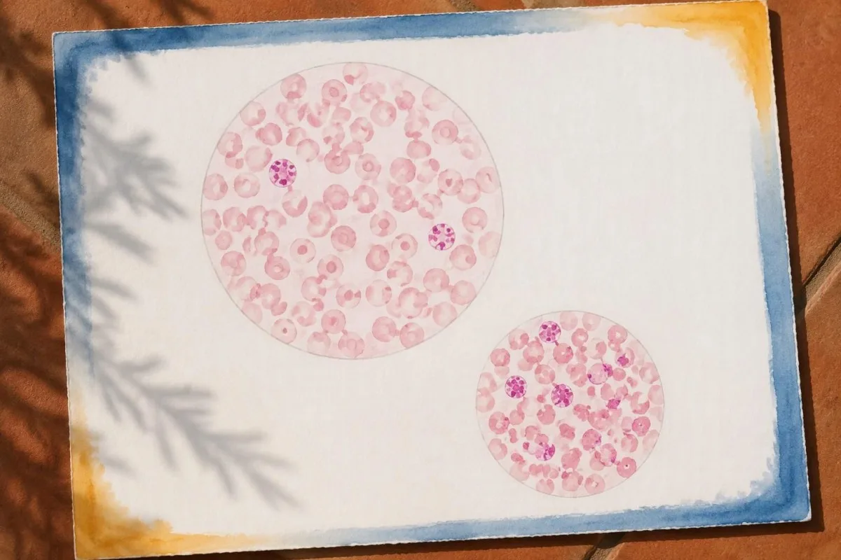
Fetomaternal hemorrhage can follow delivery, abdominal trauma, external cephalic version, or invasive pregnancy procedures. After 20 weeks, many UK protocols add fetomaternal hemorrhage testing to anti-D management after potentially sensitizing events, whereas earlier-pregnancy prophylaxis decisions follow different, event-specific rules.
Some clinically significant fetomaternal hemorrhages have no obvious precipitating event. A hypothetical positive result at 32 weeks without trauma therefore still deserves interpretation; the absence of an accident does not disprove cell transfer, and the assay does not prove that the placenta has a particular structural problem.
Fetal fibronectin and fetal red-cell testing answer different questions, despite the shared word fetal. The former assesses preterm-birth risk in selected circumstances, while the latter estimates cell transfer; our guide to a positive fetal fibronectin result explains why neither test substitutes for the other.
The consequences of fetal blood loss depend on both volume and speed. A hypothetical 20 mL loss over several days allows a different physiological response from 20 mL lost rapidly, so clinicians assess fetal movement, heart-rate patterns, and anemia rather than assigning severity from the maternal percentage alone.
When is fetomaternal hemorrhage testing ordered?
Fetomaternal hemorrhage testing is ordered to estimate fetal-cell exposure for anti-D prophylaxis or to investigate suspected fetal blood loss. Common settings include delivery, significant pregnancy trauma, and unexplained fetal anemia; after birth, many protocols collect the maternal sample at least 30 minutes later to allow mixing.

Postpartum sampling commonly occurs approximately 30 minutes to 2 hours after delivery, although the local laboratory specifies its accepted interval. A sample taken immediately at birth may not yet represent well-mixed circulation, and a later specimen can be affected by cell clearance or intervening treatment.
Rh immune globulin is generally administered as soon as practical and within 72 hours of an eligible sensitizing event or delivery. Collecting a specimen before prophylaxis is often preferred, but arranging the assay must not unnecessarily delay indicated treatment; Qureshi et al. (2014) emphasize prompt prophylaxis within a structured laboratory pathway.
The laboratory needs the event time, gestational age, maternal RhD type, and details of any recent anti-D or transfusion. There is usually no fasting requirement for this stain, unlike some metabolic tests; our first-draw preparation guide helps separate routine preparation from the time-sensitive details of pregnancy testing.
Antenatal testing after trauma has 2 purposes that should not be confused: estimating exposure for prophylaxis and supporting assessment of fetal loss. A negative maternal stain does not cancel the need for obstetric observation when the injury, contractions, pain, or fetal monitoring justify it.
How do you interpret the percentage and estimated volume?
Kleihauer-Betke test interpretation requires identifying whether the report gives a fetal-cell percentage, fetal whole-blood volume, or fetal red-cell volume. Under a common 5,000 mL maternal-volume assumption, fetal-cell percentage × 50 estimates fetal whole blood in mL; 0.1% therefore corresponds to approximately 5 mL.

The basic calculation is fetal-cell fraction × estimated maternal blood volume. For example, 0.4% becomes 0.004 × 5,000 mL = 20 mL of fetal whole blood; entering 0.4 rather than 0.004 into a fraction-based calculator produces a 100-fold error.
Fetal whole blood and fetal red cells are not interchangeable volumes. Using an illustrative fetal hematocrit of 50%, 20 mL of fetal whole blood contains about 10 mL of red cells, but laboratories may use a different validated conversion; check the report's exact wording before comparing it with a dosing threshold.
A report saying only positive provides less information than one reporting 0.4% and an estimated 20 mL. Our guide to qualitative versus quantitative results explains this distinction: a detection flag answers whether something was found, while a quantitative result supports a calculation.
In Kantesti educational content, we keep 0.4%, 20 mL of fetal whole blood, and 10 mL of fetal red cells separate because each can appear on a report. Ask whether the laboratory used a fixed maternal volume, a patient-specific estimate, or an assay-specific formula; the printed volume is an estimate, not blood collected directly from the fetus.
Who needs Rh immune globulin after a positive result?
Rh immune globulin, also called anti-D immunoglobulin, is generally indicated for an eligible RhD-negative mother without established immune anti-D when RhD-positive fetal-cell exposure is possible. A positive stain does not itself determine eligibility, and a standard 300-microgram dose prevents sensitization rather than treating fetal anemia.

An RhD-positive mother generally does not need anti-D prophylaxis, although she can still experience clinically significant fetomaternal hemorrhage. Conversely, an unsensitized RhD-negative mother may need prophylaxis even when a stain detects 0% fetal cells, because small sensitizing exposures can fall below the assay's reliable detection level.
Established immune anti-D changes management because prophylactic anti-D does not reverse existing sensitization. ACOG Practice Bulletin No. 181 (2017) distinguishes prevention from management of alloimmunization; a positive antibody screen after a recent 300-microgram injection may instead reflect passive anti-D, which requires laboratory interpretation.
Maternal antibodies and antibodies coating a baby's cells are also different measurements. A newborn's positive direct Coombs test can help investigate antibody-related red-cell effects, but it does not measure the mL of fetal blood transferred into the mother's circulation.
Eligibility can be less straightforward with weak or partial D typing results. Molecular testing may classify some weak D types, particularly types 1, 2, and 3, as not needing prophylaxis under established guidance, while other variants require specialist decisions; do not decide from a shortened blood-group label alone.
How much fetal blood does an anti-D dose cover?
A 300-microgram anti-D dose, equivalent to 1,500 IU, typically covers 30 mL of fetal whole blood or 15 mL of fetal red cells. Other countries and products use different routine doses, so the local transfusion protocol—not the number of positive cells alone—determines additional prophylaxis.

US practice commonly uses 300 micrograms after delivery of an RhD-positive infant to an eligible mother, with extra doses when the measured exposure exceeds coverage. ACOG (2017) describes the 30 mL fetal-whole-blood coverage principle; this threshold reflects prophylactic capacity, not a universal boundary between harmless and dangerous fetal loss.
The BCSH guideline by Qureshi et al. (2014) describes at least 500 IU after eligible events from 20 weeks and after eligible delivery, with testing to identify exposures requiring more anti-D. A 500 IU dose covers approximately 4 mL of fetal red cells in that framework; product characteristics and current local guidance must still be checked.
Additional-dose calculations may use an IU-per-mL rule, whole-vial dosing, and a safety allowance for measurement uncertainty. Some UK intramuscular protocols use at least 125 IU per mL of fetal red cells, while intravenous preparations may have different instructions; neither route nor dose should be inferred from an internet formula.
Blood group labels do not change the fundamental whole-blood versus red-cell distinction: 15 mL of fetal red cells and 30 mL of fetal whole blood represent equivalent coverage only under the assumed 50% hematocrit. Our blood group and hematology guide provides background without replacing the blood bank's dose authorization.
Worked example: what could a 0.8% result mean?
A hypothetical 0.8% fetal-cell result estimates 40 mL of fetal whole blood when maternal blood volume is assumed to be 5,000 mL. Dividing 40 mL by 30 mL gives 1.33 standard 300-microgram dose equivalents, but the final prescribed amount includes the laboratory's rounding and safety rules.

First, convert the percentage correctly: 0.8% = 0.008, and 0.008 × 5,000 mL = 40 mL. This example assumes the counted cells are genuinely fetal and the maternal-volume estimate is appropriate; either assumption can fail, so the calculation does not independently validate the laboratory result.
Next, calculate nominal coverage: 40 ÷ 30 = 1.33 standard doses. Under a common US safety convention, a decimal below 0.5 is rounded down before adding 1 vial, giving 2 vials in this example; other laboratories use different validated rounding or individualized dosing.
A rounding allowance intentionally exceeds the arithmetic minimum because staining and counting are imprecise. Under that same convention, an estimate of 60 mL gives 2 dose equivalents and then 1 extra vial, illustrating why the prescription can look larger than simple division suggests.
Anti-D is an immunoglobulin medicine, not a replacement transfusion for the baby. If fetal anemia requires treatment, compatibility testing and specialist fetal or neonatal care form a separate pathway; our transfusion compatibility explanation describes why giving 2 prophylactic vials to the mother does not restore fetal circulating volume.
Why is the estimated hemorrhage volume not exact?
The Kleihauer-Betke volume estimate is uncertain because manual counting, staining quality, cell classification, and estimated maternal blood volume all affect the answer. When 2 fetal-type cells are counted among 2,000 cells, the reported fraction is 0.1%; a difference of just 1 counted cell substantially changes that estimate.
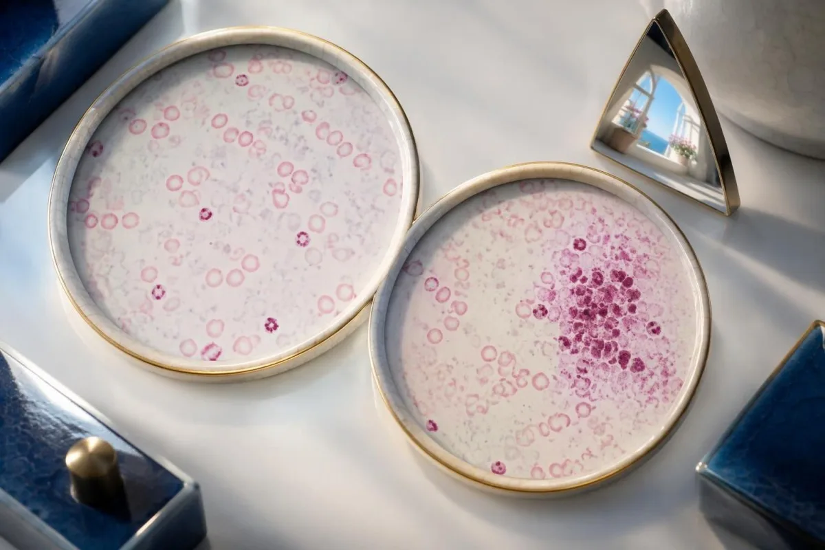
Rare-cell sampling produces unavoidable statistical variation, even with good technique. At a true fraction of 0.1%, a count of 2,000 cells would contain only about 2 fetal cells on average, whereas 10,000 cells would contain about 10; larger counts help, but they do not eliminate misclassification.
Maternal blood volume is another source of error. The same hypothetical 0.4% fraction estimates 16 mL with a 4,000 mL maternal volume and 24 mL with a 6,000 mL volume, rather than the 20 mL obtained using a fixed 5,000 mL assumption.
Red-cell percentages do not translate perfectly into whole-blood volumes when maternal and fetal hematocrits or cell sizes differ. A hypothetical maternal hematocrit of 30% and fetal hematocrit of 50% illustrates why some laboratory methods apply corrections; our hematocrit and hemoglobin comparison explains the underlying measures.
Kantesti is an AI biomarker interpretation platform that can help explain why a calculated 20 mL is an estimate rather than an exact measurement. The practical question is whether the uncertainty could change prophylaxis or fetal assessment; unexpectedly large, borderline-for-dosing, or technically doubtful results warrant discussion with the transfusion laboratory.
Can maternal fetal hemoglobin cause a misleading positive?
Maternal HbF-containing red cells, known as F cells, can retain the Kleihauer-Betke stain and be mistaken for fetal cells. Pregnancy, hereditary persistence of fetal hemoglobin, some hemoglobin disorders, and HbF-increasing treatment can complicate interpretation; an HbF result of 2% does not mean 2% of circulating cells are fetal.
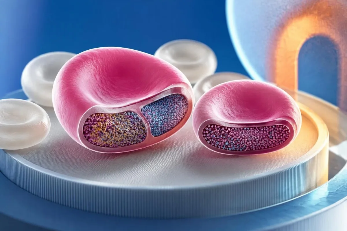
Hemoglobin analysis measures the proportion of hemoglobin types, while Kleihauer-Betke testing counts stained cells. For example, a hypothetical HbF concentration of 3% could be distributed across many maternal F cells, each containing some HbF, rather than confined to 3% distinctly fetal cells.
A history of thalassemia, sickle cell disease, or hereditary persistence of HbF should be given to the laboratory before interpreting an apparent large transfer. Our hemoglobin electrophoresis guide explains HbA, HbS, HbC, and HbF patterns; none of those fractions alone provides a hemorrhage volume in mL.
A low maternal MCV can support investigation for iron deficiency or thalassemia, but it does not prove HbF interference. A hypothetical MCV of 68 fL requires its own workup, described in our low MCV interpretation guide; only some underlying disorders produce enough maternal HbF to affect this stain.
Manual stain appearance may suggest maternal F cells, but visual distinction is not always reliable. Sandler and Sathiyamoorthy (2010) review this limitation of acid-elution testing; if an apparent 1.5% fetal-cell result conflicts with the pregnancy assessment or a known HbF disorder, a validated confirmatory method may prevent substantial overestimation.
When does flow cytometry help confirm fetal-cell transfer?
Flow cytometry helps when a Kleihauer-Betke result is unexpectedly large, technically uncertain, or potentially confounded by maternal HbF. A validated 2-marker assay can distinguish fetal cells from maternal F cells more effectively than HbF staining alone, while anti-D-based methods require a suitable maternal–fetal RhD difference.

Anti-HbF flow cytometry measures fluorescence in individual cells and can analyze far more cells than a manual count. However, an assay using only 1 HbF marker may still count maternal F cells; greater numerical precision does not automatically mean greater certainty about fetal origin.
Some dual-marker assays combine HbF with carbonic anhydrase, an adult-cell marker that is lower in fetal erythrocytes. The 2-dimensional pattern can separate HbF-containing maternal cells from fetal cells, but the laboratory must validate its gating because cell populations can overlap and developmental stage affects marker expression.
Anti-D flow cytometry identifies RhD-positive cells in an RhD-negative maternal background when that contrast is present. It cannot provide the same discrimination if both mother and fetus are RhD-negative or both are RhD-positive, and recent anti-D treatment or complex D typing should be discussed before choosing this method.
Flow cytometry is a method family, not 1 interchangeable test: a PNH flow cytometry assay uses different markers and answers a different question. Kantesti's technical validation information concerns our interpretation system, not accreditation of a hospital's fetal-cell assay; request the actual laboratory method when confirmation matters.
Does a positive result mean the baby is anemic?
A positive Kleihauer-Betke result does not diagnose fetal anemia; it estimates transferred cells in maternal circulation. Fetal assessment may include heart-rate monitoring and middle cerebral artery Doppler, where a peak systolic velocity around or above 1.5 multiples of the median raises concern for moderate-to-severe anemia in appropriate settings.
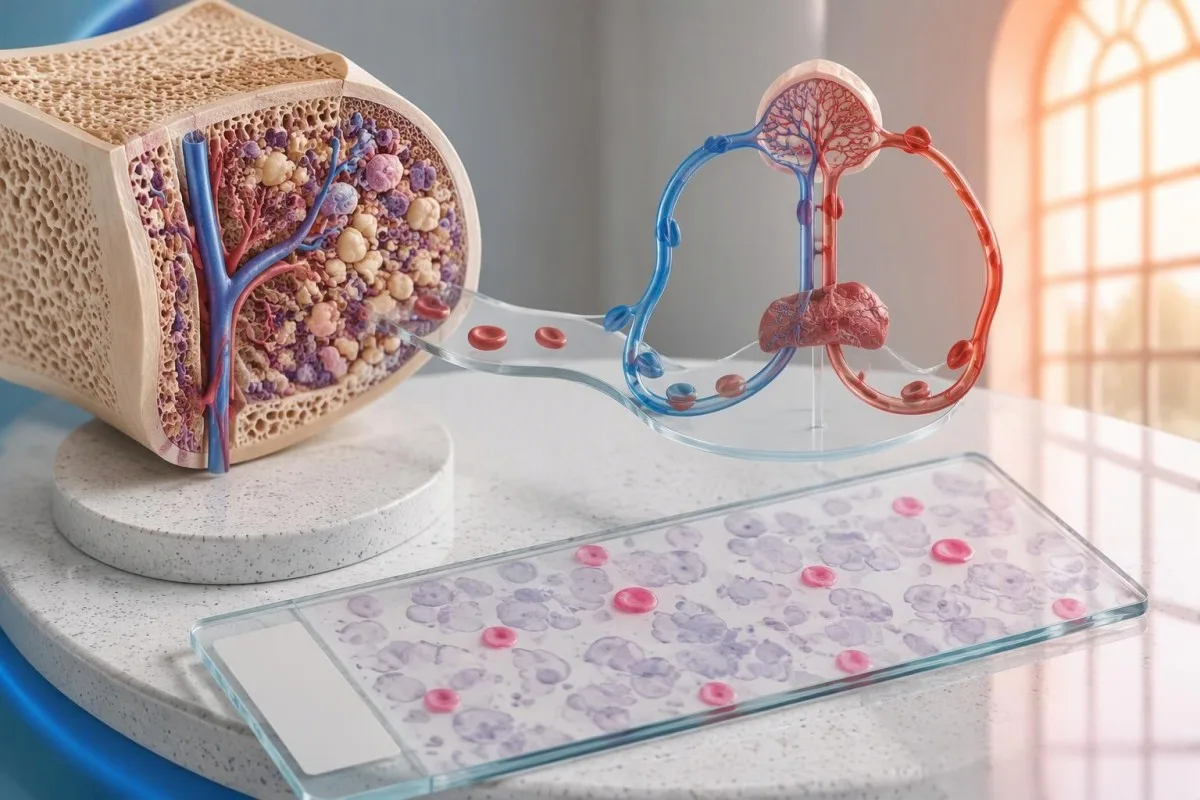
The same estimated loss can have different consequences at different gestational ages. In a purely hypothetical fetus with a 100 mL circulating volume, a 20 mL loss represents 20% of that volume; this arithmetic is illustrative, not a method for calculating an individual fetus's circulation from a maternal stain.
Middle cerebral artery Doppler measures flow velocity rather than hemoglobin directly. The 1.5 MoM threshold is a screening signal, not a stand-alone diagnosis, and performance can be less reliable after intrauterine transfusion or later in pregnancy; a fetal medicine specialist integrates gestational age and other findings.
Reduced fetal movement or significant abdominal trauma requires prompt obstetric assessment even with a negative or pending stain. Severe headache, upper abdominal pain, or other pregnancy warning symptoms may instead suggest conditions such as HELLP syndrome, which follows a different laboratory pathway and should not be explained away by 1 positive result.
Fetomaternal transfer is not synonymous with maternal hemorrhage or a clotting disorder. When obstetric complications raise concern for disseminated intravascular coagulation, platelet count, fibrinogen, and clotting times answer additional questions; a 0.5% fetal-cell fraction neither confirms nor excludes DIC.
What do negative and repeat Kleihauer-Betke results mean?
A negative Kleihauer-Betke result means fetal-type cells were not detected above that laboratory's reporting limit in that specimen. It does not prove that transfer was exactly 0 mL, exclude every clinically relevant exposure, or replace fetal assessment; repeat testing is chosen according to the event, treatment, and assay.
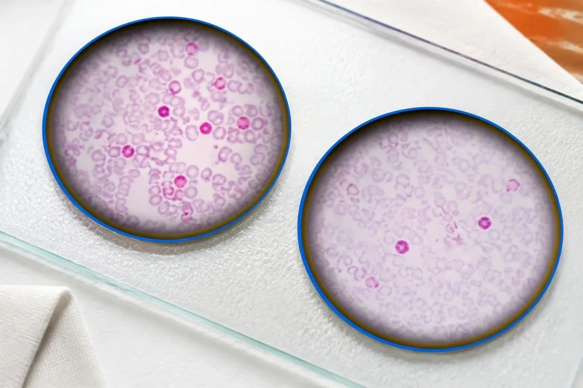
The test is less reliable for very small transfers than for larger ones, particularly when few cells are counted manually. A mother eligible for routine postpartum anti-D may still need the standard dose despite a reported 0.0%, because the assay primarily identifies exposure beyond routine coverage rather than eliminating all sensitization risk.
Large confirmed transfers may require follow-up testing to show that RhD-positive fetal cells have cleared after sufficient prophylaxis. Some UK protocols reassess approximately 48 hours after intravenous anti-D or 72 hours after intramuscular anti-D; the transfusion service chooses the interval and assay rather than applying a universal repeat schedule.
A lower percentage on repeat testing does not always mean the original estimate was wrong. Cell clearance, dilution, and sampling variation can change a hypothetical 0.4% result to 0.2%; our guide to blood test changes over time explains why chronology matters when comparing laboratory values.
A new sensitizing event needs a new clinical review even if testing was completed 1 day earlier. Previous prophylaxis, its dose, the interval since administration, and the new exposure all matter; patients should not assume that an earlier negative stain or routine antenatal injection covers every subsequent event.
What should you ask your care team about your result?
Ask your care team for the fetal-cell percentage, estimated volume and its units, maternal RhD and antibody status, and the planned action. For a result such as 0.6%, clarify whether the estimate represents 30 mL of fetal whole blood or a separately calculated red-cell volume before discussing prophylaxis.

Kantesti is an AI blood test interpretation platform operated by Kantesti Ltd, whose organizational information identifies UK Company No. 17090423. Our role is educational interpretation: explaining 0.6%, 30 mL, and their assumptions without replacing the laboratory's fetal-cell confirmation or your clinician's treatment decision.
A useful discussion separates 3 tasks: confirming cell origin, calculating prophylaxis when indicated, and assessing the fetus or newborn when clinically necessary. Our AI report explanation guide describes source checking and limitations; an AI-generated dose calculation is not an authorized prescription.
If maternal HbF is known to be elevated, ask whether the laboratory considered F-cell interference and whether a 2-marker flow assay would change management. Also provide the time of the event, any recent anti-D, prior transfusions, and hemoglobin-disorder results; missing context can matter more than an extra decimal place.
Our interpretation technology guide explains how AI supports reading laboratory information, but no software can assess fetal movement or perform Doppler imaging. Contact maternity services promptly for concerning symptoms, and contact the prescribing team if the anti-D plan is unclear rather than waiting until the 72-hour window is nearly over.
Research publications and the evidence behind interpretation
The clinical basis for Kleihauer-Betke interpretation comes from transfusion laboratory methods and obstetric anti-D guidance, not unrelated biomarker publications. The 2017 ACOG bulletin, 2014 BCSH guideline, and 2010 laboratory-methods review below address prevention, dose coverage, and assay limitations; local protocols remain necessary.

ACOG Practice Bulletin No. 181 (2017) supports RhD alloimmunization prevention and the relationship between 300 micrograms of anti-D and 30 mL of fetal whole blood. Qureshi et al. (2014) describe a UK approach using different routine doses and red-cell-volume thresholds; those differences explain why prescriptions from 2 countries may not look identical.
Sandler and Sathiyamoorthy (2010) review acid-elution and flow-based laboratory methods, including limitations relevant to maternal HbF. The enduring distinction is between detection, quantification, and identification: counting 10 stained cells more accurately does not prove they are fetal unless the method can discriminate their origin.
Two Kantesti publications are listed separately in the DOI references: the complement and ANA guide and the Nipah virus diagnostic guide. These 2 publications concern other laboratory topics and are not evidence for Kleihauer-Betke accuracy, fetal-anemia thresholds, or anti-D dosing; a DOI identifies a deposited work but does not itself establish peer review or clinical validation.
Our medical content standards distinguish educational explanations from individualized medical decisions. For related laboratory context, the coagulation testing guide covers aPTT and other clotting markers, but those tests cannot substitute for fetal-cell quantification; ask which of these 2 diagnostic questions your ordered panel is intended to answer.
Frequently asked questions
What does a positive Kleihauer-Betke test mean?
A positive Kleihauer-Betke test usually means fetal red cells have entered maternal circulation, indicating fetomaternal hemorrhage. The stain actually detects cells that retain fetal hemoglobin, so maternal HbF-containing cells can sometimes mimic fetal cells. Under a common 5,000 mL maternal-volume assumption, a 0.2% fetal-cell result estimates approximately 10 mL of fetal whole blood. Clinicians use the result alongside RhD status, antibody testing, and the pregnancy assessment.
Does a positive Kleihauer-Betke test mean my baby is in danger?
A positive Kleihauer-Betke test does not by itself show that the baby is anemic or in immediate danger. Risk depends on how much blood was lost relative to the fetus's circulation, how quickly it was lost, and the fetal assessment. Middle cerebral artery Doppler velocity around or above 1.5 MoM can raise concern for anemia in appropriate settings, but it is not a stand-alone diagnosis. Reduced fetal movement, significant abdominal trauma, or vaginal bleeding warrants prompt maternity assessment regardless of the test result.
How is the Kleihauer-Betke percentage converted to hemorrhage volume?
A common calculation estimates fetal whole-blood volume in mL by multiplying the reported fetal-cell percentage by 50, assuming 5,000 mL of maternal blood. For example, 0.8% × 50 estimates 40 mL of fetal whole blood. This shortcut does not directly calculate fetal red-cell volume and may differ from a laboratory's validated, patient-specific method. The result remains an estimate because counting, hematocrit differences, and maternal blood-volume assumptions introduce uncertainty.
How much Rh immune globulin is needed after a positive result?
The required Rh immune globulin dose depends on eligibility, estimated fetal-cell exposure, product coverage, and the local transfusion protocol. A standard 300-microgram dose, equivalent to 1,500 IU, typically covers 30 mL of fetal whole blood or 15 mL of fetal red cells. Larger exposures can require additional doses with a safety allowance for assay uncertainty. The blood bank or prescribing clinician must authorize the dose; anti-D prevents sensitization and does not treat fetal anemia.
Can the Kleihauer-Betke test be falsely positive because of maternal HbF?
Maternal red cells containing HbF can stain like fetal cells and cause an overestimated Kleihauer-Betke result. Pregnancy, hereditary persistence of HbF, certain hemoglobin disorders, and HbF-increasing treatments can contribute to this interference. A maternal HbF fraction of 3% is a hemoglobin measurement, not proof that 3% of circulating cells are fetal. A validated flow cytometry assay that distinguishes maternal F cells from fetal cells may clarify an uncertain result.
Does a negative Kleihauer-Betke test mean I do not need anti-D?
A negative Kleihauer-Betke test does not automatically remove the need for anti-D prophylaxis in an eligible RhD-negative mother. A reported 0.0% means no fetal-type cells were detected above the method's reporting limit, not that exposure was exactly zero. Routine prophylaxis can still be indicated after an eligible event or delivery, while the assay helps identify exposures requiring additional coverage. Anti-D is generally given promptly and within 72 hours, so arranging testing should not unnecessarily delay indicated treatment.
Get your blood test results explained
Upload your lab report and Kantesti's AI explains each biomarker in plain language within seconds. Used by more than 2 million people worldwide.
References
- Klein, T., Mitchell, S., & Weber, H. (2026). C3 C4 Complement Blood Test & ANA Titer Guide. Kantesti AI Medical Research. DOI
- Klein, T., Mitchell, S., & Weber, H. (2026). Nipah Virus Blood Test: Early Detection & Diagnosis Guide 2026. Kantesti AI Medical Research. DOI
- American College of Obstetricians and Gynecologists (2017). Practice Bulletin No. 181: Prevention of Rh D Alloimmunization. Obstetrics & Gynecology. DOI
- Qureshi H et al. (2014). BCSH guideline for the use of anti-D immunoglobulin for the prevention of haemolytic disease of the fetus and newborn. Transfusion Medicine. DOI
- Sandler SG and Sathiyamoorthy S (2010). Laboratory methods for Rh immunoprophylaxis: a review. Immunohematology.
- 2M+Tests analysed
- 127+Countries
- 100+Languages
About the medical team


E-E-A-T Trust Signals
Experience
Physician-led clinical review of lab interpretation workflows.
Expertise
Laboratory medicine focus on how biomarkers behave in clinical context.
Authoritativeness
Medically reviewed by Dr. Thomas Klein, with contributions from Dr. Sarah Mitchell and Prof. Dr. Hans Weber.
Trustworthiness
Evidence-based interpretation with clear follow-up pathways to reduce alarm.





Medical disclaimer
This article is for educational purposes only and does not constitute medical advice. Always consult a qualified healthcare provider for diagnosis and treatment decisions.
Published · Contact the editorial team
Published by Kantesti Ltd, registered in England & Wales, company no. 17090423, London, United Kingdom.
