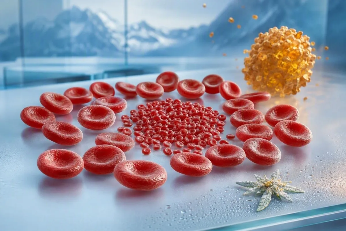
A low mean corpuscular volume usually reflects iron-restricted red-cell production or an inherited hemoglobin trait. The fastest safe triage comes from reading MCV alongside RBC count, RDW, ferritin, transferrin saturation, and the clinical story.



- Low MCV means red cells are smaller than expected; most adult laboratories flag microcytosis below 80 fL.
- Ferritin below 15 ng/mL is highly specific for depleted iron stores in an otherwise well adult, while values below 30 ng/mL commonly support iron deficiency in practice.
- RBC count above 5.0 ×10¹²/L with MCV below 75 fL and a normal RDW often points toward thalassemia trait rather than simple iron deficiency.
- RDW above 14.5% supports evolving iron deficiency because new cells become smaller before all circulating cells are affected.
- Transferrin saturation below 20% indicates limited circulating iron availability; below 15% is more persuasive when ferritin is equivocal.
- Mentzer index below 13 favors thalassemia trait, while an index above 13 favors iron deficiency, but it is a screening clue rather than a diagnosis.
- HbA2 above 3.5% on hemoglobin electrophoresis supports beta-thalassemia trait after iron deficiency has been corrected or excluded.
- New iron deficiency in men or postmenopausal women needs a cause-focused evaluation, often including gastrointestinal assessment rather than iron tablets alone.
What a Low MCV Means on a Complete Blood Count
A low MCV means the average red cell is small, usually below 80 fL in adults. The two leading low MCV causes are iron deficiency and thalassemia trait, and the practical split starts with ferritin, RBC count, RDW, and whether anemia is actually present.

MCV is an average, so it can look only mildly low at 78 fL even when two different cell populations are present. A rising RDW reveals that mixture earlier than MCV alone, which is why a CBC index comparison is more useful than chasing one flagged result.
A normal hemoglobin does not rule out a meaningful problem. I regularly see ferritin of 8 to 18 ng/mL with hemoglobin still above 120 g/L, particularly in people with heavy menstrual loss, endurance training, restrictive diets, or frequent blood donation.
Kantesti is an AI blood test analyzer that reads MCV together with hemoglobin, RBC count, RDW, ferritin, and prior results rather than assigning a cause from one red flag. As of September 9, 2026, that pattern-based approach remains safer than self-treating every low MCV as iron deficiency.
Microcytosis is a finding, not a diagnosis
Microcytosis describes cell size; anemia describes insufficient oxygen-carrying capacity. A person can have microcytosis without anemia, anemia without microcytosis, or both—each pattern has a different work-up.
Use RBC Count and RDW Before Assuming Iron Deficiency
An RBC count that is relatively high despite a low MCV favors thalassemia trait, whereas a low or ordinary RBC count with high RDW favors iron deficiency. This is the most useful first-pass CBC pattern when ferritin has not yet returned.
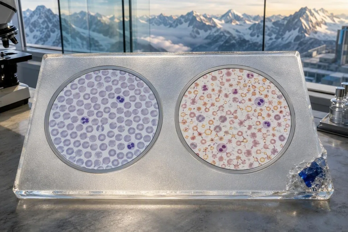
In uncomplicated iron deficiency, the marrow lacks substrate and often produces fewer cells: hemoglobin falls, RBC count may sit below 4.5 ×10¹²/L, and RDW often rises above 14.5%. In thalassemia trait, marrow output is preserved, so an RBC count of 5.2 to 6.2 ×10¹²/L is not unusual despite an MCV in the low 60s or 70s.
The Mentzer index divides MCV by RBC count. For example, 68 fL ÷ 5.8 = 11.7, a result that favors thalassemia trait; 74 fL ÷ 4.3 = 17.2 leans toward iron deficiency, though mixed disease can defeat the rule.
A low MCV with normal hemoglobin and a stable high RBC count over several years is often inherited rather than nutritional. Compare the whole complete blood count with earlier tests before deciding that a recent lifestyle change caused it.
Why RDW Often Separates Early Iron Deficiency From Trait
High RDW with low MCV usually reflects iron deficiency because old normal-sized cells circulate beside newer smaller cells. RDW may remain normal in thalassemia trait because small cells are produced consistently over time.

RDW-CV commonly has a reference interval around 11.5% to 14.5%, although the laboratory's own interval wins. A value of 16.8% with MCV 76 fL has more diagnostic weight than either result alone, especially if the prior MCV was 87 fL six months earlier.
RDW is not specific: recent transfusion, B12 deficiency, folate deficiency, hemolysis, and recovery after iron therapy can all increase it. A surprising rise after treatment may simply represent new iron-replete cells entering circulation; our guide to RDW after iron therapy explains this counterintuitive phase.
In my clinical experience, a normal RDW should slow down—not stop—the iron-deficiency work-up. Long-standing deficiency can eventually make most cells uniformly small, while alpha-thalassemia trait plus iron loss may produce a very high RDW.
Trend beats a single RDW
A shift from RDW 12.8% to 15.6% is meaningful even if both values straddle different laboratories' reference limits. The direction of change often appears before a patient feels tired or breathless.
Ferritin Is the Best First Follow-Up Test for Low MCV
Ferritin is the first test after a low MCV in most adults because it estimates stored iron. Ferritin below 15 ng/mL strongly indicates iron deficiency, and many clinicians investigate values below 30 ng/mL when symptoms or microcytosis are present.
Ferritin is an acute-phase reactant, so inflammation can conceal depleted stores. Camaschella's review of iron-deficiency anemia describes why ferritin rises with inflammatory signaling; a ferritin of 55 ng/mL does not reliably exclude iron deficiency when CRP is elevated (Camaschella, 2015).
Kantesti AI is an AI blood test interpretation platform that flags the specific mismatch of low MCV, low transferrin saturation, and apparently normal ferritin when CRP or liver markers suggest an acute-phase effect. This is one of those areas where context matters more than a bold laboratory flag.
For a useful next draw, avoid interpreting serum iron in isolation because it changes across the day and after supplements. Pair ferritin with transferrin saturation, CRP, and the iron studies guide whenever the pattern is unclear.
The Typical Iron Deficiency Pattern Behind Microcytosis
Iron deficiency usually produces low ferritin, low transferrin saturation, rising RDW, and eventually low hemoglobin. The earliest clue may be ferritin decline months before MCV falls below 80 fL.
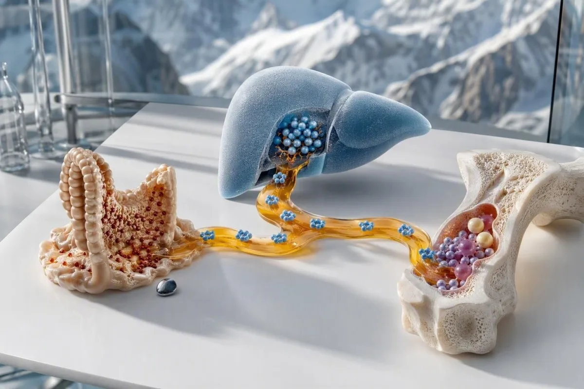
A practical pattern is ferritin 12 ng/mL, transferrin saturation 9%, MCV 74 fL, and RDW 17%. Serum iron alone is less dependable because a recent meal, iron tablet, or morning-versus-afternoon collection can move it considerably.
Menstrual loss is common, but it should not become a reflex explanation. A 38-year-old with periods described as normal, ferritin 6 ng/mL, and persistent microcytosis needs dietary, donation, medication, celiac, and gastrointestinal questions rather than reassurance; see low ferritin without heavy periods.
Oral elemental iron doses of 40 to 65 mg once daily or every other day are often better tolerated than older multiple-daily-dose schedules. Response should be checked with a clinician, because hemoglobin usually rises by roughly 10 g/L over 2 to 4 weeks if absorption and the diagnosis are right.
When Inflammation Makes Ferritin Look Better Than It Is
Inflammation can create low MCV with normal or raised ferritin by trapping iron away from the marrow. Low transferrin saturation, raised CRP, chronic kidney disease, autoimmune disease, or active infection can point toward this functional iron-restriction pattern.
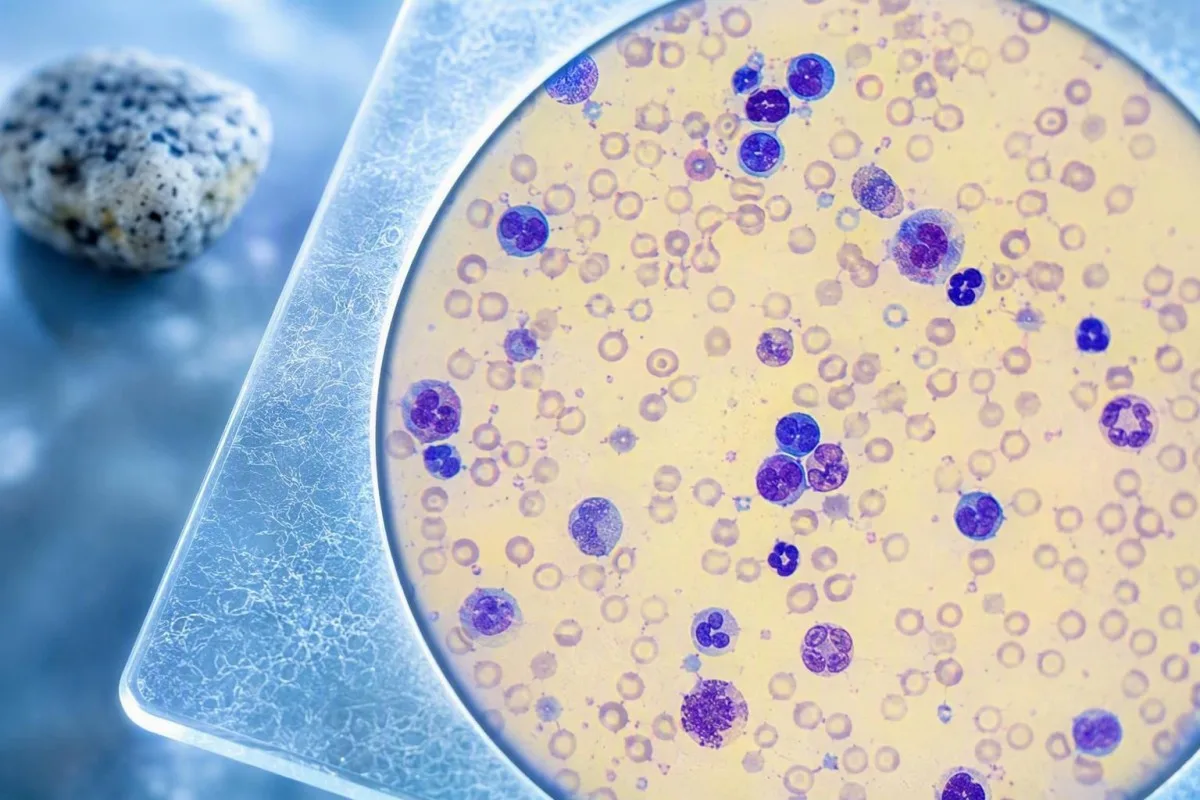
Transferrin saturation equals serum iron divided by total iron-binding capacity, multiplied by 100. A saturation below 20% suggests restricted iron availability, but values below 15% are more persuasive when ferritin sits between 30 and 100 ng/mL.
Inflammation commonly lowers transferrin while increasing ferritin, producing a deceptive combination. The transferrin and inflammation pattern matters because a low TIBC does not mean iron stores are plentiful.
Soluble transferrin receptor can help when results disagree because it is less affected by acute inflammation, although availability and assay ranges vary. I would not use a normal ferritin of 70 ng/mL as a reason to dismiss iron restriction in someone with CRP 35 mg/L and transferrin saturation 11%.
Anemia of Chronic Disease and Other Acquired Low MCV Causes
Chronic inflammatory disease, chronic kidney disease, lead exposure, and sideroblastic processes can cause low MCV, but they are less common than iron deficiency or thalassemia trait. Their iron-study patterns and clinical setting prevent unnecessary iron treatment.

Anemia of chronic inflammation is often normocytic at first and becomes microcytic in a minority of cases. Ferritin is usually normal or high, serum iron low, and TIBC low or normal—quite different from classic iron deficiency, where TIBC is often raised.
Chronic kidney disease can combine reduced erythropoietin production with iron-restricted erythropoiesis. If eGFR is below 60 mL/min/1.73 m² for at least 3 months, iron interpretation should be coordinated with the renal team; review the CKD staging guide.
Lead toxicity and sideroblastic anemia deserve targeted testing only when exposure, drugs, alcohol use, neurologic symptoms, basophilic stippling, or an unusual smear points there. A blood film reviewed by a hematology laboratory is more informative than ordering a broad panel without a clinical lead.
The CBC Pattern That Suggests Thalassemia Trait
Thalassemia trait typically causes a disproportionately low MCV with a normal or high RBC count and mild or absent anemia. Ferritin is usually normal unless iron deficiency coexists, and RDW is often normal or only slightly raised.
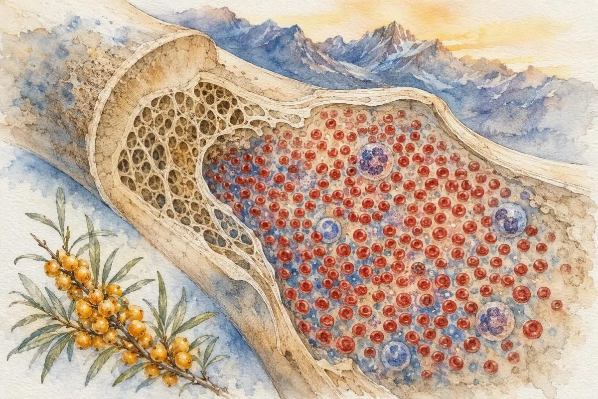
A classic beta-thalassemia trait panel might show hemoglobin 118 g/L, MCV 66 fL, RBC 5.7 ×10¹²/L, RDW 13.2%, and ferritin 48 ng/mL. The low MCV can look alarming, but the stable high RBC count is the clue many readers miss.
Trait is not an illness to correct with iron unless iron deficiency is proven. Repeated iron courses in someone with ferritin 80 ng/mL and a lifelong MCV around 68 fL rarely improve the count and can delay genetic counseling or a correct explanation.
Family origin can inform probability, but it never replaces testing; thalassemia traits occur worldwide. A seemingly high RBC count can also reflect dehydration, so compare with hematocrit, albumin, and the context in our high RBC count review.
Alpha and beta trait are not identical
Beta-thalassemia trait often raises HbA2, whereas alpha-thalassemia trait can have normal electrophoresis. Normal electrophoresis therefore does not exclude alpha-thalassemia when the CBC pattern and family history are convincing.
When Hemoglobin Electrophoresis and Genetic Testing Help
Hemoglobin electrophoresis is most useful when ferritin is normal and the CBC suggests thalassemia trait. HbA2 above approximately 3.5% supports beta-thalassemia trait, while alpha-thalassemia may require molecular testing after specialist review.
Correct iron deficiency before relying on a borderline HbA2 result, because iron deficiency can lower HbA2 enough to mask beta-thalassemia trait. A sensible sequence is iron studies first, treatment if depleted, then repeat CBC and hemoglobin analysis after the deficiency is addressed.
Galanello and Origa describe beta-thalassemia as a globin-production disorder with carrier states that are usually mild but important for reproductive counseling (Galanello and Origa, 2010). If both biological parents carry clinically relevant hemoglobin variants, each pregnancy may have a 25% chance of an affected child depending on the variants involved.
Do not assume every small-cell pattern is thalassemia. Hemoglobin variants can coexist, and a sickle-cell screening guide explains why definitive interpretation belongs with the laboratory report and a clinician familiar with hemoglobinopathy testing.
Mixed Iron Deficiency and Thalassemia: The Pattern That Confuses Most
Iron deficiency can coexist with thalassemia trait, producing low ferritin plus a higher-than-expected RBC count. This mixed pattern is common enough that a low MCV should never be labeled as trait until iron stores have been checked.
Consider a person with MCV 64 fL, RBC 5.4 ×10¹²/L, RDW 18.1%, ferritin 9 ng/mL, and hemoglobin 104 g/L. The RBC count suggests trait; the very low ferritin and high RDW prove clinically relevant iron deficiency as well.
After iron replacement, hemoglobin and RDW may improve while MCV remains low, revealing the underlying trait. That residual MCV is not treatment failure if ferritin has repleted and the clinical cause of iron loss has been addressed.
Kantesti is an AI-powered blood test analysis tool that compares pre- and post-treatment CBC patterns so a persistent inherited microcytosis is not mistaken for ongoing deficiency. Our longitudinal lab guide explains why two well-timed results can answer more than one oversized panel.
Less Common Nutritional, Drug, and Marrow Causes of Low MCV
Copper deficiency, excess zinc exposure, alcohol-related sideroblastic change, certain medicines, and rare marrow disorders can cause microcytosis or a mixed red-cell picture. These causes become more plausible when ferritin is not low and the usual iron-versus-trait pattern does not fit.

Copper deficiency more often causes neutropenia and anemia that is normocytic or macrocytic, but microcytosis can occur. It is particularly relevant after bariatric surgery, severe malabsorption, or prolonged high-dose zinc use; zinc intake above 40 mg/day for months can impair copper absorption.
Sideroblastic anemia often features high serum iron and ferritin rather than depleted stores, so giving more iron can be the wrong move. Medication review matters: isoniazid, linezolid, chloramphenicol, and heavy alcohol exposure are among recognized clinical leads, not routine explanations.
When low MCV occurs with low neutrophils, unexplained neurologic symptoms, or zinc supplementation, ask about copper deficiency clues and involve a clinician. These are not diagnoses to make from an online result alone.
Finding the Source of Proven Iron Deficiency Matters
Proven iron deficiency requires a cause-focused history, not just replacement therapy. Heavy menstrual loss, pregnancy, diet, blood donation, medications, malabsorption, and gastrointestinal blood loss are all common routes to depleted iron stores.

The British Society of Gastroenterology recommends gastrointestinal investigation for unexplained iron-deficiency anemia in men and postmenopausal women because significant gastrointestinal pathology may be present (Snook et al., 2021). This does not mean cancer is the usual explanation; it means occult loss is too important to overlook.
Black stools, visible rectal bleeding, unexplained weight loss, persistent abdominal pain, new change in bowel habit, fainting, or hemoglobin below 80 g/L deserve prompt clinical assessment. A positive stool screen needs timely follow-up; read our guide to a positive FIT result.
For menstruating adults, quantify bleeding instead of accepting the phrase “heavy periods.” Changing protection every 1 to 2 hours, passing clots larger than roughly 2.5 cm, or bleeding longer than 7 days makes iron loss more plausible and merits a gynecologic discussion.
What Should Change After Iron Treatment and When to Retest
With effective oral iron therapy, reticulocytes begin to increase within about 7 to 10 days and hemoglobin usually rises by around 10 g/L within 2 to 4 weeks. MCV normalizes more slowly because older microcytic cells remain in circulation for up to 120 days.
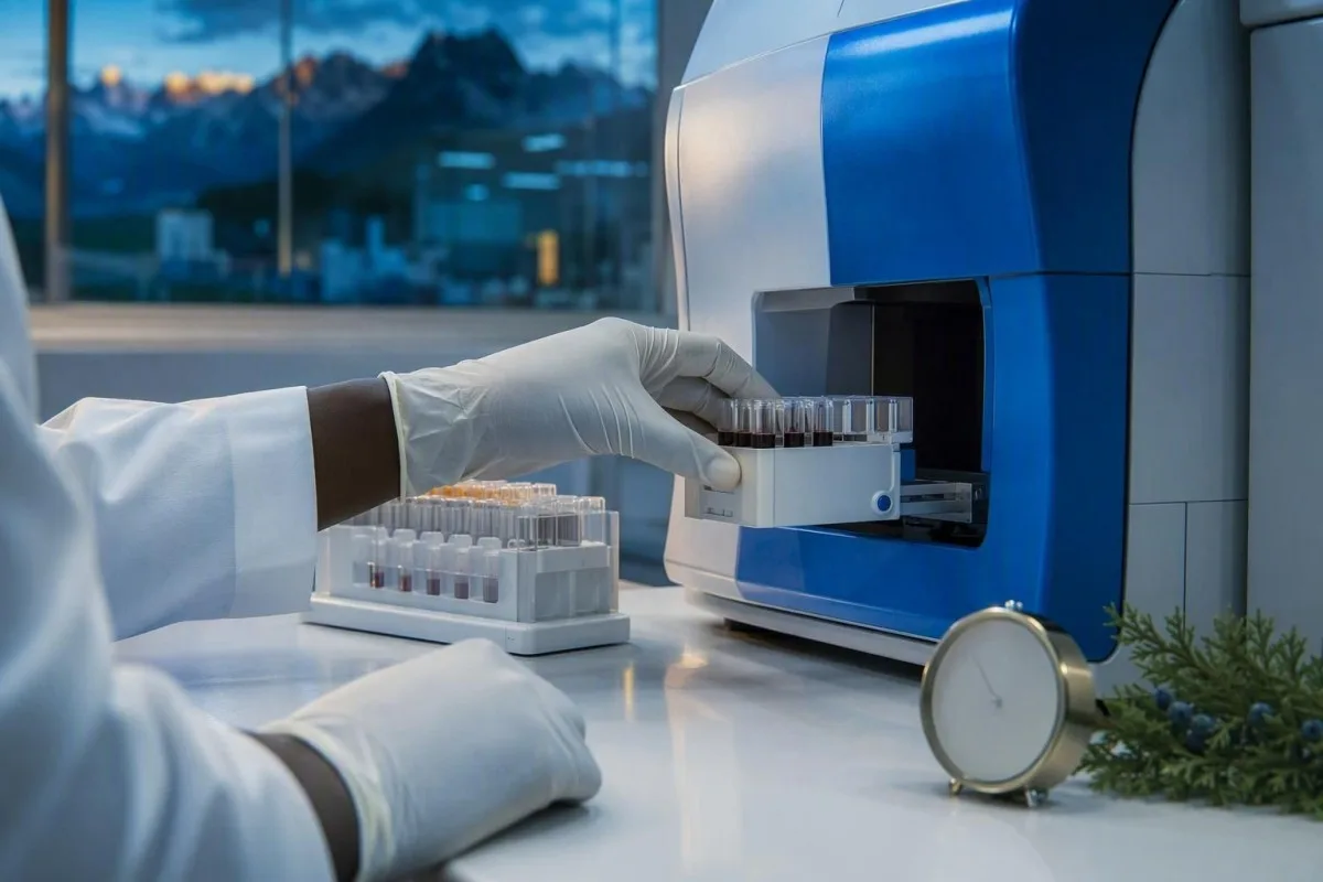
A practical check at 2 to 4 weeks looks for hemoglobin response, adherence, side effects, and ongoing loss; ferritin is often rechecked after hemoglobin recovery or roughly 8 to 12 weeks later, depending on the clinician's plan. Taking iron with tea, coffee, calcium, or acid-suppressing medication can reduce absorption.
Lack of a meaningful hemoglobin rise should prompt questions before increasing the dose: Was the diagnosis correct? Is there celiac disease, ongoing loss, poor adherence, inflammation, or a coexisting trait? The celiac testing preparation guide is relevant when malabsorption is suspected.
Thomas Klein, MD, sees a recurring mistake here: stopping iron when hemoglobin first reaches normal but before stores are rebuilt. Conversely, continuing indefinitely without a repeat ferritin and a source assessment is not good medicine either.
When Low MCV Needs Urgent Rather Than Routine Care
Low MCV itself is rarely an emergency, but severe anemia, active blood loss, chest pain, breathlessness at rest, fainting, or pregnancy changes the urgency. Symptoms and hemoglobin level matter more than whether MCV is 72 fL or 78 fL.
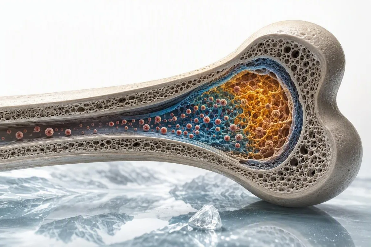
Seek urgent evaluation for chest pain, new confusion, collapse, black tarry stool, vomiting material that resembles coffee grounds, severe shortness of breath, or a rapidly falling hemoglobin. A hemoglobin below 70 g/L is often a threshold for urgent assessment in stable adults, though transfusion decisions depend on symptoms, bleeding, heart disease, and local protocols.
Pregnancy deserves earlier review because iron demand rises substantially, and untreated deficiency is associated with maternal fatigue and adverse obstetric outcomes. Ferritin below 30 ng/mL in pregnancy is commonly used as a treatment threshold in many obstetric pathways; see pregnancy ferritin ranges.
Kantesti AI interprets microcytosis results as a follow-up signal, not a substitute for acute care or diagnosis. If a report contains severe anemia or concerning symptoms, our clinical standards are reviewed against the principles described in medical validation.
A Practical Low MCV Follow-Up Plan to Take to Your Clinician
The safest low MCV follow-up is CBC review, ferritin, transferrin saturation, CRP when inflammation is possible, and hemoglobin testing when iron stores are not clearly low. Repeating MCV alone rarely answers the actual question.

Bring prior CBCs, iron supplements and doses, diet changes, donation history, menstrual or gastrointestinal symptoms, family anemia history, ethnicity only if you wish to share it, and pregnancy plans. One prior MCV of 69 fL from adolescence can immediately shift the probability toward an inherited trait.
Thomas Klein, MD, recommends writing down the exact laboratory units and reference intervals before an appointment. Kantesti AI can organize the CBC and iron pattern from a PDF or photo, but a clinician still decides whether the next step is iron treatment, stool testing, electrophoresis, genetic counseling, or observation.
For readers who want to understand how result-context tools are built and medically reviewed, the AI technology guide and our Medical Advisory Board describe the boundaries of interpretation. The goal is a clear question for your clinician, not a self-diagnosis.
Frequently Asked Questions
What is the most common cause of low MCV?
Iron deficiency is the most common cause of low MCV worldwide, particularly when ferritin is below 15 to 30 ng/mL and RDW is above the laboratory reference range. Thalassemia trait is another frequent cause, especially when the RBC count remains high at more than 5.0 ×10¹²/L despite an MCV below 80 fL. Ferritin, transferrin saturation, RBC count, and RDW distinguish these patterns more reliably than MCV alone. A clinician should investigate why iron is low rather than simply assuming diet is responsible.
Can low MCV be normal if hemoglobin is normal?
Low MCV can occur with normal hemoglobin, especially in early iron deficiency and thalassemia trait. An adult with hemoglobin above 120 g/L may still have ferritin below 15 ng/mL or an inherited trait causing a stable MCV of 65 to 78 fL. Normal hemoglobin means oxygen-carrying capacity is preserved at that moment; it does not identify the cause of small red cells. Ferritin and the RBC-count/RDW pattern remain appropriate follow-up tests.
What RBC count suggests thalassemia trait?
An RBC count above about 5.0 ×10¹²/L combined with low MCV, normal ferritin, and a normal or mildly raised RDW supports thalassemia trait. For example, an MCV of 67 fL with RBC count 5.8 ×10¹²/L is more typical of trait than simple iron deficiency. This is not diagnostic because dehydration, mixed iron deficiency, and laboratory variation can alter the count. Hemoglobin electrophoresis and, in some cases, genetic testing confirm the cause.
Does a low MCV always mean iron deficiency?
A low MCV does not always mean iron deficiency. Thalassemia trait, chronic inflammation, chronic kidney disease, lead exposure, sideroblastic disorders, and occasional copper-related problems can also produce microcytosis. Ferritin below 15 ng/mL strongly supports iron deficiency in a well adult, while a normal ferritin with an RBC count above 5.0 ×10¹²/L shifts attention toward thalassemia trait. Mixed iron deficiency and thalassemia can occur together, so one normal-looking clue should not end the assessment.
What ferritin level rules out iron deficiency?
No single ferritin level rules out iron deficiency in every clinical setting because ferritin rises during inflammation, liver disease, infection, and metabolic illness. Ferritin above 100 ng/mL generally makes absolute iron deficiency less likely in a well adult, but a value of 50 to 100 ng/mL can coexist with iron restriction when CRP is elevated and transferrin saturation is below 20%. Ferritin below 15 ng/mL is highly specific for depleted stores, and many clinicians treat a value below 30 ng/mL as probable deficiency when low MCV is present. Transferrin saturation and CRP clarify borderline results.
How quickly should MCV improve after taking iron?
MCV often improves slowly after iron treatment because existing small red cells circulate for up to 120 days. Reticulocyte production usually changes within 7 to 10 days, and hemoglobin commonly rises by about 10 g/L over 2 to 4 weeks when treatment is absorbed and ongoing loss is controlled. A persistently low MCV after ferritin and hemoglobin recover may reveal thalassemia trait rather than treatment failure. Repeat testing should be planned with the clinician who is treating the underlying cause.
Get AI-Powered Blood Test Analysis Today
Join over 2 million users worldwide who trust Kantesti for instant, accurate lab test analysis. Upload your blood test results and receive comprehensive interpretation of 15,000+ biomarkers in seconds.
📚 Referenced Research Publications
Klein, T., Mitchell, S., & Weber, H. (2026). Women's Health Guide: Ovulation, Menopause & Hormonal Symptoms. Kantesti AI Medical Research.
Klein, T., Mitchell, S., & Weber, H. (2026). Multilingual AI Assisted Clinical Decision Support for Early Hantavirus Triage: Design, Engineering Validation, and Real-World Deployment Across 50,000 Interpreted Blood Test Reports. Kantesti AI Medical Research.
📖 External Medical References





⚕️ Medical Disclaimer
This article is for educational purposes only and does not constitute medical advice. Always consult a qualified healthcare provider for diagnosis and treatment decisions.
E-E-A-T Trust Signals
Experience
Physician-led clinical review of lab interpretation workflows.
Expertise
Laboratory medicine focus on how biomarkers behave in clinical context.
Authoritativeness
Written by Dr. Thomas Klein with review by Dr. Sarah Mitchell and Prof. Dr. Hans Weber.
Trustworthiness
Evidence-based interpretation with clear follow-up pathways to reduce alarm.