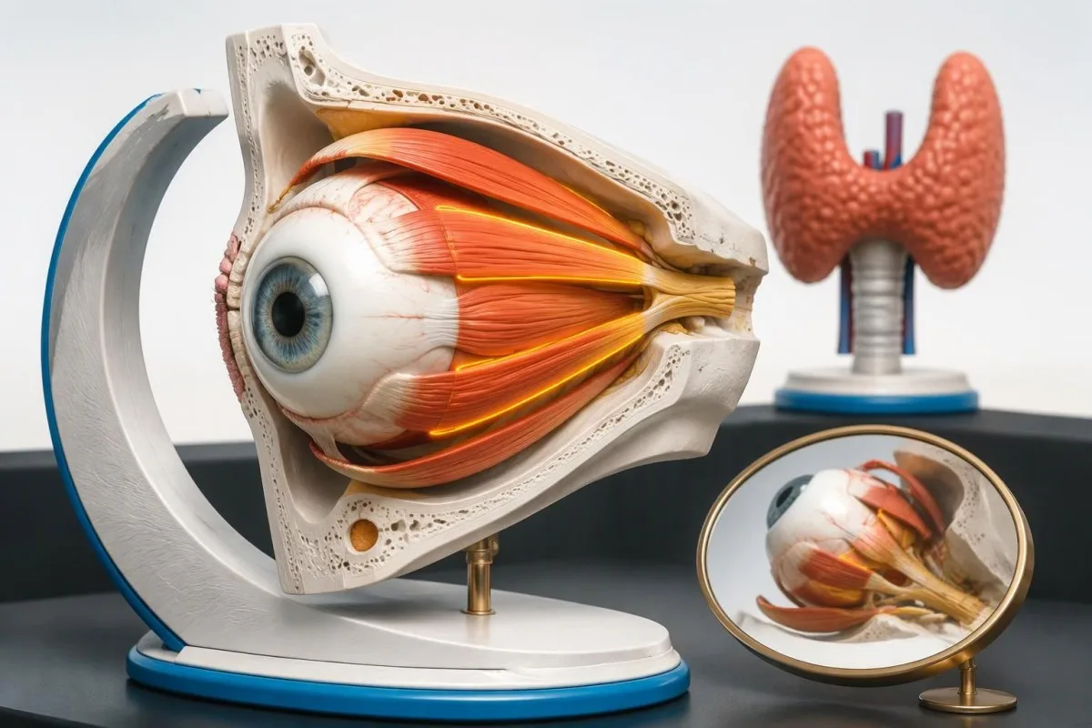
Eye pain needs immediate assessment if vision becomes dimmer, colors look washed out, or part of your visual field disappears. Thyroid eye disease can occur with normal thyroid hormone levels; new difficulty closing an eyelid also needs prompt assessment because the cornea may be exposed.

Key takeaways
- Vision loss with suspected thyroid eye disease needs immediate eye assessment, even without severe pain or obvious bulging.
- Color changes such as red appearing faded in 1 eye can indicate optic nerve dysfunction; do not wait for another thyroid blood test.
- Eyelid closure that is newly incomplete needs same-day clinical advice; accompanying pain, light sensitivity, or blurred vision warrants emergency eye care.
- Normal thyroid levels do not exclude thyroid eye disease because orbital autoimmunity and thyroid hormone production are related but separate processes.
- Early signs include persistent grittiness, eyelid swelling, deep orbital aching, and a changing eyelid position—not just Graves disease bulging eyes.
- Active disease is commonly identified using a Clinical Activity Score of at least 3 out of 7, but a lower score does not exclude sight-threatening disease.
- Thyroid results commonly use adult TSH reference intervals around 0.4–4.0 mIU/L; the laboratory's own interval and treatment history take priority.
- Selenium was studied at 100 µg twice daily for 6 months in mild disease, but benefit in selenium-sufficient populations remains uncertain.
When do Graves ophthalmopathy symptoms need emergency care?
Graves ophthalmopathy symptoms are urgent when eye pain accompanies reduced vision, washed-out colors, a missing area of vision, or a newly exposed eye surface. Seek an emergency eye service or emergency department immediately; do not wait 24 hours for thyroid results or an online interpretation.
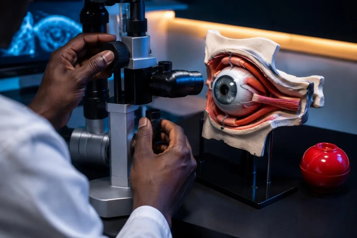
Dysthyroid optic neuropathy occurs when thyroid eye disease compromises the optic nerve, often through crowding behind the eye. It affects roughly 3–5% of people with thyroid eye disease in commonly cited clinical estimates, and the 2021 EUGOGO guideline classifies optic neuropathy and corneal breakdown as sight-threatening disease (Bartalena et al., 2021).
Pain intensity does not reliably measure danger. Someone with severe grittiness may have surface irritation, while someone with only mild discomfort may have declining optic nerve function; a new difference between the 2 eyes in brightness, sharpness, or color deserves immediate assessment rather than reassurance from the pain score.
A newly incomplete blink or eyelid closure needs same-day clinical advice, especially if the eye remains open during sleep. If incomplete closure comes with persistent blur, marked redness, light sensitivity, or increasing pain, use emergency eye care; lubricating drops cannot rule out an affected corneal surface or replace examination.
Kantesti is an AI blood test analyzer that helps explain thyroid results, but no TSH value can clear an eye emergency. Pressure measurements are also only part of the picture: our guide to glaucoma testing and vision explains why 1 pressure reading does not establish the cause of visual symptoms.
How do thyroid eye disease early signs differ from irritation?
Thyroid eye disease early signs combine surface discomfort with changes in eyelid position, orbital fullness, or eye movement. Ordinary irritation more often causes burning or itching without progressive bulging or restricted movement, although the 2 conditions can coexist and cannot always be distinguished at home.
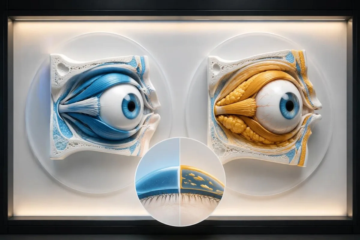
Dryness can cause watering. When the tear film breaks up, reflex tears may overflow without adequately stabilizing the eye surface; persistent watering therefore does not exclude dryness, and irritation that improves after 1 or 2 blinks can still occur in thyroid eye disease with exposure.
Deep aching behind the eye, discomfort when looking upward, and morning eyelid swelling suggest an orbital component rather than isolated tear-film disturbance. None proves thyroid eye disease, but a cluster developing over 2–3 weeks is more concerning than transient burning after several hours of screen use.
Changing eyelid height is an easily missed clue. An upper eyelid that sits higher than before can expose a strip of white above the iris, while swelling may conceal that change; compare photographs taken several months apart rather than judging appearance from 1 brightly lit selfie.
Persistent dry eyes plus dry mouth raise a different autoimmune possibility, particularly when there is no altered eye position or restricted movement. Our explanation of Sjögren dryness and testing describes this overlap; needing water to swallow dry food adds information that 1 eye symptom alone cannot provide.
What causes Graves disease bulging eyes and double vision?
Graves disease bulging eyes result from increased orbital tissue volume, including enlarged eye muscles and expanded fat. Double vision develops when the 2 eyes no longer align normally, often because affected muscles restrict movement; either change can occur without dramatic pain.
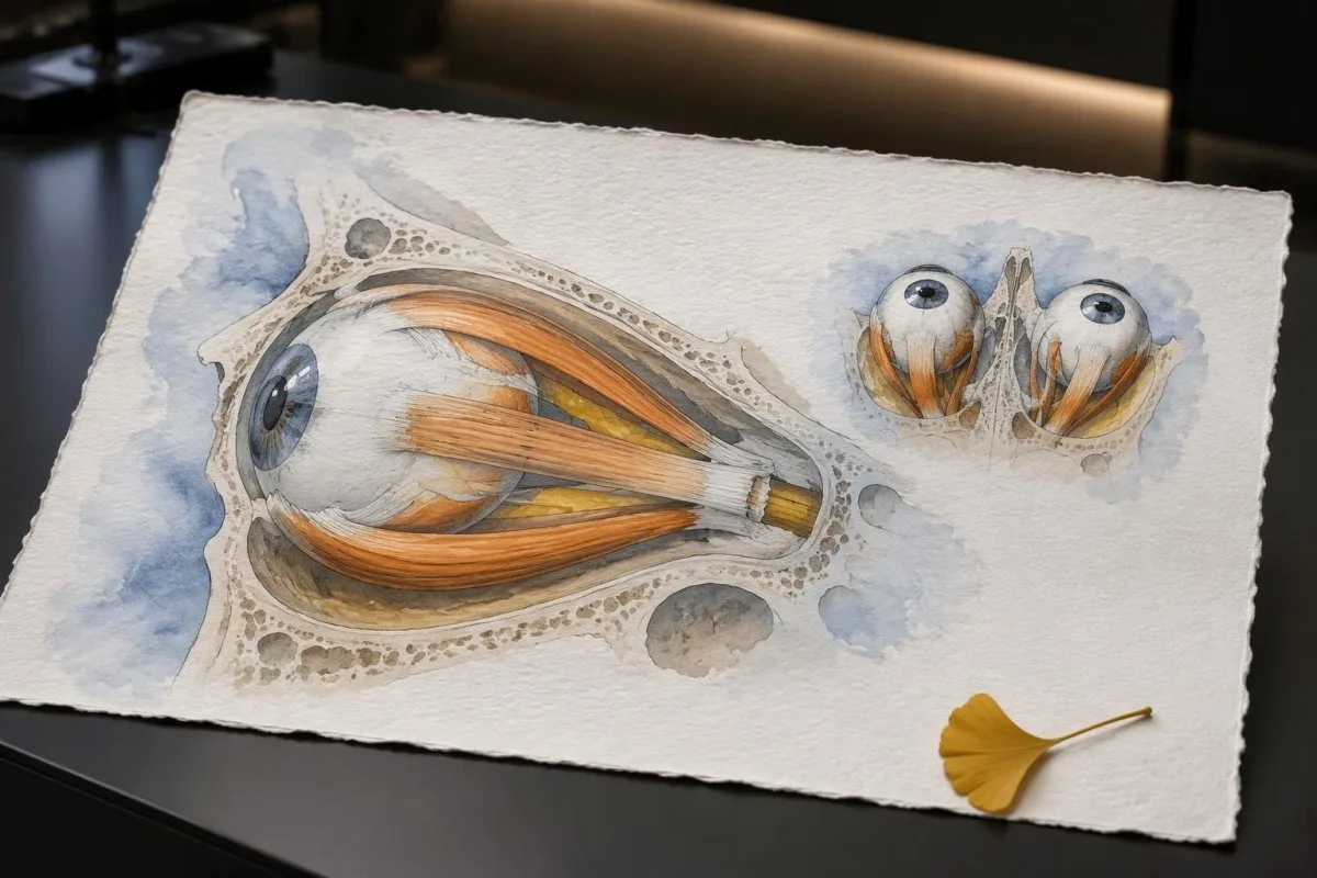
Proptosis describes forward displacement of the eyeball; eyelid retraction can make an eye look prominent even without substantial displacement. EUGOGO severity categories compare protrusion with population-specific norms, using a difference of at least 3 mm above those norms as one moderate-to-severe feature—not as an emergency threshold by itself.
Binocular double vision usually disappears when either eye is covered, because the symptom depends on misalignment between 2 eyes. Persistent doubling in the uncovered eye can instead arise from tear-film, corneal, or lens problems; this distinction helps the clinician, but neither pattern should be self-diagnosed when newly developed.
Thyroid-related muscle restriction often makes double vision worse in a particular direction, such as looking upward. A hypothetical person may initially notice 2 road signs only when checking an overhead signal, then later notice doubling straight ahead; that progression matters more than whether the eyes look obviously prominent.
Fluctuating double vision with a drooping rather than retracted eyelid raises another possibility: ocular myasthenia. Our guide to myasthenia antibody testing limits explains why a negative result does not exclude it; symptoms affecting 1 eye position can have very different mechanisms, and some people have overlapping autoimmune conditions.
Why can thyroid eye disease occur with normal thyroid levels?
Thyroid eye disease with normal thyroid levels is possible because orbital autoimmunity is not simply a consequence of excess thyroid hormone. Normal TSH and free T4 describe thyroid function at that moment; they do not establish whether the tissues around either of the 2 eyes are affected.
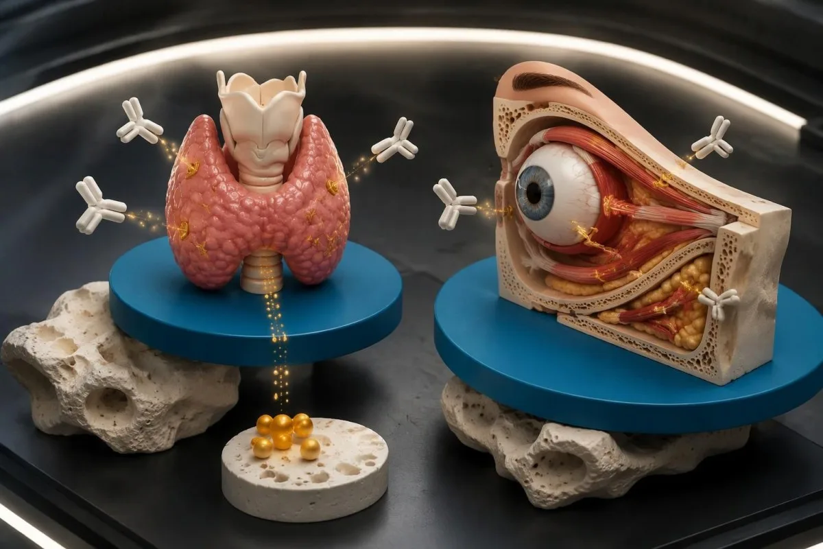
TSH receptor autoantibodies can interact with orbital fibroblasts through signaling networks that include the TSH receptor and IGF-1 receptor. These cells can contribute to tissue expansion and remodeling; the ATA/ETA consensus recognizes that thyroid eye disease can occur in euthyroid people and, less commonly, with hypothyroid autoimmune thyroid disease (Burch et al., 2022).
A treated person can have active eye disease despite a TSH of 1.8 mIU/L and free T4 of 16 pmol/L—both within many adult reference intervals. Those hypothetical numbers show controlled hormone production, not resolved orbital autoimmunity; review T4, T3 and TSH if the distinction on your report is unclear.
Eye symptoms can precede recognized thyroid dysfunction, appear around its diagnosis, or persist after hormone levels normalize. A single normal panel is therefore weaker evidence against thyroid eye disease than it may seem, particularly when 1 eye develops new retraction, movement restriction, or unexplained prominence.
TPO antibodies are not interchangeable with TRAb or TSI. TPO positivity supports thyroid autoimmunity but does not diagnose orbital involvement, and no single antibody result grades visual risk; our guide to high TPO antibody results explains why 2 different antibody tests can answer different clinical questions.
Which thyroid blood tests help explain eye symptoms?
TSH, free T4, and sometimes T3 establish thyroid function; TRAb or TSI can support an autoimmune explanation for suspicious eye findings. None measures optic nerve safety or corneal protection, and values must be interpreted against the reporting laboratory's own intervals rather than 1 universal cutoff.
Many adult laboratories use a TSH interval around 0.4–4.0 mIU/L and a free T4 interval around 10–22 pmol/L, approximately 0.8–1.7 ng/dL. Method, pregnancy, age, and local validation change these intervals, so the laboratory's printed range takes priority over a generic online table.
A suppressed TSH with normal free T4 may prompt T3 measurement because some hyperthyroidism is T3-predominant. Total T3 is influenced by binding proteins as well as thyroid output; our explanation of total T3 interpretation limits shows why 1 hormone result should not be separated from medication and physiological context.
Biotin can distort some thyroid immunoassays, producing a misleading combination of low TSH and high thyroid hormones. A hair supplement containing 5 mg supplies 5,000 µg of biotin; tell the laboratory about it and follow assay-specific withholding instructions rather than assuming a fixed interval, as discussed in biotin and thyroid assay errors.
Kantesti, our AI blood test interpretation platform, can help organize TSH, free T4, antibody results, and collection dates into a coherent explanation. For example, interpreting 3 successive panels requires knowing whether antithyroid treatment changed between them; the same improving laboratory trend cannot establish that new double vision is harmless.
| Level | Range | What it means |
|---|---|---|
| Common adult TSH interval | Approximately 0.4–4.0 mIU/L | Describes thyroid regulation; a normal result does not exclude thyroid eye disease. |
| Common adult free T4 interval | Approximately 10–22 pmol/L, or 0.8–1.7 ng/dL | Describes circulating free thyroxine; use the laboratory's own reference interval. |
| Markedly suppressed TSH | Below 0.1 mIU/L | May warrant evaluation for hyperthyroidism, treatment effects, or assay interference; not an eye-emergency threshold. |
| TRAb or TSI positivity | Assay-specific cutoff and units | Supports receptor-directed thyroid autoimmunity; does not measure visual function or prove orbital involvement alone. |
Which vision and color changes suggest optic nerve danger?
New dimming, reduced sharpness, washed-out colors, or a missing patch of vision can signal optic nerve dysfunction and require immediate assessment. A new difference between the 2 eyes is especially useful to report, but similar changes in both eyes are also urgent.
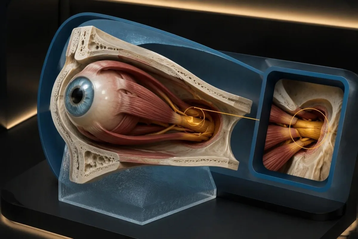
Apical crowding means enlarged tissues occupy the narrow space near the back of the orbit, where the optic nerve passes. This explains why someone can have relatively little visible bulging yet develop serious visual dysfunction; roughly 3–5% estimates for dysthyroid optic neuropathy describe a minority, not a reason to ignore its warning signs.
Red desaturation is a clue, not a reliable home screening test. If a familiar red object looks newly dull in 1 eye, seek emergency assessment rather than repeating comparisons under different lights; screens, cataracts, lighting, and congenital color differences can also influence perception and make a reassuring home comparison misleading.
A clinician may assess visual acuity, pupils, color discrimination, visual fields, and orbital imaging together. Normal reading-chart performance alone does not exclude early optic nerve compromise, because 1 central acuity measurement can miss changes in color or the wider visual field.
Optic neuritis and other optic nerve disorders can also produce movement-related pain and color loss. Our explanation of MOG antibody diagnostic context describes a different mechanism; the urgent action is an eye examination, not choosing between autoimmune diagnoses based on 1 symptom or antibody panel.
When is incomplete eyelid closure urgent?
New inability to close an eyelid fully needs same-day clinical advice because an exposed cornea can dry and become damaged. If closure difficulty comes with pain, light sensitivity, persistent blur, or marked redness, seek emergency eye care immediately rather than waiting for the next 1-day follow-up window.
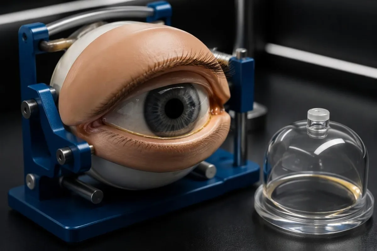
Lagophthalmos is incomplete eyelid closure; it may arise from proptosis, eyelid retraction, or both. The gap's size is not a universal safety cutoff: a 1 mm gap can matter if the tear film is poor or the eye remains exposed for hours during sleep.
Blur that clears after blinking often reflects tear-film instability, whereas blur that persists needs a different level of concern. This is not a rule-out test; checking each eye separately only once can help describe the symptom, but it must not delay care when vision has changed or the surface is painful.
Preservative-free lubricants can provide temporary protection while care is arranged, and a clinician may recommend nighttime ointment or a moisture chamber. Do not force a bulging eye closed, apply a tight patch, or improvise eyelid taping without instruction; prolonged pressure over 1 affected eye can create additional problems.
Eyelid symptoms are not always thyroid-related. A new rash around the forehead or nose with eye pain can suggest ocular shingles, even if thyroid tests are normal; our guide to shingles before the rash explains why an evolving symptom pattern over 1–3 days can change the diagnosis and urgency.
What other causes of eye pain need urgent assessment?
Severe eye pain, light sensitivity, sudden blur, or pain with systemic illness should not automatically be attributed to Graves disease. Acute glaucoma, uveitis, corneal disease, orbital infection, and neurological disorders are alternative concerns; a change in 1 eye may need emergency care even with established thyroid eye disease.
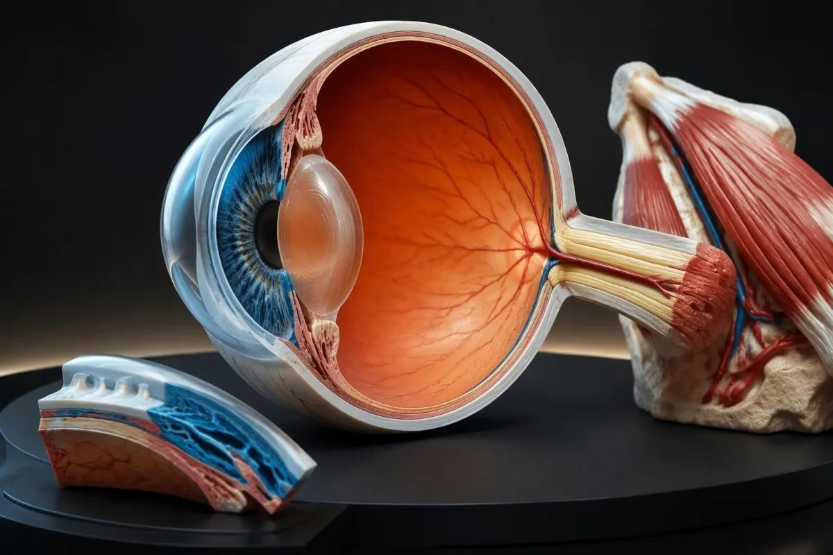
A painful red eye with halos, headache, nausea, or rapidly declining vision can indicate acute angle closure. That combination warrants emergency assessment, not an extra dose of thyroid medication; pressure-related emergencies and orbital thyroid disease affect different structures, even though a person can have more than 1 eye condition.
Uveitis commonly causes light sensitivity, redness, and pain, sometimes with blurred vision. An HLA-B27 result does not diagnose it, and a negative result does not exclude it; our guide to HLA-B27 and painful eyes explains why 1 genetic marker cannot substitute for a slit-lamp examination.
Fever with rapidly increasing eyelid swelling, painful eye movement, or a prominent eye raises concern for an orbital infection. Seek emergency care the same day; an established Graves diagnosis should not make a rapidly changing 24-hour pattern seem routine, especially if a sinus illness or recent procedure preceded it.
Sudden double vision with a severe headache, a new drooping eyelid, unequal pupils, weakness, or speech difficulty needs emergency assessment for neurological causes. Do not drive with new double vision or visual loss; arrange transport, and use the local emergency number if additional neurological symptoms develop within minutes rather than assuming 2 misaligned eyes reflect thyroid disease.
How do specialists assess activity, severity, and orbital crowding?
Specialists assess thyroid eye disease using eye function, eyelid and corneal findings, movement, proptosis measurements, and activity scoring. The 7-point Clinical Activity Score estimates inflammatory activity, while severity assessment asks whether daily function or sight is threatened; those are related but different questions.
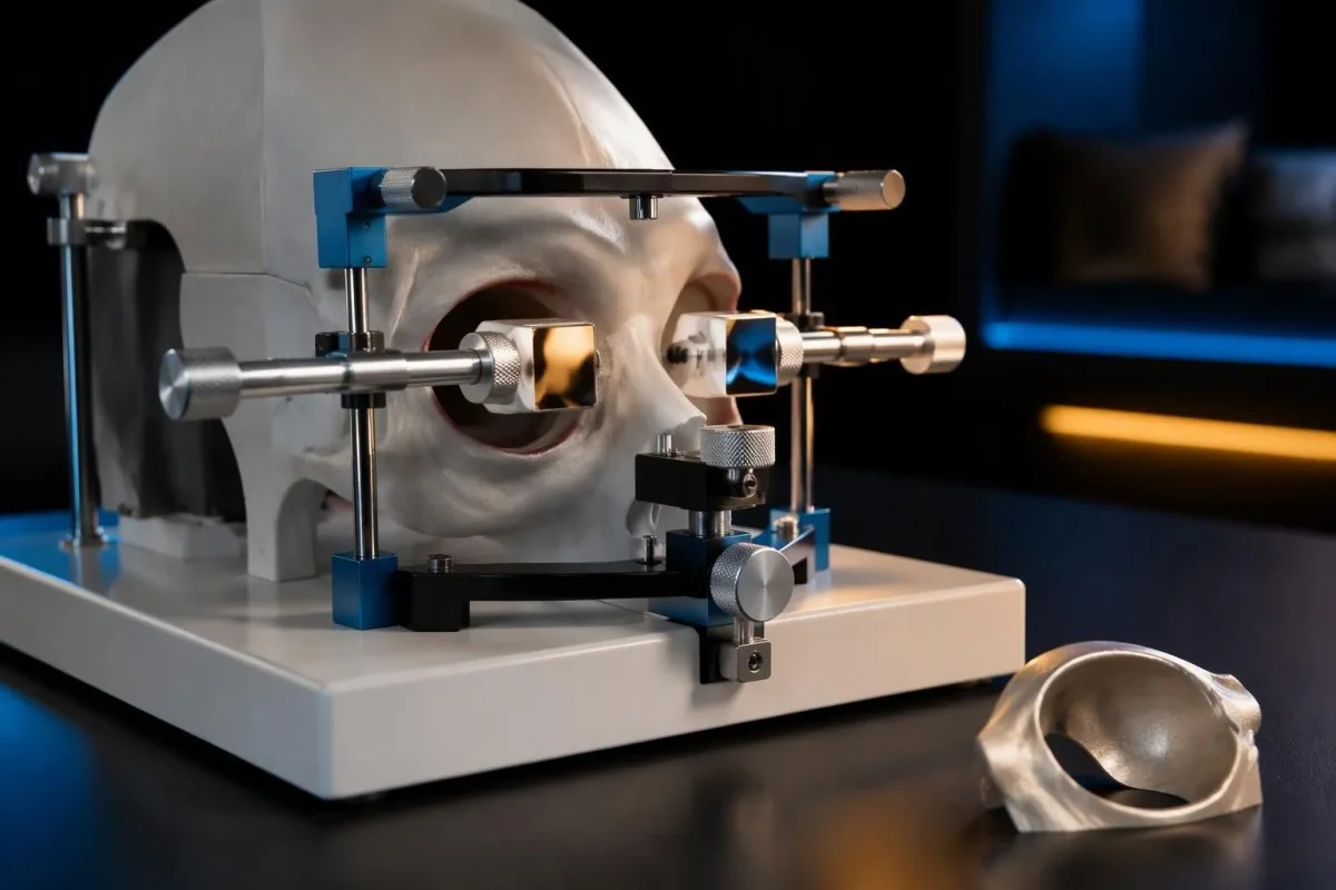
A Clinical Activity Score of at least 3 out of 7 commonly defines active disease, using features such as spontaneous orbital pain, movement-related pain, redness, and swelling. The 2021 EUGOGO guideline uses this framework, but a low score does not exclude optic neuropathy or substantial restrictive double vision (Bartalena et al., 2021).
An exophthalmometer measures eye prominence relative to the orbital rim; repeat measurements are most useful with consistent technique and the same instrument. A 2 mm change may attract attention during follow-up, but instrument differences, facial anatomy, and measurement variability mean the number must be interpreted alongside symptoms and visual testing.
Orbital CT or MRI can identify muscle enlargement, fat expansion, and crowding near the optic nerve. Imaging is especially useful when presentation is unusual, involvement is markedly one-sided, or optic neuropathy is suspected; the choice between these 2 modalities depends on the clinical question, radiation considerations, and available expertise.
Other orbital disorders can resemble thyroid eye disease, including IgG4-related disease, and broad autoimmune panels rarely resolve the distinction alone. Our guides to IgG4 result interpretation and autoimmune panel interpretation explain different laboratory limitations; neither 1 elevated marker nor normal complement establishes the cause of orbital enlargement.
What increases the risk of thyroid eye disease worsening?
Smoking, unstable thyroid function, and selected radioiodine treatment situations increase concern about thyroid eye disease progression. Active disease often evolves over roughly 6–24 months, but timing varies considerably; a presumed phase of recovery cannot override a new visual change in either of the 2 eyes.
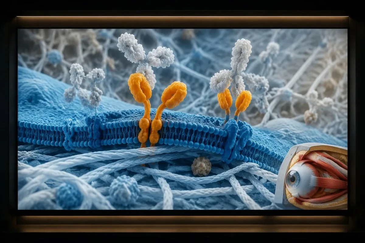
Smoking is a major modifiable risk factor for thyroid eye disease and is associated with poorer treatment response. Risk varies with exposure and population, so 1 universal risk multiplier is misleading; stopping smoking matters whether hormone levels are high, normal, or low, and reducing secondhand smoke exposure is also sensible.
Cotinine measures recent nicotine exposure rather than the specific injury caused by smoke. Nicotine replacement can produce a positive result, so it should not be interpreted as proof that someone is still smoking; our guide to cotinine and nicotine exposure explains why 2 exposure sources can produce similar laboratory findings.
Both persistent hyperthyroidism and hypothyroidism can complicate eye disease management, making stable thyroid function a treatment goal. After treatment changes, a TSH result may lag behind free T4 improvement; clinicians therefore examine the direction of several results rather than adjusting care around 1 isolated TSH measurement.
Radioiodine can worsen thyroid eye disease in susceptible people, particularly with smoking or existing active eye involvement. The EUGOGO and ATA/ETA documents discuss risk assessment and glucocorticoid protection in selected situations; this decision belongs with the treating team, and a person considering 1 radioiodine course should report eye symptoms before treatment is scheduled.
What helps mild symptoms, and when is selenium appropriate?
Mild thyroid eye disease symptoms may improve with lubricants, exposure protection, smoking cessation, and stable thyroid function. Selenium is an adjunct for selected mild, active disease—not a treatment for urgent vision changes—and evidence for a 6-month course is strongest in settings with relatively low selenium intake.
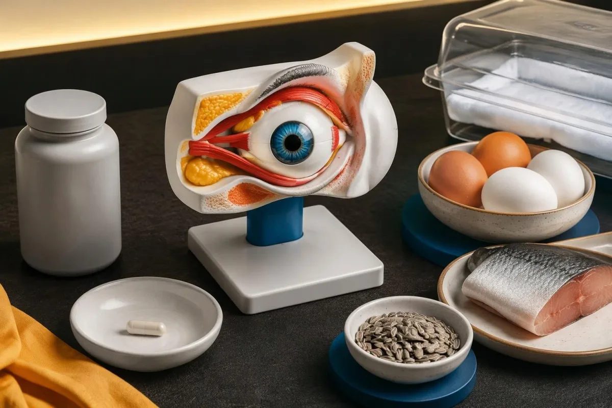
Preservative-free artificial tears are often useful when drops are needed repeatedly, and nighttime protection may help exposure symptoms. Frequency should match the product and eye-care advice rather than a rigid schedule; if 1 eye develops persistent blur, the priority shifts from adding more drops to checking the cornea and optic nerve.
The Marcocci trial studied selenium at 100 µg twice daily for 6 months in 159 people with mild Graves orbitopathy and reported improvements in quality of life and eye outcomes (Marcocci et al., 2011). The study's European setting limits certainty about benefit in selenium-sufficient populations, and its findings should not be extended to sight-threatening disease.
Do not combine a prescribed selenium course with several supplements containing the same nutrient. A 200 µg/day course plus a 100 µg multivitamin already supplies 300 µg before food; product formulations and regional upper intake limits differ, as explained in our guide to selenium dose and toxicity.
Extra iodine does not treat orbital autoimmunity and can destabilize thyroid function in susceptible people. Review thyroid diet and iodine before adding kelp or high-dose supplements; no food plan can reverse optic nerve compression, and evidence from 1 selenium trial does not validate a broader collection of thyroid supplements.
How should symptoms and thyroid results be tracked between visits?
Track visual symptoms separately from thyroid laboratory trends, because improvement in 1 does not prove improvement in the other. Record when double vision occurs, whether eyelids close, and whether either eye sees less clearly; worsening vision requires immediate care rather than waiting for a planned blood-test appointment.
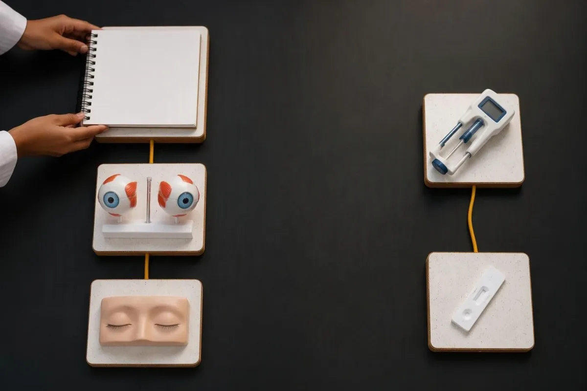
A useful record describes function rather than appearance alone: double vision straight ahead versus only looking upward, morning blur versus persistent blur, and whether both eyelids close. Take comparison photographs under similar lighting if requested, but avoid turning 1 daily photograph into a home severity score or a substitute for clinical measurement.
Consider a hypothetical 46-year-old whose TSH improves from below 0.01 to 1.6 mIU/L and whose free T4 falls from 34 to 15 pmol/L. If red suddenly looks faded in one eye, the improved thyroid numbers do not reduce urgency; the new visual symptom needs emergency assessment that day.
In a second hypothetical example, a 58-year-old has TSH 2.2 mIU/L, free T4 14 pmol/L, persistent lid retraction, and slowly increasing upward-gaze double vision. This pattern warrants specialist evaluation despite normal hormones; new straight-ahead doubling or declining vision would accelerate the assessment rather than justify waiting for another routine panel.
Kantesti can help organize dated laboratory results, but the treating team sets follow-up according to medication, disease activity, and eye findings. Thyroid panels are often repeated about 4–6 weeks after relevant treatment changes, with exceptions; our guide to thyroid testing intervals explains why 1 fixed schedule does not fit every thyroid condition.
What can AI explain, and what requires an eye examination?
AI can help explain thyroid biomarkers, units, and trends, but it cannot diagnose optic nerve compression or determine whether an exposed cornea is safe. Those questions require direct examination, and sometimes imaging; an interpretation of 3 normal thyroid results is not clearance for new eye symptoms.
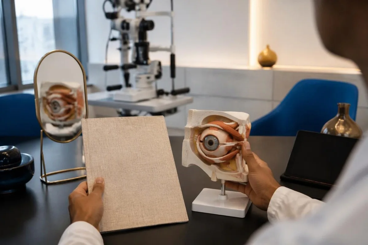
Kantesti is an AI biomarker interpretation platform that can help put TSH, free T4, T3, and thyroid antibodies into laboratory context. A useful explanation distinguishes 2 questions—whether thyroid function is controlled and whether autoimmunity is supported—while explicitly leaving visual safety to an appropriate eye-care professional.
Our AI interpretation technology guide describes the laboratory-report workflow rather than a camera-based eye diagnosis. If a report shows free T4 of 16 pmol/L, the next interpretive questions include the laboratory range, treatment timing, and previous values; none can establish whether a newly dimmer eye has normal optic nerve function.
Kantesti's organization and service scope provide background on the company behind the interpretation service. For this topic, the practical boundary is simple: 1 uploaded report can support a discussion about thyroid status, but a reassuring explanation must never postpone care for color loss, persistent visual blur, or incomplete painful eyelid closure.
Thyroid reports contain sensitive information, and family access should depend on the individual's consent rather than a shared surname or household. Review our privacy and data handling before sharing 2 family members' reports; keep each person's results separate so medication histories and abnormal values are not accidentally attributed to someone else.
Research publications and the limits of laboratory-based evidence
Clinical thyroid eye disease guidance comes from eye-specific evidence, not technical benchmarks of laboratory interpretation. The 2 Figshare publications listed below concern the interpretation engine and a different triage application; neither establishes that AI can diagnose Graves ophthalmopathy, detect optic neuropathy, or safely classify eye pain.
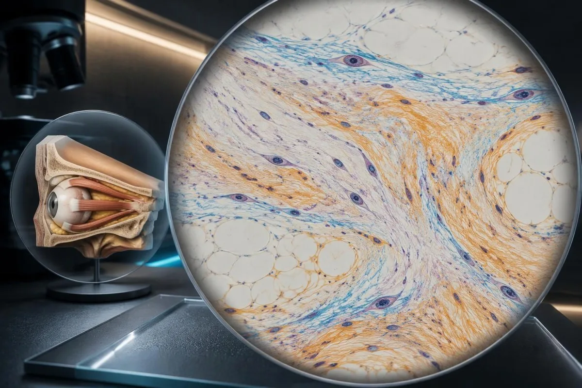
The 2021 EUGOGO guideline and 2022 ATA/ETA consensus support separating activity, severity, and sight-threatening complications. Their treatment frameworks overlap but are not identical, particularly for moderate-to-severe disease, where availability, local approval, patient characteristics, and the strongest treatment evidence differ; neither framework uses normal TSH as a reason to dismiss visual deterioration.
The selenium trial supports a defined intervention in a defined population: 159 participants with mild disease followed through a 6-month treatment course. That evidence does not establish efficacy for every supplement formulation, every nutritional setting, or severe optic nerve disease; the uncertainty is clinically relevant, not simply a caveat attached to an otherwise universal recommendation.
The listed Kantesti publications address engineering and technical evaluation questions. A benchmark described as using 100,000 synthetic cases can assess performance under its stated rubric, but synthetic-case results cannot establish sensitivity for optic neuropathy in real patients; our technical and clinical oversight should be read with that distinction in mind.
Our medical content standards distinguish educational explanations from individualized diagnosis. For this article, the safety rule remains unchanged by any laboratory result or technical score: new visual loss, new color desaturation, or a painful exposed corneal surface requires immediate clinical assessment, even when 2 successive thyroid panels are normal.
Frequently asked questions
When is eye pain from Graves disease an emergency?
Eye pain with reduced vision, washed-out colors, a missing area of vision, or painful incomplete eyelid closure requires emergency eye assessment. Dysthyroid optic neuropathy affects roughly 3–5% of people with thyroid eye disease in commonly cited clinical estimates, and severe pain is not necessary for it to occur. Seek an emergency eye service or emergency department immediately rather than waiting 24 hours for thyroid testing. Do not drive if vision is impaired or you have new double vision.
Can you have thyroid eye disease with normal thyroid levels?
Thyroid eye disease can occur with normal thyroid hormone levels because orbital autoimmunity is not the same process as excessive thyroid hormone production. A TSH of 2.0 mIU/L and free T4 of 15 pmol/L would fall within many adult laboratory intervals but would not exclude thyroid eye disease. TRAb or TSI may support an autoimmune explanation, while eye examination establishes the orbital findings. Normal blood tests must not delay urgent assessment of declining vision or newly faded colors.
What are the earliest symptoms of thyroid eye disease?
Early thyroid eye disease can cause persistent grittiness, watering, eyelid swelling, deep orbital aching, and a changing upper eyelid position. Symptoms may affect 1 eye more noticeably before obvious bulging develops. Dry-eye symptoms alone are nonspecific, but accompanying lid retraction or restricted eye movement increases concern about an orbital process. New visual dimming, color loss, or persistent double vision needs faster assessment than stable irritation alone.
Does faded red vision mean thyroid eye disease is damaging the optic nerve?
Newly faded red vision can indicate optic nerve dysfunction, including dysthyroid optic neuropathy, but it does not identify the cause on its own. A new difference between the 2 eyes is useful to report and warrants immediate assessment when thyroid eye disease is suspected. Clinicians combine color testing with visual acuity, pupils, visual fields, and sometimes orbital imaging. A reassuring home color comparison cannot exclude optic nerve compromise.
What should I do if my eyelid will not close completely?
New incomplete eyelid closure needs same-day clinical advice because the corneal surface may be exposed. Even a 1 mm gap can matter when the tear film is poor or exposure continues for hours. Pain, light sensitivity, marked redness, or persistent blurred vision makes emergency eye assessment necessary. Lubrication may provide temporary protection, but do not force the eyelid closed or apply tight pressure over the eye.
Does selenium help Graves ophthalmopathy symptoms?
Selenium can help selected people with mild, active Graves orbitopathy, but it is not a treatment for urgent visual deterioration. A 2011 randomized trial studied 100 µg twice daily for 6 months in 159 participants and reported improved quality of life and eye outcomes. Benefit in selenium-sufficient populations is less certain, and total intake from supplements and food must be considered. Discuss supplementation with the treating team rather than combining several selenium-containing products.
Get your blood test results explained
Upload your lab report and Kantesti's AI explains each biomarker in plain language within seconds. Used by more than 2 million people worldwide.
References
- Klein, T., Mitchell, S., & Weber, H. (2026). Multilingual AI Assisted Clinical Decision Support for Early Hantavirus Triage: Design, Engineering Validation, and Real-World Deployment Across 50,000 Interpreted Blood Test Reports. Kantesti AI Medical Research. DOI
- Klein, T., Mitchell, S., & Weber, H. (2026). A Pre-Registered, Rubric-Based Automated Technical Benchmark of the Kantesti Blood-Test Interpretation Engine on 100,000 Synthetic Test Cases. Kantesti AI Medical Research. DOI
- Bartalena L et al. (2021). The 2021 European Group on Graves' orbitopathy (EUGOGO) clinical practice guidelines for the medical management of Graves' orbitopathy. European Journal of Endocrinology. DOI
- Burch HB et al. (2022). Management of Thyroid Eye Disease: A Consensus Statement by the American Thyroid Association and the European Thyroid Association. Thyroid. DOI
- Marcocci C et al. (2011). Selenium and the Course of Mild Graves' Orbitopathy. The New England Journal of Medicine. DOI
- 2M+Tests analysed
- 127+Countries
- 100+Languages
About the medical team


E-E-A-T Trust Signals
Experience
Physician-led clinical review of lab interpretation workflows.
Expertise
Laboratory medicine focus on how biomarkers behave in clinical context.
Authoritativeness
Medically reviewed by Dr. Thomas Klein, with contributions from Dr. Sarah Mitchell and Prof. Dr. Hans Weber.
Trustworthiness
Evidence-based interpretation with clear follow-up pathways to reduce alarm.


Medical disclaimer
This article is for educational purposes only and does not constitute medical advice. Always consult a qualified healthcare provider for diagnosis and treatment decisions.
Published · Contact the editorial team
Published by Kantesti Ltd, registered in England & Wales, company no. 17090423, London, United Kingdom.
