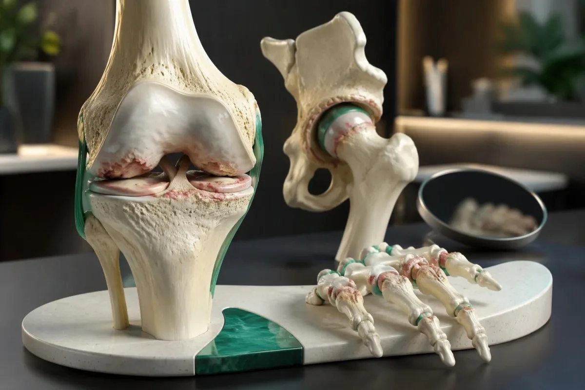
The first warning signs are usually joint pain during or after activity, stiffness after rest that generally lasts under 30 minutes, and gradually reduced movement. Knees may hurt on stairs, hips when putting on socks, and hands when gripping. Blood tests cannot confirm osteoarthritis; a hot, rapidly swollen joint needs urgent assessment.

Key takeaways
- Activity-related pain commonly appears with walking, stairs, gripping, or repeated joint loading; one painful activity alone does not establish the diagnosis.
- Brief stiffness after waking or sitting usually lasts no longer than 30 minutes in a typical osteoarthritis presentation.
- Clinical diagnosis is often sufficient in adults aged 45 or older with activity-related joint pain and absent or brief morning stiffness, according to NICE.
- Knee symptoms include stair pain, difficulty fully straightening the joint, and intermittent swelling; painless clicking alone is not diagnostic.
- Hip symptoms can include groin pain, difficulty putting on socks, and pain referred to the knee—even when the hip does not feel obviously painful.
- Hand symptoms commonly affect the thumb base and finger joints nearest the nail or in the middle of the finger; numbness suggests an additional or different problem.
- Blood test limits mean that no routine CRP, ESR, rheumatoid factor, or metabolic panel result can confirm or exclude osteoarthritis.
- Urgent warning signs include a suddenly hot, swollen joint, inability to bear weight after injury, or leg swelling with chest pain or breathlessness; do not wait for an AI interpretation.
What are the early signs of osteoarthritis?
Early signs of osteoarthritis are activity-related joint pain, brief stiffness after rest, and a gradual loss of comfortable movement. The pattern matters more than one symptom: NICE permits a clinical diagnosis in adults aged 45 or older with activity-related pain and no morning stiffness, or stiffness lasting no longer than 30 minutes.
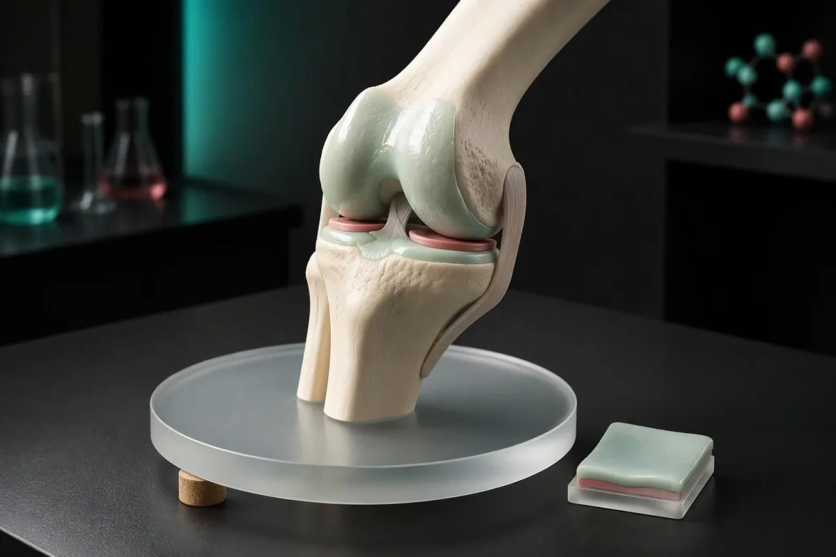
Osteoarthritis affects the whole joint, not just the cartilage covering its bones. Changes in the joint lining, underlying bone, supporting muscles, and ligaments can all contribute to symptoms; the NICE age threshold of 45 years describes a practical diagnostic pathway, not a biological rule that excludes younger adults (NICE, 2022).
For example, a hypothetical 56-year-old notices knee aching after a longer walk, stiffness for 8 minutes after breakfast, and difficulty kneeling that was absent a few months earlier. Together, these findings support an osteoarthritis assessment more than an isolated click would, but examination still needs to check for tendon problems, injury, and inflammatory joint disease.
Kantesti is an AI blood test analyzer that explains laboratory findings without treating them as proof of osteoarthritis. Our organization and clinical scope are separate from an in-person joint examination: recording 3 details—the triggering activity, stiffness duration, and lost function—often gives a clinician more useful starting information than ordering a broad laboratory panel.
How does activity-related joint pain usually behave?
Osteoarthritis pain typically increases during or after loading the affected joint and eases with reduced loading, especially early in the condition. A useful distinction is between stiffness during the first few steps after rest and aching that builds after 20 minutes of walking; both can occur in the same joint.
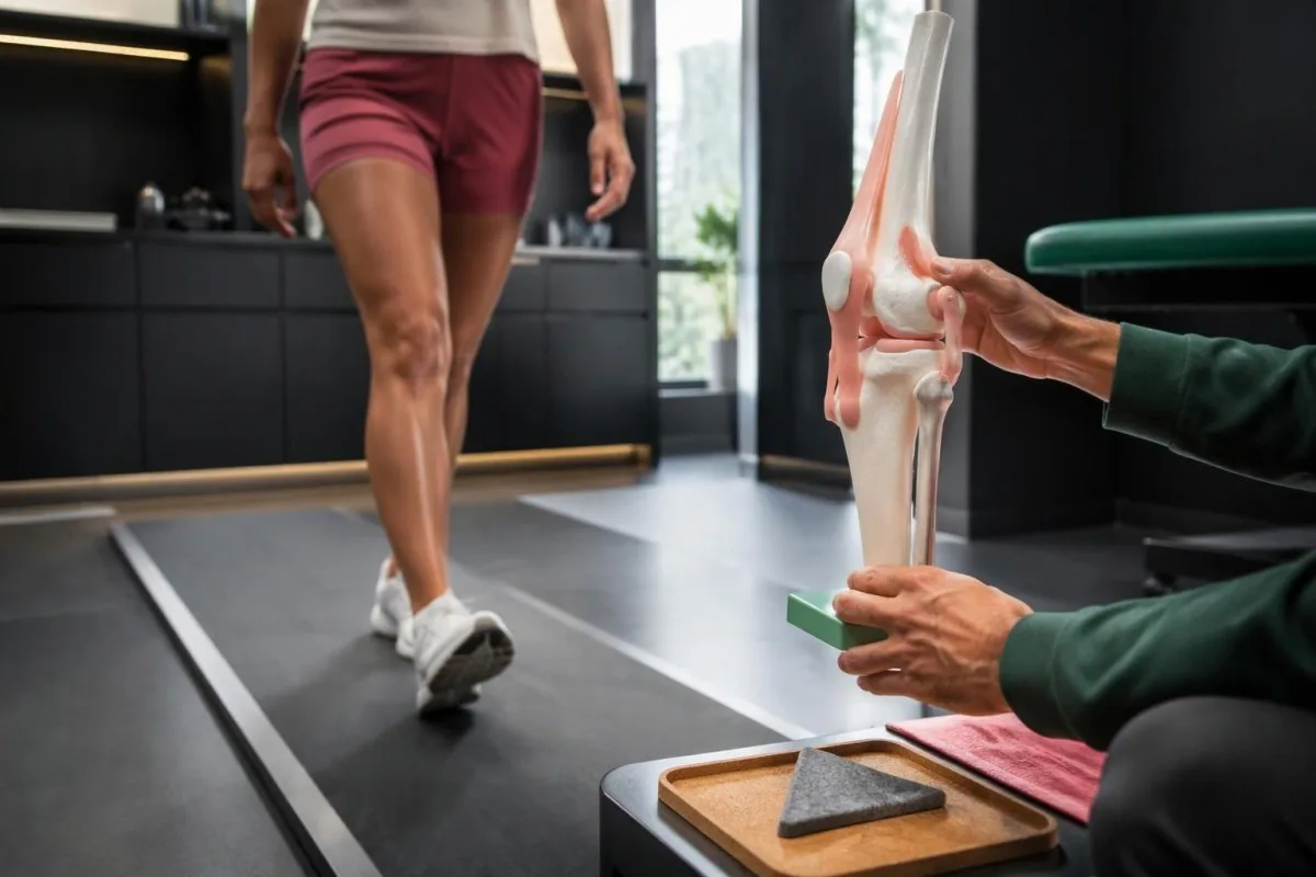
A reproducible trigger helps locate the problem, but does not identify its cause by itself. In a hypothetical diary, knee pain rising from 2/10 to 5/10 during stair climbing suggests load sensitivity; pain from a tendon, a previous injury, or the kneecap joint can produce a similar pattern and may coexist with osteoarthritis.
Pain at rest does not automatically rule out osteoarthritis because more established disease can disturb sleep or ache after a busy day. Nevertheless, new persistent rest pain, rapid worsening over several days, or pain accompanied by fever or unexplained weight loss deserves assessment rather than being attributed to an existing X-ray finding.
Calf pain that predictably starts after a similar walking distance and settles within a few minutes of stopping may reflect circulation rather than the knee joint. Our guide to arterial leg pain explains that distinction; note whether stopping alone helps or whether changing the joint position is what brings relief.
Which osteoarthritis knee symptoms appear first?
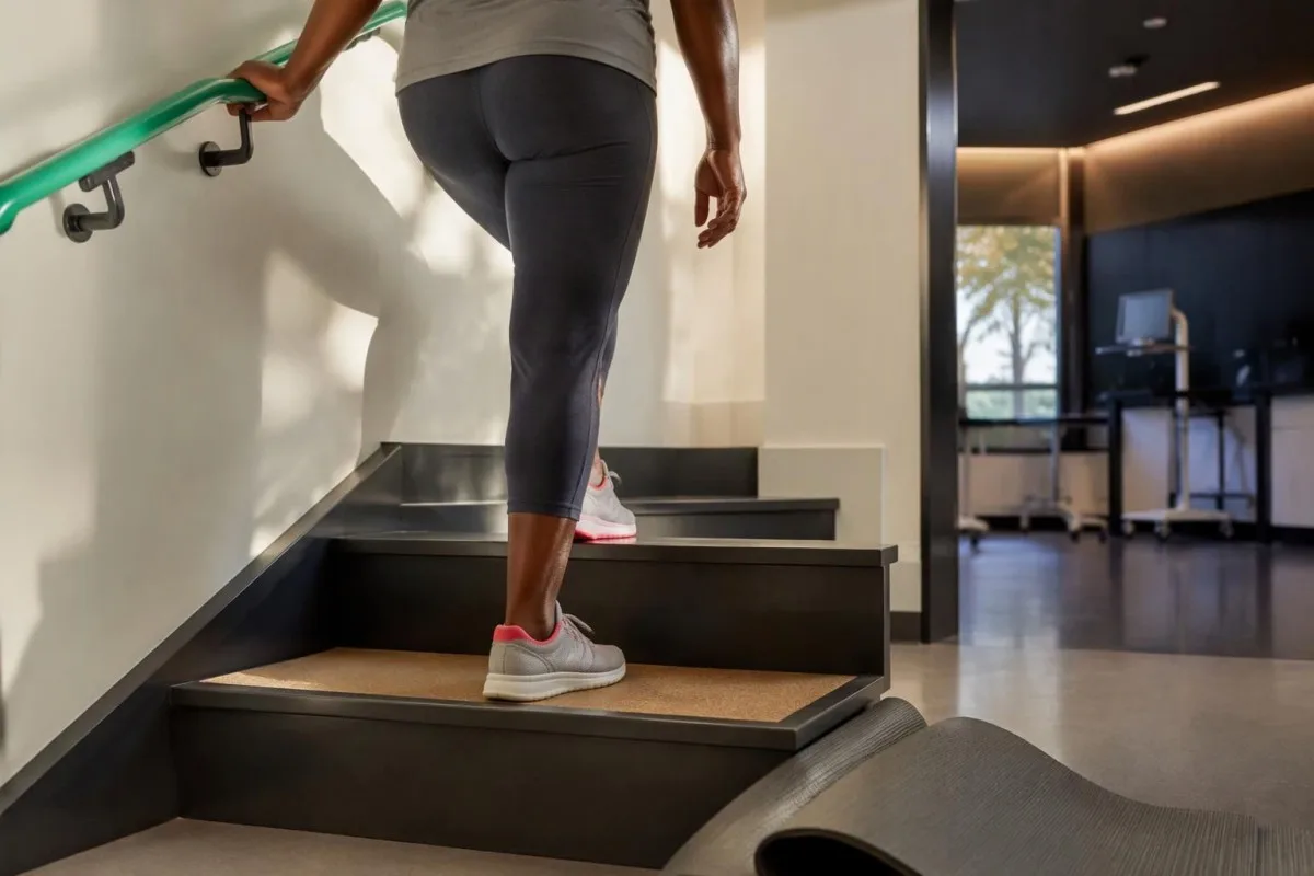
Osteoarthritis knee symptoms often begin with pain on stairs, rising from a chair, squatting, or walking farther than usual. Early discomfort may be around the kneecap or along either joint line; a hypothetical 60-year-old struggling with one flight of stairs needs the pain pattern examined, not diagnosed from its location alone.
Grinding or crackling can accompany knee osteoarthritis, but noise alone does not show cartilage loss or establish pain severity. Compare 2 features instead: whether the noise is painful and whether movement has changed; painless clicking in a knee that still functions normally is less informative than a new inability to straighten it comfortably.
Intermittent puffiness may reflect fluid within the joint, while a sense of giving way can result from pain-related muscle inhibition rather than a torn ligament. A knee that becomes physically stuck and cannot straighten—a true locked knee, even after one episode—needs prompt assessment because a mechanical obstruction may require a different pathway.
Swelling extending into the calf should not automatically be blamed on knee arthritis or a fluid-filled cyst behind the knee. New swelling in 1 leg, especially with calf tenderness or clot risk factors, warrants urgent evaluation; our urgent leg swelling guide explains why examination and appropriate testing are needed before assuming a harmless cause.
Can hip osteoarthritis cause groin or knee pain?
Hip osteoarthritis commonly causes groin pain and difficulty rotating the hip, but pain can also reach the thigh, buttock, or knee. If 1 knee hurts without a convincing knee explanation, examining the hip can uncover the source; putting on socks or getting into a car may reveal restricted movement.
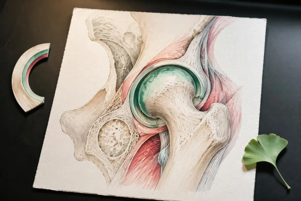
Loss of comfortable inward hip rotation can be an examination clue, but no single movement confirms the diagnosis. In a hypothetical 58-year-old, groin aching after walking plus difficulty reaching the foot is more suggestive of a hip joint problem than isolated tenderness over the outer hip, which often points toward surrounding tendons.
Burning pain travelling below the knee, tingling, or weakness suggests a nerve contribution rather than osteoarthritis alone. Our explanation of sciatica and nerve signs separates these patterns; a person can have 2 simultaneous problems, so an arthritic hip on an X-ray should not erase neurological symptoms from the assessment.
A sudden change in hip function deserves a different level of attention from gradual stiffness. For example, a hypothetical 67-year-old who develops marked groin pain and cannot bear weight after a minor fall needs urgent fracture assessment, even if previous imaging showed osteoarthritis; some fractures are not visible on the first plain X-ray.
What do osteoarthritis hand symptoms feel like?
Osteoarthritis hand symptoms often include aching during pinching or gripping, stiffness after inactivity, and reduced dexterity. The thumb-base joint and 2 groups of finger joints—those nearest the nail and those in the middle of the finger—are common sites; firm bony enlargements may appear later rather than being the first warning.
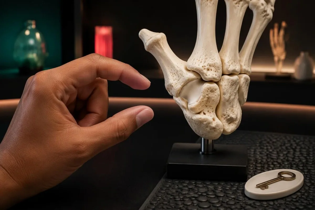
Thumb-base osteoarthritis may first become noticeable when turning a key, opening a jar, or holding a phone for a prolonged period. A hypothetical 62-year-old can still grip strongly with the whole hand yet struggle with precise pinching, because those tasks distribute force differently; reduced function is not always equivalent to generalized weakness.
Numbness is not a typical direct effect of finger-joint osteoarthritis. Tingling in the thumb, index finger, middle finger, and part of the ring finger can suggest median nerve compression; our guide to carpal tunnel finger patterns explains why symptoms in roughly 3½ fingers lead to a different examination from painful bony finger joints.
The distribution of swelling changes the differential diagnosis: persistent soft swelling across the knuckles is less typical than firm enlargement near the fingertips. If hand enlargement accompanies 2 additional changes, such as increasing shoe size and altered facial features, our hand enlargement warning signs explain why an endocrine cause may need consideration rather than an arthritis assumption.
How long should osteoarthritis stiffness last?
Osteoarthritis stiffness usually lasts no longer than 30 minutes after waking and is often shorter after sitting. This brief “gelling” differs from sustained inflammatory stiffness, but the cutoff is a clinical guide rather than a stopwatch test: duration, swelling, affected joints, and general symptoms must be considered together.
Measure stiffness until the joint moves reasonably freely, not until every trace of pain disappears. In a hypothetical 7-day diary, stiffness lasting 5–12 minutes followed by later activity-related aching fits a different pattern from an hour of difficulty moving every morning; separating pain from stiffness prevents an inflated estimate.
Prolonged shoulder and hip-girdle stiffness in someone older than 50 years raises consideration of polymyalgia rheumatica, particularly when morning stiffness exceeds 45 minutes. Our guide to polymyalgia testing limits explains why normal inflammatory markers do not always settle that question; the pattern is broader than restricted motion in one hip joint.
Back pain beginning before 45 years, lasting over 3 months, and improving with movement rather than rest can suggest inflammatory spinal disease. The HLA-B27 clinical context matters more than the genetic result alone; neither a positive test nor a stiffness duration independently establishes the diagnosis.
Which movement changes are worth recording?
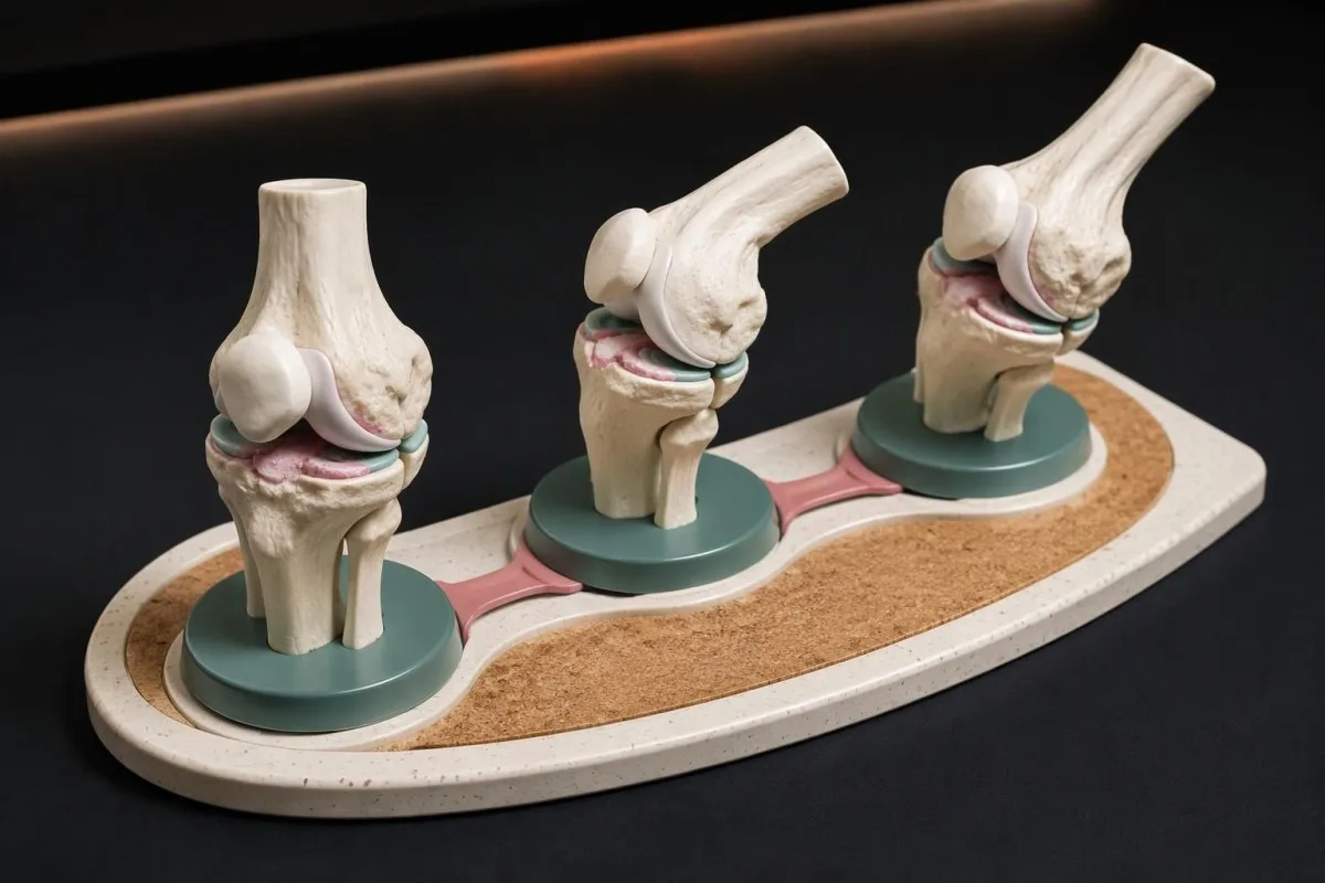
Reduced movement may appear as difficulty fully straightening a knee, rotating a hip, or closing the hand comfortably. Compare the same 2 or 3 everyday tasks over time rather than forcing a joint through its limits; a change in function often communicates the problem more clearly than a home estimate of movement angles.
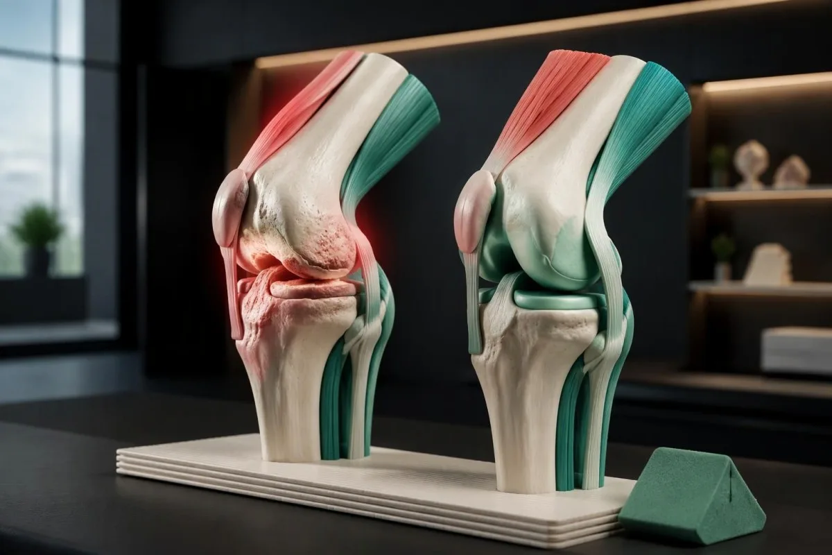
A knee that remains slightly bent while standing can alter walking and increase the effort required from surrounding muscles. Clinicians may measure extension against a straight position of approximately 0 degrees, but body variation, previous injury, and examination technique matter; a small measured difference is not an independent diagnosis or a reason to force stretching.
Choose tasks that reflect the affected joint: 1 flight of stairs for a knee, putting on a sock for a hip, or turning a familiar key for the thumb. At Kantesti, we distinguish functional observations from laboratory interpretation because a stable CRP result cannot show whether a person has lost the ability to perform those movements.
Osteoporosis and osteoarthritis describe different problems, despite their similar names. Osteoporosis often remains silent until a fracture, whereas osteoarthritis usually produces joint-related symptoms; our bone loss testing guide explains why even 1 low-trauma fracture can justify bone-health assessment without implying that stiff finger joints are caused by osteoporosis.
Can osteoarthritis cause swelling or warmth?
Osteoarthritis can cause intermittent joint swelling and mild warmth, because local joint-lining changes are part of the condition. A suddenly very hot, markedly swollen single joint is different: it needs urgent assessment for infection, crystal arthritis, or another acute cause, even when the person has established osteoarthritis.
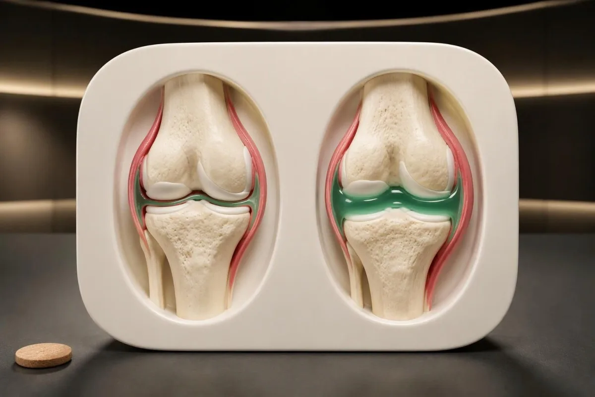
Firm bony enlargement and soft fluid-related swelling are not the same finding. In a hypothetical 64-year-old, a long-standing hard finger-joint bump may reflect osteoarthritis, whereas a knee enlarging over 12 hours deserves a fresh assessment; the speed of change can be more useful than whether the joint was previously labelled arthritic.
Normal blood results cannot safely dismiss an acutely hot joint. Our explanation of WBC and CRP disagreement shows why timing and clinical findings matter; a result collected early on day 1 may not reflect the full response, and joint infection can occur without striking systemic laboratory abnormalities.
Joint-fluid testing is sometimes needed because examination and blood markers cannot reliably separate all acute causes. Fluid may be checked for crystals, cellular counts, and culture; identifying 1 crystal type does not automatically exclude simultaneous infection, so a clinician must interpret the sample alongside symptoms, risk factors, and the course of the illness.
Why can blood tests not confirm osteoarthritis?
No routine blood test confirms or excludes osteoarthritis. Diagnosis depends on symptoms, examination, and sometimes imaging; CRP, ESR, and antibody tests can investigate alternative explanations but cannot measure whether a particular knee or thumb joint has osteoarthritis. There are 0 standard laboratory confirmation tests for routine clinical osteoarthritis diagnosis.
CRP is a nonspecific marker of systemic inflammation, not a cartilage-loss measurement. If a hypothetical laboratory uses a reference interval below 5 mg/L, a CRP of 18 mg/L calls for clinical context rather than proving inflammatory arthritis; local osteoarthritis changes, another illness, and other contributing factors can coexist, and reference intervals differ between laboratories.
Rheumatoid factor and anti-CCP testing are most useful when the history and examination suggest inflammatory arthritis, especially persistent synovitis. Our guide to rheumatoid factor interpretation explains why 1 positive antibody result cannot replace the examination; negative antibodies also do not exclude rheumatoid arthritis or turn an uncertain joint problem into confirmed osteoarthritis.
Kantesti is an AI blood test interpretation platform that helps explain reported markers without claiming to identify osteoarthritis from them. Our biomarker interpretation guide supports checking 3 report details—the units, laboratory reference interval, and collection date—before discussing an abnormal result with a clinician; broad screening panels are not automatically useful for a typical osteoarthritis presentation.
Which symptom patterns suggest another diagnosis?
Persistent soft joint swelling, prolonged morning stiffness, acute redness, nerve symptoms, or illness outside the joints can suggest something other than osteoarthritis. Pattern recognition helps select testing: swelling across several knuckles points toward a different assessment from isolated thumb-base pain, while abrupt swelling in 1 joint raises crystal or infectious causes.
Symmetrical knuckle swelling with prolonged stiffness can suggest rheumatoid arthritis, while Raynaud symptoms, rashes, or muscle weakness may broaden the autoimmune assessment. Our connective tissue testing guide explains why 1 antibody, such as anti-RNP, is insufficient by itself; ordering many antibodies without a compatible symptom pattern can create misleading incidental positives.
Gout and calcium pyrophosphate crystal arthritis can cause sudden attacks, and calcium pyrophosphate deposition may also coexist with osteoarthritis. A serum urate below 6 mg/dL during an acute episode does not reliably rule out gout; when the diagnosis or infection risk is uncertain, examining joint fluid can be more informative than repeating a routine blood panel.
Iron overload can produce an osteoarthritis-like pattern, sometimes involving the second and third knuckles. Persistent transferrin saturation around 45% or higher can prompt iron-overload assessment, but does not establish its cause; Kantesti does not recommend this test for every aching joint, and our complete iron study explanation distinguishes saturation from ferritin, which can also rise with inflammation or liver disease.
When do X-rays or MRI scans help?
Imaging helps when symptoms are atypical, the diagnosis is uncertain, or the result could change treatment. NICE does not recommend routine diagnostic imaging for a typical clinical presentation; EULAR likewise advises that imaging is unnecessary in typical osteoarthritis and identifies plain radiography as the first imaging choice when imaging is needed.
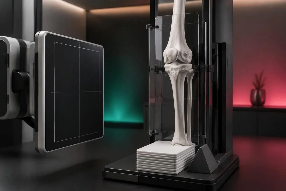
For the knee, weight-bearing radiographs can make joint-space narrowing more apparent than a non-weight-bearing view. EULAR recommends appropriate views and techniques rather than indiscriminate scanning (Sakellariou et al., 2017); importantly, 1 X-ray cannot directly measure pain, and prominent structural changes may coexist with modest symptoms while early symptoms can precede obvious radiographic changes.
MRI is not a routine second step simply because an osteoarthritis X-ray looks mild. It can be useful for a suspected occult fracture, another structural disorder, or selected mechanical symptoms; in a hypothetical 66-year-old with abrupt severe pain and difficulty bearing weight despite an unrevealing X-ray, the clinical concern—not the desire to grade arthritis—determines further imaging.
Repeat scans are usually less useful than reassessing symptoms and function when management remains unchanged. NICE advises against routine follow-up imaging for nonsurgical management (NICE, 2022); Kantesti's methodology and clinical limits should be read with the same distinction in mind: a technical interpretation benchmark is not proof that an AI can diagnose joint disease from 1 laboratory report.
Which joint symptoms need urgent medical evaluation?
A suddenly hot, swollen joint, severe pain with inability to bear weight, or rapid loss of function needs urgent medical evaluation. Seek same-day care for an unexplained acute hot joint, even without fever; a temperature below 38°C does not safely exclude joint infection, particularly in older or immunosuppressed adults.
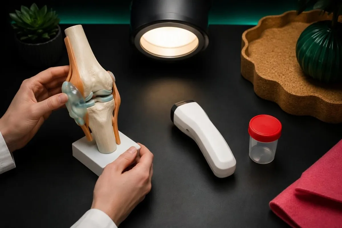
After an injury, inability to bear weight, a visible change in joint shape, or rapidly increasing swelling requires prompt assessment for fracture or another significant injury. Even a low-height fall can matter in someone with fragile bones; an existing diagnosis of osteoarthritis should not delay evaluation of a new event or justify walking through severe pain.
Emergency symptoms can occur near an arthritic joint without being caused by it. Leg swelling with chest pain or breathlessness warrants emergency care; new bladder retention, saddle-area numbness, or rapidly worsening leg weakness also requires immediate assessment, and 1 new neurological red flag is enough to change the plan from a routine arthritis appointment.
A swollen knee developing over days to weeks after plausible tick exposure may require an infection-focused assessment rather than an osteoarthritis label. Our guide to Lyme test interpretation explains why exposure history and validated testing matter; a hot acute joint still needs urgent assessment, while a random positive antibody result does not establish the cause of swelling.
What can you do while arranging a routine assessment?
For nonurgent, suspected osteoarthritis, keep moving within tolerable limits and arrange an assessment if symptoms persist or restrict daily life. Exercise is a core treatment once the diagnosis is established; a hypothetical starting plan might use 5–10 minutes of comfortable walking rather than alternating complete rest with a suddenly much longer session.

Exercise should be adapted to the affected joint, current capacity, and any warning signs rather than prescribed as one universal routine. The ACR/Arthritis Foundation guideline strongly recommends exercise for hand, hip, and knee osteoarthritis (Kolasinski et al., 2020); try changing 1 variable at a time, such as duration, so a clinician or physiotherapist can identify what is tolerable.
Weight loss can reduce symptoms for people with knee or hip osteoarthritis who are overweight, but it is not a prerequisite for receiving care. NICE states that losing 10% of body weight is likely to help more than 5%; our evidence-based food choices can support balanced eating, although no specific food has been shown to rebuild an osteoarthritic joint.
Pain treatment needs a safety review, particularly before regularly taking an oral anti-inflammatory medicine. NICE recommends topical NSAIDs for knee osteoarthritis, while oral NSAIDs require consideration of gastrointestinal, kidney, and cardiovascular risks; at Kantesti, we encourage recording the exact mg dose and frequency, and our medication-related laboratory guide explains why accompanying medicines and kidney results matter.
How should you use lab explanations and related research?
Use laboratory explanations to prepare better questions, not to self-confirm osteoarthritis or postpone a joint examination. Kantesti is an AI-powered blood test analysis tool that supports understanding of reported results; it cannot determine the cause of a painful knee from 1 CRP value, assess joint warmth remotely, or replace urgent care.

Bring a concise symptom record to a routine appointment: the joint involved, trigger, stiffness duration, swelling, injury history, and lost function. A 7-day diary is a practical example, not a requirement to wait; our responsible AI guidance explains why an automated result should not override a clinician's examination or an urgent change in symptoms.
Laboratory reports sometimes contain unrelated findings that still need their own context. For example, 1 urine dipstick abnormality cannot explain knee pain without a relevant history; our urinalysis interpretation guide addresses urinary findings separately, preventing an incidental result from being presented as evidence for osteoarthritis or used to justify unnecessary treatment.
The 2 DOI-linked publications below are supplemental laboratory education resources on urinalysis and iron studies, not osteoarthritis diagnostic studies. The directly relevant clinical references are NICE NG226, EULAR's imaging recommendations, and the ACR/Arthritis Foundation management guideline; a DOI makes a document identifiable, but does not itself establish peer review, clinical validation, or relevance to a joint diagnosis.
Frequently asked questions
What is usually the first symptom of osteoarthritis?
The first noticeable osteoarthritis symptom is often joint pain during or after activity, accompanied by brief stiffness after rest. Morning stiffness generally lasts no longer than 30 minutes in a typical presentation. A knee may hurt on stairs, a hip when putting on socks, and a thumb when opening containers. These patterns support assessment but are not enough to diagnose every painful joint without an examination.
Can blood tests show whether I have osteoarthritis?
No routine blood test can confirm or exclude osteoarthritis. A CRP within a laboratory's reference interval, often below 5 mg/L, does not prove that a joint is healthy or explain its pain. Blood tests may help investigate rheumatoid arthritis, infection, iron overload, or other conditions when the symptoms suggest them. Diagnosis usually relies on the clinical pattern and examination, with imaging used selectively.
How can I tell osteoarthritis stiffness from rheumatoid arthritis stiffness?
Osteoarthritis stiffness usually lasts no longer than 30 minutes, whereas rheumatoid arthritis more often causes prolonged morning stiffness and persistent soft joint swelling. The duration is not an absolute dividing line, and either condition can have atypical features. Symmetrical swelling across several knuckles strengthens the reason to assess for inflammatory arthritis. Negative rheumatoid factor or anti-CCP results do not exclude rheumatoid arthritis.
Do I need an X-ray if I have early osteoarthritis symptoms?
An X-ray is not always needed for early osteoarthritis symptoms. NICE supports clinical diagnosis in adults aged 45 or older with activity-related joint pain and absent morning stiffness or stiffness lasting no longer than 30 minutes. Imaging becomes more useful when the pattern is atypical, the diagnosis remains uncertain, or the result could change treatment. A normal-looking X-ray does not exclude all early joint changes or other causes of pain.
Can hip osteoarthritis feel like knee pain?
Hip osteoarthritis can refer pain to the knee, even when hip discomfort is not the main complaint. Assessment of 1 unexplained painful knee may therefore include hip movement and a check for groin pain or difficulty putting on socks. Tingling, burning pain below the knee, or weakness suggests a possible nerve contribution instead. Sudden severe hip or groin pain with inability to bear weight needs urgent assessment rather than a routine arthritis assumption.
When should I seek urgent help for a swollen joint?
Seek same-day medical assessment for a suddenly hot, swollen, very painful joint, especially if movement or weight-bearing becomes difficult. A temperature below 38°C and normal blood markers do not safely rule out joint infection. After an injury, inability to bear weight or a changed joint shape also needs prompt evaluation. Call emergency services for accompanying chest pain, breathlessness, or new severe neurological symptoms.
Get your blood test results explained
Upload your lab report and Kantesti's AI explains each biomarker in plain language within seconds. Used by more than 2 million people worldwide.
References
- Klein, T., Mitchell, S., & Weber, H. (2026). Urobilinogen in Urine Test: Complete Urinalysis Guide 2026. Kantesti AI Medical Research. DOI
- Klein, T., Mitchell, S., & Weber, H. (2026). Iron Studies Guide: TIBC, Iron Saturation & Binding Capacity. Kantesti AI Medical Research. DOI
- National Institute for Health and Care Excellence (2022). Osteoarthritis in over 16s: diagnosis and management. NICE guideline NG226.
- Sakellariou G et al. (2017). EULAR recommendations for the use of imaging in the clinical management of peripheral joint osteoarthritis. Annals of the Rheumatic Diseases. DOI
- Kolasinski SL et al. (2020). 2019 American College of Rheumatology/Arthritis Foundation Guideline for the Management of Osteoarthritis of the Hand, Hip, and Knee. Arthritis Care & Research. DOI
- 2M+Tests analysed
- 127+Countries
- 100+Languages
About the medical team


E-E-A-T Trust Signals
Experience
Physician-led clinical review of lab interpretation workflows.
Expertise
Laboratory medicine focus on how biomarkers behave in clinical context.
Authoritativeness
Medically reviewed by Dr. Thomas Klein, with contributions from Dr. Sarah Mitchell and Prof. Dr. Hans Weber.
Trustworthiness
Evidence-based interpretation with clear follow-up pathways to reduce alarm.

Medical disclaimer
This article is for educational purposes only and does not constitute medical advice. Always consult a qualified healthcare provider for diagnosis and treatment decisions.
Published · Contact the editorial team
Published by Kantesti Ltd, registered in England & Wales, company no. 17090423, London, United Kingdom.
