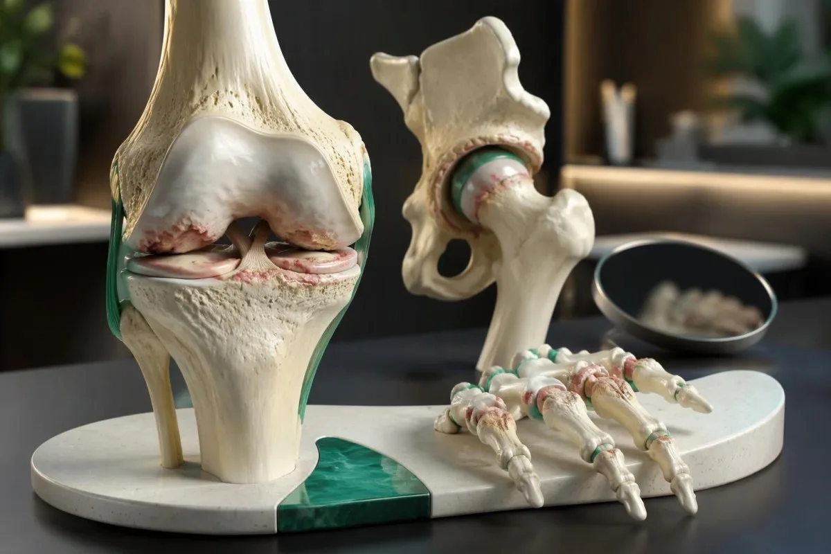
ပထမဆုံး သတိပြုမိနိုင်သော လက္ခဏာများမှာ လှုပ်ရှားမှုလုပ်နေစဉ် သို့မဟုတ် လုပ်ပြီးနောက် အဆစ်နာခြင်း၊ အနားယူပြီးနောက် အများအားဖြင့် မိနစ် ၃၀ အောက်သာ ကြာတတ်သော တောင့်တင်းခြင်းနှင့် လှုပ်ရှားနိုင်မှု တဖြည်းဖြည်း လျော့နည်းလာခြင်းတို့ ဖြစ်သည်။ လှေကားတက်သည့်အခါ ဒူးနာနိုင်ပြီး၊ ခြေအိတ်ဝတ်သည့်အခါ တင်ပါးနာနိုင်ကာ၊ ပစ္စည်းကို ဆုပ်ကိုင်သည့်အခါ လက်နာနိုင်သည်။ သွေးစစ်ဆေးမှုများဖြင့် အရိုးအဆစ်ရောင်ရောဂါကို အတည်ပြု၍မရပါ။ အဆစ်ပူပြီး လျင်မြန်စွာ ဖောင်းလာပါက အရေးပေါ် စစ်ဆေးမှုခံယူရန် လိုအပ်သည်။

အဓိက မှတ်သားစရာများ
- လှုပ်ရှားမှုနှင့်ဆက်စပ်သော နာကျင်မှု လမ်းလျှောက်ခြင်း၊ လှေကားတက်ခြင်း၊ ဆုပ်ကိုင်ခြင်း သို့မဟုတ် အဆစ်ကို ထပ်ခါတလဲလဲ ဖိအားပေးခြင်းတို့နှင့်အတူ အများအားဖြင့် ဖြစ်ပေါ်တတ်သည်။ လှုပ်ရှားမှုတစ်ခုတည်းကြောင့် နာကျင်ခြင်းသည် ရောဂါအမည်သတ်မှတ်ရန် မလုံလောက်ပါ။
- ခဏတာ တောင့်တင်းခြင်း အိပ်ရာနိုးပြီးနောက် သို့မဟုတ် ထိုင်နေပြီးနောက် ဖြစ်ပါက၊ အရိုးအဆစ်ရောင်ရောဂါ၏ ပုံမှန်လက္ခဏာပုံစံတွင် များသောအားဖြင့် မိနစ် ၃၀ ထက် မပိုကြာတတ်ပါ။
- လက်တွေ့ရောဂါရှာဖွေခြင်း NICE အရ၊ လှုပ်ရှားမှုနှင့်ဆက်စပ်သော အဆစ်နာခြင်းရှိပြီး မနက်ပိုင်းတောင့်တင်းမှု မရှိခြင်း သို့မဟုတ် ခဏတာသာရှိခြင်းတို့ရှိသော အသက် ၄၅ နှစ်နှင့်အထက် အရွယ်ရောက်သူများတွင် ၎င်းသည် များသောအားဖြင့် လုံလောက်သည်။
- ဒူးလက္ခဏာများ လှေကားတက်သည့်အခါ နာကျင်ခြင်း၊ အဆစ်ကို အပြည့်အဝ ဆန့်တန်းရန် ခက်ခဲခြင်းနှင့် တစ်ခါတစ်ရံ ရောင်ရမ်းခြင်းတို့ ပါဝင်သည်။ နာကျင်မှုမရှိဘဲ အဆစ်မှ အသံမြည်ခြင်းတစ်ခုတည်းဖြင့် ရောဂါအမည်သတ်မှတ်၍ မရပါ။
- တင်ပါးဆုံရိုးလက္ခဏာများ ပေါင်ခြံနာခြင်း၊ ခြေအိတ်ဝတ်ရန် ခက်ခဲခြင်းနှင့် ဒူးသို့ ပျံ့နှံ့နာကျင်ခြင်းတို့ ပါဝင်နိုင်သည်—တင်ပါးဆုံရိုးမှာ သိသာစွာ မနာသလို ခံစားရသည့်အခါတွင်ပင် ဖြစ်နိုင်သည်။
- လက်တွင်ဖြစ်သော လက္ခဏာများ သည် များသောအားဖြင့် လက်မခြေရင်းနှင့် လက်သည်းအနီးရှိ လက်ချောင်းအဆစ်များ သို့မဟုတ် လက်ချောင်းအလယ်ပိုင်းရှိ အဆစ်များကို ထိခိုက်စေသည်။ ထုံကျင်ခြင်းရှိပါက အခြားပြဿနာတစ်ခု ထပ်ရှိခြင်း သို့မဟုတ် ကွဲပြားသောပြဿနာတစ်ခု ဖြစ်ခြင်းကို ညွှန်ပြသည်။
- သွေးစစ်ဆေးမှု၏ ကန့်သတ်ချက်များ ကြောင့် ပုံမှန်စစ်ဆေးသော CRP၊ ESR၊ rheumatoid factor သို့မဟုတ် metabolic panel ရလဒ်များဖြင့် အရိုးအဆစ်ရောင်ရောဂါရှိကြောင်း အတည်ပြုခြင်း သို့မဟုတ် မရှိကြောင်း ဖယ်ထုတ်ခြင်း မပြုနိုင်ပါ။
- အရေးပေါ် သတိပေးလက္ခဏာများ တွင် အဆစ်ရုတ်တရက် ပူလာခြင်း၊ ဖောင်းလာခြင်း၊ ဒဏ်ရာရပြီးနောက် ခြေထောက်ပေါ် အလေးချိန်မတင်နိုင်ခြင်း သို့မဟုတ် ရင်ဘတ်အောင့်ခြင်း၊ အသက်ရှူမဝခြင်းနှင့်အတူ ခြေထောက်ဖောရောင်ခြင်းတို့ ပါဝင်သည်။ AI ၏ အဓိပ္ပာယ်ကောက်ချက်ကို စောင့်မနေပါနှင့်။
အရိုးအဆစ်ရောင်ရောဂါ၏ အစောပိုင်းလက္ခဏာများမှာ အဘယ်နည်း။
အရိုးအဆစ်ရောင်ရောဂါ၏ အစောပိုင်းလက္ခဏာများ သည် လှုပ်ရှားမှုလုပ်စဉ် အဆစ်နာခြင်း၊ အနားယူပြီးနောက် အချိန်တိုအတွင်း အဆစ်တောင့်တင်းခြင်းနှင့် သက်သောင့်သက်သာ လှုပ်ရှားနိုင်မှု တဖြည်းဖြည်း လျော့နည်းလာခြင်းတို့ ဖြစ်သည်။ လက္ခဏာတစ်ခုတည်းထက် လက္ခဏာပုံစံက ပိုအရေးကြီးသည်။ NICE အရ အသက် ၄၅ နှစ်နှင့်အထက် ရှိပြီး လှုပ်ရှားမှုလုပ်စဉ် အဆစ်နာကာ မနက်ပိုင်းအဆစ်တောင့်တင်းခြင်းမရှိသူများ၊ သို့မဟုတ် အဆစ်တောင့်တင်းခြင်းသည် ၃၀ မိနစ်.
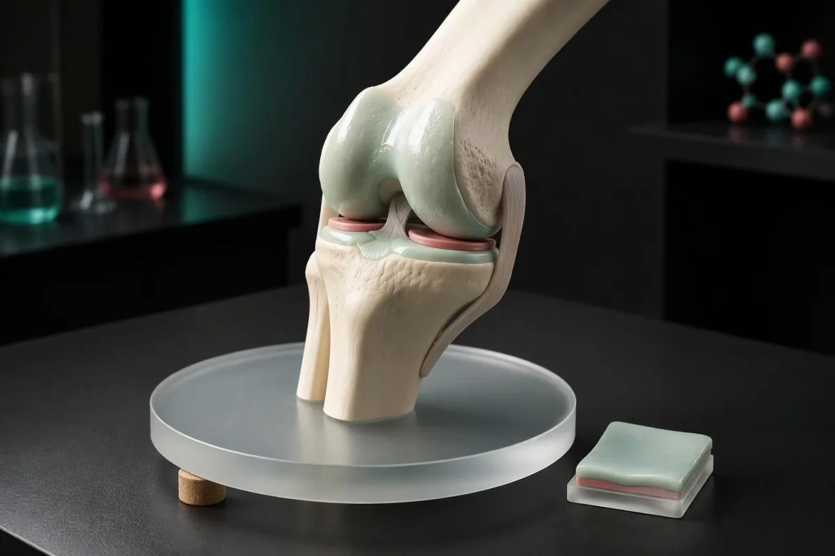
အရိုးအဆစ်ရောင်ရောဂါသည် အရိုးများကို ဖုံးအုပ်ထားသော အရိုးနုတင်မက အဆစ်တစ်ခုလုံးကို ထိခိုက်စေသည်။ အဆစ်အတွင်းလွှာ၊ အောက်ခံအရိုး၊ ထောက်ပံ့ပေးသော ကြွက်သားများနှင့် အရွတ်များတွင် ဖြစ်ပေါ်သော ပြောင်းလဲမှုများအားလုံးသည် လက္ခဏာများကို ဖြစ်စေနိုင်သည်။ NICE ၏ အသက်သတ်မှတ်ချက်ဖြစ်သော ၄၅ နှစ် သည် လက်တွေ့ကျသော ရောဂါရှာဖွေရေးနည်းလမ်းကို ဖော်ပြခြင်းသာဖြစ်ပြီး ငယ်ရွယ်သူများတွင် မဖြစ်နိုင်ကြောင်း သတ်မှတ်သည့် ဇီဝဗေဒဆိုင်ရာ စည်းမျဉ်းမဟုတ်ပါ (NICE, 2022)။
ဥပမာအားဖြင့်၊ စိတ်ကူးယဉ် အသက် ၅၆ နှစ်အရွယ် လူတစ်ဦးသည် လမ်းပိုလျှောက်ပြီးနောက် ဒူးနာကို သတိထားမိပြီး၊ မနက်စာစားပြီးနောက် ၈ မိနစ်ထက်ပိုသောအခါ အထိ တောင့်တင်းကာ လွန်ခဲ့သော လအနည်းငယ်က မရှိခဲ့သည့် ဒူးထောက်ရခက်ခဲမှုကို ကြုံတွေ့ရသည်။ ဤအချက်များကို စုပေါင်းကြည့်လျှင် တစ်ချက်တည်း အသံမြည်ခြင်းထက် အရိုးအဆစ်ရောင်ရောဂါကို စစ်ဆေးရန် ပိုမိုထောက်ခံသော်လည်း စစ်ဆေးရာတွင် အရွတ်ပြဿနာများ၊ ဒဏ်ရာနှင့် ရောင်ရမ်းသော အဆစ်ရောဂါတို့ကိုလည်း ဆက်လက်စစ်ဆေးရန် လိုအပ်သည်။
ကန်တက်စတီသည် AI သွေးစမ်းသပ်မှုခွဲခြမ်းစိတ်ဖြာ ဓာတ်ခွဲခန်းရလဒ်များကို အရိုးအဆစ်ရောင်ရောဂါရှိကြောင်း သက်သေအဖြစ် မယူဘဲ ရှင်းပြပေးသော အဖွဲ့အစည်းနှင့် ဆေးဘက်ဆိုင်ရာ အကန့်အသတ် သည် လူကိုယ်တိုင် အဆစ်စစ်ဆေးခြင်းနှင့် သီးခြားဖြစ်သည်။ မှတ်တမ်းတင်ထားသော အချက် ၃ ချက်ဖြစ်သည့် လက္ခဏာဖြစ်စေသော လှုပ်ရှားမှု၊ အဆစ်တောင့်တင်းသည့်ကြာချိန်နှင့် ဆုံးရှုံးသွားသော လုပ်ဆောင်နိုင်မှုတို့သည် ကျယ်ပြန့်သော ဓာတ်ခွဲခန်းစစ်ဆေးမှုများ မှာယူခြင်းထက် ဆရာဝန်အတွက် ပိုမိုအသုံးဝင်သော အစပြုအချက်အလက်များကို ပေးလေ့ရှိသည်။
လှုပ်ရှားမှုနှင့်ဆက်စပ်သော အဆစ်နာခြင်းသည် များသောအားဖြင့် မည်သို့ဖြစ်တတ်သနည်း။
Osteoarthritis နာကျင်မှု အထူးသဖြင့် ရောဂါအခြေအနေ အစောပိုင်းတွင် ထိခိုက်နေသော အဆစ်ကို ဝန်တင်သည့်အခါ သို့မဟုတ် ဝန်တင်ပြီးနောက် ပိုဆိုးလာကာ ဝန်လျှော့လိုက်လျှင် သက်သာလာတတ်သည်။ ခွဲခြားသိနိုင်ရန် အသုံးဝင်သောအချက်မှာ အနားယူပြီးနောက် ပထမ ခြေလှမ်းအနည်းငယ် တွင် ဖြစ်သော တောင့်တင်းမှုနှင့် လမ်းလျှောက်ပြီးနောက် တဖြည်းဖြည်း ပိုနာလာခြင်းတို့ကြား ကွာခြားချက်ဖြစ်သည်။ 20 minutes နှစ်မျိုးစလုံးသည် အဆစ်တစ်ခုတည်းတွင် ဖြစ်ပွားနိုင်သည်။
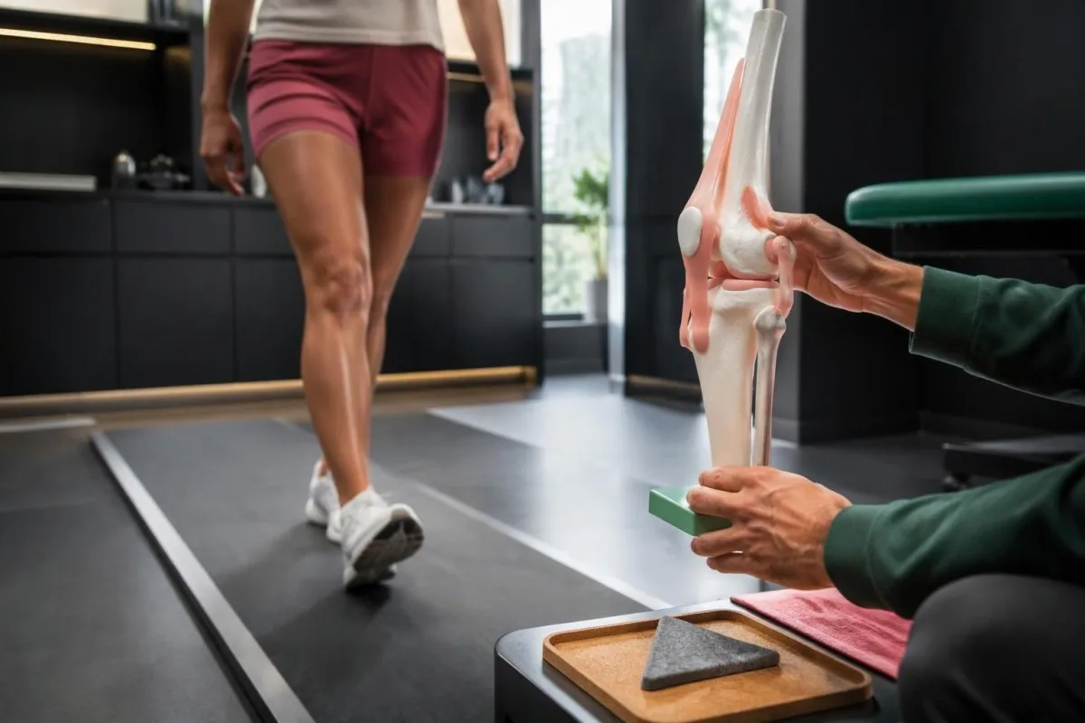
ပြန်လည်ဖြစ်ပေါ်စေနိုင်သည့် အကြောင်းရင်းတစ်ခုက ပြဿနာကို ရှာဖွေဖော်ထုတ်ရန် အထောက်အကူပြုသော်လည်း၊ ၎င်းတစ်ခုတည်းဖြင့် ပြဿနာ၏အကြောင်းရင်းကို မဖော်ထုတ်နိုင်ပါ။ စိတ်ကူးဖြင့်ရေးထားသော နေ့စဉ်မှတ်တမ်းတစ်ခုတွင် ဒူးနာကျင်မှုသည် 2/10 မှ 5/10 သို့ မြင့်တက်လာခြင်း လှေကားတက်စဉ်တွင် ဝန်ပိမှုကို ခံနိုင်ရည်နည်းကြောင်း ညွှန်ပြနိုင်သည်။ အရွတ်၊ ယခင်ဒဏ်ရာ သို့မဟုတ် ဒူးခေါင်းဆစ်မှ နာကျင်မှုသည်လည်း အလားတူပုံစံဖြစ်နိုင်ပြီး အရိုးအဆစ်ရောင်ရမ်းနာနှင့်အတူ တွဲရှိနိုင်သည်။
အနားယူနေစဉ် နာကျင်ခြင်းသည် အရိုးအဆစ်ရောင်ရမ်းနာကို အလိုအလျောက် ပယ်ချနိုင်သည့်အချက်မဟုတ်ပါ၊ အကြောင်းမှာ ပိုမိုဖြစ်ပွားနေသော ရောဂါအခြေအနေသည် အိပ်စက်မှုကို အနှောင့်အယှက်ဖြစ်စေနိုင်သလို တစ်နေ့လုံး လှုပ်ရှားမှုများပြီးနောက် ကိုက်ခဲစေနိုင်သောကြောင့်ဖြစ်သည်။ သို့သော် အသစ်ဖြစ်ပေါ်လာသော အနားယူနေစဉ် နာကျင်မှုသည် ဆက်တိုက်ဖြစ်နေခြင်း၊ ရက်အတော်ကြာ, အတွင်း လျင်မြန်စွာ ပိုဆိုးလာခြင်း၊ သို့မဟုတ် အဖျားတက်ခြင်း၊ အကြောင်းရင်းမသိ ကိုယ်အလေးချိန်ကျခြင်းတို့နှင့်အတူ နာကျင်ခြင်းရှိပါက၊ ရှိပြီးသား X-ray တွေ့ရှိချက်ကြောင့်ဟု သတ်မှတ်မည့်အစား စစ်ဆေးအကဲဖြတ်သင့်သည်။
လမ်းလျှောက်သည့်အကွာအဝေး အလားတူရောက်တိုင်း ခြေသလုံးနာကျင်မှု စတင်ဖြစ်ပေါ်ပြီး မိနစ်အနည်းငယ် ရပ်လိုက်ပြီးနောက် သက်သာသွားပါက ဒူးဆစ်အစား သွေးလည်ပတ်မှုပြဿနာကြောင့် ဖြစ်နိုင်သည်။ ကျွန်ုပ်တို့၏ ခြေထောက်သွေးလွှတ်ကြောဆိုင်ရာ နာကျင်မှု လမ်းညွှန်တွင် ထိုကွာခြားချက်ကို ရှင်းပြထားသည်။ ရပ်လိုက်ရုံဖြင့် သက်သာသလား၊ သို့မဟုတ် အဆစ်အနေအထား ပြောင်းလဲလိုက်မှ သက်သာသလားကို မှတ်သားပါ။
အရိုးအဆစ်ရောင်ရောဂါကြောင့်ဖြစ်သော ဒူးလက္ခဏာများအနက် မည်သည့်လက္ခဏာများက အရင်ပေါ်လာသနည်း။
ဒူးအရိုးအဆစ်ရောင်ရမ်းနာ၏ လက္ခဏာများ သည် လှေကားတက်ခြင်း၊ ကုလားထိုင်မှထခြင်း၊ ထိုင်ထလုပ်ခြင်း သို့မဟုတ် ပုံမှန်ထက် ပိုဝေးဝေးလမ်းလျှောက်ခြင်းတို့တွင် နာကျင်မှုဖြင့် မကြာခဏ စတင်တတ်သည်။ အစောပိုင်းမသက်မသာဖြစ်မှုသည် ဒူးခေါင်းတစ်ဝိုက် သို့မဟုတ် အဆစ်မျဉ်းတစ်ဖက်ဖက်တစ်လျှောက်တွင် ဖြစ်နိုင်သည်။ စိတ်ကူးဖြင့်ဖော်ပြထားသော အသက် 60 အရွယ် လူတစ်ဦးသည် လှေကားတစ်ထစ်တက်ရန်ပင် ခက်ခဲနေပါက နာကျင်မှုတည်နေရာတစ်ခုတည်းကိုကြည့်၍ ရောဂါသတ်မှတ်ခြင်းမဟုတ်ဘဲ နာကျင်မှုပုံစံကို စစ်ဆေးရမည်။
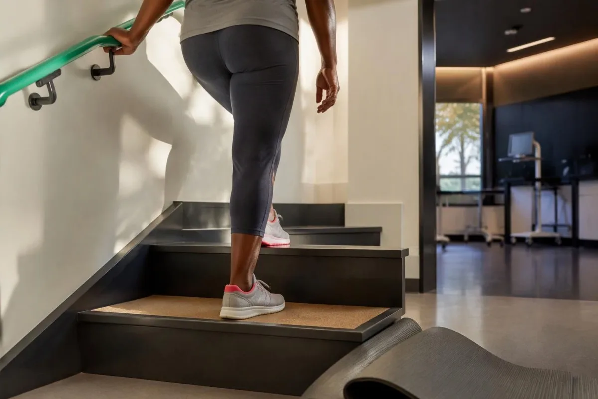
ဒူးအရိုးအဆစ်ရောင်ရမ်းနာတွင် ကြိတ်သံ သို့မဟုတ် တဂွတ်ဂွတ်မြည်သံ ဖြစ်နိုင်သော်လည်း အသံတစ်ခုတည်းဖြင့် အရိုးနုဆုံးရှုံးမှုရှိကြောင်း သို့မဟုတ် နာကျင်မှုပြင်းထန်မှုကို မသတ်မှတ်နိုင်ပါ။ ထိုအစား အချက် 2 ချက် ကို နှိုင်းယှဉ်ပါ။ အသံထွက်ခြင်းက နာကျင်စေသလား၊ လှုပ်ရှားမှုပြောင်းလဲသွားသလားဟူ၍ဖြစ်သည်။ ပုံမှန်အတိုင်း လုပ်ဆောင်နိုင်သေးသော ဒူးတွင် နာကျင်မှုမရှိဘဲ တကျွတ်ကျွတ်မြည်ခြင်းသည် ဒူးကို သက်သောင့်သက်သာ ဖြောင့်မရတော့ခြင်းအသစ်ဖြစ်ပေါ်လာခြင်းလောက် အရေးမကြီးပါ။
အခါအားလျော်စွာ ဖောင်းလာခြင်းသည် အဆစ်အတွင်း အရည်ရှိနေခြင်းကို ညွှန်ပြနိုင်ပြီး၊ ဒူးမခိုင်သလိုဖြစ်ခြင်းသည် အရွတ်ပြဲခြင်းထက် နာကျင်မှုကြောင့် ကြွက်သားလှုပ်ရှားမှု ဟန့်တားခံရခြင်းကြောင့် ဖြစ်နိုင်သည်။ ဒူးသည် ကိုယ်ထိလက်ရောက် ကပ်နေပြီး ဖြောင့်မရတော့ခြင်း—ဆိုလိုသည်မှာ ဒူးအဆစ် အမှန်တကယ် ကပ်နေခြင်း, —တစ်ကြိမ်သာဖြစ်ခဲ့သော်လည်း အမြန်စစ်ဆေးအကဲဖြတ်ရန် လိုအပ်သည်၊ အကြောင်းမှာ စက်ပိုင်းဆိုင်ရာ ပိတ်ဆို့မှုဖြစ်ပါက ကုသမှုလမ်းကြောင်း မတူညီနိုင်သောကြောင့်ဖြစ်သည်။
ခြေသလုံးအထိ ရောင်ရမ်းလာခြင်းကို ဒူးအဆစ်ရောင်ခြင်း သို့မဟုတ် ဒူးနောက်ဘက်ရှိ အရည်အိတ်ကြောင့်ဟု အလိုအလျောက် မယူဆသင့်ပါ။ အသစ်ဖြစ်လာသော ခြေထောက် 1 ဖက်, ရောင်ရမ်းခြင်း၊ အထူးသဖြင့် ခြေသလုံးကိုထိလျှင်နာခြင်း သို့မဟုတ် သွေးခဲဖြစ်နိုင်ခြေ အန္တရာယ်အချက်များရှိပါက အရေးပေါ် စစ်ဆေးအကဲဖြတ်ရန် လိုအပ်သည်။ ကျွန်ုပ်တို့၏ ခြေထောက်ရောင်ရမ်းမှု အရေးပေါ်လမ်းညွှန် ရောဂါအကြောင်းရင်းကို အန္တရာယ်မရှိဟု ယူဆမထားမီ စစ်ဆေးမှုနှင့် သင့်လျော်သော စမ်းသပ်စစ်ဆေးမှုများ အဘယ်ကြောင့် လိုအပ်ကြောင်း ရှင်းပြထားသည်။
တင်ပါးဆုံရိုးအရိုးအဆစ်ရောင်ရောဂါကြောင့် ပေါင်ခြံ သို့မဟုတ် ဒူးနာနိုင်ပါသလား။
တင်ပါးဆုံရိုး အရိုးအဆစ်ရောင်ခြင်း ပုံမှန်အားဖြင့် ပေါင်ခြံနာကျင်ခြင်းနှင့် တင်ပါးကို လှည့်ရန်ခက်ခဲခြင်းတို့ကို ဖြစ်စေသော်လည်း နာကျင်မှုသည် ပေါင်၊ တင်ပါးနောက်ပိုင်း သို့မဟုတ် ဒူးအထိလည်း ရောက်နိုင်သည်။ အကယ်၍ ဒူးတစ်ဖက် သည် ဒူးပြဿနာဟု ယုံကြည်လောက်သော အကြောင်းရင်းမရှိဘဲ နာကျင်ပါက တင်ပါးကို စစ်ဆေးခြင်းဖြင့် နာကျင်မှု၏ အရင်းအမြစ်ကို ရှာဖွေတွေ့ရှိနိုင်သည်။ ခြေအိတ်စွပ်ခြင်း သို့မဟုတ် ကားပေါ်တက်ခြင်းတို့က လှုပ်ရှားနိုင်မှု ကန့်သတ်နေခြင်းကို ဖော်ပြနိုင်သည်။
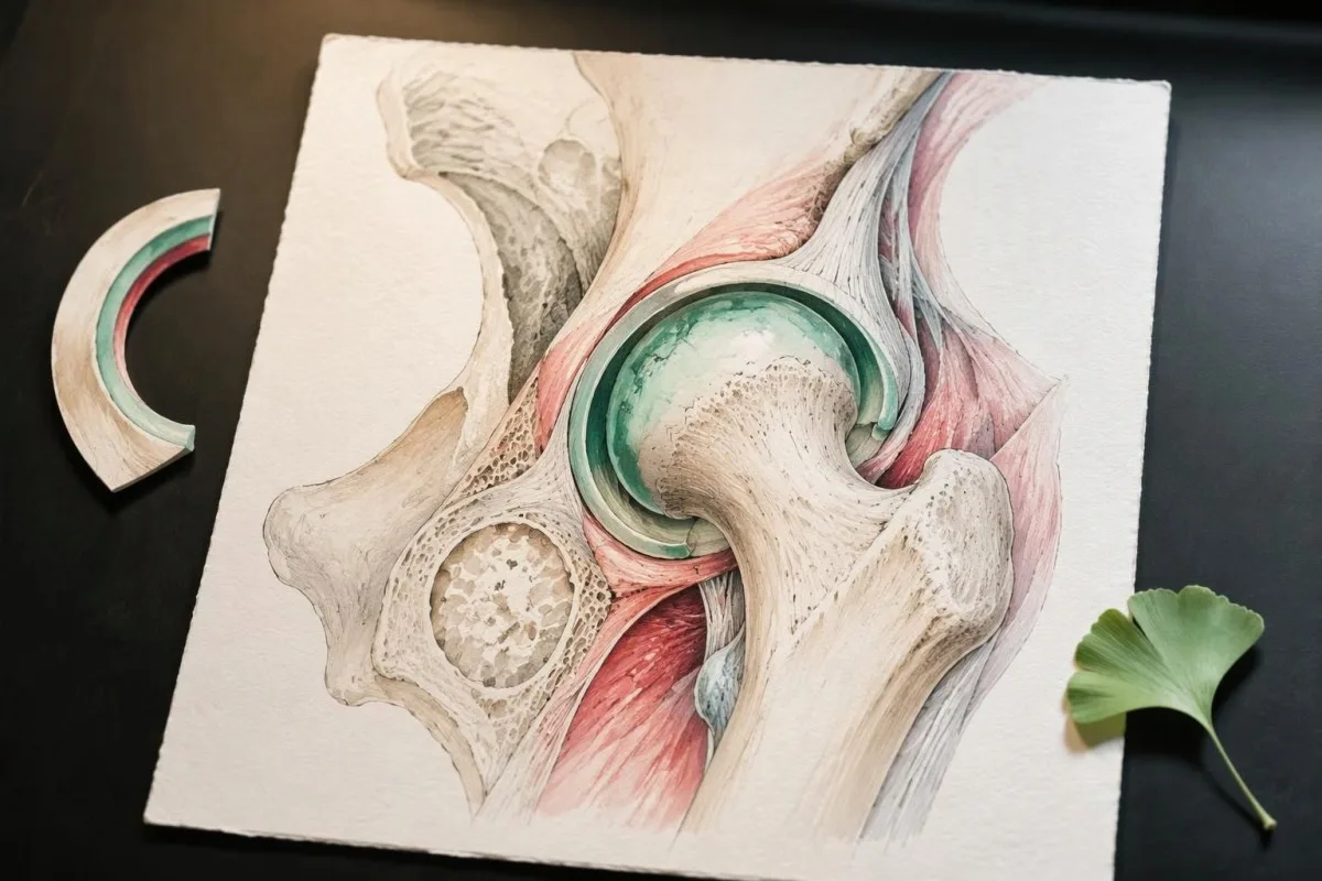
တင်ပါးကို အတွင်းဘက်သို့ သက်သောင့်သက်သာ လှည့်နိုင်မှု ဆုံးရှုံးခြင်းသည် စမ်းသပ်စစ်ဆေးရာတွင် သဲလွန်စဖြစ်နိုင်သော်လည်း လှုပ်ရှားမှုတစ်ခုတည်းဖြင့် ရောဂါအတည်ပြု၍ မရပါ။ ဥပမာအဖြစ် စိတ်ကူးကြည့်ထားသော အသက် ၅၈ နှစ်အရွယ်, လူတစ်ဦးတွင် လမ်းလျှောက်ပြီးနောက် ပေါင်ခြံကိုက်ခဲခြင်းနှင့် ခြေဖဝါးကို လက်ဖြင့်လှမ်းထိရန် ခက်ခဲခြင်းတို့သည် တင်ပါးအပြင်ဘက်ကိုသာ ထိလျှင် နာကျင်ခြင်းထက် တင်ပါးဆုံရိုးအဆစ်ပြဿနာကို ပိုမိုညွှန်ပြသည်။ အပြင်ဘက်ကို ထိလျှင်နာခြင်းသည် ပတ်ဝန်းကျင်ရှိ အရွတ်များနှင့် ဆက်စပ်နေတတ်သည်။
ဒူးအောက်ဘက်အထိ ပူလောင်နာကျင်မှု ပျံ့သွားခြင်း၊ ထုံကျင်စူးရှခြင်း သို့မဟုတ် အားနည်းခြင်းတို့သည် အရိုးအဆစ်ရောင်ခြင်းတစ်ခုတည်းထက် အာရုံကြောပါဝင်ပတ်သက်မှုကို ညွှန်ပြသည်။ ကျွန်ုပ်တို့၏ ဆိုင်ယာတစ်ကာနှင့် အာရုံကြောဆိုင်ရာ လက္ခဏာများ အကြောင်း ရှင်းလင်းချက်တွင် ဤပုံစံများကို ခွဲခြားဖော်ပြထားသည်။ လူတစ်ဦးတွင် တစ်ပြိုင်နက်တည်း ပြဿနာ ၂ ခု, ရှိနိုင်သောကြောင့် X-ray တွင် တင်ပါးအဆစ်ရောင်ခြင်း တွေ့ရသည်ဆိုရုံဖြင့် အာရုံကြောဆိုင်ရာ လက္ခဏာများကို စစ်ဆေးသုံးသပ်မှုမှ ချန်လှပ်မထားသင့်ပါ။
တင်ပါး၏ လုပ်ဆောင်နိုင်မှု ရုတ်တရက်ပြောင်းလဲလာခြင်းကို တဖြည်းဖြည်းတောင့်တင်းလာခြင်းထက် ပိုမိုအလေးထား စစ်ဆေးရန်လိုသည်။ ဥပမာအားဖြင့် စိတ်ကူးယဉ်ထားသော အသက် ၆၇ နှစ်အရွယ် လူတစ်ဦးသည် အနည်းငယ်လဲကျပြီးနောက် ပေါင်ခြံတွင် ပြင်းထန်စွာနာကျင်ကာ ကိုယ်အလေးချိန်ကို ခြေထောက်ပေါ် မတင်နိုင်ပါက အရေးပေါ်အရိုးကျိုးမှု စစ်ဆေးရန်လိုသည်။ ယခင်ပုံရိပ်စစ်ဆေးမှုတွင် အရိုးအဆစ်ရောင်ခြင်း တွေ့ထားခဲ့သော်လည်း ထိုသို့စစ်ဆေးရမည်ဖြစ်သည်။ အချို့အရိုးကျိုးမှုများကို ပထမဆုံး ရိုးရိုး X-ray တွင် မမြင်ရနိုင်ပါ။
အရိုးအဆစ်ရောင်ရောဂါကြောင့်ဖြစ်သော လက်လက္ခဏာများကို မည်သို့ခံစားရသနည်း။
လက်အရိုးအဆစ်ရောင်ခြင်း၏ လက္ခဏာများ တွင် မကြာခဏ ညှပ်ကိုင်ခြင်း သို့မဟုတ် ဆုပ်ကိုင်ခြင်းအတွင်း ကိုက်ခဲခြင်း၊ လှုပ်ရှားမှုမရှိဘဲ နေပြီးနောက် တောင့်တင်းခြင်းနှင့် လက်ချောင်းအသုံးပြုနိုင်မှု လျော့နည်းခြင်းတို့ ပါဝင်သည်။ လက်မခြေရင်းရှိ အဆစ်နှင့် လက်ချောင်းအဆစ်အုပ်စု ၂ ခု—လက်သည်းနှင့် အနီးဆုံးရှိ အဆစ်များနှင့် လက်ချောင်းအလယ်ပိုင်းရှိ အဆစ်များ—သည် အဖြစ်များသော နေရာများဖြစ်သည်။ မာကျောသော အရိုးဖုများသည် ပထမဆုံးသတိပေးလက္ခဏာအဖြစ်မဟုတ်ဘဲ နောက်ပိုင်းမှ ပေါ်လာနိုင်သည်။
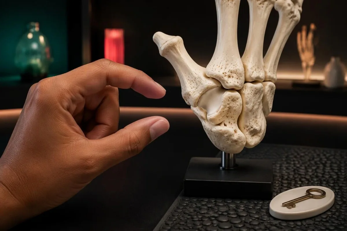
လက်မခြေရင်းအဆစ် အရိုးအဆစ်ရောင်ခြင်းကို သော့လှည့်ခြင်း၊ ဘူးအဖုံးဖွင့်ခြင်း သို့မဟုတ် ဖုန်းကို ကြာရှည်ကိုင်ထားခြင်းတို့ ပြုလုပ်ချိန်တွင် ပထမဆုံး သတိပြုမိနိုင်သည်။ စိတ်ကူးယဉ်ထားသော အသက် ၆၂ နှစ်အရွယ် လူတစ်ဦးသည် လက်တစ်ဖက်လုံးဖြင့် အားကောင်းစွာ ဆုပ်ကိုင်နိုင်သေးသော်လည်း တိကျသော ညှပ်ကိုင်မှုတွင် အခက်အခဲရှိနိုင်သည်။ အကြောင်းမှာ ထိုအလုပ်များတွင် အားကို ပုံစံမတူဘဲ ဖြန့်ဝေသုံးစွဲရသောကြောင့်ဖြစ်သည်။ လုပ်ဆောင်နိုင်မှု လျော့နည်းခြင်းသည် ယေဘုယျအားနည်းခြင်းနှင့် အမြဲတမ်း တူညီနေသည်မဟုတ်ပါ။
ထုံကျင်ခြင်းသည် လက်ချောင်းအဆစ် အရိုးအဆစ်ရောင်ခြင်း၏ ပုံမှန်တိုက်ရိုက်အကျိုးဆက် မဟုတ်ပါ။ လက်မ၊ လက်ညှိုး၊ လက်ခလယ်နှင့် လက်သူကြွယ်၏ တစ်စိတ်တစ်ပိုင်းတွင် စူးရှထုံကျင်ခြင်းသည် အလယ်လက်မောင်းကြော ဖိမိခြင်းကို ညွှန်ပြနိုင်သည်။ ကျွန်ုပ်တို့၏ လမ်းညွှန်ချက်သည် ကာပါလ်တန်နယ်လ်ကြောင့် လက်ချောင်းများတွင် ဖြစ်ပေါ်သည့်ပုံစံများအကြောင်း လမ်းညွှန်ချက် လက္ခဏာများသည် ခန့်မှန်းခြေအားဖြင့် အဘယ်ကြောင့် လက်ချောင်း ၃ လက်ခွဲ တွင် ဖြစ်ပေါ်ပါက အရိုးဆစ်များ နာကျင်ခြင်းနှင့် မတူသော စမ်းသပ်စစ်ဆေးမှု လိုအပ်ကြောင်းကို ရှင်းပြသည်။
ရောင်ရမ်းမှု ပျံ့နှံ့နေသည့်ပုံစံက ထည့်သွင်းစဉ်းစားရမည့် ရောဂါအကြောင်းရင်းများကို ပြောင်းလဲစေသည်။ လက်ဆစ်များတစ်လျှောက် အချိန်ကြာကြာ ပျော့ပျောင်းစွာ ရောင်နေခြင်းသည် လက်ချောင်းထိပ်နားတွင် မာကျောစွာ ကြီးလာခြင်းထက် ပိုမိုပုံမှန်မဟုတ်ပါ။ လက်ကြီးလာခြင်းနှင့်အတူ အခြားပြောင်းလဲမှု ၂ ခု, ဖြစ်သည့် ဖိနပ်အရွယ်အစား တိုးလာခြင်းနှင့် မျက်နှာအသွင်အပြင် ပြောင်းလဲခြင်းတို့ပါ တွေ့ရလျှင်၊ ကျွန်ုပ်တို့၏ လက်ကြီးလာခြင်း သတိပေးလက္ခဏာများ သည် အဆစ်ရောင်ရောဂါဟု ယူဆရမည့်အစား ဟော်မုန်းဆိုင်ရာ အကြောင်းရင်းကို ထည့်သွင်းစဉ်းစားရန် လိုအပ်နိုင်သည့်အကြောင်းကို ရှင်းပြသည်။
အရိုးအဆစ်ရောင်ရောဂါကြောင့်ဖြစ်သော တောင့်တင်းမှုသည် မည်မျှကြာသင့်သနည်း။
အရိုးအဆစ်ရောင်ရောဂါကြောင့် တောင့်တင်းခြင်း သည် အများအားဖြင့် ၃၀ မိနစ် နိုးထပြီးနောက်ထက် ပိုမကြာဘဲ၊ ထိုင်ပြီးနောက်တွင်လည်း ပိုတိုလေ့ရှိသည်။ ဤခဏတာ “တောင့်တင်းနေခြင်း” သည် ကြာရှည်ခံသော ရောင်ရမ်းမှုကြောင့် တောင့်တင်းခြင်းနှင့် ကွဲပြားသော်လည်း သတ်မှတ်ချက်မှာ အချိန်တိုင်းကိရိယာဖြင့် တိုင်းတာသည့် စမ်းသပ်မှုမဟုတ်ဘဲ ဆေးခန်းဆိုင်ရာ လမ်းညွှန်ချက်သာဖြစ်သည်။ ကြာချိန်၊ ရောင်ရမ်းမှု၊ ထိခိုက်သည့်အဆစ်များနှင့် အထွေထွေလက္ခဏာများကို အတူတကွ ထည့်သွင်းစဉ်းစားရမည်။
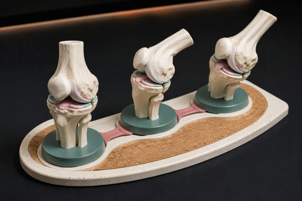
နာကျင်မှုလုံးဝ ပျောက်သည်အထိ မဟုတ်ဘဲ အဆစ်ကို လွတ်လပ်စွာ လှုပ်ရှားနိုင်လောက်သည်အထိ တောင့်တင်းမှုကို တိုင်းတာပါ။ စိတ်ကူးဖြင့်ယူဆထားသော ၇ ရက် မှတ်တမ်းတွင် တောင့်တင်းမှုသည် ၅–၁၂ မိနစ် ကြာပြီးနောက် လှုပ်ရှားမှုကြောင့် နာကျင်ကိုက်ခဲလာခြင်းသည် မနက်တိုင်း တစ်နာရီကြာ လှုပ်ရှားရခက်ခဲခြင်းနှင့် မတူသော ပုံစံဖြစ်သည်။ နာကျင်မှုနှင့် တောင့်တင်းမှုကို ခွဲခြားခြင်းဖြင့် ကြာချိန်ကို ပိုမိုများပြားစွာ ခန့်မှန်းမိခြင်းမှ ကာကွယ်နိုင်သည်။
အသက် နှစ်ပေါင်း 50 ကျော်သူတစ်ဦးတွင် ပခုံးနှင့် တင်ပါးဆုံပတ်လည် ကြာရှည်တောင့်တင်းမှု ရှိပါက အထူးသဖြင့် မနက်ခင်း တောင့်တင်းမှုသည် 45 မိနစ်. နှင့် တွဲဖက်ကြသည်။ polymyalgia စစ်ဆေးမှု ကန့်သတ်ချက်များ ထက် ကျော်လွန်သောအခါ polymyalgia rheumatica ကို ထည့်သွင်းစဉ်းစားသင့်သည်။ ရောင်ရမ်းမှု အမှတ်အသားများ ပုံမှန်ဖြစ်နေခြင်းက ထိုမေးခွန်းကို အမြဲတမ်း အဆုံးအဖြတ်မပေးနိုင်သည့် အကြောင်းရင်းကို ရှင်းပြသည်။ ဤပုံစံသည် တင်ပါးဆုံအဆစ်တစ်ခုတည်း လှုပ်ရှားမှုကန့်သတ်ခြင်းထက် ပိုမိုကျယ်ပြန့်သည်။
မတိုင်မီ စတင်ဖြစ်ပွားသော ၄၅ နှစ်, ထက်ပိုကြာပြီး 3 months, အနားယူခြင်းထက် လှုပ်ရှားခြင်းဖြင့် သက်သာလာသည့် ခါးနာခြင်းသည် ကျောရိုးရောင်ရမ်းရောဂါကို ညွှန်ပြနိုင်သည်။ ထို HLA-B27 ဆိုင်ရာ ဆေးခန်းအခြေအနေ သည် မျိုးရိုးဗီဇစစ်ဆေးမှုရလဒ်တစ်ခုတည်းထက် ပိုအရေးကြီးသည်။ စစ်ဆေးမှုအဖြေ အပေါင်းဖြစ်ခြင်းဖြစ်စေ၊ တောင့်တင်းမှုကြာချိန်ဖြစ်စေ တစ်ခုတည်းဖြင့် ရောဂါကို အတည်မပြုနိုင်ပါ။
မည်သည့်လှုပ်ရှားမှုပြောင်းလဲမှုများကို မှတ်တမ်းတင်သင့်သနည်း။
လှုပ်ရှားမှု လျော့နည်းခြင်း ဒူးကို အပြည့်အဝဆန့်ရန် ခက်ခဲခြင်း၊ တင်ပါးကို လှည့်ရန် ခက်ခဲခြင်း၊ သို့မဟုတ် လက်ကို သက်သောင့်သက်သာ ပိတ်ရန် ခက်ခဲခြင်းအဖြစ် ပေါ်လာနိုင်သည်။ တူညီသော နေ့စဉ်လုပ်ငန်း ၂ ခု သို့မဟုတ် ၃ ခု ကို အချိန်ကြာလာသည်နှင့်အမျှ နှိုင်းယှဉ်ပါ၊ အဆစ်ကို ၎င်း၏ ကန့်သတ်ချက်ထက် ကျော်လွန်၍ အတင်းမရွှေ့ပါနှင့်။ လုပ်ဆောင်နိုင်စွမ်း ပြောင်းလဲမှုသည် အိမ်တွင် လှုပ်ရှားမှုထောင့်ကို ခန့်မှန်းခြင်းထက် ပြဿနာကို ပိုမိုရှင်းလင်းစွာ ဖော်ပြလေ့ရှိသည်။
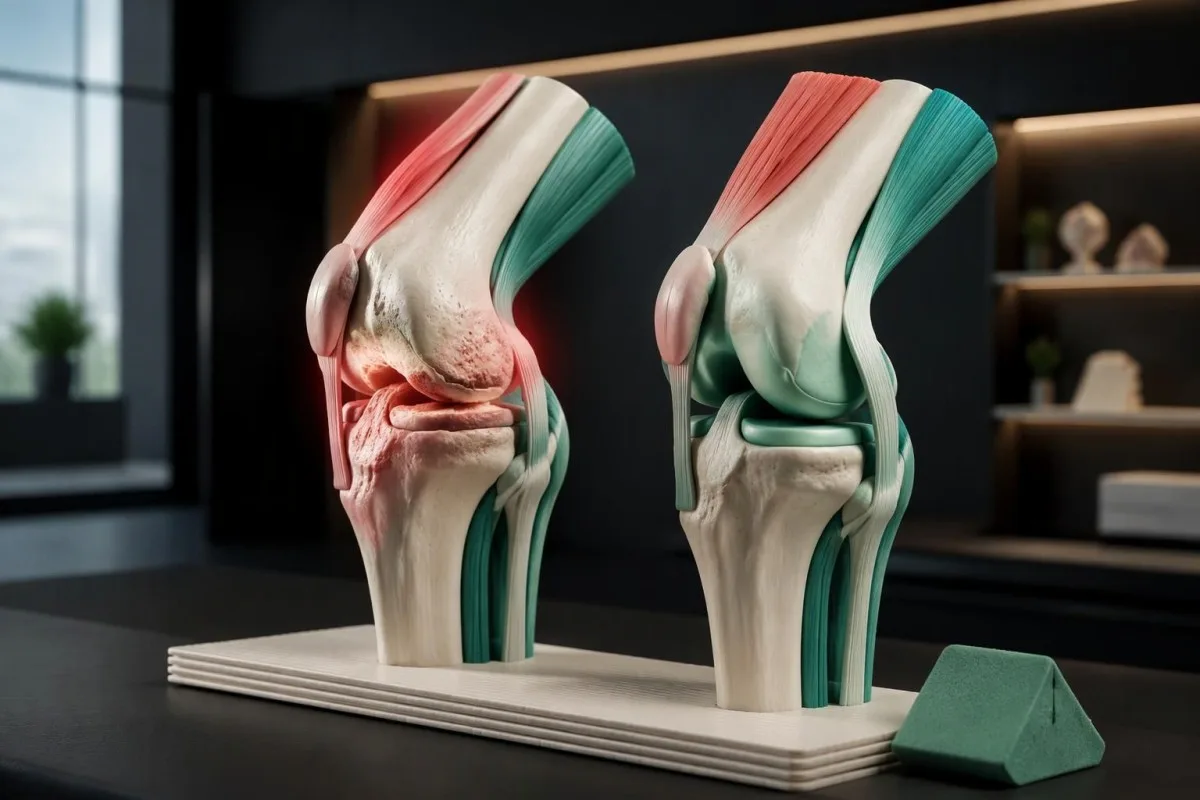
မတ်တပ်ရပ်နေစဉ် အနည်းငယ်ကွေးနေဆဲဖြစ်သော ဒူးတစ်ဖက်သည် လမ်းလျှောက်ပုံကို ပြောင်းလဲစေပြီး ပတ်ဝန်းကျင်ရှိ ကြွက်သားများမှ လိုအပ်သော အားထုတ်မှုကို တိုးမြှင့်စေနိုင်သည်။ ဆေးဘက်ဆိုင်ရာ ကျွမ်းကျင်သူများသည် ဒူးဆန့်မှုကို ခန့်မှန်းခြေအားဖြင့် ဖြောင့်သောအနေအထားဖြစ်သည့် ၀ ဒီဂရီ, နှင့် နှိုင်းယှဉ်၍ တိုင်းတာနိုင်သော်လည်း ခန္ဓာကိုယ်တစ်ဦးချင်း ကွဲပြားမှု၊ ယခင်ဒဏ်ရာနှင့် စမ်းသပ်မှုပြုလုပ်ပုံတို့သည် အရေးကြီးသည်။ တိုင်းတာရရှိသော ကွာခြားချက် အနည်းငယ်သည် သီးခြားရောဂါသတ်မှတ်ချက် မဟုတ်သကဲ့သို့ အတင်းဆန့်ထုတ်ရန် အကြောင်းပြချက်လည်း မဟုတ်ပါ။
ထိခိုက်နေသော အဆစ်နှင့် ကိုက်ညီသည့် လုပ်ငန်းများကို ရွေးချယ်ပါ။ လှေကားတစ်ထပ် ဒူးအတွက်၊ တင်ပါးအတွက် ခြေအိတ်ဝတ်ခြင်း၊ သို့မဟုတ် လက်မအတွက် မကြာခဏသုံးနေကျသော သော့ကို လှည့်ခြင်းတို့ကို ရွေးချယ်ပါ။ Kantesti တွင် CRP ရလဒ် တည်ငြိမ်နေခြင်းက လူတစ်ဦးသည် ထိုလှုပ်ရှားမှုများကို လုပ်ဆောင်နိုင်စွမ်း ဆုံးရှုံးသွားခြင်း ရှိ၊ မရှိကို မပြသနိုင်သောကြောင့် လုပ်ဆောင်မှုဆိုင်ရာ လေ့လာတွေ့ရှိချက်များကို ဓာတ်ခွဲခန်းဆိုင်ရာ အဓိပ္ပာယ်ဖော်ခြင်းနှင့် သီးခြားခွဲခြားထားသည်။
အရိုးပွရောဂါနှင့် အရိုးအဆစ်ရောင်ရောဂါတို့သည် အမည်ဆင်တူသော်လည်း ကွဲပြားသော ပြဿနာများကို ဖော်ပြသည်။ အရိုးပွရောဂါသည် အရိုးကျိုးသည်အထိ လက္ခဏာမပြဘဲ ရှိနေလေ့ရှိသော်လည်း အရိုးအဆစ်ရောင်ရောဂါသည် အများအားဖြင့် အဆစ်နှင့်ဆိုင်သော လက္ခဏာများ ဖြစ်စေသည်။ ကျွန်ုပ်တို့၏ အရိုးဆုံးရှုံးမှု စစ်ဆေးခြင်း လမ်းညွှန် တွင် ထိခိုက်မှုနည်းသော အရိုးကျိုးမှု ၁ ကြိမ် ပင်လျှင် အရိုးကျန်းမာရေး စစ်ဆေးမှုကို ခိုင်လုံစေနိုင်သည့် အကြောင်းရင်းကို ရှင်းပြထားပြီး၊ လက်ချောင်းအဆစ်တောင့်တင်းခြင်းသည် အရိုးပွရောဂါကြောင့်ဖြစ်သည်ဟု ဆိုလိုခြင်းမဟုတ်ပါ။
အရိုးအဆစ်ရောင်ရောဂါကြောင့် ရောင်ရမ်းခြင်း သို့မဟုတ် ပူနွေးခြင်း ဖြစ်နိုင်ပါသလား။
အရိုးအဆစ်ရောင်ရောဂါသည် အဆစ်ဖောင်းခြင်းနှင့် အနည်းငယ်ပူနွေးခြင်းတို့ကို တစ်ခါတစ်ရံ ဖြစ်စေနိုင်သည်၊ အကြောင်းမှာ အဆစ်အတွင်းနံရံတွင် ဖြစ်ပေါ်သည့် ပြောင်းလဲမှုများသည် ဤရောဂါ၏ တစ်စိတ်တစ်ပိုင်းဖြစ်သောကြောင့် ဖြစ်သည်။ ရုတ်တရက် အလွန်ပူနွေးလာပြီး သိသိသာသာ ဖောင်းလာသော အဆစ်တစ်ဆစ်တည်း သည် ကွဲပြားသည်။ ထိုအခြေအနေတွင် အရိုးအဆစ်ရောင်ရောဂါ ရှိနှင့်ပြီးသူဖြစ်သော်လည်း ပိုးဝင်ခြင်း၊ ပုံဆောင်ခဲအဆစ်ရောင်ရောဂါ သို့မဟုတ် အခြားရုတ်တရက်ဖြစ်ပွားသော အကြောင်းရင်းအတွက် အရေးပေါ်စစ်ဆေးမှု လိုအပ်သည်။
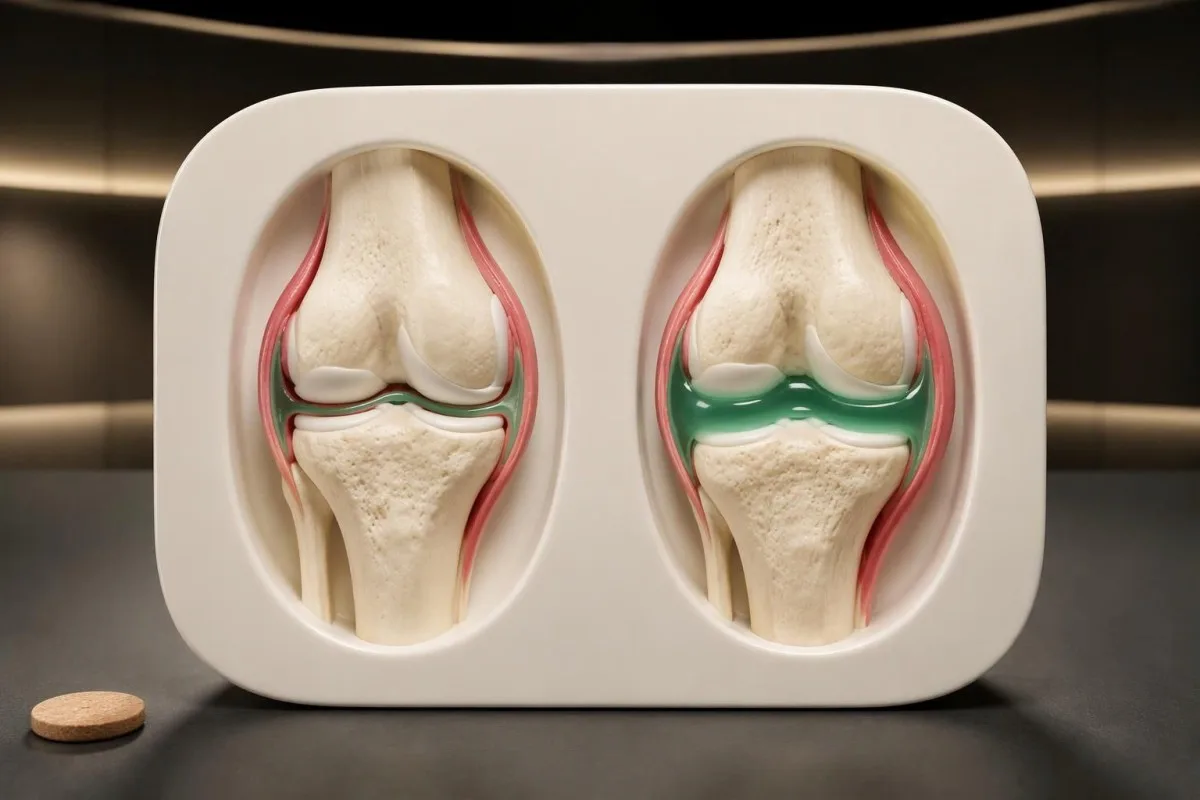
မာကျောသော အရိုးဖုထခြင်းနှင့် အရည်ကြောင့် ဖြစ်သော ပျော့ပျောင်းဖောင်းခြင်းတို့သည် တွေ့ရှိချက်တစ်မျိုးတည်း မဟုတ်ပါ။ ဥပမာအားဖြင့် အသက် ၆၄ နှစ်အရွယ်, လူတစ်ဦးတွင် ကာလကြာရှည်ရှိနေသော လက်ချောင်းအဆစ်အဖုမာမာသည် အရိုးအဆစ်ရောင်ရောဂါကြောင့် ဖြစ်နိုင်သော်လည်း၊ 12 နာရီ အချိန်ကာလအတွင်း ပိုမိုကြီးလာသော ဒူးတစ်ဖက်ကို ပြန်လည်စစ်ဆေးအကဲဖြတ်သင့်သည်။ အဆစ်ကို ယခင်က အဆစ်ရောင်ရောဂါဟု သတ်မှတ်ထားခြင်း ရှိ၊ မရှိထက် ပြောင်းလဲမှုဖြစ်ပွားသည့် အရှိန်က ပိုမိုအသုံးဝင်သော အချက်အလက် ဖြစ်နိုင်သည်။
Normal blood results cannot safely dismiss an acutely hot joint. Our explanation of WBC နှင့် CRP မတူညီမှု shows why timing and clinical findings matter; a result collected early on day 1 may not reflect the full response, and joint infection can occur without striking systemic laboratory abnormalities.
Joint-fluid testing is sometimes needed because examination and blood markers cannot reliably separate all acute causes. Fluid may be checked for crystals, cellular counts, and culture; identifying 1 crystal type does not automatically exclude simultaneous infection, so a clinician must interpret the sample alongside symptoms, risk factors, and the course of the illness.
သွေးစစ်ဆေးမှုများဖြင့် အရိုးအဆစ်ရောင်ရောဂါကို အဘယ်ကြောင့် အတည်ပြု၍မရသနည်း။
No routine blood test confirms or excludes osteoarthritis. Diagnosis depends on symptoms, examination, and sometimes imaging; CRP, ESR, and antibody tests can investigate alternative explanations but cannot measure whether a particular knee or thumb joint has osteoarthritis. There are 0 standard laboratory confirmation tests for routine clinical osteoarthritis diagnosis.
CRP is a nonspecific marker of systemic inflammation, not a cartilage-loss measurement. If a hypothetical laboratory uses a reference interval below 5 mg/L, a CRP of 18 mg/L calls for clinical context rather than proving inflammatory arthritis; local osteoarthritis changes, another illness, and other contributing factors can coexist, and reference intervals differ between laboratories.
Rheumatoid factor and anti-CCP testing are most useful when the history and examination suggest inflammatory arthritis, especially persistent synovitis. Our guide to နေထိုင်သော ကပ်ပါးကောင်များ၏ အဓိပ္ပာယ် ကျွန်ုပ်တို့၏ သုံးသပ်ချက်က အဘယ်ကြောင့်ဆိုသည်ကို ရှင်းပြထားသည်။ 1 positive antibody result cannot replace the examination; negative antibodies also do not exclude rheumatoid arthritis or turn an uncertain joint problem into confirmed osteoarthritis.
ကန်တက်စတီသည် AI သွေးစစ်ဆေးမှု ရလဒ် ပလက်ဖောင်း that helps explain reported markers without claiming to identify osteoarthritis from them. Our ဇီဝအမှတ်အသား အနက်ဖွင့်လမ်းညွှန် supports checking 3 report details—the units, laboratory reference interval, and collection date—before discussing an abnormal result with a clinician; broad screening panels are not automatically useful for a typical osteoarthritis presentation.
မည်သည့်လက္ခဏာပုံစံများက အခြားရောဂါတစ်ခုကို ညွှန်ပြသနည်း။
Persistent soft joint swelling, prolonged morning stiffness, acute redness, nerve symptoms, or illness outside the joints can suggest something other than osteoarthritis. Pattern recognition helps select testing: swelling across several knuckles points toward a different assessment from isolated thumb-base pain, while abrupt swelling in 1 joint raises crystal or infectious causes.
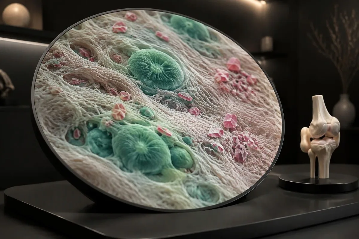
Symmetrical knuckle swelling with prolonged stiffness can suggest rheumatoid arthritis, while Raynaud symptoms, rashes, or muscle weakness may broaden the autoimmune assessment. Our connective tissue testing guide ကျွန်ုပ်တို့၏ သုံးသပ်ချက်က အဘယ်ကြောင့်ဆိုသည်ကို ရှင်းပြထားသည်။ 1 antibody, such as anti-RNP, is insufficient by itself; ordering many antibodies without a compatible symptom pattern can create misleading incidental positives.
Gout and calcium pyrophosphate crystal arthritis can cause sudden attacks, and calcium pyrophosphate deposition may also coexist with osteoarthritis. A serum urate below 6 mg/dL during an acute episode does not reliably rule out gout; when the diagnosis or infection risk is uncertain, examining joint fluid can be more informative than repeating a routine blood panel.
Iron overload can produce an osteoarthritis-like pattern, sometimes involving the second and third knuckles. Persistent transferrin saturation around 45% or higher can prompt iron-overload assessment, but does not establish its cause; Kantesti does not recommend this test for every aching joint, and our complete iron study explanation distinguishes saturation from ferritin, which can also rise with inflammation or liver disease.
ဓာတ်မှန် သို့မဟုတ် MRI စစ်ဆေးမှုများက မည်သည့်အခါတွင် အထောက်အကူပြုသနည်း။
Imaging helps when symptoms are atypical, the diagnosis is uncertain, or the result could change treatment. NICE does not recommend routine diagnostic imaging for a typical clinical presentation; EULAR likewise advises that imaging is unnecessary in typical osteoarthritis and identifies plain radiography as the first imaging choice when imaging is needed.
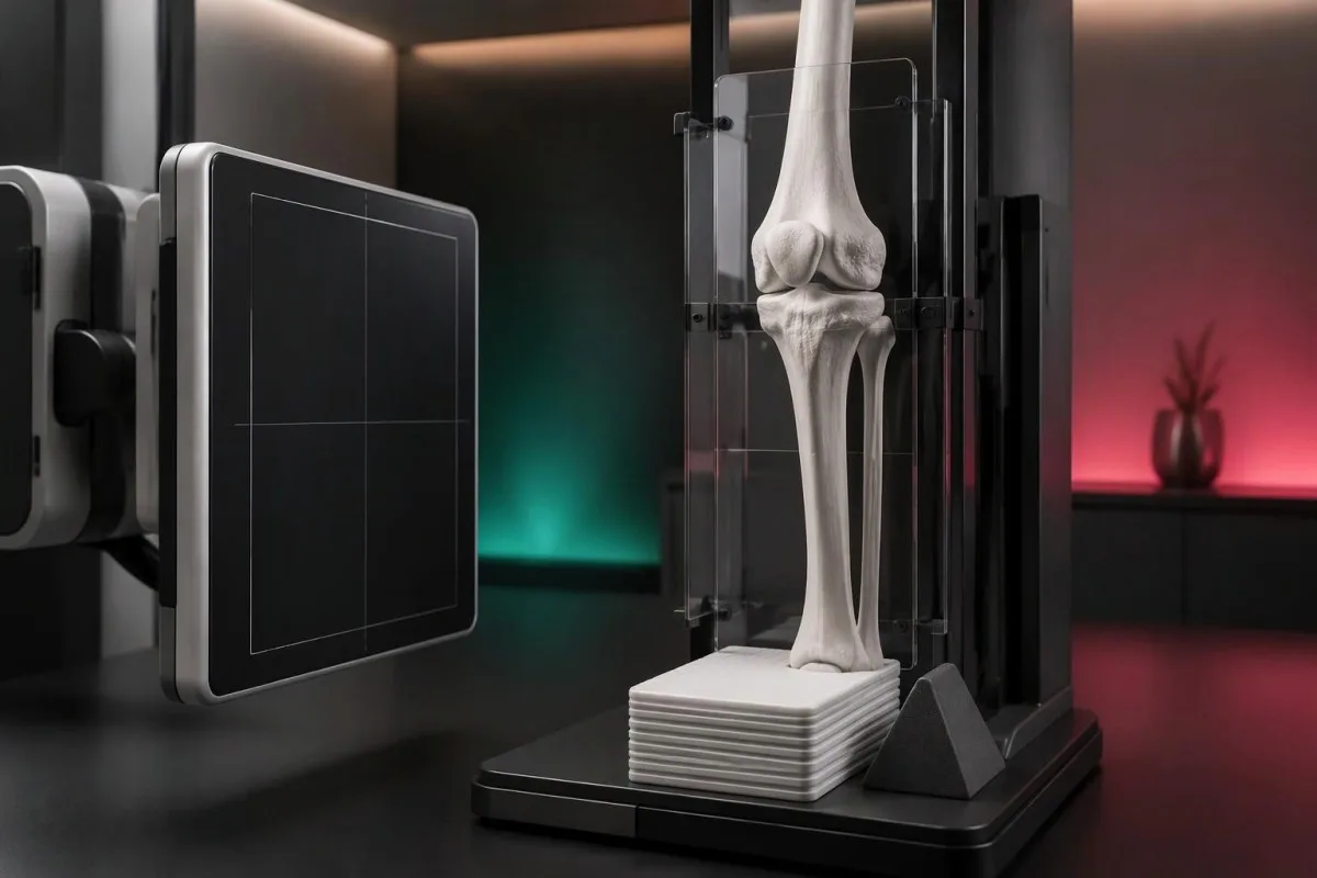
For the knee, weight-bearing radiographs can make joint-space narrowing more apparent than a non-weight-bearing view. EULAR recommends appropriate views and techniques rather than indiscriminate scanning (Sakellariou et al., 2017); importantly, 1 X-ray cannot directly measure pain, and prominent structural changes may coexist with modest symptoms while early symptoms can precede obvious radiographic changes.
MRI is not a routine second step simply because an osteoarthritis X-ray looks mild. It can be useful for a suspected occult fracture, another structural disorder, or selected mechanical symptoms; in a hypothetical 66-year-old with abrupt severe pain and difficulty bearing weight despite an unrevealing X-ray, the clinical concern—not the desire to grade arthritis—determines further imaging.
Repeat scans are usually less useful than reassessing symptoms and function when management remains unchanged. NICE advises against routine follow-up imaging for nonsurgical management (NICE, 2022); Kantesti's နည်းစနစ်နှင့် ဆေးကုသမှုဆိုင်ရာ ကန့်သတ်ချက်များ should be read with the same distinction in mind: a technical interpretation benchmark is not proof that an AI can diagnose joint disease from 1 laboratory report.
မည်သည့်အဆစ်လက္ခဏာများအတွက် အရေးပေါ် ဆေးဘက်ဆိုင်ရာ စစ်ဆေးမှုခံယူရန် လိုအပ်သနည်း။
A suddenly hot, swollen joint, severe pain with inability to bear weight, or rapid loss of function needs urgent medical evaluation. Seek same-day care for an unexplained acute hot joint, even without fever; a temperature below 38°C does not safely exclude joint infection, particularly in older or immunosuppressed adults.
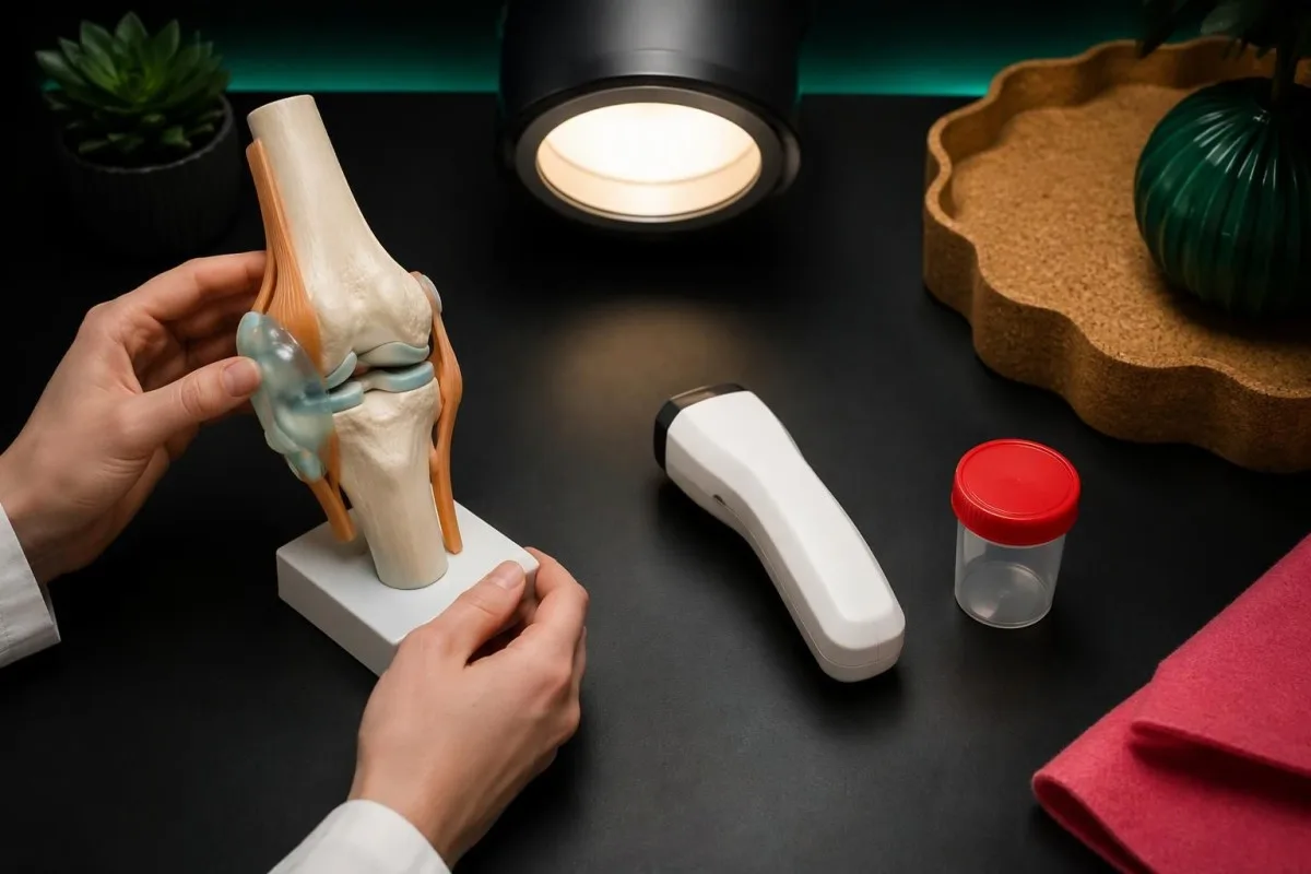
After an injury, inability to bear weight, a visible change in joint shape, or rapidly increasing swelling requires prompt assessment for fracture or another significant injury. Even a low-height fall can matter in someone with fragile bones; an existing diagnosis of osteoarthritis should not delay evaluation of a new event or justify walking through severe pain.
Emergency symptoms can occur near an arthritic joint without being caused by it. Leg swelling with chest pain or breathlessness warrants emergency care; new bladder retention, saddle-area numbness, or rapidly worsening leg weakness also requires immediate assessment, and 1 new neurological red flag is enough to change the plan from a routine arthritis appointment.
A swollen knee developing over နေ့များမှ ရက်များအထိ after plausible tick exposure may require an infection-focused assessment rather than an osteoarthritis label. Our guide to Lyme test interpretation explains why exposure history and validated testing matter; a hot acute joint still needs urgent assessment, while a random positive antibody result does not establish the cause of swelling.
ပုံမှန်စစ်ဆေးမှုအတွက် စီစဉ်နေစဉ်အတွင်း မည်သည့်အရာများ လုပ်ဆောင်နိုင်သနည်း။
For nonurgent, suspected osteoarthritis, keep moving within tolerable limits and arrange an assessment if symptoms persist or restrict daily life. Exercise is a core treatment once the diagnosis is established; a hypothetical starting plan might use 5–10 မိနစ် of comfortable walking rather than alternating complete rest with a suddenly much longer session.
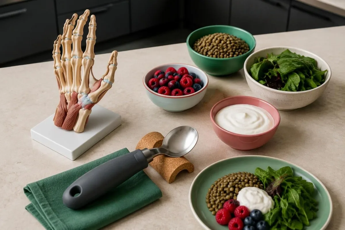
Exercise should be adapted to the affected joint, current capacity, and any warning signs rather than prescribed as one universal routine. The ACR/Arthritis Foundation guideline strongly recommends exercise for hand, hip, and knee osteoarthritis (Kolasinski et al., 2020); try changing 1 variable at a time, such as duration, so a clinician or physiotherapist can identify what is tolerable.
Weight loss can reduce symptoms for people with knee or hip osteoarthritis who are overweight, but it is not a prerequisite for receiving care. NICE states that losing 10% of body weight is likely to help more than 5%; ကျွန်တော်တို့ရဲ့ အထောက်အထား-အခြေခံ အစားအသောက် ရွေးချယ်မှုများ can support balanced eating, although no specific food has been shown to rebuild an osteoarthritic joint.
Pain treatment needs a safety review, particularly before regularly taking an oral anti-inflammatory medicine. NICE recommends topical NSAIDs for knee osteoarthritis, while oral NSAIDs require consideration of gastrointestinal, kidney, and cardiovascular risks; at Kantesti, we encourage recording the exact mg dose and frequency, and our medication-related laboratory guide explains why accompanying medicines and kidney results matter.
ဓာတ်ခွဲခန်းဆိုင်ရာ ရှင်းလင်းချက်များနှင့် ဆက်စပ်သုတေသနများကို မည်သို့အသုံးပြုသင့်သနည်း။
Use laboratory explanations to prepare better questions, not to self-confirm osteoarthritis or postpone a joint examination. ကန်တက်စတီသည် AI-powered သွေးစစ်ဆေးမှု ခွဲခြမ်းစိတ်ဖြာ ကိရိယာ that supports understanding of reported results; it cannot determine the cause of a painful knee from 1 CRP value, assess joint warmth remotely, or replace urgent care.

Bring a concise symptom record to a routine appointment: the joint involved, trigger, stiffness duration, swelling, injury history, and lost function. A ၇ ရက်တာမှတ်တမ်း is a practical example, not a requirement to wait; our တာဝန်ယူမှုရှိသော AI လမ်းညွှန်ချက် explains why an automated result should not override a clinician's examination or an urgent change in symptoms.
Laboratory reports sometimes contain unrelated findings that still need their own context. For example, 1 urine dipstick abnormality cannot explain knee pain without a relevant history; our ဆီးစစ်ဆေးမှု (urinalysis) အဓိပ္ပာယ်ဖော် လမ်းညွှန် addresses urinary findings separately, preventing an incidental result from being presented as evidence for osteoarthritis or used to justify unnecessary treatment.
ဟိ 2 DOI-linked publications below are supplemental laboratory education resources on urinalysis and iron studies, not osteoarthritis diagnostic studies. The directly relevant clinical references are NICE NG226, EULAR's imaging recommendations, and the ACR/Arthritis Foundation management guideline; a DOI makes a document identifiable, but does not itself establish peer review, clinical validation, or relevance to a joint diagnosis.
ተደጋግሞ የሚጠየቁ ጥያቄዎች
အရိုးအဆစ်ရောင်ရောဂါ၏ ပုံမှန်အားဖြင့် ပထမဆုံးတွေ့ရသော လက္ခဏာမှာ အဘယ်နည်း။
သတိပြုမိနိုင်သော အရိုးအဆစ်ရောင်ရောဂါ၏ ပထမဆုံးလက္ခဏာမှာ လှုပ်ရှားမှုပြုလုပ်နေစဉ် သို့မဟုတ် ပြုလုပ်ပြီးနောက် အဆစ်နာကျင်ခြင်းဖြစ်လေ့ရှိပြီး အနားယူပြီးနောက် ခဏတာတောင့်တင်းခြင်းလည်း တွဲဖြစ်တတ်သည်။ ပုံမှန်တွေ့ရသောအခြေအနေတွင် နံနက်ပိုင်းတောင့်တင်းမှုသည် ယေဘုယျအားဖြင့် မိနစ် ၃၀ ထက် မကြာတတ်ပါ။ လှေကားတက်ချိန်တွင် ဒူးနာခြင်း၊ ခြေအိတ်ဝတ်ချိန်တွင် တင်ပါးဆုံနာခြင်း၊ ဘူးအဖုံးဖွင့်ချိန်တွင် လက်မနာခြင်းတို့ ဖြစ်နိုင်သည်။ ဤပုံစံများသည် အကဲဖြတ်ရာတွင် အထောက်အကူပြုသော်လည်း စမ်းသပ်စစ်ဆေးမှုမပြုဘဲ နာကျင်နေသော အဆစ်တိုင်းကို ရောဂါရှာဖွေရန် မလုံလောက်ပါ။
သွေးစစ်ဆေးမှုတွေက ကျွန်ုပ်မှာ အရိုးအဆစ်ရောင်ရောဂါရှိမရှိ ပြနိုင်ပါသလား?
No routine blood test can confirm or exclude osteoarthritis. A CRP within a laboratory's reference interval, often below 5 mg/L, does not prove that a joint is healthy or explain its pain. Blood tests may help investigate rheumatoid arthritis, infection, iron overload, or other conditions when the symptoms suggest them. Diagnosis usually relies on the clinical pattern and examination, with imaging used selectively.
အရိုးအဆစ်ရောင်ရမ်းနာကြောင့် ဖြစ်သော တောင့်တင်းမှုနှင့် ရူမတွိုက်အဆစ်ရောင်ရောဂါကြောင့် ဖြစ်သော တောင့်တင်းမှုကို ဘယ်လိုခွဲခြားသိနိုင်မလဲ?
အရိုးအဆစ်ရောင်ရမ်းခြင်းကြောင့် ဖြစ်သော တောင့်တင်းမှုသည် ပုံမှန်အားဖြင့် မိနစ် ၃၀ ထက် မပိုကြာတတ်သော်လည်း၊ ရူမတွိုက်အဆစ်ရောင်ရမ်းခြင်းတွင်မူ မနက်ပိုင်းတောင့်တင်းမှု ကြာရှည်ခြင်းနှင့် အဆစ်များ ပျော့ပျောင်းစွာ ဖောင်းရောင်နေခြင်းတို့ ပိုမိုဖြစ်တတ်သည်။ ကြာချိန်သည် ရောဂါနှစ်မျိုးကို အတိအကျခွဲခြားပေးသည့် စည်းမျဉ်းမဟုတ်သကဲ့သို့၊ ရောဂါတစ်မျိုးစီတွင် ပုံမှန်မဟုတ်သော လက္ခဏာများလည်း ရှိနိုင်သည်။ လက်ဆစ်အများအပြားတွင် နှစ်ဖက်ညီညီ ဖောင်းရောင်ခြင်းက ရောင်ရမ်းအဆစ်ရောဂါ ရှိမရှိ စစ်ဆေးရန် ပိုမိုခိုင်လုံသော အကြောင်းပြချက်ဖြစ်သည်။ rheumatoid factor သို့မဟုတ် anti-CCP ရလဒ်များ အနုတ်လက္ခဏာဖြစ်ခြင်းသည် ရူမတွိုက်အဆစ်ရောင်ရမ်းခြင်းကို ပယ်ချမပေးပါ။
အရိုးအဆစ်ရောင်ရောဂါ၏ အစောပိုင်းလက္ခဏာများရှိပါက X-ray ရိုက်ဖို့ လိုအပ်ပါသလား?
အရိုးအဆစ်ရောင်ရမ်းမှု အစောပိုင်းလက္ခဏာများအတွက် ဓာတ်မှန်ရိုက်ရန် အမြဲမလိုအပ်ပါ။ လှုပ်ရှားမှုနှင့်ဆက်စပ်သော အဆစ်နာကျင်မှုရှိပြီး နံနက်ပိုင်း အဆစ်တောင့်တင်းမှု မရှိသူ၊ သို့မဟုတ် အဆစ်တောင့်တင်းမှု ၃၀ မိနစ်ထက် မကြာသူ အသက် ၄၅ နှစ်နှင့်အထက် အရွယ်ရောက်သူများတွင် လက်တွေ့စမ်းသပ်စစ်ဆေးမှုဖြင့် ရောဂါသတ်မှတ်နိုင်ကြောင်း NICE က ထောက်ခံထားသည်။ ရောဂါလက္ခဏာပုံစံ ပုံမှန်မဟုတ်သည့်အခါ၊ ရောဂါသတ်မှတ်ချက် မသေချာသေးသည့်အခါ၊ သို့မဟုတ် စစ်ဆေးမှုရလဒ်ကြောင့် ကုသမှု ပြောင်းလဲနိုင်သည့်အခါ ပုံရိပ်ဖော်စစ်ဆေးမှုက ပိုအသုံးဝင်လာသည်။ ဓာတ်မှန်တွင် ပုံမှန်အတိုင်းမြင်ရခြင်းသည် အဆစ်အတွင်း အစောပိုင်းပြောင်းလဲမှုအားလုံး သို့မဟုတ် နာကျင်မှုဖြစ်စေသည့် အခြားအကြောင်းရင်းများကို ဖယ်ထုတ်နိုင်သည်ဟု မဆိုလိုပါ။
တင်ပါးဆုံရိုး အရိုးအဆစ်ရောင်ရောဂါက ဒူးနာသလို ခံစားရနိုင်ပါသလား?
တင်ပါးဆုံရိုး အရိုးအဆစ်ရောင်ရမ်းမှုကြောင့် တင်ပါးဆုံရိုးမအီမသာဖြစ်မှုကို အဓိကမတိုင်ကြားသည့်အခါတွင်ပင် ဒူးဆီသို့ နာကျင်မှုကူးစက်နိုင်သည်။ ထို့ကြောင့် အကြောင်းရင်းမရှင်းလင်းသော နာကျင်နေသည့် ဒူး 1 ဖက်ကို စစ်ဆေးရာတွင် တင်ပါးဆုံရိုးလှုပ်ရှားမှုကို အကဲဖြတ်ခြင်းနှင့် ပေါင်ခြံနာကျင်မှုရှိမရှိ၊ ခြေအိတ်ဝတ်ရန် ခက်ခဲမှုရှိမရှိ စစ်ဆေးခြင်းတို့ ပါဝင်နိုင်သည်။ ဒူးအောက်ပိုင်းတွင် ကျဉ်ခြင်း၊ ပူလောင်နာကျင်ခြင်း သို့မဟုတ် အားနည်းခြင်းတို့ရှိပါက အာရုံကြောပါဝင်နိုင်ခြေကို ညွှန်ပြသည်။ ကိုယ်အလေးချိန် မထောက်နိုင်သည့်အထိ တင်ပါးဆုံရိုး သို့မဟုတ် ပေါင်ခြံတွင် ရုတ်တရက် ပြင်းထန်စွာ နာကျင်ပါက ပုံမှန်အဆစ်ရောင်ရမ်းမှုဟု ယူဆမနေဘဲ အရေးပေါ်စစ်ဆေးမှုခံယူရန် လိုအပ်သည်။
အဆစ်ရောင်ရမ်းဖောင်းလာလျှင် ဘယ်အချိန်တွင် အရေးပေါ်အကူအညီ ရယူသင့်သလဲ?
ရုတ်တရက် ပူလာပြီး ရောင်ရမ်းကာ အလွန်နာကျင်သော အဆစ်တစ်ခု ဖြစ်လာပါက၊ အထူးသဖြင့် လှုပ်ရှားရန် သို့မဟုတ် ကိုယ်အလေးချိန်ထောက်ရန် ခက်ခဲလာပါက ထိုနေ့အတွင်း ဆေးဘက်ဆိုင်ရာ စစ်ဆေးမှုခံယူပါ။ အပူချိန် ၃၈°C အောက်ရှိခြင်းနှင့် သွေးတွင်းရောင်ရမ်းမှုညွှန်းကိန်းများ ပုံမှန်ဖြစ်ခြင်းတို့ကြောင့် အဆစ်ပိုးဝင်ခြင်းမရှိဟု စိတ်ချစွာ မသတ်မှတ်နိုင်ပါ။ ထိခိုက်ဒဏ်ရာရပြီးနောက် ကိုယ်အလေးချိန်ထောက်မရခြင်း သို့မဟုတ် အဆစ်ပုံသဏ္ဌာန် ပြောင်းလဲသွားခြင်းရှိပါကလည်း အမြန်စစ်ဆေးမှုခံယူရန် လိုအပ်သည်။ ရင်ဘတ်နာခြင်း၊ အသက်ရှူမဝခြင်း သို့မဟုတ် အသစ်ဖြစ်ပေါ်လာသော ပြင်းထန်သည့် အာရုံကြောဆိုင်ရာ လက္ခဏာများပါရှိပါက အရေးပေါ်ဝန်ဆောင်မှုများကို ခေါ်ဆိုပါ။
သင့်သွေးစစ်ဆေးမှု ရလဒ်ကို ရှင်းလင်းချက်ရယူပါ
သင့်ဓာတ်ခွဲခန်းအစီရင်ခံစာကို တင်ပါ၊ Kantesti ၏ AI က ဇီဝအမှတ်အသားတစ်ခုစီကို နားလည်လွယ်သော ဘာသာစကားဖြင့် စက္ကန့်ပိုင်းအတွင်း ရှင်းပြပေးမည်။ ကမ္ဘာတစ်ဝန်းရှိ လူ ၂ သန်းကျော် အသုံးပြုထားသည်။.
ကိုးကားချက်များ
- Klein, T., Mitchell, S., & Weber, H. (2026)။. အစာအိမ်ထဲ Urobilinogen စမ်းသပ်မှု: ၂၀၂၆ အပြည့်အစုံ သွေးမလှိုင်း စမ်းသပ်မှု လမ်းညွှန်။. Kantesti AI ဆေးဘက်ဆိုင်ရာသုတေသန။. DOI
- Klein, T., Mitchell, S., & Weber, H. (2026)။. သံဓာတ်စစ်ဆေးမှုလမ်းညွှန်- TIBC၊ သံဓာတ်ပြည့်ဝမှုနှင့် ချိတ်ဆက်နိုင်စွမ်း။. Kantesti AI ဆေးဘက်ဆိုင်ရာသုတေသန။. DOI
- National Institute for Health and Care Excellence (2022). Osteoarthritis in over 16s: diagnosis and management. NICE guideline NG226.
- Sakellariou G et al. (2017). EULAR recommendations for the use of imaging in the clinical management of peripheral joint osteoarthritis. Annals of the Rheumatic Diseases။. DOI
- Kolasinski SL et al. (2020). 2019 American College of Rheumatology/Arthritis Foundation Guideline for the Management of Osteoarthritis of the Hand, Hip, and Knee. Arthritis Care & Research. DOI
- 2M+ခွဲခြမ်းစိတ်ဖြာထားသော စစ်ဆေးမှုများ
- 127+နိုင်ငံတွေ
- 100+ဘာသာစကားများ
ဆေးဘက်ဆိုင်ရာအဖွဲ့အကြောင်း


E-E-A-T ယုံကြည်မှု အချက်ပြများ
အတွေ့အကြုံ
ဆရာဝန်ဦးဆောင်သည့် ဓာတ်ခွဲခန်း အဓိပ္ပာယ်ဖတ်ခြင်း လုပ်ငန်းစဉ်များကို ပြန်လည်သုံးသပ်ခြင်း။.
ကျွမ်းကျင်မှု
biomarker များသည် ဆေးဘက်ဆိုင်ရာ အခြေအနေတွင် မည်သို့ ပြုမူနေသည်ကို အာရုံစိုက်သည့် ဓာတ်ခွဲခန်း ဆေးပညာ။.
အခွင့်အာဏာရှိခြင်း
ဒေါက်တာ Sarah Mitchell နှင့် ပါမောက္ခ ဒေါက်တာ Hans Weber တို့၏ ပံ့ပိုးကူညီမှုဖြင့် ဒေါက်တာ Thomas Klein က ဆေးဘက်ဆိုင်ရာသုံးသပ်မှုပြုလုပ်ထားသည်။.
ယုံကြည်စိတ်ချရမှု
စိတ်ပူစရာများကို လျော့ချရန်အတွက် ရှင်းလင်းသော နောက်ဆက်တွဲ လမ်းကြောင်းများပါဝင်သည့် သက်သေအထောက်အထားအခြေပြု အဓိပ္ပာယ်ဖတ်ချက်။.

ဆေးဘက်ဆိုင်ရာ ငြင်းဆိုချက်
ဤဆောင်းပါးသည် ပညာရေးဆိုင်ရာ ရည်ရွယ်ချက်အတွက်သာ ဖြစ်ပြီး ဆေးဘက်ဆိုင်ရာ အကြံဉာဏ် မဟုတ်ပါ။ ရောဂါရှာဖွေခြင်းနှင့် ကုသမှု ဆုံးဖြတ်ချက်များအတွက် အရည်အချင်းပြည့်မီသော ကျန်းမာရေးစောင့်ရှောက်မှုပေးသူနှင့် အမြဲတမ်း တိုင်ပင်ပါ။.
ထုတ်ဝေသည်။ · အယ်ဒီတာအဖွဲ့ကို ဆက်သွယ်ပါ
Kantesti Ltd မှ ထုတ်ဝေသည်၊ အင်္ဂလန်နှင့် ဝေလနယ်တွင် မှတ်ပုံတင်ထားပြီး ကုမ္ပဏီအမှတ်. 17090423, ၊ လန်ဒန်၊ ယူနိုက်တက်ကင်းဒမ်း။.
