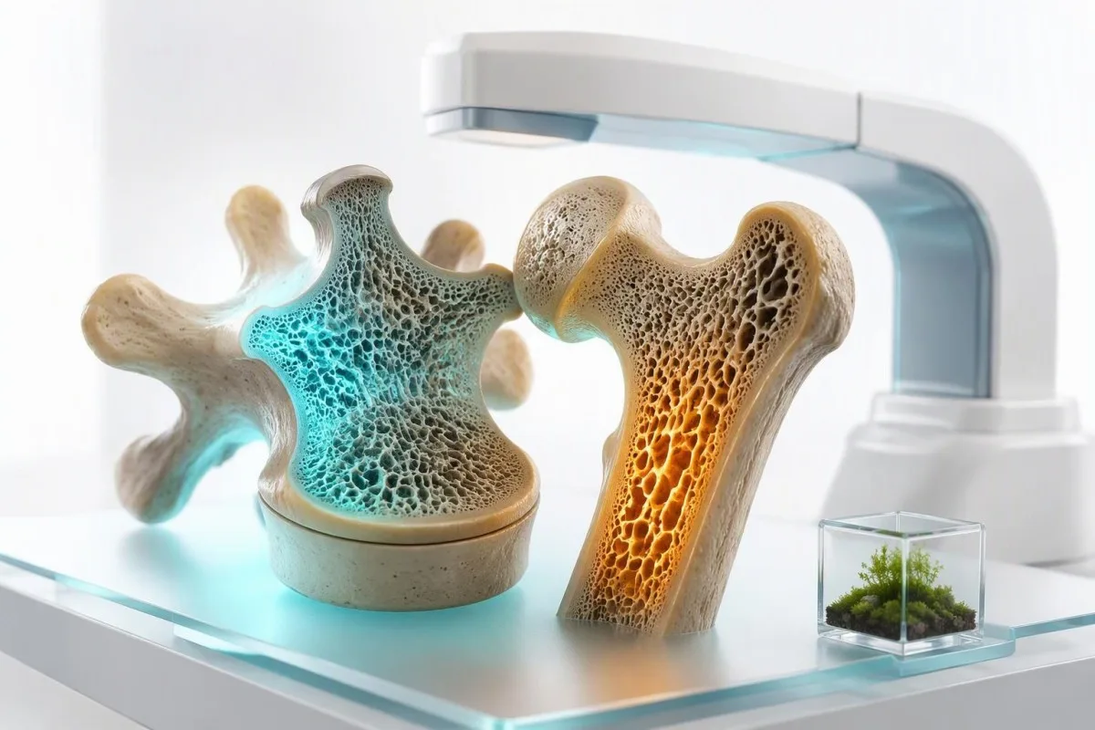
သွေး စစ် ဆေး မှု များ က အရိုး သိပ် သည်း မှု ကို တိုက် ရိုက် တိုင်း တာ မဟုတ် သ လို အရိုး ပွ ရောဂါ ကို လည်း တ ခု တည်း နှင့် ရောဂါ ရှာ ဖွေ နိုင် ခြင်း မ ရှိ ပါ ။ သို့ သော် လည်း ဗီတာမင်ဒီ ချို့တဲ့ခြင်း၊ ပါရာ သ ရွိုက် ရောဂါ ၊ ကျောက် ကပ် ၏ လုပ် ငန်း ဆောင် တာ မ မှန် ခြင်း ၊ ဟ ော် မုန်း ဆုံး ရှုံး ခြင်း နှင့် အ ရိုး များ ကို ဖျက် စီး နေ သည့် အ ရိုး ဆုံး ရှုံး မှု ကို တ ဖြည်း ဖြည်း အ မြန် ဆန် ဆုံး ဖြစ် စေ နိုင် သည့် အ စာ စ affected ဖြစ် ခြင်း တို့ ကို ဖော် ထုတ် ပြ နိုင် ပါ သည် ။.


- DXA စ က န် အရိုး ပွ ရောဂါ ကို ရောဂါ ရှာ ဖွေ ရန် လိုအပ် သည် ။ T-score -၂.၅ သို့ မ ဟု .
- 25-ဟိုက်ဒရိုစီဗီတာမင်ဒီ ၂၀ ng/mL (၅၀ nmol/L) အောက် သည် ချို့ တဲ့ ခြင်း ကို ဖော် ပြ ပြီး ကယ် လ် စီ ယမ် စုပ် ယူ .
- ကယ် လ် စီ ယမ် နှင့် PTH ပ .
- သွေး တွင်း ကယ် လ် စီ ယမ် မှာ ပုံ မှန် အတိုင်း အရိုး ပွ ရောဂါ တွင် .
- ကရီတီနင်းနှင့် eGFR နာ တာ ရှည် ကျောက် ကပ် ရောဂါ ကို ဖော် ထုတ် နိုင် သည် ။ ၎င်း သည် အ .
- TSH သည် 0.1 mIU/L အောက် ပ .
- ၂၄ နာရီ ဆီးကယ်လ်စီယမ် သည် ကယ်လ်စီယမ်နည်းပါးခြင်းနှင့် ဆီးတွင်းကယ်လ်စီယမ်ဆုံးရှုံးခြင်းနှင့် ပါရာသိုင်းရွတ်ရောဂါအချို့ကို ခွဲခြားရန် ကူညီသည်။.
- DXA သည် ပုံမှန်အားဖြင့် သင့်လျော်သည်။ ကျိုးလွယ်သော အရိုးကျိုးပြီးနောက်၊ သက်တမ်းရှည်စတီရွိုက်သုံးစွဲပြီးနောက် သို့မဟုတ် အသက် 65 နှစ်နှင့်အထက် အမျိုးသမီးများအတွက်။.
အရိုး ဆုံး ရှုံး မှု ကို ရောဂါ ရှာ ဖွေ ရန် အတွက် အရိုး ပွ ရောဂါ သွေး စစ် ဆေး မှု ကို လုပ် နိုင် ပါ သ လား ။
အရိုးပွရောဂါကို မည်သည့်သွေးစစ်ဆေးမှုကမျှ မခွဲခြားနိုင်ပါ၊ အဘယ်ကြောင့်ဆိုသော် သွေးစစ်ဆေးမှုများသည် အရိုးသတ္တုသိပ်သည်းဆကို မတိုင်းတာသောကြောင့်ဖြစ်သည်။. DXA စကင်သည် အရိုးသိပ်သည်းဆ -2.5 သို့မဟုတ် ထို့ထက်နည်းသော T-score အနည်းဆုံးရှိသောအခါတွင် အရိုးပွရောဂါကို ခွဲခြားသတ်မှတ်ပေးပြီး သွေးနှင့် ဆီးစစ်ဆေးမှုများသည် အရိုးကို အားနည်းစေနိုင်သော ကုသနိုင်သော အကြောင်းရင်းများကို ဖော်ထုတ်ပေးသည်။.
Serum calcium, phosphate, vitamin D, and parathyroid hormone reflect mineral regulation, not the amount of mineral already present in the hip or spine. A normal panel therefore does not rule out osteoporosis; in fact, many patients with a T-score of -3.0 have entirely ordinary calcium results. ဒေါက်တာ သောမတ်စ် ကလိုင်း၊ MD ကနဦး DXA တောင်းဆိုမှုများကို နှစ်ပေါင်းများစွာ နှောင့်နှေးစေသည့် ဤမှားယွင်းသော အယူအဆကို မြင်တွေ့ခဲ့ရသည်။.
ကန်တက်စတီသည် AI သွေးစမ်းသပ်မှုခွဲခြမ်းစိတ်ဖြာ ကယ်လ်စီယမ်၊ PTH၊ ဗီတာမင် D၊ ကျောက်ကပ်လုပ်ဆောင်ချက်နှင့် သိုင်းရွတ်ဒ်ရလဒ်များကို ရောဂါရှာဖွေရေးအဖြစ် အမှတ်တစ်မှတ်ကို အizzazione မပြုဘဲ ဆေးဘက်ဆိုင်ရာ ပုံစံတစ်ခုသို့ ထည့်သွင်းပေးသည်။ ကျွန်ုပ်တို့၏ ပြန်လည်သုံးသပ်မှု လုပ်ငန်းစဉ်တွင်၊ ဗီတာမင် D နည်းပါးသော ရလဒ်သည် ကုသမှုကို ပံ့ပိုးပေးသော်လည်း၊ ၎င်းသည် DXA ရလဒ် သို့မဟုတ် အရိုးကျိုးမှု-အန္တရာယ် အကဲဖြတ်မှုအတွက် ဘယ်သောအခါမျှ အစားထိုးမပေးနိုင်ပါ။.
A fragility fracture—one occurring from a standing-height fall or less—can establish clinical osteoporosis even before DXA is available. The 2020 AACE guideline recommends evaluation for secondary causes in people with osteoporosis or fragility fractures, particularly when bone loss is early, severe, or unexpectedly rapid (Camacho et al., 2020). اسان جي بائيو مارڪر گائيڊ ရည်ညွှန်း-အတန်းစား ရလဒ်တစ်ခုသည် အဘယ်ကြောင့် ဆေးဘက်ဆိုင်ရာ အကြောင်းအရာ လိုအပ်သည်ကို ရှင်းပြသည်။.
စမ်းသပ်မှု နှစ်မျိုးလုံးသည် မည်သည့်အရာကို ဖြေကြားသနည်း
DXA က“ဒီနေ့ အရိုး ဘယ်လောက် သိပ်သည်းသလဲ” လို့ မေးတယ်။ ဓာတ်ခွဲခန်း စမ်းသပ်မှုက “ဟော်မုန်း၊ အာဟာရ၊ ကျောက်ကပ်၊ အစာအိမ်-အူလမ်းကြောင်း၊ ဒါမှမဟုတ် ဆေး-နှင့်သက်ဆိုင်သော လုပ်ငန်းစဉ်က ဆုံးရှုံးမှုကို မောင်းနှင်ရန် ကူညီပေးနေသလား” လို့ မေးတယ်။ ကယ်လ်စီယမ် တစ်ခုတည်း လုံလောက်ပါသလား၊ ပြောင်းလဲနိုင်သော အခြေအနေတစ်ခုကို ကုသရန် လိုအပ်ပါသလား၊ ဒါမှမဟုတ် အရိုးကျိုး-ကာကွယ်ဆေးကို ထည့်သွင်းစဉ်းစားသင့်သလား ဆုံးဖြတ်သည့်အခါ အဖြေနှစ်ခုစလုံးသည် အရေးကြီးပါသည်။.
ဓာတ် ခွဲ ခန်း စ စ် ဆေး မှု ထက် DXA စ က န် ကို ဘယ် အ ခါ တွင် လိုအပ် သ လဲ ။
အသက်၊ အရိုးကျိုးမှုသမိုင်း၊ ဆေးဝါးထိတွေ့မှု သို့မဟုတ် အန္တရာယ်ရှိသော အချက်များက အရိုးသိပ်သည်းဆ လျော့နည်းခြင်းကို အကြံပြုသောအခါ DXA စကင်သည် လိုအပ်ပြီး သွေးစစ်ဆေးမှုများသည် ၎င်းကို အစားထိုး၍မရပါ။. DXA သည် ခါးဆစ်နှင့် တင်ပါးဆုံရိုးတွင် အရိုးသတ္တုသိပ်သည်းဆကို အလွန်နည်းပါးသော ရေဒီယေးရှင်း ထိတွေ့မှုဖြင့်၊ ပုံမှန်အားဖြင့် 10 မိုက်ခရိုစီဗတ်ထ်အောက်တွင် တိုင်းတာသည်။.
အသက် 65 နှစ်နှင့် အထက်ရှိသော အမျိုးသမီးများသည် ပုံမှန်အားဖြင့် DXA ဖြင့် အရိုးပွရောဂါ စစ်ဆေးသင့်ပြီး၊ ပိုငယ်သော သွေးဆုံးပြီးနောက် အမျိုးသမီးများသည် အန္တရာယ်-အခြေခံ အကဲဖြတ်မှု လိုအပ်သည်။ အရိုးနည်းပါးသော ကျိုးလွယ်သော အရိုးကျိုးပြီးနောက်၊ အရပ်အမြင့် 4 စင်တီမီတာထက် ပိုမိုကျဆင်းခြင်း၊ အသက် 40 နှစ်မတိုင်မီ သွေးဆုံးခြင်း သို့မဟုတ် 3 လ သို့မဟုတ် ထို့ထက်ပို၍ တစ်နေ့လျှင် 5 mg ၏ ပရက်ဒီနီဆုန်း-ညီမျှသော စတီရွိုက်ကုသမှုခံယူပြီးနောက် ပိုမိုစောသော စစ်ဆေးမှုသည် အကျိုးရှိသည်။.
A T-score သည် အရိုးသိပ်သည်းဆကို ကျန်းမာသော လူငယ်-လူကြီး အကိုးအကား ပြည်သူများနှင့် နှိုင်းယှဉ်သည်- -1.0 သို့မဟုတ် အထက်မှာ ပုံမှန်ဖြစ်သည်၊ -1.0 မှ -2.5 မှာ အရိုးပွရောဂါဖြစ်သည်၊ -2.5 သို့မဟုတ် အောက်မှာ အရိုးပွရောဂါဖြစ်သည်။ A Z-score သည် အလားတူ အသက်နှင့် ကျားမ ကွဲပြားသူများနှင့် နှိုင်းယှဉ်သည်၊ သွေးဆုံးခါစ အရွယ်ရောက်ပြီးသူ သို့မဟုတ် အသက် 50 နှစ်အောက် အမျိုးသားတွင် -2.0 သို့မဟုတ် အောက်တွင် ရှိသော Z-score သည် ဒုတိယ အရိုးပွရောဂါကို ပိုမိုရှာဖွေရန် အချက်ပြသင့်သည်။.
DXA သည် အသက်ကြီးသူများတွင် အဆစ်အမြစ်ရောင်ခြင်း၊ အေအော်တစ် အဆီကပ်ခြင်း သို့မဟုတ် ကျောရိုး ကျိုးမှု အပြောင်းအလဲများ ရှိနေပါက ခါးဆစ်တွင် လွဲမှားစွာ အာမခံချက်ပေးနိုင်ပါသည်။ ထို့ကြောင့် ကျွန်ုပ်သည် တင်ပါးဆုံရိုး၊ ကျောရိုး၊ အရိုးကျိုးမှုသမိုင်းနှင့် အခါအားလျော်စွာ ကျောရိုး-အရိုးကျိုးမှု အကဲဖြတ်မှုတို့ကို နှိုင်းယှဉ်ပေးသည်၊ “ကောင်း” သော ကျောရိုး နံပါတ်တစ်ခုမှ မည်သူ့ကိုမျှ အာမခံချက် မပေးပါ။. Osteocalcin ရလဒ်များ သည် လည်ပတ်မှု အကြောင်းအရာကို ထည့်သွင်းနိုင်သော်လည်း ရုပ်ပုံဖော်ခြင်းကို အစားထိုး၍မရပါ။.
ဒု တိ ယ အ ရိုး ပွ ရောဂါ ဓာတ် ခွဲ ခန်း စ စ် ဆေး မှု များ ကို ပုံ မှန် အား ဖြင့် ပ ထ မ ဦး ဆုံး အ နေ နှင့် ဘယ် အ ရာ များ ကို အ သုံး ပြု သ လဲ ။
The core secondary osteoporosis lab panel usually includes calcium, albumin, creatinine with eGFR, alkaline phosphatase, 25-hydroxyvitamin D, CBC, phosphate, and PTH when calcium is abnormal. The exact panel changes with age, symptoms, medicines, fracture pattern, and the DXA result.
A အပြည့်အစုံ ဇီဝဓာတုစစ်ဆေးမှုအစု (comprehensive metabolic panel) supplies calcium, albumin, creatinine, bicarbonate, and liver markers, while alkaline phosphatase gives a broad clue to bone formation or hepatobiliary disease. Adult total calcium is commonly 8.5 to 10.5 mg/dL (2.12 to 2.62 mmol/L), but the laboratory interval and albumin concentration must be checked before calling a result high or low.
A CBC can expose anemia that points toward celiac disease, chronic inflammation, kidney disease, marrow disorders, or nutritional deficiency. In a patient with a low Z-score and unexplained anemia, I am much more likely to add celiac serology and protein studies than to simply advise more dairy.
The 2017 UK guideline advises baseline blood testing to exclude secondary osteoporosis in patients being evaluated for fragility fractures (Compston et al., 2017). How often to recheck vitamin D depends on the starting value, dose, absorption, and whether a condition such as coeliac disease is still active.
Tests that are not routine for everyone
Magnesium, celiac antibodies, serum protein electrophoresis, cortisol testing, sex hormones, and 24-hour urine calcium are targeted tests rather than universal screening. Ordering everything at once often creates incidental abnormalities; a good work-up follows the clues rather than chasing every borderline result.
အ ရိုး ဆုံး ရှုံး မှု အတွက် ကယ် လ် စီ ယမ် နှင့် အ ယ် လ ဘူ မင် ကို ဘ ယ် လို အ န інтерпретация လုပ် သင့် သ လဲ ။
High calcium with an inappropriately normal or high PTH raises concern for primary hyperparathyroidism, a treatable cause of bone loss. Low calcium is less typical of uncomplicated osteoporosis and usually points toward vitamin D deficiency, malabsorption, kidney disease, hypoparathyroidism, or medication effects.
About 40% of circulating calcium is albumin-bound, so a low albumin can make total calcium appear low even when biologically active calcium is adequate. Laboratories may report albumin-adjusted calcium, but direct ionized calcium သည် ပြင်ဆင်ထားသော စုစုပေါင်းကယ်လ်စီယမ်ထက် ပိုမိုယုံကြည်ရတတ်ပါသည်။ ကျွန်ုပ်၏ အတွေ့အကြုံအရ၊ အွန်လိုင်းတွင် လူနာများကို မှားယွင်းစွာ အမျိုးအစားခွဲခြားခံရစေသည့် အများဆုံးအကြောင်းရင်းများထဲမှ တစ်ခုမှာ ယင်းဖြစ်ပါသည်။ is preferable when the result is borderline, albumin is markedly abnormal, or an acid-base disturbance is present.
A persistent adjusted calcium above roughly 10.2 to 10.5 mg/dL (2.55 to 2.62 mmol/L), depending on the laboratory, deserves repeat testing with PTH. PTH should be suppressed when calcium is high; if it is not, primary hyperparathyroidism becomes more likely. Slightly elevated calcium is often not an emergency, but repeated results should not be ignored.
One clinical trap: thiazide diuretics, lithium, dehydration, and calcium carbonate antacids can raise calcium modestly. I ask patients to bring every supplement bottle, because “bone support” products may contain 1,200 mg of calcium plus vitamin D, and that changes both the result and the next decision.
Why normal calcium does not protect the skeleton
Normal serum calcium is tightly defended by PTH, vitamin D, kidneys, and the skeleton. When dietary calcium is inadequate or vitamin D absorption fails, bone resorption can help preserve a blood calcium concentration within the 8.5 to 10.5 mg/dL range—an effective short-term survival mechanism with a long-term skeletal cost.
PTH နှင့် ဗီတာမင် ဒီ စ စ် ဆေး မှု များ က အ ဘယ် အ ရာ ကို ဖော် ထုတ် ပြ သ သ လဲ ။
The most informative calcium PTH vitamin D testing pattern is low 25-hydroxyvitamin D with high PTH and normal or low-normal calcium, which often indicates secondary hyperparathyroidism. This pattern means the body is working harder to maintain calcium balance and may be drawing on bone stores.
A 25-ဟိုက်ဒရိုစီဗီတာမင်ဒီ level below 20 ng/mL (50 nmol/L) is generally considered deficiency; 20 to 29 ng/mL is often called insufficiency, although professional groups disagree about whether every value in this interval needs treatment. The 25-hydroxy test is the correct measure of body stores, not 1,25-dihydroxyvitamin D, which can be normal or elevated in deficiency.
PTH reference intervals are assay-specific, commonly about 15 to 65 pg/mL (1.6 to 6.9 pmol/L). A PTH of 78 pg/mL alongside vitamin D of 12 ng/mL and calcium of 9.1 mg/dL usually suggests compensatory secondary hyperparathyroidism; a PTH of 78 with calcium of 10.8 mg/dL suggests a very different pathway.
ကန်တက်စတီသည် AI သွေးစစ်ဆေးမှု ရလဒ် ပလက်ဖောင်း that reads vitamin D, calcium, albumin, eGFR, and PTH as a linked physiology problem, including whether the pattern merits clinician follow-up rather than self-prescribed high-dose supplements. For a practical safety discussion, see vitamin D and K2 dosing.
A sensible recheck interval
After starting vitamin D, retesting 25-hydroxyvitamin D and calcium in about 8 to 12 weeks is reasonable for deficiency, malabsorption, high doses, kidney disease, or a prior high calcium result. Daily maintenance doses of 800 to 2,000 IU suit many adults, while loading regimens should be individualized by the treating clinician.
နာ ရီ ၂၄ နာ ရီ ကာ လ အ တွင်း အ ဆီး ခ ရီး မှ ကယ် လ် စီ ယမ် စ စ် ဆေး မှု က ဘယ် အ ခါ တွင် အ ရေး ပါ သ လဲ ။
A 24-hour urine calcium test is most useful when osteoporosis occurs with kidney stones, high PTH, unexplained low calcium intake, or suspected renal calcium loss. It does not diagnose osteoporosis, but it can explain why the calcium balance is negative despite a seemingly good diet.
Many laboratories consider roughly 100 to 250 mg of calcium per 24 hours typical for adults eating a usual diet, but the useful interpretation depends on sodium intake, collection completeness, sex, body size, and local methods. A value above 250 mg/day in women or 300 mg/day in men is often called hypercalciuria, though thresholds vary.
High urine calcium can accompany high sodium intake, loop diuretics, idiopathic hypercalciuria, primary hyperparathyroidism, sarcoidosis, or excessive vitamin D. The combination of recurrent calcium stones, urine calcium of 350 mg/day, and declining hip density is a stronger signal than a single elevated collection alone. High urine calcium causes are worth reviewing before simply increasing supplements.
The collection is easy to spoil: missing one daytime sample, collecting for 20 rather than 24 hours, or changing diet dramatically makes the number less useful. Kantesti AI's trend review can help people preserve the collection date, supplement dose, eGFR, and serum calcium beside the report; our မော်ဒယ် reasoning လုပ်ဆောင်ပုံအတွက်တော့ describes how structured context improves lab interpretation.
အ ရိုး ဆုံး ရှုံး မှု သွေး စ စ် ဆေး မှု များ တွင် သိုင်းရွိုက် စစ်ဆေးမှု များ ကို အ ဘယ် အ ကြောင့် ထည့် သွင်း ထား ပါ သ လဲ ။
Thyroid hormone excess accelerates bone turnover, so a suppressed TSH should be investigated in people with osteoporosis or unexplained fractures. A TSH below 0.1 mIU/L is particularly concerning in postmenopausal women and older adults, especially if free T4 or free T3 is elevated.
TSH is usually the first thyroid test because it responds sensitively to excess thyroid hormone. The common adult reference interval is about 0.4 to 4.0 mIU/L, although pregnancy, age, assay design, acute illness, and thyroid treatment change how a result should be read.
Over-replacement with levothyroxine is a surprisingly practical cause of low TSH. A 68-year-old patient may feel perfectly well with TSH 0.06 mIU/L, yet repeated suppression over years can be relevant to fracture risk; the target should be reviewed rather than assuming a “low-normal dose” is automatically safe.
Subclinical hyperthyroidism means low TSH with normal free T4 and free T3, and its skeletal effect is real but varies by duration and degree of suppression. Subclinical thyroid disease is different from hyperthyroidism, but both articles illustrate why one thyroid result cannot be interpreted without symptoms, medicines, and repeat testing.
Why hypothyroidism is not the mirror image
Untreated overt hypothyroidism slows bone remodeling rather than directly causing rapid bone loss, but it can increase falls through fatigue, myopathy, and impaired balance. The bigger skeletal concern is usually excessive replacement therapy, not a mildly raised TSH by itself.
အ ရိုး ပွ ရောဂါ ၏ အ ကြောင်း ခံ အ စာ စ affected ဖြစ် ခြင်း ကို ဘ ယ် အ
Celiac serology is appropriate when low bone density occurs with iron deficiency, chronic diarrhea, low BMI, unexplained vitamin D deficiency, or recurrent fractures. Celiac disease can reduce absorption of calcium and vitamin D even when gastrointestinal symptoms are absent.
The usual first test is tissue transglutaminase IgA plus total IgA. A positive tTG-IgA needs clinical confirmation, and an isolated negative test can mislead when total IgA is low or the person has already removed gluten from the diet.
Ferritin below 15 ng/mL is strongly suggestive of depleted iron stores in most adults, while ferritin may appear normal or high during inflammation. Iron deficiency plus low vitamin D, low body weight, and low bone density is a combination that should move celiac disease higher on the differential rather than being treated as four unrelated findings.
Patients should not start a gluten-free diet before initial celiac testing unless a clinician advises it, because antibody levels can fall and obscure diagnosis. Gluten challenge timing explains why the diet history belongs on the requisition.
Other nutritional signals
Low phosphate, low magnesium, low albumin, folate deficiency, and unexplained anemia may support an absorption or nutrition concern, but none identifies a specific gastrointestinal diagnosis alone. A dietary history can be as revealing as another tube of laboratory sample—particularly in restrictive diets, eating disorders, and endurance athletes with low energy availability.
ကျောက် ကပ် စ စ် ဆေး မှု များ က အ ရိုး ပွ ရောဂါ ကို အ ကဲ ခတ် နည်း ကို ဘ ယ် လို အ
An eGFR below 60 mL/min/1.73 m² for 3 months or longer can disrupt phosphate, vitamin D activation, and PTH regulation, creating chronic kidney disease–mineral and bone disorder. Kidney function also affects which osteoporosis medicines are suitable and how calcium results are interpreted.
Creatinine alone is not a reliable measure of kidney health in frail older adults or highly muscular athletes; eGFR provides a better starting estimate. Persistent eGFR below 60 mL/min/1.73 m² meets a core criterion for chronic kidney disease, while a result below 30 requires particularly careful bone and medicine review.
As kidney function falls, phosphate may rise and the kidney produces less active vitamin D, contributing to high PTH. In early chronic kidney disease, phosphate can still be within the usual 2.5 to 4.5 mg/dL range, so a normal phosphate does not exclude a developing mineral disorder.
A low bicarbonate, often below 22 mmol/L, can signal metabolic acidosis that promotes mineral release from bone over time. Kidney disease stages and urine albumin testing help separate a one-off eGFR dip from chronic disease.
Why medication choices need review
Bisphosphonate prescribing, denosumab monitoring, calcium dosing, and vitamin D strategy can all change at lower eGFR levels. This is a situation where self-treatment is risky: severe hypocalcemia after antiresorptive therapy is uncommon but more likely when advanced kidney disease and vitamin D deficiency coexist.
အ ရိုး ပွ ရောဂါ နှင့် အ
Serum protein electrophoresis and serum free light chains are targeted tests when bone loss is accompanied by anemia, elevated total protein, kidney impairment, unexplained bone pain, or vertebral fractures out of proportion to age. They look for monoclonal proteins, not routine osteoporosis.
A ဂမ်မာ ကွာဟချက်—total protein minus albumin—above about 4.0 g/dL can prompt further review, although dehydration, infection, autoimmune conditions, and chronic liver disease can also widen it. A normal gamma gap does not exclude a small monoclonal protein, which is why test selection should follow the full clinical picture.
Multiple myeloma can cause bone damage, anemia, renal dysfunction, and high calcium, but osteoporosis itself is vastly more common. The red-flag pattern is not “a low DXA score”; it is a low score plus persistent hemoglobin decline, creatinine change, calcium elevation, focal pain, or compression fractures without an adequate explanation.
Serum protein electrophoresis should usually be paired with immunofixation and serum free light chains when a plasma-cell disorder is being considered. Free light-chain ratio interpretation is especially nuanced in chronic kidney disease, where both light chains may rise without malignancy.
အ
Sex-hormone testing is most useful for early menopause, missed periods, suspected hypogonadism, pituitary symptoms, or osteoporosis before the usual screening age. It is not routinely needed for every postmenopausal woman with a low DXA score.
Estrogen decline increases osteoclast activity, which helps explain the rapid bone loss seen in the first 5 to 10 years after menopause. A single estradiol result is often difficult to interpret in perimenopause because levels can change substantially across days and cycles.
In men, a fasting morning total testosterone drawn between roughly 7 and 11 AM is the usual starting test when symptoms and low bone density suggest hypogonadism. A total testosterone below 300 ng/dL (10.4 nmol/L) on two separate morning samples is a commonly used threshold for further evaluation, not an automatic treatment decision.
High SHBG can make total testosterone look adequate while free testosterone is lower, particularly with aging, hyperthyroidism, liver disease, and some medications. Hormone results in men are most informative when the sample time and symptoms are recorded.
Medication and reproductive history matter
GnRH analogues, aromatase inhibitors, androgen-deprivation therapy, some anti-seizure medicines, and prolonged depot medroxyprogesterone can affect bone density. The medication timeline often provides more actionable information than an isolated hormone level, particularly after cancer treatment or gender-affirming care.
အ
Bone turnover markers can show whether bone remodeling is relatively fast, but they do not diagnose osteoporosis or predict an individual fracture with enough certainty to use alone. Serum CTX reflects bone resorption and P1NP reflects bone formation; their strongest role is monitoring treatment adherence and response.
CTX changes markedly with food intake and time of day, so fasting morning collection is preferred when results are being compared. P1NP is generally less affected by meals and is often practical for monitoring anabolic or antiresorptive treatment, although assay methods and reference intervals differ.
A high marker can occur after a recent fracture, during hyperthyroidism, with vitamin D deficiency, in hyperparathyroidism, and in high-turnover osteoporosis. The result therefore answers “is remodeling active?” rather than “where is the cause?”—a distinction that prevents expensive but unhelpful testing.
ကန်တက်စတီသည် AI-powered သွေးစစ်ဆေးမှု ခွဲခြမ်းစိတ်ဖြာ ကိရိယာ that can place serial P1NP or CTX results beside calcium, vitamin D, eGFR, and treatment dates, but the trend only means something when samples were taken under comparable conditions. Osteocalcin testing has similar limitations and should not be used as a home bone-density test.
A useful monitoring clue
A meaningful decline in a resorption marker after antiresorptive treatment can occur within 3 to 6 months, before a repeat DXA can show a substantial density change. That earlier biological signal can be helpful when adherence is uncertain, but clinicians should use the same assay and collection timing.
အ
Glucocorticoids, aromatase inhibitors, androgen-deprivation therapy, some antiseizure medicines, proton-pump inhibitors, and excess thyroid hormone can contribute to bone loss. A medication review is often more diagnostic than adding another broad laboratory panel.
Oral glucocorticoids can reduce bone formation within months, and fracture risk rises before DXA decline fully captures the effect. Prednisone-equivalent doses of 2.5 to 7.5 mg daily are not harmless when used long term; risk also depends on age, prior fractures, and cumulative exposure.
Proton-pump inhibitors do not reliably cause osteoporosis in every user, but long-term treatment can coexist with low magnesium, vitamin B12 deficiency, and reduced calcium absorption in selected patients. A magnesium level below 1.7 mg/dL (0.70 mmol/L) deserves review, especially with muscle cramps, arrhythmia risk, or diuretic use.
Do not stop prescribed medication solely because of a bone-health article. Instead, make a timeline of every prescription, injection, inhaler, supplement, and dose change, then use annual blood work planning to discuss a targeted monitoring plan with the prescribing clinician.
က
Most calcium, PTH, and 25-hydroxyvitamin D tests do not require fasting, but fasting morning testing improves comparability for CTX, phosphate, and some paired metabolic evaluations. Keep your usual diet unless the clinician specifically requests preparation for a timed urine collection or absorption study.
Biotin can interfere with certain immunoassays, including some PTH and thyroid tests, although the degree depends on the laboratory method. Many clinicians advise stopping high-dose biotin supplements—often 5,000 to 10,000 mcg daily—for at least 48 hours before testing; people using prescribed high doses should confirm this with their care team.
Record calcium, vitamin D, magnesium, antacid, diuretic, thyroid medication, and osteoporosis-treatment doses taken during the prior week. Calcium taken shortly before a draw can modestly change serum calcium, while a missed dose of levothyroxine or an acute dehydrating illness can distort the broader pattern.
Kantesti AI lets users retain original laboratory units, collection dates, medicines, and repeated results together, which is more useful than comparing screenshots from different laboratories. Our clinical use cases show why trend interpretation must retain the original reference interval and test method.
The question to bring to the appointment
Ask, “Does this pattern suggest a reversible contributor, and do I need DXA or vertebral imaging now?” That question keeps the visit focused on fracture prevention rather than on chasing a single borderline laboratory flag.
အ
High calcium with confusion, severe weakness, vomiting, dehydration, palpitations, or constipation needs urgent assessment, especially when calcium is above 12 mg/dL (3.0 mmol/L). New severe back pain, sudden height loss, or a low-trauma fracture also warrants timely clinical review even when blood tests are normal.
Calcium above 14 mg/dL (3.5 mmol/L) can be a medical emergency, particularly when symptoms, kidney dysfunction, or rapid rise are present. A total calcium result should be verified against albumin and, where needed, ionized calcium, but symptoms should never wait for perfect laboratory certainty.
Phosphate below 1.0 mg/dL (0.32 mmol/L) can cause marked weakness and requires urgent evaluation, while persistently low phosphate around 1.5 to 2.0 mg/dL may point toward vitamin D deficiency, hyperparathyroidism, renal phosphate wasting, or medication effects. ဖော့စဖိတ်နည်းတဲ့ လက္ခဏာများ deserve attention because osteomalacia can be mistaken for routine osteoporosis.
As of September 23, 2026, the practical message remains straightforward: normal blood tests do not clear someone from osteoporosis, and abnormal tests do not establish it. Thomas Klein, MD, recommends using laboratory patterns to find reversible causes while confirming density and vertebral risk with appropriate imaging; our ဆေးဘက်ဆိုင်ရာ အတည်ပြု စံနှုန်းများ describe the clinical boundaries of Kantesti AI interpretation.
Research and clinical oversight
Kantesti works with physician oversight and privacy-focused lab interpretation across 127+ countries, but it does not replace fracture assessment, emergency care, or an endocrinology consultation. Readers who want to understand the people behind the medical review process can meet our ဆေးဘက်ဆိုင်ရာအကြံပေးဘုတ်အဖွဲ့.
အမေးများသောမေးခွန်းများ
သွေးစစ်ဆေးမှုဖြင့် အရိုးပွရောဂါရှိ/မရှိ သိနိုင်သလား။
No, a blood test cannot tell whether you have osteoporosis because osteoporosis is defined by bone mineral density or a qualifying fragility fracture, not by a serum biomarker. A DXA scan diagnoses osteoporosis when the relevant T-score is -2.5 or lower. Blood tests such as calcium, PTH, 25-hydroxyvitamin D, creatinine, and TSH identify reversible contributors to bone loss. Normal blood results do not rule out osteoporosis.
အရိုးပွရောဂါအတွက် မည်သည့်သွေးစစ်ဆေးမှုများ ပြုလုပ်သင့်သနည်း။
A first-line secondary osteoporosis panel commonly includes calcium, albumin, creatinine with eGFR, alkaline phosphatase, 25-hydroxyvitamin D, phosphate, and a complete blood count. PTH is especially useful when calcium is high, low, or repeatedly borderline; many assays use an approximate reference interval of 15 to 65 pg/mL. Thyroid tests, celiac antibodies, serum protein electrophoresis, sex hormones, and 24-hour urine calcium are added when symptoms or baseline results point in that direction. The test list should be tailored rather than ordered indiscriminately.
အရိုးကျန်းမာရေးအတွက် ဗီတာမင် D အဆင့်သည် မည်မျှ နိမ့်ကျနေလျှင် အလွန်နည်းသည်ဟု ဆိုနိုင်သနည်း။
A 25-hydroxyvitamin D level below 20 ng/mL, equivalent to 50 nmol/L, is generally considered vitamin D deficiency and can impair calcium absorption. Values from 20 to 29 ng/mL are often called insufficient, although experts differ on whether every value in this range requires treatment for bone health. Low vitamin D can raise PTH while calcium remains normal, which may increase bone turnover. Retesting is often considered 8 to 12 weeks after treatment begins when deficiency, malabsorption, kidney disease, or high-dose supplementation is involved.
ပုံမှန် ကယ်လ်စီယမ် ပမာဏဖြင့် အရိုးပွရောဂါကို မသိနိုင်ပါသလား။
No, normal serum calcium does not rule out bone loss because the body tightly maintains calcium in the blood using PTH, vitamin D, kidney handling, and calcium released from bone. Most laboratories use a total calcium range near 8.5 to 10.5 mg/dL, but albumin affects the total result. Osteoporosis can be severe while calcium remains within that interval. A DXA scan is the test that measures whether bone density is low.
ကိုယ်တွင်း PTH များခြင်းက အရိုးပွရောဂါကို ဖြစ်စေသလား။
Persistently high PTH can contribute to bone loss because PTH increases calcium release and remodeling in bone, particularly when the cause is primary hyperparathyroidism or prolonged vitamin D deficiency. High calcium plus a PTH that is normal or elevated is an especially meaningful pattern because PTH should normally be suppressed when calcium is high. A low vitamin D level, such as 12 ng/mL, with elevated PTH and normal calcium more often suggests secondary hyperparathyroidism. Diagnosis requires repeat results, medication review, kidney assessment, and clinician interpretation.
အရိုးပွရောဂါအတွက် ၂၄ နာရီ ဆီးတွင်း ကယ်လ်စီယမ် စစ်ဆေးသင့်သလား။
A 24-hour urine calcium test is not necessary for every person with osteoporosis, but it is useful with kidney stones, high PTH, unusually early bone loss, suspected malabsorption, or possible renal calcium wasting. Values above roughly 250 mg/day in women and 300 mg/day in men are often considered high, although sodium intake and collection quality affect interpretation. A timed urine collection should include every urine sample for the full 24 hours. The result is usually interpreted alongside serum calcium, PTH, vitamin D, and creatinine.
AI ပါဝါသုံး သွေးစစ်ဆေးမှု ရလဒ်ကို ယနေ့ စတင် ခွဲခြမ်းစိတ်ဖြာပါ
Kantesti ကို အချိန်မီ၊ တိကျသော ဓာတ်ခွဲခန်း စစ်ဆေးမှု ရလဒ်ခွဲခြမ်းစိတ်ဖြာမှုအတွက် ယုံကြည်သည့် ကမ္ဘာတစ်ဝန်း အသုံးပြုသူ 2 သန်းကျော်နှင့် ပူးပေါင်းပါ။ သင့် သွေးစစ်ဆေးမှု ရလဒ်များကို တင်ပြီး စက္ကန့်ပိုင်းအတွင်း 15,000+ biomarker များ၏ ပြည့်စုံသော အဓိပ္ပာယ်ဖတ်ချက်ကို ရယူပါ။.
📚 ကိုးကားထားသော သုတေသန ထုတ်ဝေမှုများ
Klein, T., Mitchell, S., & Weber, H. (2026). Kantesti LTD (2026)။ ဆီးအတွင်း Urobilinogen စစ်ဆေးမှု- ပြီးပြည့်စုံသော ဆီးခွဲခြမ်းစိတ်ဖြာမှု လမ်းညွှန် 2026။ Zenodo။.. Kantesti AI Medical Research.
Klein, T., Mitchell, S., & Weber, H. (2026). Kantesti LTD. (2026). သံဓာတ်စစ်ဆေးမှုလမ်းညွှန်- TIBC, သံဓာတ်ပြည့်ဝမှုနှင့် ပမာဏ။ Zenodo.. Kantesti AI Medical Research.
📖 ပြင်ပ ဆေးဘက်ဆိုင်ရာ ကိုးကားချက်များ
⚕️ ဆေးဘက်ဆိုင်ရာ ရှင်းလင်းချက်
ဤဆောင်းပါးသည် ပညာရေးဆိုင်ရာ ရည်ရွယ်ချက်အတွက်သာ ဖြစ်ပြီး ဆေးဘက်ဆိုင်ရာ အကြံဉာဏ် မဟုတ်ပါ။ ရောဂါရှာဖွေခြင်းနှင့် ကုသမှု ဆုံးဖြတ်ချက်များအတွက် အရည်အချင်းပြည့်မီသော ကျန်းမာရေးစောင့်ရှောက်မှုပေးသူနှင့် အမြဲတမ်း တိုင်ပင်ပါ။.
E-E-A-T ယုံကြည်မှု အချက်ပြများ
အတွေ့အကြုံ
ဆရာဝန်ဦးဆောင်သည့် ဓာတ်ခွဲခန်း အဓိပ္ပာယ်ဖတ်ခြင်း လုပ်ငန်းစဉ်များကို ပြန်လည်သုံးသပ်ခြင်း။.
ကျွမ်းကျင်မှု
biomarker များသည် ဆေးဘက်ဆိုင်ရာ အခြေအနေတွင် မည်သို့ ပြုမူနေသည်ကို အာရုံစိုက်သည့် ဓာတ်ခွဲခန်း ဆေးပညာ။.
အခွင့်အာဏာရှိခြင်း
ဒေါက်တာ Thomas Klein မှ ရေးသားပြီး ဒေါက်တာ Sarah Mitchell နှင့် ပါမောက္ခ ဒေါက်တာ Hans Weber တို့က ပြန်လည်သုံးသပ်ထားသည်။.
ယုံကြည်စိတ်ချရမှု
စိတ်ပူစရာများကို လျော့ချရန်အတွက် ရှင်းလင်းသော နောက်ဆက်တွဲ လမ်းကြောင်းများပါဝင်သည့် သက်သေအထောက်အထားအခြေပြု အဓိပ္ပာယ်ဖတ်ချက်။.