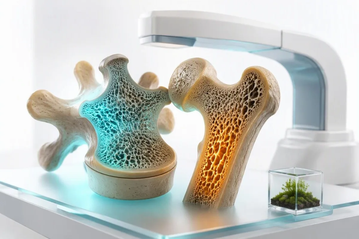
Кандагы анализлар сөяк тыгызлыгын үлчи алмый яки үзләре генә остеопорозны диагнозлый алмый. Шулай да, алар D витамины җитешсезлеге, паратиреоид авыруы, бөернең эшчәнлеген бозу, гормон югалтү һәм сөяк югалтуны тыныч кына тизләтүче мальабсорбцияне ачыкларга мөмкин.


- DXA сканы остеопорозны диагнозлау өчен кирәк; -2.5 яки аннан азрак T-очкыны диагноз чигенә туры килә.
- 25-гидрокси D витамины 20 нг/мл (50 нмоль/л) дан түбән җитешсезлекне күрсәтә һәм кальцийны үзләштерүне начарлатырга мөмкин.
- Кальций плюс PTH беренчел гиперпаратиреоидизмны ачыклау өчен иң файдалы кан анализы тоташуы.
- Кандагы кальций нормаль булырга мөмкин остеопороз кебек, чөнки скелет кан дәрәҗәсен стабиль тоту өчен кальций бирә.
- Креатинин һәм eGFR хроник бөер авыруын ачыклагыз, ул минерал метаболизмын һәм дәвалау сайлауларын үзгәртә.
- TSH 0.1 мИУ/лдан түбән менопаузадан соң, бигрәк тә, сыну куркынычлыгын арттыручы тиреоид гормоны артык булуын күрсәтергә мөмкин.
- 24 сәгатьлек сидектә кальций кальцийнең аз керүен сидек аша кальций югалудан һәм кайбер паратиреоид авыруларыннан аерырга ярдәм итә.
- DXA гадәттә кирәк җиңелчә сынудан, озак вакыт стерроид кулланганнан яки 65 яшь яки аннан өлкәрәк хатын-кызлар өчен.
Сөяк югалтуны остеопороз өчен кан анализы диагнозлый аламы?
Остеопорозны диагнозлау өчен кан анализы юк, чөнки кан анализы сөяк минераллары тыгызлыгын үлчәми. DXA скане остеопорозны диагнозлый, иң түбән тиешле T-күрсәткече -2.5 яки аннан да түбән булганда, ә кан һәм сидек анализлары сөякне йомшартучы сәбәпләрне ачыклый.
Канда кальций, фосфат, витамин D һәм паратиреоид гормоны минералларның регуляциясен чагылдыра, терсәк яки умыртка баганасында инде булган минераллар күләмен түгел. Шуңа күрә нормаль күрсәткечләр остеопорозны инкяр итми; чынлыкта, T-күрсәткече -3.0 булган күп пациентларда кальций күрсәткечләре гадәти була. Доктор Томас Клейн, медицина фәннәре докторы бу ялгыш фикернең DXA үткәрүне еллар буена кичектергәнен күргән.
Кантести - ЯИ анализы кальций, ПТГ, витамин D, бөер функциясе һәм калкансыман биз нәтиҗәләрен бер клиник яктан бәйле схемада куллана, бер генә күрсәткечне диагноз итеп карамыйча. Безнең эш агымында, түбән витамин D күрсәткече дәвалауны хуплый ала, ләкин ул беркайчан да DXA нәтиҗәсен яки сыну куркынычлыгын бәяләүне алмаштыра алмый.
Еңелчә сыну — өйрәнү биеклегеннән яки аннан да түбәнрәк егылудан килә торган сыну — DXA булганчы ук клиник остеопорозны ачыкларга мөмкин. 2020 AACE кулланмасы остеопороз яки җиңелчә сыну булган кешеләрдә икенчел сәбәпләрне тикшерүне тәкъдим итә, аеруча сөяк югалту иртә, көчле яки көтелмәгәнчә тиз булганда (Camacho et al., 2020). Mūsu biomarķieru ceļvedis ни өчен чикләр эчендәге нәтиҗә клиник контекстны таләп итә.
Ике төрле тест нинди сорауларга җавап бирә
DXA соравы: “Бүген сөяк никадәр тыгыз?” Лаборатория тесты соравы: “Гормональ, туклану, бөер, ашказаны-эчәклек яки дарулар белән бәйле процесс югалтуга ярдәм итәме?” Кальцийның гына җитәрлекме, дәвалау таләп иткән искә төшереп булмый торган хәл бармы, яки сынуны профилактикалаучы дару карарга кирәкме икәнлеген билгеләгәндә ике җавап та мөһим.
Кайчан лаборатория анализларына караганда DXA сканын күбрәк кирәк була?
DXA сканы кирәк, әгәр яшь, сыну тарихы, дару куллану яки куркынычлык факторлары сөяк тыгызлыгы кимүен күрсәтсә; кан анализлары аны алмаштыра алмый. DXA бил һәм терсәк сөякләренең минерал тыгызлыгын бик аз радиация йогынтысы белән, гадәттә 10 микрозиверттан да кимрәк үлчи.
65 яшь яки аннан өлкәрәк хатын-кызлар гадәттә DXA белән остеопорозны тикшерү узарга тиеш, ә яшьрәк постменопауза хатын-кызларына куркынычлыкка нигезләнгән бәяләү кирәк. Түбән травмалы сынудан соң, буе 4 см артык кимегәндә, 40 яшькә кадәр иртә менопауза булганда, яки 3 ай яки аннан да күбрәк вакыт эчендә көненә 5 мг prednisone-эквивалент стероид белән дәвалану вакытында иртәрәк тикшерү акыллы.
A T-күрсәткече сөяк тыгызлыгын сәламәт яшь өлкәннәр төркеме белән чагыштыра: -1.0 яки аннан югарырак — нормаль, -1.0 — -2.5 — остеопения, һәм -2.5 яки аннан түбәнрәк — остеопороз. А Z-күрсәткече шул ук яшьтәге һәм җенестәге кешеләр белән чагыштыра; яшь постменопаузалы хатын-кыз яки 50 яшькә кадәрге ир-атта -2.0 яки аннан да түбән Z-күрсәткече икенчел остеопорозны тирәнтенрәк эзләргә этәрә.
Остеоартрит, аорта кальцификациясе яки умырткалык компрессион үзгәрешләре булган өлкәннәрдә DXA бил умырткасы буенча ялгыш ышанычлы күрсәтелергә мөмкин. Шуңа күрә мин терсәк, умырткалык, сыну тарихы һәм кайвакыт умырткалык сынуын бәяләүне чагыштырам, бер “яхшы” саннан ышанычлы уйламыйча. Остеокальцин нәтиҗәләре әйләнеш контекстын өсти ала, ләкин аларны иммагшнг алмаштыра алмый.
Ничә остеопорозның икенчел лаборатория анализы гадәттә беренче чиратта була?
The core secondary osteoporosis lab panel usually includes calcium, albumin, creatinine with eGFR, alkaline phosphatase, 25-hydroxyvitamin D, CBC, phosphate, and PTH when calcium is abnormal. The exact panel changes with age, symptoms, medicines, fracture pattern, and the DXA result.
A матдәләр алмашуның комплекслы панеле supplies calcium, albumin, creatinine, bicarbonate, and liver markers, while alkaline phosphatase gives a broad clue to bone formation or hepatobiliary disease. Adult total calcium is commonly 8.5 to 10.5 mg/dL (2.12 to 2.62 mmol/L), but the laboratory interval and albumin concentration must be checked before calling a result high or low.
A CBC can expose anemia that points toward celiac disease, chronic inflammation, kidney disease, marrow disorders, or nutritional deficiency. In a patient with a low Z-score and unexplained anemia, I am much more likely to add celiac serology and protein studies than to simply advise more dairy.
The 2017 UK guideline advises baseline blood testing to exclude secondary osteoporosis in patients being evaluated for fragility fractures (Compston et al., 2017). How often to recheck vitamin D depends on the starting value, dose, absorption, and whether a condition such as coeliac disease is still active.
Tests that are not routine for everyone
Magnesium, celiac antibodies, serum protein electrophoresis, cortisol testing, sex hormones, and 24-hour urine calcium are targeted tests rather than universal screening. Ordering everything at once often creates incidental abnormalities; a good work-up follows the clues rather than chasing every borderline result.
Сөяк югалту өчен кальций һәм альбуминны ничек интерпретацияләргә?
High calcium with an inappropriately normal or high PTH raises concern for primary hyperparathyroidism, a treatable cause of bone loss. Low calcium is less typical of uncomplicated osteoporosis and usually points toward vitamin D deficiency, malabsorption, kidney disease, hypoparathyroidism, or medication effects.
About 40% of circulating calcium is albumin-bound, so a low albumin can make total calcium appear low even when biologically active calcium is adequate. Laboratories may report albumin-adjusted calcium, but direct ионлаштырылган кальций is preferable when the result is borderline, albumin is markedly abnormal, or an acid-base disturbance is present.
A persistent adjusted calcium above roughly 10.2 to 10.5 mg/dL (2.55 to 2.62 mmol/L), depending on the laboratory, deserves repeat testing with PTH. PTH should be suppressed when calcium is high; if it is not, primary hyperparathyroidism becomes more likely. Slightly elevated calcium is often not an emergency, but repeated results should not be ignored.
One clinical trap: thiazide diuretics, lithium, dehydration, and calcium carbonate antacids can raise calcium modestly. I ask patients to bring every supplement bottle, because “bone support” products may contain 1,200 mg of calcium plus vitamin D, and that changes both the result and the next decision.
Why normal calcium does not protect the skeleton
Normal serum calcium is tightly defended by PTH, vitamin D, kidneys, and the skeleton. When dietary calcium is inadequate or vitamin D absorption fails, bone resorption can help preserve a blood calcium concentration within the 8.5 to 10.5 mg/dL range—an effective short-term survival mechanism with a long-term skeletal cost.
PTH һәм D витамины анализы нәрсә ачыклый?
The most informative calcium PTH vitamin D testing pattern is low 25-hydroxyvitamin D with high PTH and normal or low-normal calcium, which often indicates secondary hyperparathyroidism. This pattern means the body is working harder to maintain calcium balance and may be drawing on bone stores.
A 25-гидрокси D витамины level below 20 ng/mL (50 nmol/L) is generally considered deficiency; 20 to 29 ng/mL is often called insufficiency, although professional groups disagree about whether every value in this interval needs treatment. The 25-hydroxy test is the correct measure of body stores, not 1,25-dihydroxyvitamin D, which can be normal or elevated in deficiency.
PTH reference intervals are assay-specific, commonly about 15 to 65 pg/mL (1.6 to 6.9 pmol/L). A PTH of 78 pg/mL alongside vitamin D of 12 ng/mL and calcium of 9.1 mg/dL usually suggests compensatory secondary hyperparathyroidism; a PTH of 78 with calcium of 10.8 mg/dL suggests a very different pathway.
Кантести - AI кан анализы нәтиҗәләрен аңлату платформасы that reads vitamin D, calcium, albumin, eGFR, and PTH as a linked physiology problem, including whether the pattern merits clinician follow-up rather than self-prescribed high-dose supplements. For a practical safety discussion, see vitamin D and K2 dosing.
A sensible recheck interval
After starting vitamin D, retesting 25-hydroxyvitamin D and calcium in about 8 to 12 weeks is reasonable for deficiency, malabsorption, high doses, kidney disease, or a prior high calcium result. Daily maintenance doses of 800 to 2,000 IU suit many adults, while loading regimens should be individualized by the treating clinician.
Кайчан 24 сәгатьлек сидек кальций анализы мөһим?
A 24-hour urine calcium test is most useful when osteoporosis occurs with kidney stones, high PTH, unexplained low calcium intake, or suspected renal calcium loss. It does not diagnose osteoporosis, but it can explain why the calcium balance is negative despite a seemingly good diet.
Many laboratories consider roughly 100 to 250 mg of calcium per 24 hours typical for adults eating a usual diet, but the useful interpretation depends on sodium intake, collection completeness, sex, body size, and local methods. A value above 250 mg/day in women or 300 mg/day in men is often called hypercalciuria, though thresholds vary.
High urine calcium can accompany high sodium intake, loop diuretics, idiopathic hypercalciuria, primary hyperparathyroidism, sarcoidosis, or excessive vitamin D. The combination of recurrent calcium stones, urine calcium of 350 mg/day, and declining hip density is a stronger signal than a single elevated collection alone. High urine calcium causes are worth reviewing before simply increasing supplements.
The collection is easy to spoil: missing one daytime sample, collecting for 20 rather than 24 hours, or changing diet dramatically makes the number less useful. Kantesti AI's trend review can help people preserve the collection date, supplement dose, eGFR, and serum calcium beside the report; our технология кулланмасы describes how structured context improves lab interpretation.
Ни өчен тиреоид анализы сөяк югалту өчен кан анализына кертелгән?
Thyroid hormone excess accelerates bone turnover, so a suppressed TSH should be investigated in people with osteoporosis or unexplained fractures. A TSH below 0.1 mIU/L is particularly concerning in postmenopausal women and older adults, especially if free T4 or free T3 is elevated.
TSH is usually the first thyroid test because it responds sensitively to excess thyroid hormone. The common adult reference interval is about 0.4 to 4.0 mIU/L, although pregnancy, age, assay design, acute illness, and thyroid treatment change how a result should be read.
Over-replacement with levothyroxine is a surprisingly practical cause of low TSH. A 68-year-old patient may feel perfectly well with TSH 0.06 mIU/L, yet repeated suppression over years can be relevant to fracture risk; the target should be reviewed rather than assuming a “low-normal dose” is automatically safe.
Subclinical hyperthyroidism means low TSH with normal free T4 and free T3, and its skeletal effect is real but varies by duration and degree of suppression. Subclinical thyroid disease is different from hyperthyroidism, but both articles illustrate why one thyroid result cannot be interpreted without symptoms, medicines, and repeat testing.
Why hypothyroidism is not the mirror image
Untreated overt hypothyroidism slows bone remodeling rather than directly causing rapid bone loss, but it can increase falls through fatigue, myopathy, and impaired balance. The bigger skeletal concern is usually excessive replacement therapy, not a mildly raised TSH by itself.
Ничә анализ остеопороз артындагы мальабсорбцияне таба?
Celiac serology is appropriate when low bone density occurs with iron deficiency, chronic diarrhea, low BMI, unexplained vitamin D deficiency, or recurrent fractures. Celiac disease can reduce absorption of calcium and vitamin D even when gastrointestinal symptoms are absent.
The usual first test is tissue transglutaminase IgA plus total IgA. A positive tTG-IgA needs clinical confirmation, and an isolated negative test can mislead when total IgA is low or the person has already removed gluten from the diet.
Ferritin below 15 ng/mL is strongly suggestive of depleted iron stores in most adults, while ferritin may appear normal or high during inflammation. Iron deficiency plus low vitamin D, low body weight, and low bone density is a combination that should move celiac disease higher on the differential rather than being treated as four unrelated findings.
Patients should not start a gluten-free diet before initial celiac testing unless a clinician advises it, because antibody levels can fall and obscure diagnosis. Gluten challenge timing explains why the diet history belongs on the requisition.
Other nutritional signals
Low phosphate, low magnesium, low albumin, folate deficiency, and unexplained anemia may support an absorption or nutrition concern, but none identifies a specific gastrointestinal diagnosis alone. A dietary history can be as revealing as another tube of laboratory sample—particularly in restrictive diets, eating disorders, and endurance athletes with low energy availability.
Бөер анализлары остеопорозны бәяләүне ничек үзгәртә?
An eGFR below 60 mL/min/1.73 m² for 3 months or longer can disrupt phosphate, vitamin D activation, and PTH regulation, creating chronic kidney disease–mineral and bone disorder. Kidney function also affects which osteoporosis medicines are suitable and how calcium results are interpreted.
Creatinine alone is not a reliable measure of kidney health in frail older adults or highly muscular athletes; eGFR provides a better starting estimate. Persistent eGFR below 60 mL/min/1.73 m² meets a core criterion for chronic kidney disease, while a result below 30 requires particularly careful bone and medicine review.
As kidney function falls, phosphate may rise and the kidney produces less active vitamin D, contributing to high PTH. In early chronic kidney disease, phosphate can still be within the usual 2.5 to 4.5 mg/dL range, so a normal phosphate does not exclude a developing mineral disorder.
A low bicarbonate, often below 22 mmol/L, can signal metabolic acidosis that promotes mineral release from bone over time. Kidney disease stages and urine albumin testing help separate a one-off eGFR dip from chronic disease.
Why medication choices need review
Bisphosphonate prescribing, denosumab monitoring, calcium dosing, and vitamin D strategy can all change at lower eGFR levels. This is a situation where self-treatment is risky: severe hypocalcemia after antiresorptive therapy is uncommon but more likely when advanced kidney disease and vitamin D deficiency coexist.
Кайчан протеин анализларын остеопороз белән карарга кирәк?
Serum protein electrophoresis and serum free light chains are targeted tests when bone loss is accompanied by anemia, elevated total protein, kidney impairment, unexplained bone pain, or vertebral fractures out of proportion to age. They look for monoclonal proteins, not routine osteoporosis.
A гамма аралыгы—total protein minus albumin—above about 4.0 g/dL can prompt further review, although dehydration, infection, autoimmune conditions, and chronic liver disease can also widen it. A normal gamma gap does not exclude a small monoclonal protein, which is why test selection should follow the full clinical picture.
Multiple myeloma can cause bone damage, anemia, renal dysfunction, and high calcium, but osteoporosis itself is vastly more common. The red-flag pattern is not “a low DXA score”; it is a low score plus persistent hemoglobin decline, creatinine change, calcium elevation, focal pain, or compression fractures without an adequate explanation.
Serum protein electrophoresis should usually be paired with immunofixation and serum free light chains when a plasma-cell disorder is being considered. Free light-chain ratio interpretation is especially nuanced in chronic kidney disease, where both light chains may rise without malignancy.
Кайчан эстроген һәм тестостерон анализлары ярдәм итә?
Sex-hormone testing is most useful for early menopause, missed periods, suspected hypogonadism, pituitary symptoms, or osteoporosis before the usual screening age. It is not routinely needed for every postmenopausal woman with a low DXA score.
Estrogen decline increases osteoclast activity, which helps explain the rapid bone loss seen in the first 5 to 10 years after menopause. A single estradiol result is often difficult to interpret in perimenopause because levels can change substantially across days and cycles.
In men, a fasting morning total testosterone drawn between roughly 7 and 11 AM is the usual starting test when symptoms and low bone density suggest hypogonadism. A total testosterone below 300 ng/dL (10.4 nmol/L) on two separate morning samples is a commonly used threshold for further evaluation, not an automatic treatment decision.
High SHBG can make total testosterone look adequate while free testosterone is lower, particularly with aging, hyperthyroidism, liver disease, and some medications. Hormone results in men are most informative when the sample time and symptoms are recorded.
Medication and reproductive history matter
GnRH analogues, aromatase inhibitors, androgen-deprivation therapy, some anti-seizure medicines, and prolonged depot medroxyprogesterone can affect bone density. The medication timeline often provides more actionable information than an isolated hormone level, particularly after cancer treatment or gender-affirming care.
Сөяк әйләнеше маркерлары актив сөяк югалтуны күрсәтәме?
Bone turnover markers can show whether bone remodeling is relatively fast, but they do not diagnose osteoporosis or predict an individual fracture with enough certainty to use alone. Serum CTX reflects bone resorption and P1NP reflects bone formation; their strongest role is monitoring treatment adherence and response.
CTX changes markedly with food intake and time of day, so fasting morning collection is preferred when results are being compared. P1NP is generally less affected by meals and is often practical for monitoring anabolic or antiresorptive treatment, although assay methods and reference intervals differ.
A high marker can occur after a recent fracture, during hyperthyroidism, with vitamin D deficiency, in hyperparathyroidism, and in high-turnover osteoporosis. The result therefore answers “is remodeling active?” rather than “where is the cause?”—a distinction that prevents expensive but unhelpful testing.
Кантести - AI нигезендәге кан анализы тикшерү коралы that can place serial P1NP or CTX results beside calcium, vitamin D, eGFR, and treatment dates, but the trend only means something when samples were taken under comparable conditions. Osteocalcin testing has similar limitations and should not be used as a home bone-density test.
A useful monitoring clue
A meaningful decline in a resorption marker after antiresorptive treatment can occur within 3 to 6 months, before a repeat DXA can show a substantial density change. That earlier biological signal can be helpful when adherence is uncertain, but clinicians should use the same assay and collection timing.
Ничә дару сөяк югалтуга китерә яки сөяк анализларын үзгәртә?
Glucocorticoids, aromatase inhibitors, androgen-deprivation therapy, some antiseizure medicines, proton-pump inhibitors, and excess thyroid hormone can contribute to bone loss. A medication review is often more diagnostic than adding another broad laboratory panel.
Oral glucocorticoids can reduce bone formation within months, and fracture risk rises before DXA decline fully captures the effect. Prednisone-equivalent doses of 2.5 to 7.5 mg daily are not harmless when used long term; risk also depends on age, prior fractures, and cumulative exposure.
Proton-pump inhibitors do not reliably cause osteoporosis in every user, but long-term treatment can coexist with low magnesium, vitamin B12 deficiency, and reduced calcium absorption in selected patients. A magnesium level below 1.7 mg/dL (0.70 mmol/L) deserves review, especially with muscle cramps, arrhythmia risk, or diuretic use.
Do not stop prescribed medication solely because of a bone-health article. Instead, make a timeline of every prescription, injection, inhaler, supplement, and dose change, then use annual blood work planning to discuss a targeted monitoring plan with the prescribing clinician.
Кальций, PTH һәм D витамины анализына ничек әзерләнергә?
Most calcium, PTH, and 25-hydroxyvitamin D tests do not require fasting, but fasting morning testing improves comparability for CTX, phosphate, and some paired metabolic evaluations. Keep your usual diet unless the clinician specifically requests preparation for a timed urine collection or absorption study.
Biotin can interfere with certain immunoassays, including some PTH and thyroid tests, although the degree depends on the laboratory method. Many clinicians advise stopping high-dose biotin supplements—often 5,000 to 10,000 mcg daily—for at least 48 hours before testing; people using prescribed high doses should confirm this with their care team.
Record calcium, vitamin D, magnesium, antacid, diuretic, thyroid medication, and osteoporosis-treatment doses taken during the prior week. Calcium taken shortly before a draw can modestly change serum calcium, while a missed dose of levothyroxine or an acute dehydrating illness can distort the broader pattern.
Kantesti AI lets users retain original laboratory units, collection dates, medicines, and repeated results together, which is more useful than comparing screenshots from different laboratories. Our clinical use cases show why trend interpretation must retain the original reference interval and test method.
The question to bring to the appointment
Ask, “Does this pattern suggest a reversible contributor, and do I need DXA or vertebral imaging now?” That question keeps the visit focused on fracture prevention rather than on chasing a single borderline laboratory flag.
Сөяк белән бәйле нинди лаборатория нәтиҗәләрен тиз медицина тикшерүе таләп итә?
High calcium with confusion, severe weakness, vomiting, dehydration, palpitations, or constipation needs urgent assessment, especially when calcium is above 12 mg/dL (3.0 mmol/L). New severe back pain, sudden height loss, or a low-trauma fracture also warrants timely clinical review even when blood tests are normal.
Calcium above 14 mg/dL (3.5 mmol/L) can be a medical emergency, particularly when symptoms, kidney dysfunction, or rapid rise are present. A total calcium result should be verified against albumin and, where needed, ionized calcium, but symptoms should never wait for perfect laboratory certainty.
Phosphate below 1.0 mg/dL (0.32 mmol/L) can cause marked weakness and requires urgent evaluation, while persistently low phosphate around 1.5 to 2.0 mg/dL may point toward vitamin D deficiency, hyperparathyroidism, renal phosphate wasting, or medication effects. Фосфат түбәнлеге симптомнары deserve attention because osteomalacia can be mistaken for routine osteoporosis.
As of September 23, 2026, the practical message remains straightforward: normal blood tests do not clear someone from osteoporosis, and abnormal tests do not establish it. Thomas Klein, MD, recommends using laboratory patterns to find reversible causes while confirming density and vertebral risk with appropriate imaging; our медицина валидация стандартлары describe the clinical boundaries of Kantesti AI interpretation.
Research and clinical oversight
Kantesti works with physician oversight and privacy-focused lab interpretation across 127+ countries, but it does not replace fracture assessment, emergency care, or an endocrinology consultation. Readers who want to understand the people behind the medical review process can meet our Медицина консультатив советы.
Еш бирелә торган сораулар
Кандағы анализ остеопороз бармы, юкмы икәнен ачыклый аламы?
No, a blood test cannot tell whether you have osteoporosis because osteoporosis is defined by bone mineral density or a qualifying fragility fracture, not by a serum biomarker. A DXA scan diagnoses osteoporosis when the relevant T-score is -2.5 or lower. Blood tests such as calcium, PTH, 25-hydroxyvitamin D, creatinine, and TSH identify reversible contributors to bone loss. Normal blood results do not rule out osteoporosis.
Сөяк چېпләрне ачыклау өчен нинди кан сынаулары узарга кирәк?
A first-line secondary osteoporosis panel commonly includes calcium, albumin, creatinine with eGFR, alkaline phosphatase, 25-hydroxyvitamin D, phosphate, and a complete blood count. PTH is especially useful when calcium is high, low, or repeatedly borderline; many assays use an approximate reference interval of 15 to 65 pg/mL. Thyroid tests, celiac antibodies, serum protein electrophoresis, sex hormones, and 24-hour urine calcium are added when symptoms or baseline results point in that direction. The test list should be tailored rather than ordered indiscriminately.
Сөяк сәламәтлеге өчен D витаминының бик түбән дәрәҗәсе нинди?
A 25-hydroxyvitamin D level below 20 ng/mL, equivalent to 50 nmol/L, is generally considered vitamin D deficiency and can impair calcium absorption. Values from 20 to 29 ng/mL are often called insufficient, although experts differ on whether every value in this range requires treatment for bone health. Low vitamin D can raise PTH while calcium remains normal, which may increase bone turnover. Retesting is often considered 8 to 12 weeks after treatment begins when deficiency, malabsorption, kidney disease, or high-dose supplementation is involved.
Нормаль кальций дәрәҗәләре сөяк югалтуны бетерә аламы?
No, normal serum calcium does not rule out bone loss because the body tightly maintains calcium in the blood using PTH, vitamin D, kidney handling, and calcium released from bone. Most laboratories use a total calcium range near 8.5 to 10.5 mg/dL, but albumin affects the total result. Osteoporosis can be severe while calcium remains within that interval. A DXA scan is the test that measures whether bone density is low.
Сезнең калкансыман биз алды безләрегез сөякләрнең сынучан булуына сәбәп буламы?
Persistently high PTH can contribute to bone loss because PTH increases calcium release and remodeling in bone, particularly when the cause is primary hyperparathyroidism or prolonged vitamin D deficiency. High calcium plus a PTH that is normal or elevated is an especially meaningful pattern because PTH should normally be suppressed when calcium is high. A low vitamin D level, such as 12 ng/mL, with elevated PTH and normal calcium more often suggests secondary hyperparathyroidism. Diagnosis requires repeat results, medication review, kidney assessment, and clinician interpretation.
Сөяк челтәренә бәйле остеопорозны ачыклау өчен 24 сәгатьлек сидек кальцийын тикшертергә кирәкме?
A 24-hour urine calcium test is not necessary for every person with osteoporosis, but it is useful with kidney stones, high PTH, unusually early bone loss, suspected malabsorption, or possible renal calcium wasting. Values above roughly 250 mg/day in women and 300 mg/day in men are often considered high, although sodium intake and collection quality affect interpretation. A timed urine collection should include every urine sample for the full 24 hours. The result is usually interpreted alongside serum calcium, PTH, vitamin D, and creatinine.
Бүген үк AI белән эшләнгән кан анализы тикшерүе
Дөнья буенча 2 миллионнан артык кулланучы кушылыгыз: алар Kantestiны тиз һәм төгәл лаборатория анализы өчен ышана. Кан анализы нәтиҗәләрегезне йөкләгез һәм 15,000+ биомаркерларын секундлар эчендә тулы аңлатма белән алыгыз.
📚 Сылтама бирелгән тикшеренү басмалары
Klein, T., Mitchell, S., & Weber, H. (2026). Kantesti LTD. (2026). Урина тестында уробилиноген: Тулы уриналь анализ кулланмасы 2026. Zenodo.. Kantesti AI медицина тикшеренүе.
Klein, T., Mitchell, S., & Weber, H. (2026). Kantesti LTD. (2026). Тимерне өйрәнү җитәкчелеге: TIBC, тимер туену һәм бәйләү сыйдырышлыгы. Zenodo.. Kantesti AI медицина тикшеренүе.
📖 Тышкы медицина белешмәләре
⚕️ Медицина кисәтүе
Бу мәкалә бары тик белем бирү максатларында гына һәм медицина киңәше булып тормый. Диагноз һәм дәвалау карарлары өчен һәрвакыт квалификацияле сәламәтлек саклау белгеченә мөрәҗәгать итегез.
E-E-A-T ышаныч сигналлары
Тәҗрибә
Табиб җитәкчелегендә лаборатория нәтиҗәләрен аңлату эш процессларын клиник тикшерү.
Белгечлек
Лаборатория медицинасы: биомаркерларның клиник контекстта үз-үзләрен тотышын аңлау.
Авторититет
Доктор Томас Кляйн тарафыннан язылган, доктор Сара Митчелл һәм профессор доктор Ханс Вебер тарафыннан тикшерелгән.
Ышанычлылык
Ачык күзәтү юллары белән куркуны киметү өчен дәлилләргә нигезләнгән аңлату.