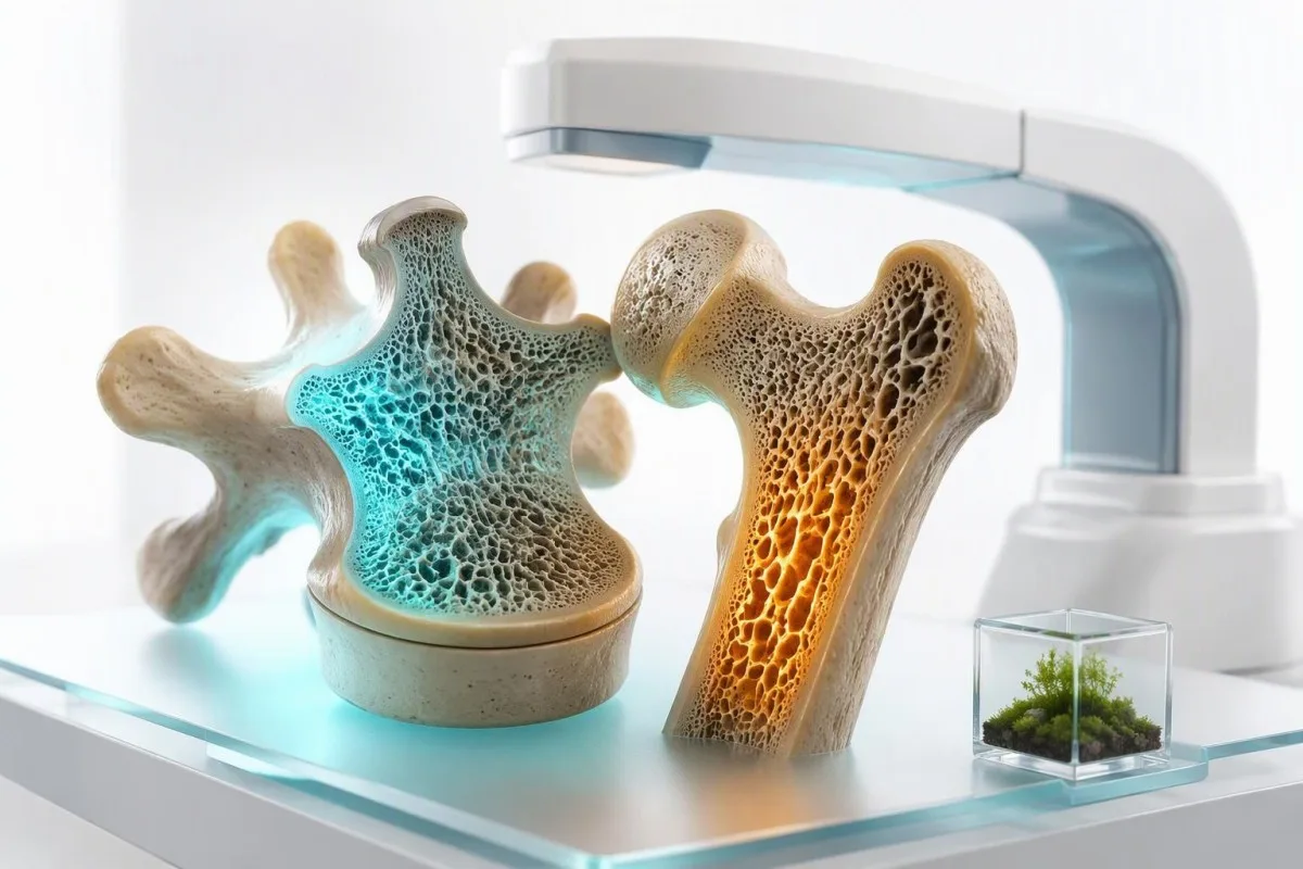
ການກວດເລືອດບໍ່ສາມາດວັດແທກຄວາມໜາແໜ້ນຂອງກະດູກຫຼືວິນິດໄສໂລກກະດູກພຸນໄດ້ດ້ວຍຕົນເອງ. ເຖິງຢ່າງໃດກໍ່ຕາມ, ພວກມັນສາມາດເປີດເຜີຍ vitamin D deficiency, ພະຍາດຕ່ອມ parathyroid, ການເຮັດວຽກຂອງไตທີ່ຜິດປົກກະຕິ, ການສູນເສຍຮໍໂມນ, ແລະ malabsorption ທີ່ອາດຈະກຳລັງເລັ່ງການສູນເສຍກະດູກຢ່າງຊ້າໆ.


- DXA scan ຕ້ອງມີເພື່ອກວດພົບ osteoporosis; T-score 2.5- ຫລືຕໍ່າກວ່າແມ່ນໄດ້ມາດຕະຖານການວິນິດໄສ.
- 25-ຮິໂດຣອກຊີວິຕາມິນດີ ຕ່ຳກວ່າ 20 ng/mL (50 nmol/L) ສະແດງເຖິງການຂາດ ແລະ ສາມາດຂັດຂວາງການດູດຊຶມແຄລຊຽມໄດ້.
- Calcium ບວກກັບ PTH ແມ່ນການຈັບຄູ່ການກວດເລືອດທີ່ມີປະໂຫຍດຫຼາຍທີ່ສຸດສຳລັບການຊອກຫາ primary hyperparathyroidism.
- Serum calcium ສາມາດເປັນປົກກະຕິ ໃນ osteoporosis ເພາະວ່າໂຄງກະດູກສະໜອງ calcium ເພື່ອຮັກສາລະດັບເລືອດໃຫ້ຄົງທີ່.
- Creatinine ແລະ eGFR ລະບຸພະຍາດไตຊໍາເຮື້ອ, ເຊິ່ງປ່ຽນແປງການເຜົາຜະຫລາຍແຮ່ທາດ ແລະ ທາງເລືອກໃນການປິ່ນປົວ.
- TSH ຕໍ່າກວ່າ 0.1 mIU/L ອາດເປັນສັນຍານບອກເຖິງການມີຮໍໂມນ thyroid ເກີນ, ເຊິ່ງເພີ່ມຄວາມສ່ຽງຕໍ່ການແຕກຫັກ, ໂດຍສະເພາະຫຼັງຈາກໝົດປະຈຳເດືອນ.
- ຄວາມເຂັ້ມຂຸ້ນແຄວຊຽມໃນຍ່ຽວ 24 ຊົ່ວໂມງ ໄດ້ຊ່ວຍແຍກຄວາມແຕກຕ່າງລະຫວ່າງການກິນແຄລຊຽມໜ້ອຍກັບການຂັບແຄລຊຽມອອກທາງປັດສະວະ ແລະ ຄວາມຜິດປົກກະຕິຂອງຕ່ອມ parathyroid.
- DXA ໂດຍທົ່ວໄປແມ່ນເໝາະສົມ ຫລັງຈາກກະດູກຫັກ, ໃຊ້ສະເຕີຣອຍດໃນໄລຍະຍາວ, ຫລື ສຳລັບແມ່ຍິງອາຍຸ 65 ປີຂຶ້ນໄປ.
ການກວດເລືອດໂລກກະດູກພຸນສາມາດວິນິດໄສການສູນເສຍກະດູກໄດ້ບໍ?
ບໍ່ມີການກວດເລືອດໃດສາມາດວິນິດສัย ໂລກກະດູກພຸນໄດ້ ເພາະການກວດເລືອດບໍ່ໄດ້ວັດແທກຄວາມໜາແໜ້ນຂອງແຮ່ທາດໃນກະດູກ. ການສະແກນ DXA ວິນິດສัย ໂລກກະດູກພຸນ ເມື່ອຄະແນນ T-score ຕ່ຳສຸດທີ່ກ່ຽວຂ້ອງແມ່ນ -2.5 ຫລືຕ່ຳກວ່າ, ໃນຂະນະທີ່ການກວດເລືອດແລະປັດສະວະ ລະບຸຜູ້ປະກອບສ່ວນທີ່ສາມາດປິ່ນປົວໄດ້ ທີ່ອາດຈະເຮັດໃຫ້ກະດູກອ່ອນແອລົງ.
ແຄລຊຽມໃນເລືອດ, ຟอสຟໍ, ວິຕາມິນ D, ແລະຮໍໂມນ parathyroid ສະທ້ອນເຖິງການຄວບຄຸມແຮ່ທາດ, ບໍ່ແມ່ນປະລິມານແຮ່ທາດທີ່ມີຢູ່ແລ້ວໃນສະໂພກຫລືກະດູກສັນຫລັງ. ດັ່ງນັ້ນ, ຜົນການກວດແບບປົກກະຕິ ບໍ່ໄດ້ຍົກເວັ້ນໂລກກະດູກພຸນ; ຄວາມຈິງແລ້ວ, ຜູ້ປ່ວຍຫລາຍຄົນທີ່ມີ T-score -3.0 ມີຜົນການກວດແຄລຊຽມປົກກະຕິທັງໝົດ. ດຣ. ທອມັສ ໄຄລນ໌, MD ໄດ້ເຫັນຄວາມເຂົ້າໃຈຜິດນີ້ ເຮັດໃຫ້ການສົ່ງຕໍ່ DXA ຊັກຊ້າໄປຫລາຍປີ.
ແຄນເທສຕີ ເປັນ AI ເຄື່ອງວິເຄາະເລືອດ ທີ່ວາງແຄລຊຽມ, PTH, ວິຕາມິນ D, ການເຮັດວຽກຂອງไต, ແລະຜົນການກວດຕ່ອມໄທລອຍ ໃຫ້ເປັນຮູບແບບທາງຄລີນິກທີ່ສອດຄ່ອງກັນໜຶ່ງດຽວ ແທນທີ່ຈະຮັກສາອາການໃດໜຶ່ງເປັນການວິນິດສัย. ໃນຂັ້ນຕອນການກວດສອບຂອງພວກເຮົາ, ຜົນການກວດວິຕາມິນ D ຕ່ຳ ຈະສະໜັບສະໜູນການປິ່ນປົວ, ແຕ່ບໍ່ເຄີຍແທນທີ່ຜົນການກວດ DXA ຫລືການປະເມີນຄວາມສ່ຽງກະດູກຫັກ.
ກະດູກຫັກເນື່ອງຈາກຄວາມອ່ອນແອ—ຄືການຫັກຈາກການລົ້ມລະດັບຄວາມສູງຢືນຫລືຕ່ຳກວ່າ—ສາມາດວິນິດສัย ໂລກກະດູກພຸນທາງຄລີນິກໄດ້ ເຖິງແມ່ນກ່ອນທີ່ຈະມີ DXA. ຂໍ້ແນະນຳ AACE ປີ 2020 ແນະນຳໃຫ້ປະເມີນສາເຫດທຸງສະນອງໃນຜູ້ທີ່ມີໂລກກະດູກພຸນ ຫລືກະດູກຫັກເນື່ອງຈາກຄວາມອ່ອນແອ, ໂດຍສະເພາະເມື່ອການສູນເສຍກະດູກເກີດຂຶ້ນໃນໄລຍະເລີ່ມຕົ້ນ, ຮຸນແຮງ, ຫລືໄວຜິດປົກກະຕິ (Camacho et al., 2020). ຄູ່ມື biomarkers ຂອງພວກເຮົາ ອະທິບາຍວ່າເປັນຫຍັງຜົນການກວດໃນຂອບເຂດປົກກະຕິ ຍັງຕ້ອງການບໍລິບົດທາງຄລີນິກ.
ສິ່ງທີ່ການກວດສອງປະເພດຕອບ
DXA ຕອບວ່າ, “ມື້ນີ້ກະດູກໜາແໜ້ນເທົ່າໃດ?” ການກວດຫ້ອງທົດລອງຕອບວ່າ, “ມີຂະບວນການກ່ຽວກັບຮໍໂມນ, ໂພຊະນາການ, ໝາກໄຂ່ຫລັງ, ລະບົບຍ່ອຍອາຫານ, ຫລືຢາ ທີ່ຊ່ວຍເພີ່ມການສູນເສຍບໍ?” ທັງສອງຄຳຕອບມີຄວາມສຳຄັນເມື່ອຕັດສິນໃຈວ່າແຄລຊຽມຢ່າງດຽວພຽງພໍ, ภาวะที่สามารถพลิกกลับได้ต้องการการรักษา, ຫລືຄວນພິຈາລະນາຢາປ້ອງກັນກະດູກຫັກ.
ເມື່ອໃດທີ່ທ່ານຕ້ອງການ DXA scan ແທນການກວດເລືອດ?
ການສະແກນ DXA ຕ້ອງການເມື່ອອາຍຸ, ປະຫວັດກະດູກຫັກ, ການໄດ້ຮັບຢາ, ຫລືປັດໄຈສ່ຽງ ຊີ້ບອກເຖິງຄວາມໜາແໜ້ນຂອງກະດູກຫຼຸດລົງ; ການກວດເລືອດບໍ່ສາມາດແທນທີ່ໄດ້. DXA ວັດແທກຄວາມໜາແໜ້ນຂອງແຮ່ທາດໃນກະດູກທີ່ກະດູກສັນຫລັງສ່ວນເອິກ ແລະສະໂພກ ດ້ວຍການຊູນລັງສີຕ່ຳຫຼາຍ, ໂດຍທົ່ວໄປແມ່ນຕ່ຳກວ່າ 10 microsieverts.
ແມ່ຍິງອາຍຸ 65 ປີຂຶ້ນໄປ ໂດຍທົ່ວໄປຄວນໄດ້ຮັບການຄັດເລືອກໂລກກະດູກພຸນດ້ວຍ DXA, ແລະແມ່ຍິງໃນໄວຖື່ງທີ່ອາຍຸໜ້ອຍກວ່າ ຕ້ອງການການປະເມີນຕາມຄວາມສ່ຽງ. ການກວດກ່ອນໜ້ານີ້ມີເຫດຜົນຫລັງຈາກກະດູກຫັກຈາກການບາດເຈັບຕ່ຳ, ການສູນເສຍຄວາມສູງຫຼາຍກວ່າ 4 ຊມ, ໄວຖື່ງກ່ອນໄວອາຍຸ 40 ປີ, ຫລືການປິ່ນປົວດ້ວຍສະເຕີຣອຍດເທົ່າກັບ prednisone 5 ມລກ ຕໍ່ມື້ ເປັນເວລາ 3 ເດືອນຂຶ້ນໄປ.
A T-score ປຽບທຽບຄວາມໜາແໜ້ນຂອງກະດູກກັບປະຊາກອນອ້າງອີງຂອງຜູ້ໃຫຍ່ໜຸ່ມສຸຂະພາບດີ: -1.0 ຫລືສູງກວ່າແມ່ນປົກກະຕິ, -1.0 ຫາ -2.5 ແມ່ນ osteopenia, ແລະ -2.5 ຫລືຕ່ຳກວ່າແມ່ນ osteoporosis. A Z-score ປຽບທຽບກັບຄົນທີ່ມີອາຍຸແລະເພດດຽວກັນ; Z-score ຢູ່ທີ່ -2.0 ຫລືຕ່ຳກວ່າໃນຜູ້ໃຫຍ່ທີ່ຍັງບໍ່ທັນຫມົດປະຈໍາເດືອນ ຫລືຜູ້ຊາຍອາຍຸຕ່ຳກວ່າ 50 ປີ ຄວນໄດ້ຮັບການຄົ້ນຫາໂລກກະດູກພຸນທຸງສະນອງຢ່າງລະອຽດ.
DXA ສາມາດເບິ່ງຄືວ່າເປັນການປອບໃຈທີ່ຜິດພາດຢູ່ກະດູກສັນຫລັງໃນຜູ້ສູງອາຍຸທີ່ມີໂລກຂໍ້ອັກເສບ, ຫີນປູນໃນເສັ້ນເລືອດແດງ, ຫລືການປ່ຽນແປງຂອງການບີບຕົວຂອງກະດູກສັນຫລັງ. ນັ້ນຄືເຫດຜົນທີ່ຂ້ອຍປຽບທຽບສະໂພກ, ກະດູກສັນຫລັງ, ປະຫວັດການກະດູກຫັກ, ແລະບາງຄັ້ງການປະເມີນກະດູກສັນຫລັງທີ່ກະດູກຫັກ ແທນທີ່ຈະປອບໃຈຄົນຈາກຕົວເລກກະດູກສັນຫລັງໜຶ່ງທີ່ເບິ່ງຄືວ່າ “ດີ”. ຜົນ Osteocalcin ອາດຈະເພີ່ມບໍລິບົດການປ່ຽນແປງ, ແຕ່ມັນບໍ່ໄດ້ແທນທີ່ການຖ່າຍພາບ.
ການກວດເລືອດ osteoporosis ທີ່ສອງອັນໃດທີ່ເປັນເສັ້ນທຳອິດ?
ແຜນການທົດລອງກະດູກພຸນຂັ້ນສອງຫຼັກ ໂດຍທົ່ວໄປປະກອບມີແຄລຊຽມ, ອາບູມິນ, ຄຣີເອຕີນິນ ດ້ວຍ eGFR, ຟอสຟາເທດ, ວິຕາມິນ D 25-hydroxy, CBC, ຟอสຟໍ, ແລະ PTH ເມື່ອແຄລຊຽມຜິດປົກກະຕິ. ແຜນການທີ່ແນ່ນອນມີການປ່ຽນແປງຕາມອາຍຸ, ອາການ, ຢາ, ຮູບແບບການກະດູກຫັກ, ແລະຜົນ DXA.
A ກວດສະພາບທາງເຄມີທີ່ຄົບຖ້ວນ ສະໜອງແຄລຊຽມ, ອາບູມິນ, ຄຣີເອຕີນິນ, ໄບຄາບໍເນດ, ແລະຕົວຊີ້ວັດຕັບ, ໃນຂະນະທີ່ຟอสຟາເທດໃຫ້ຄຳໃບ້ກວ້າງຂວາງກ່ຽວກັບການສ້າງກະດູກຫຼືພະຍາດຕັບ/ຖົງນ້ຳດີ. ໂດຍທົ່ວໄປແລ້ວແຄລຊຽມທັງໝົດຂອງຜູ້ໃຫຍ່ແມ່ນ 8.5 ຫາ 10.5 mg/dL (2.12 ຫາ 2.62 mmol/L), ແຕ່ຊ່ວງຂອງຫ້ອງທົດລອງ ແລະ ຄວາມເຂັ້ມຂຸ້ນຂອງອາບູມິນຕ້ອງໄດ້ກວດສອບກ່ອນທີ່ຈະຕີລາຄາຜົນລັບວ່າສູງຫຼືຕ່ຳ.
CBC ສາມາດເປີດເຜີຍອາການເລືອດຈາງທີ່ຊີ້ບອກເຖິງພະຍາດ celiac, ການອັກເສບຊໍາເຮື້ອ, ພະຍາດไต, ຄວາມຜິດປົກກະຕິຂອງໄຂກະດູກ, ຫຼືການຂາດສານອາຫານ. ໃນຄົນເຈັບທີ່ມີຄະແນນ Z ຕ່ ຳ ແລະອາການເລືອດຈາງທີ່ບໍ່ສາມາດອະທິາຍໄດ້, ຂ້ອຍມີແນວໂນ້ມຫຼາຍທີ່ຈະເພີ່ມ celiac serology ແລະການສຶກສາທາດໂປຼຕີນຫຼາຍກວ່າການແນະນຳຜະລິດຕະພັນນົມເພີ່ມເຕີມ.
ຂໍ້ແນະນຳຂອງ UK ປີ 2017 ແນະນຳໃຫ້ທົດລອງເລືອດໃນເບື້ອງຕົ້ນເພື່ອຍົກເວັ້ນໂລກກະດູກພຸນຂັ້ນສອງໃນຄົນເຈັບທີ່ຖືກປະເມີນຫາການກະດູກຫັກທີ່ອ່ອນແອ (Compston et al., 2017). ຄວນກວດວິຕາມິນ D ຄືນເລື້ອຍປານໃດ ຂຶ້ນກັບຄ່າເລີ່ມຕົ້ນ, ປະລິມານ, ການດູດຊຶມ, ແລະບໍ່ວ່າສະພາບການເຊັ່ນພະຍາດ coeliac ຍັງມີການເຄື່ອນໄຫວຢູ່.
ການທົດສອບທີ່ບໍ່ແມ່ນປົກກະຕິສຳລັບທຸກຄົນ
ແມກນີຊຽມ, antibodies celiac, serum protein electrophoresis, ການທົດສອບ cortisol, ຮໍໂມນເພດ, ແລະ 24-hour urine calcium ໄດ້ຮັບການທົດສອບແບບເປົ້າໝາຍຫຼາຍກວ່າການກວດຫາທົ່ວໄປ. ການສັ່ງທຸກຢ່າງພ້ອມກັນມັກຈະສ້າງຄວາມຜິດປົກກະຕິທີ່ບໍ່ໄດ້ຕັ້ງໃຈ; ການກວດສອບທີ່ດີແມ່ນການຕິດຕາມຄຳໃບ້ຫຼາຍກວ່າການໄລ່ຕາມຜົນທີ່ຂອບເຂດ.
ຄວນຕີຄວາມໝາຍ calcium ແລະ albumin ແນວໃດສຳລັບການສູນເສຍກະດູກ?
ແຄລຊຽມສູງດ້ວຍ PTH ທີ່ເປັນປົກກະຕິຫຼືສູງຢ່າງບໍ່ເໝາະສົມ ເຮັດໃຫ້ເກີດຄວາມກັງວົນກ່ຽວກັບ primary hyperparathyroidism, ເຊິ່ງເປັນສາເຫດຂອງການສູນເສຍກະດູກທີ່ສາມາດປິ່ນປົວໄດ້. ແຄລຊຽມຕ່ ຳ ບໍ່ແມ່ນເລື່ອງທຳມະດາຂອງໂລກກະດູກພຸນທີ່ບໍ່ສັບສົນ ແລະ ໂດຍທົ່ວໄປແລ້ວຊີ້ບອກເຖິງການຂາດວິຕາມິນດີ, ການດູດຊຶມ, ພະຍາດไต, hypoparathyroidism, ຫຼືຜົນກະທົບຂອງຢາ.
ປະມານ 40% ຂອງແຄລຊຽມໃນການໄຫຼວຽນແມ່ນຕິດກັບອາບູມິນ, ດັ່ງນັ້ນອາບູມິນຕ່ ຳ ສາມາດເຮັດໃຫ້ແຄລຊຽມທັງໝົດເບິ່ງຄືຕ່ ຳ ເຖິງແມ່ນວ່າແຄລຊຽມທີ່ຫ້າວຫັນທາງຊີວະວິທະຍາກໍ່ພຽງພໍ. ຫ້ອງທົດລອງອາດຈະລາຍງານແຄລຊຽມທີ່ປັບດ້ວຍອາບູມິນ, ແຕ່ໂດຍກົງ ແຄວຊຽມແບບທີ່ຢູ່ໃນຮູບແບບ ionized ດີກວ່າເມື່ອຜົນລັບຢູ່ຂອບເຂດ, ອາບູມິນຜິດປົກກະຕິຫຼາຍ, ຫຼືມີການລົບກວນສົມດຸນອາຊິດ-ດ່າງ.
ພະຍາດແຄລຊຽມທີ່ປັບແລ້ວສູງຢ່າງຕໍ່ເນື່ອງປະມານ 10.2 ຫາ 10.5 mg/dL (2.55 ຫາ 2.62 mmol/L), ຂຶ້ນກັບຫ້ອງທົດລອງ, ຄວນໄດ້ຮັບການທົດສອບຊ້ຳດ້ວຍ PTH. PTH ຄວນຖືກສະກັດກັ້ນເມື່ອແຄລຊຽມສູງ; ຖ້າບໍ່ດັ່ງນັ້ນ, hyperparathyroidism ປະຖົມຈະເປັນໄປໄດ້ຫຼາຍຂຶ້ນ. ແຄລຊຽມສູງເລັກນ້ອຍ ມັກຈະບໍ່ແມ່ນເລື່ອງຮີບດ່ວນ, ແຕ່ຜົນການກວດຊ້ຳຄວນໄດ້ຮັບການເອົາໃຈໃສ່.
ກົນລະຍຸດທາງຄລີນິກໜຶ່ງ: ຢາຂັບປັດສະວະ thiazide, lithium, dehydration, ແລະຢາ antacids ທີ່ບັນຈຸທາດແຄລຊຽມຄາບໍເນດສາມາດເພີ່ມແຄລຊຽມໄດ້ເລັກໜ້ອຍ. ຂ້ອຍຂໍໃຫ້ຄົນເຈັບເອົາຂວດອາຫານເສີມທຸກຢ່າງມາ, ເພາະຜະລິດຕະພັນ “ສະໜັບສະໜູນກະດູກ” ອາດມີແຄລຊຽມ 1,200 mg ບວກກັບວິຕາມິນ D, ແລະນັ້ນຈະປ່ຽນທັງຜົນໄດ້ຮັບ ແລະ ການຕັດສິນໃຈຄັ້ງຕໍ່ໄປ.
ເປັນຫຍັງແຄລຊຽມປົກກະຕິຈຶ່ງບໍ່ສາມາດປົກປ້ອງໂຄງກະດູກໄດ້
ແຄລຊຽມໃນເລືອດປົກກະຕິຖືກຄວບຄຸມຢ່າງໃກ້ຊິດໂດຍ PTH, ວິຕາມິນ D, ໝາກໄຂ່ຫຼັງ, ແລະໂຄງກະດູກ. ເມື່ອການໄດ້ຮັບແຄລຊຽມໃນອາຫານບໍ່ພຽງພໍຫຼືການດູດຊຶມວິຕາມິນ D ລົ້ມເຫຼວ, ການຊຸດໂຊມຂອງກະດູກສາມາດຊ່ວຍຮັກສາຄວາມເຂັ້ມຂົ້ນຂອງແຄລຊຽມໃນເລືອດໃນລະຫວ່າງ 8.5 ຫາ 10.5 mg/dL—ເປັນກົນໄກການຢູ່ລອດໃນໄລຍະສັ້ນທີ່ມີປະສິດທິພາບ, ແຕ່ສົ່ງຜົນເສຍຕໍ່ກະດູກໃນໄລຍະຍາວ.
ການກວດ PTH ແລະ vitamin D ເປີດເຜີຍຫຍັງແດ່?
ຮູບແບບການທົດສອບວິຕາມິນ D, ແຄລຊຽມ, PTH ທີ່ໃຫ້ຂໍ້ມູນຫຼາຍທີ່ສຸດແມ່ນວິຕາມິນ D 25-hydroxy vitamin D ຕ່ຳ, PTH ສູງ, ແລະແຄລຊຽມປົກກະຕິ ຫຼື ຕ່ຳ-ປົກກະຕິ, ເຊິ່ງມັກຊີ້ບອກເຖິງ hyperparathyroidism ທີສອງ. ຮູບແບບນີ້ໝາຍຄວາມວ່າຮ່າງກາຍກຳລັງເຮັດວຽກໜັກຂຶ້ນເພື່ອຮັກສາຄວາມສົມດຸນຂອງແຄລຊຽມ ແລະອາດຈະດຶງເອົາສານອາຫານຈາກກະດູກ.
A 25-ຮິໂດຣອກຊີວິຕາມິນດີ ລະດັບຕ່ຳກວ່າ 20 ng/mL (50 nmol/L) ໂດຍທົ່ວໄປຖືວ່າຂາດ, 20 ຫາ 29 ng/mL ມັກຖືກເອີ້ນວ່າບໍ່ພຽງພໍ, ເຖິງແມ່ນວ່າກຸ່ມວິຊາຊີບຈະບໍ່ເຫັນດີວ່າຄ່າທຸກຄ່າໃນຊ່ວງນີ້ຕ້ອງການການປິ່ນປົວ. ການທົດສອບ 25-hydroxy vitamin D ເປັນການວັດແທກສານສະສົມໃນຮ່າງກາຍທີ່ຖືກຕ້ອງ, ບໍ່ແມ່ນ 1,25-dihydroxyvitamin D, ເຊິ່ງອາດຈະເປັນປົກກະຕິຫຼືເພີ່ມຂຶ້ນໃນການຂາດ.
ຊ່ວງອ້າງອີງຂອງ PTH ແມ່ນສະເພາະກັບການວິເຄາະ, ໂດຍທົ່ວໄປປະມານ 15 ຫາ 65 pg/mL (1.6 ຫາ 6.9 pmol/L). PTH 78 pg/mL ພ້ອມກັບວິຕາມິນ D 12 ng/mL ແລະແຄລຊຽມ 9.1 mg/dL ມັກຊີ້ບອກເຖິງ hyperparathyroidism ທີສອງຊົດເຊີຍ; PTH 78 ພ້ອມກັບແຄລຊຽມ 10.8 mg/dL ຊີ້ບອກເຖິງເສັ້ນທາງທີ່ແຕກຕ່າງກັນຫຼາຍ.
ແຄນເທສຕີ ເປັນ ແພລດຟອມການອ່ານຜົນກວດເລືອດຂອງ AI ທີ່ອ່ານຄ່າວິຕາມິນ D, ແຄລຊຽມ, albumin, eGFR, ແລະ PTH ເປັນບັນຫາການເຮັດວຽກຂອງຮ່າງກາຍທີ່ເຊື່ອມໂຍງກັນ, ລວມມີວ່າຮູບແບບນັ້ນສົມຄວນໄດ້ຮັບການຕິດຕາມຈາກແພດໝໍແທນການກິນອາຫານເສີມໃນປະລິมาณສູງດ້ວຍຕົນເອງ. ສໍາລັບການສົນທະນາຄວາມປອດໄພໃນທາງປະຕິບັດ, ເບິ່ງ ປະລິມານວິຕາມິນ D ແລະ K2.
ໄລยะห่างการตรวจซ้ำที่เหมาะสม
ຫຼັງຈາກເລີ່ມກິນວິຕາມິນ D, ການກວດວິຕາມິນ D 25-hydroxy vitamin D ແລະແຄລຊຽມຄືນອີກໃນອີກປະມານ 8 ຫາ 12 ອາທິດແມ່ນສົມເຫດສົມຜົນສຳລັບການຂາດ, ການດູດຊຶມ, ປະລິມານສູງ, ພະຍາດໝາກໄຂ່ຫຼັງ, ຫຼືຜົນແຄລຊຽມສູງກ່ອນໜ້ານີ້. ປະລິມານຮັກສາປະຈໍາວັນ 800 ຫາ 2,000 IU ເໝາະສົມສໍາລັບຜູ້ໃຫຍ່ຈໍານວນຫຼາຍ, ໃນຂະນະທີ່ລະບອບການໃຫ້ຢາຄວນໄດ້ຮັບການປັບແຕ່ງເປັນສ່ວນບຸກຄົນໂດຍແພດທີ່ປິ່ນປົວ.
ການກວດ calcium 24 ຊົ່ວໂມງໃນປັດສະວະມີຄວາມສຳຄັນເມື່ອໃດ?
ການກວດແຄລຊຽມໃນປັດສະວະ 24 ຊົ່ວໂມງມີປະໂຫຍດຫຼາຍທີ່ສຸດເມື່ອໂລກກະດູກພຸນເກີດຂຶ້ນພ້ອມກັບບັນຫາກ່ຽວກັບຕ່ອມຂີ້ມູກ, PTH ສູງ, ການໄດ້ຮັບແຄລຊຽມຕ່ຳທີ່ບໍ່ສາມາດອະທິບາຍໄດ້, ຫຼືການສົງໄສການສູນເສຍແຄລຊຽມຂອງໝາກໄຂ່ຫຼັງ. ມັນບໍ່ໄດ້ວິນິດໄສໂລກກະດູກພຸນ, ແຕ່ມັນສາມາດອະທິບາຍໄດ້ວ່າເປັນຫຍັງຄວາມສົມດຸນຂອງແຄລຊຽມຈຶ່ງຕິດລົບເຖິງແມ່ນວ່າຈະໄດ້ຮັບອາຫານທີ່ດີກໍຕາມ.
ຫ້ອງທົດລອງຈໍານວນຫຼາຍຖືວ່າປະມານ 100 ຫາ 250 mg ຂອງແຄລຊຽມຕໍ່ 24 ຊົ່ວໂມງເປັນປົກກະຕິສໍາລັບຜູ້ໃຫຍ່ທີ່ກິນອາຫານທົ່ວໄປ, ແຕ່ການຕີຄວາມຫມາຍທີ່ເປັນປະໂຫຍດແມ່ນຂຶ້ນກັບການໄດ້ຮັບໂຊດຽມ, ຄວາມສົມບູນຂອງການເກັບ, ເພດ, ຂະຫນາດຮ່າງກາຍ, ແລະວິທີການທ້ອງຖິ່ນ. ຄ່າສູງກວ່າ 250 mg/ມື້ໃນຜູ້ຍິງ ຫຼື 300 mg/ມື້ໃນຜູ້ຊາຍ ມັກຖືກເອີ້ນວ່າ hypercalciuria, ເຖິງແມ່ນວ່າຂອບເຂດຈະແຕກຕ່າງກັນ.
ແຄລຊຽມໃນປັດສະວະສູງອາດມາພ້ອມກັບການໄດ້ຮັບໂຊດຽມສູງ, ຢາຂັບປັດສະວະ loop, idiopathic hypercalciuria, primary hyperparathyroidism, sarcoidosis, ຫຼືວິຕາມິນ D ຫຼາຍເກີນໄປ. ການລວມກັນຂອງຕ່ອມຂີ້ມູກແຄລຊຽມທີ່ເກີດຂຶ້ນຊ້ຳ, ແຄລຊຽມໃນປັດສະວະ 350 mg/ມື້, ແລະຄວາມໜາແໜ້ນຂອງສະໂພກຫຼຸດລົງເປັນສັນຍານທີ່ແຂງແຮງກວ່າການເກັບຄັ້ງດຽວທີ່ສູງຂຶ້ນ. ສາເຫດຂອງແຄລຊຽມໃນປັດສະວະສູງ ຄວນໄດ້ຮັບການທົບທວນກ່ອນທີ່ຈະເພີ່ມອາຫານເສີມ.
ການເກັບຕົວຢ່າງງ່າຍທີ່ຈະເສຍຫາຍ: ບໍ່ໄດ້ຕົວຢ່າງໜຶ່ງຄັ້ງໃນລະຫວ່າງມື້, ເກັບຕົວຢ່າງເປັນເວລາ 20 ຊົ່ວໂມງແທນ 24 ຊົ່ວໂມງ, ຫຼືປ່ຽນອາຫານຢ່າງຫຼວງຫຼາຍເຮັດໃຫ້ຕົວເລກນັ້ນມີປະໂຫຍດໜ້ອຍລົງ. Kantesti AI's trend review ສາມາດຊ່ວຍຄົນເຮົາເກັບວັນທີເກັບຕົວຢ່າງ, ປະລິມານອາຫານເສີມ, eGFR, ແລະແຄລຊຽມໃນເລືອດຄຽງຄູ່ກັບລາຍງານ; ຂອງພວກເຮົາ ຄູ່ມືດ້ານເທັກໂນໂລຍີ ອະທິບາຍວ່າບໍລິບົດທີ່ມີໂຄງສ້າງຊ່ວຍປັບປຸງການຕີຄວາມໝາຍຂອງຫ້ອງທົດລອງແນວໃດ.
ເປັນຫຍັງການກວດ thyroid ຈຶ່ງລວມຢູ່ໃນການກວດເລືອດການສູນເສຍກະດູກ?
ຄວາມເປັນພິດຂອງຮໍໂມນໄທລອຍ تسریعการหมุนเวียนของกระดูก ดังนั้น TSH ที่ถูกกดควรได้รับการตรวจสอบในผู้ที่มีภาวะกระดูกพรุนหรือกระดูกหักที่อธิบายไม่ได้. A TSH below 0.1 mIU/L is particularly concerning in postmenopausal women and older adults, especially if free T4 or free T3 is elevated.
TSH is usually the first thyroid test because it responds sensitively to excess thyroid hormone. The common adult reference interval is about 0.4 to 4.0 mIU/L, although pregnancy, age, assay design, acute illness, and thyroid treatment change how a result should be read.
Over-replacement with levothyroxine is a surprisingly practical cause of low TSH. A 68-year-old patient may feel perfectly well with TSH 0.06 mIU/L, yet repeated suppression over years can be relevant to fracture risk; the target should be reviewed rather than assuming a “low-normal dose” is automatically safe.
Subclinical hyperthyroidism means low TSH with normal free T4 and free T3, and its skeletal effect is real but varies by duration and degree of suppression. Subclinical thyroid disease is different from hyperthyroidism, but both articles illustrate why one thyroid result cannot be interpreted without symptoms, medicines, and repeat testing.
Why hypothyroidism is not the mirror image
Untreated overt hypothyroidism slows bone remodeling rather than directly causing rapid bone loss, but it can increase falls through fatigue, myopathy, and impaired balance. The bigger skeletal concern is usually excessive replacement therapy, not a mildly raised TSH by itself.
ການກວດຫຍັງແດ່ທີ່ຊອກຫາ malabsorption ທີ່ຢູ່ເບື້ອງຫລັງ osteoporosis?
Celiac serology is appropriate when low bone density occurs with iron deficiency, chronic diarrhea, low BMI, unexplained vitamin D deficiency, or recurrent fractures. Celiac disease can reduce absorption of calcium and vitamin D even when gastrointestinal symptoms are absent.
The usual first test is tissue transglutaminase IgA plus total IgA. A positive tTG-IgA needs clinical confirmation, and an isolated negative test can mislead when total IgA is low or the person has already removed gluten from the diet.
Ferritin below 15 ng/mL is strongly suggestive of depleted iron stores in most adults, while ferritin may appear normal or high during inflammation. Iron deficiency plus low vitamin D, low body weight, and low bone density is a combination that should move celiac disease higher on the differential rather than being treated as four unrelated findings.
Patients should not start a gluten-free diet before initial celiac testing unless a clinician advises it, because antibody levels can fall and obscure diagnosis. Gluten challenge timing explains why the diet history belongs on the requisition.
Other nutritional signals
Low phosphate, low magnesium, low albumin, folate deficiency, and unexplained anemia may support an absorption or nutrition concern, but none identifies a specific gastrointestinal diagnosis alone. A dietary history can be as revealing as another tube of laboratory sample—particularly in restrictive diets, eating disorders, and endurance athletes with low energy availability.
ການກວດไตປ່ຽນແປງການປະເມີນ osteoporosis ແນວໃດ?
An eGFR below 60 mL/min/1.73 m² for 3 months or longer can disrupt phosphate, vitamin D activation, and PTH regulation, creating chronic kidney disease–mineral and bone disorder. Kidney function also affects which osteoporosis medicines are suitable and how calcium results are interpreted.
Creatinine alone is not a reliable measure of kidney health in frail older adults or highly muscular athletes; eGFR provides a better starting estimate. Persistent eGFR below 60 mL/min/1.73 m² meets a core criterion for chronic kidney disease, while a result below 30 requires particularly careful bone and medicine review.
As kidney function falls, phosphate may rise and the kidney produces less active vitamin D, contributing to high PTH. In early chronic kidney disease, phosphate can still be within the usual 2.5 to 4.5 mg/dL range, so a normal phosphate does not exclude a developing mineral disorder.
A low bicarbonate, often below 22 mmol/L, can signal metabolic acidosis that promotes mineral release from bone over time. Kidney disease stages and urine albumin testing help separate a one-off eGFR dip from chronic disease.
Why medication choices need review
Bisphosphonate prescribing, denosumab monitoring, calcium dosing, and vitamin D strategy can all change at lower eGFR levels. This is a situation where self-treatment is risky: severe hypocalcemia after antiresorptive therapy is uncommon but more likely when advanced kidney disease and vitamin D deficiency coexist.
ຄວນພິຈາລະນາການກວດ protein ເມື່ອໃດກັບ osteoporosis?
Serum protein electrophoresis and serum free light chains are targeted tests when bone loss is accompanied by anemia, elevated total protein, kidney impairment, unexplained bone pain, or vertebral fractures out of proportion to age. They look for monoclonal proteins, not routine osteoporosis.
A gamma gap—total protein minus albumin—above about 4.0 g/dL can prompt further review, although dehydration, infection, autoimmune conditions, and chronic liver disease can also widen it. A normal gamma gap does not exclude a small monoclonal protein, which is why test selection should follow the full clinical picture.
Multiple myeloma can cause bone damage, anemia, renal dysfunction, and high calcium, but osteoporosis itself is vastly more common. The red-flag pattern is not “a low DXA score”; it is a low score plus persistent hemoglobin decline, creatinine change, calcium elevation, focal pain, or compression fractures without an adequate explanation.
Serum protein electrophoresis should usually be paired with immunofixation and serum free light chains when a plasma-cell disorder is being considered. Free light-chain ratio interpretation is especially nuanced in chronic kidney disease, where both light chains may rise without malignancy.
ການກວດ estrogen ແລະ testosterone ຊ່ວຍເມື່ອໃດ?
Sex-hormone testing is most useful for early menopause, missed periods, suspected hypogonadism, pituitary symptoms, or osteoporosis before the usual screening age. It is not routinely needed for every postmenopausal woman with a low DXA score.
Estrogen decline increases osteoclast activity, which helps explain the rapid bone loss seen in the first 5 to 10 years after menopause. A single estradiol result is often difficult to interpret in perimenopause because levels can change substantially across days and cycles.
In men, a fasting morning total testosterone drawn between roughly 7 and 11 AM is the usual starting test when symptoms and low bone density suggest hypogonadism. A total testosterone below 300 ng/dL (10.4 nmol/L) on two separate morning samples is a commonly used threshold for further evaluation, not an automatic treatment decision.
High SHBG can make total testosterone look adequate while free testosterone is lower, particularly with aging, hyperthyroidism, liver disease, and some medications. Hormone results in men are most informative when the sample time and symptoms are recorded.
Medication and reproductive history matter
GnRH analogues, aromatase inhibitors, androgen-deprivation therapy, some anti-seizure medicines, and prolonged depot medroxyprogesterone can affect bone density. The medication timeline often provides more actionable information than an isolated hormone level, particularly after cancer treatment or gender-affirming care.
ຕົວຊີ້ວັດການປ່ຽນແປງຂອງກະດູກສະແດງການສູນເສຍກະດູກທີ່ແຈ້ງຊັດບໍ?
Bone turnover markers can show whether bone remodeling is relatively fast, but they do not diagnose osteoporosis or predict an individual fracture with enough certainty to use alone. Serum CTX reflects bone resorption and P1NP reflects bone formation; their strongest role is monitoring treatment adherence and response.
CTX changes markedly with food intake and time of day, so fasting morning collection is preferred when results are being compared. P1NP is generally less affected by meals and is often practical for monitoring anabolic or antiresorptive treatment, although assay methods and reference intervals differ.
A high marker can occur after a recent fracture, during hyperthyroidism, with vitamin D deficiency, in hyperparathyroidism, and in high-turnover osteoporosis. The result therefore answers “is remodeling active?” rather than “where is the cause?”—a distinction that prevents expensive but unhelpful testing.
ແຄນເທສຕີ ເປັນ ឧបករណ៍វិភាគតេស្តឈាមដែលដំណើរការដោយ AI that can place serial P1NP or CTX results beside calcium, vitamin D, eGFR, and treatment dates, but the trend only means something when samples were taken under comparable conditions. Osteocalcin testing has similar limitations and should not be used as a home bone-density test.
A useful monitoring clue
A meaningful decline in a resorption marker after antiresorptive treatment can occur within 3 to 6 months, before a repeat DXA can show a substantial density change. That earlier biological signal can be helpful when adherence is uncertain, but clinicians should use the same assay and collection timing.
ຢາຊະນິດໃດເຮັດໃຫ້ກະດູກສູນເສຍໄປຫຼືປ່ຽນແປງຜົນການກວດເລືອດກະດູກ?
Glucocorticoids, aromatase inhibitors, androgen-deprivation therapy, some antiseizure medicines, proton-pump inhibitors, and excess thyroid hormone can contribute to bone loss. A medication review is often more diagnostic than adding another broad laboratory panel.
Oral glucocorticoids can reduce bone formation within months, and fracture risk rises before DXA decline fully captures the effect. Prednisone-equivalent doses of 2.5 to 7.5 mg daily are not harmless when used long term; risk also depends on age, prior fractures, and cumulative exposure.
Proton-pump inhibitors do not reliably cause osteoporosis in every user, but long-term treatment can coexist with low magnesium, vitamin B12 deficiency, and reduced calcium absorption in selected patients. A magnesium level below 1.7 mg/dL (0.70 mmol/L) deserves review, especially with muscle cramps, arrhythmia risk, or diuretic use.
Do not stop prescribed medication solely because of a bone-health article. Instead, make a timeline of every prescription, injection, inhaler, supplement, and dose change, then use annual blood work planning to discuss a targeted monitoring plan with the prescribing clinician.
ທ່ານຄວນກຽມຕົວສຳລັບການກວດ calcium, PTH, ແລະ vitamin D ແນວໃດ?
Most calcium, PTH, and 25-hydroxyvitamin D tests do not require fasting, but fasting morning testing improves comparability for CTX, phosphate, and some paired metabolic evaluations. Keep your usual diet unless the clinician specifically requests preparation for a timed urine collection or absorption study.
Biotin can interfere with certain immunoassays, including some PTH and thyroid tests, although the degree depends on the laboratory method. Many clinicians advise stopping high-dose biotin supplements—often 5,000 to 10,000 mcg daily—for at least 48 hours before testing; people using prescribed high doses should confirm this with their care team.
Record calcium, vitamin D, magnesium, antacid, diuretic, thyroid medication, and osteoporosis-treatment doses taken during the prior week. Calcium taken shortly before a draw can modestly change serum calcium, while a missed dose of levothyroxine or an acute dehydrating illness can distort the broader pattern.
Kantesti AI lets users retain original laboratory units, collection dates, medicines, and repeated results together, which is more useful than comparing screenshots from different laboratories. Our clinical use cases show why trend interpretation must retain the original reference interval and test method.
The question to bring to the appointment
Ask, “Does this pattern suggest a reversible contributor, and do I need DXA or vertebral imaging now?” That question keeps the visit focused on fracture prevention rather than on chasing a single borderline laboratory flag.
ຜົນການກວດເລືອດທີ່ກ່ຽວຂ້ອງກັບກະດູກອັນໃດທີ່ຕ້ອງການການກວດສຸຂະພາບຢ່າງຮີບດ່ວນ?
High calcium with confusion, severe weakness, vomiting, dehydration, palpitations, or constipation needs urgent assessment, especially when calcium is above 12 mg/dL (3.0 mmol/L). New severe back pain, sudden height loss, or a low-trauma fracture also warrants timely clinical review even when blood tests are normal.
Calcium above 14 mg/dL (3.5 mmol/L) can be a medical emergency, particularly when symptoms, kidney dysfunction, or rapid rise are present. A total calcium result should be verified against albumin and, where needed, ionized calcium, but symptoms should never wait for perfect laboratory certainty.
Phosphate below 1.0 mg/dL (0.32 mmol/L) can cause marked weakness and requires urgent evaluation, while persistently low phosphate around 1.5 to 2.0 mg/dL may point toward vitamin D deficiency, hyperparathyroidism, renal phosphate wasting, or medication effects. ອາການ phosphate ຕໍ່າ deserve attention because osteomalacia can be mistaken for routine osteoporosis.
As of September 23, 2026, the practical message remains straightforward: normal blood tests do not clear someone from osteoporosis, and abnormal tests do not establish it. Thomas Klein, MD, recommends using laboratory patterns to find reversible causes while confirming density and vertebral risk with appropriate imaging; our ມາດຕະຖານການຢັ້ງຢືນທາງການແພດ describe the clinical boundaries of Kantesti AI interpretation.
Research and clinical oversight
Kantesti works with physician oversight and privacy-focused lab interpretation across 127+ countries, but it does not replace fracture assessment, emergency care, or an endocrinology consultation. Readers who want to understand the people behind the medical review process can meet our ຄະນະທີ່ປຶກສາທາງການແພດ.
ຄໍາຖາມທີ່ຖາມເລື້ອຍໆ
ການກວດເລືອດສາມາດບອກໄດ້ບໍວ່າທ່ານເປັນໂລກກະດູກພຸນ?
No, a blood test cannot tell whether you have osteoporosis because osteoporosis is defined by bone mineral density or a qualifying fragility fracture, not by a serum biomarker. A DXA scan diagnoses osteoporosis when the relevant T-score is -2.5 or lower. Blood tests such as calcium, PTH, 25-hydroxyvitamin D, creatinine, and TSH identify reversible contributors to bone loss. Normal blood results do not rule out osteoporosis.
ຄວນກວດເລືອດຫຍັງແດ່ສຳລັບພະຍາດກະດູກພຸນ?
A first-line secondary osteoporosis panel commonly includes calcium, albumin, creatinine with eGFR, alkaline phosphatase, 25-hydroxyvitamin D, phosphate, and a complete blood count. PTH is especially useful when calcium is high, low, or repeatedly borderline; many assays use an approximate reference interval of 15 to 65 pg/mL. Thyroid tests, celiac antibodies, serum protein electrophoresis, sex hormones, and 24-hour urine calcium are added when symptoms or baseline results point in that direction. The test list should be tailored rather than ordered indiscriminately.
ລະດັບວິຕາມິນ D ທີ່ຕ່ ຳ ໂພດສຳລັບສຸຂະພາບກະດູກແມ່ນເທົ່າໃດ?
A 25-hydroxyvitamin D level below 20 ng/mL, equivalent to 50 nmol/L, is generally considered vitamin D deficiency and can impair calcium absorption. Values from 20 to 29 ng/mL are often called insufficient, although experts differ on whether every value in this range requires treatment for bone health. Low vitamin D can raise PTH while calcium remains normal, which may increase bone turnover. Retesting is often considered 8 to 12 weeks after treatment begins when deficiency, malabsorption, kidney disease, or high-dose supplementation is involved.
ระดับแคลเซียมปกติสามารถบ่งชี้ว่าไม่มีภาวะกระดูกบางได้หรือไม่?
No, normal serum calcium does not rule out bone loss because the body tightly maintains calcium in the blood using PTH, vitamin D, kidney handling, and calcium released from bone. Most laboratories use a total calcium range near 8.5 to 10.5 mg/dL, but albumin affects the total result. Osteoporosis can be severe while calcium remains within that interval. A DXA scan is the test that measures whether bone density is low.
PTH สูงทำให้เกิดโรคกระดูกพรุนหรือไม่?
Persistently high PTH can contribute to bone loss because PTH increases calcium release and remodeling in bone, particularly when the cause is primary hyperparathyroidism or prolonged vitamin D deficiency. High calcium plus a PTH that is normal or elevated is an especially meaningful pattern because PTH should normally be suppressed when calcium is high. A low vitamin D level, such as 12 ng/mL, with elevated PTH and normal calcium more often suggests secondary hyperparathyroidism. Diagnosis requires repeat results, medication review, kidney assessment, and clinician interpretation.
ຂ້ອຍຄວນກວດພະຍາດກະດູກພຸນດ້ວຍການກວດປັດສະວະ 24 ຊົ່ວໂມງບໍ?
A 24-hour urine calcium test is not necessary for every person with osteoporosis, but it is useful with kidney stones, high PTH, unusually early bone loss, suspected malabsorption, or possible renal calcium wasting. Values above roughly 250 mg/day in women and 300 mg/day in men are often considered high, although sodium intake and collection quality affect interpretation. A timed urine collection should include every urine sample for the full 24 hours. The result is usually interpreted alongside serum calcium, PTH, vitamin D, and creatinine.
ຮັບການວິເຄາະຜົນກວດເລືອດດ້ວຍ AI ທັນທີ
ເຂົ້າຮ່ວມຜູ້ໃຊ້ຫຼາຍກວ່າ 2 ລ້ານຄົນທົ່ວໂລກ ທີ່ໄວ້ໃຈ Kantesti ສຳລັບການວິເຄາະການກວດເລືອດທີ່ທັນທີ ແລະຖືກຕ້ອງ. ອັບໂຫຼດຜົນກວດເລືອດຂອງທ່ານ ແລະຮັບການຕີຄວາມໝາຍຢ່າງຄົບຖ້ວນຂອງ biomarker 15,000+ ໃນວິນາທີ.
📚 ບົດຄວາມວິຈັຍທີ່ອ້າງອີງ
Klein, T., Mitchell, S., & Weber, H. (2026). Kantesti LTD. (2026). ປັດສະວະ urobillinogen: ຄູ່ມືການວິເຄາະປັດສະວະຄົບຖ້ວນ 2026. Zenodo.. ການຄົ້ນຄວ້າທາງການແພດຂອງ AI Kantesti.
Klein, T., Mitchell, S., & Weber, H. (2026). Kantesti LTD. (2026). ຄູ່ມືການສຶກສາທາດເຫຼັກ: TIBC, ການອີ່ມຕົວຂອງທາດເຫຼັກ & ຄວາມສາມາດໃນການຜູກ. Zenodo.. ການຄົ້ນຄວ້າທາງການແພດຂອງ AI Kantesti.
📖 ເອກະສານອ້າງອີງທາງການແພດພາຍນອກ
⚕️ ຂໍ້ສັງເກດທາງການແພດ
ບົດຄວາມນີ້ມີຈຸດປະສົງເພື່ອການສຶກສາເທົ່ານັ້ນ ແລະບໍ່ແມ່ນຄຳແນະນຳທາງການແພດ. ຄວນປຶກສາຜູ້ໃຫ້ບໍລິການດ້ານສຸຂະພາບທີ່ມີຄຸນວຸດທິສະເໝີ ສຳລັບການວິນິດໄຊ ແລະ ການຕັດສິນໃຈດ້ານການຮັກສາ.
ສັນຍານຄວາມໄວ້ໃຈ E-E-A-T
ປະສົບການ
ການທົບທວນຄລີນິກຂອງແພດຜູ້ນຳພາ ກ່ຽວກັບຂັ້ນຕອນການຕີຄວາມໝາຍຜົນການກວດໃນຫ້ອງທົດລອງ.
ຄວາມຊ່ຽວຊານ
ວິຊາການແພດທົດລອງ (ການແພດທາງຫ້ອງທົດລອງ) ເນັ້ນໃສ່ວ່າຕົວຊີ້ວັດ (biomarkers) ມີພຶດຕິກຳແນວໃດໃນບັນບົດທາງຄລີນິກ.
ຄວາມເປັນອຳນາດ
ຂຽນໂດຍທ່ານດຣ. Thomas Klein ໂດຍມີການກວດທານໂດຍທ່ານດຣ. Sarah Mitchell ແລະ ສາດສະດາຈານດຣ. Hans Weber.
ຄວາມໜ້າເຊື່ອຖື
ການຕີຄວາມໝາຍອີງຕາມຫຼັກຖານດ້ວຍເສັ້ນທາງຕິດຕາມທີ່ຊັດເຈນ ເພື່ອຫຼຸດການຕົກໃຈ.