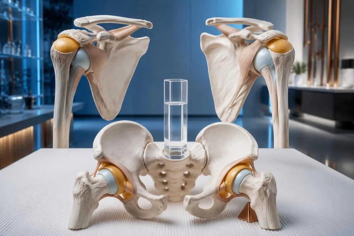
ဟုတ်ပါတယ်—ပိုလီမိုင်အယ်ဂျီယာ ရူမက်တစ်ကာတွင် သွေးစစ်ချက်ရလဒ်များ ပုံမှန်ဖြစ်နိုင်သော်လည်း၊ ကုသမှုမစတင်မီ ESR နှင့် CRP နှစ်မျိုးလုံး ပုံမှန်ဖြစ်ခြင်းမှာ ရှားပါးသည်။ ရောဂါသတ်မှတ်ချက်သည် လက္ခဏာပုံစံ၊ စမ်းသပ်စစ်ဆေးမှုနှင့် အခြားအကြောင်းရင်းများကို ဖယ်ထုတ်ခြင်းအပေါ် မူတည်ပြီး၊ ဓာတ်ခွဲခန်းရလဒ်တစ်ခုတည်းအပေါ် မမူတည်ပါ။.

အဓိက မှတ်သားစရာများ
- ပုံမှန် ရလဒ်များ ပိုလီမိုင်အယ်လ်ဂျီယာ ရူမက်တစ်ကာကို လုံးဝဖယ်ထုတ်၍မရပါ။ ESR နှင့် CRP နှစ်မျိုးလုံး ပုံမှန်ဖြစ်နေပါက ကုသမှုမခံယူရသေးသော ရောဂါလက္ခဏာသတ်မှတ်ချက်သည် ပုံမှန်မဟုတ်သဖြင့် အခြားဖြစ်နိုင်ခြေများကို စစ်ဆေးသင့်သည်။.
- အသက် ၅၀ နှစ်နှင့်အထက် သည် ရောဂါသတ်မှတ်ရာတွင် အဓိကကျသော သဲလွန်စတစ်ခုဖြစ်သည်။ ပိုလီမိုင်အယ်လ်ဂျီယာ ရူမက်တစ်ကာသည် အသက် ၅၀ အောက်တွင် အလွန်ရှားပါးသည်။.
- မိနစ် ၄၅ ထက်ပိုကြာသော နံနက်ပိုင်း အကြောတင်းခြင်း ပခုံးနှစ်ဖက်လုံး ထိခိုက်နေသည့်အခါ၊ အထူးသဖြင့် တင်ပါးဆုံခါးပတ်ပိုင်းဆိုင်ရာ လက္ခဏာများပါရှိလျှင် ရောဂါသတ်မှတ်ချက်ကို ထောက်ခံသည်။.
- CRP သည် 5 mg/L အောက် ဓာတ်ခွဲခန်းအများအပြားတွင် သတ်မှတ်ထားသော ပုံမှန်အတိုင်းအတာအတွင်း ပါဝင်သော်လည်း၊ အချို့ဓာတ်ခွဲခန်းများက အပေါ်ကန့်သတ်တန်ဖိုးကို 10 mg/L အဖြစ် သတ်မှတ်သည်။ မည်သည့်ကန့်သတ်တန်ဖိုးမျှ PMR ရောဂါသတ်မှတ်ရန် သတ်မှတ်ချက်မဟုတ်ပါ။.
- ESR နှင့် CRP မကိုက်ညီမှု အခြေအနေအရ ထည့်သွင်းစဉ်းစားရန် လိုအပ်သည်။ သွေးအားနည်းခြင်းနှင့် ပလာစမာပရိုတိန်းများကြောင့် CRP တူညီစွာ မမြင့်တက်ဘဲ ESR မြင့်တက်နိုင်သည်။.
- ပခုံးအာထရာဆောင်း bursitis သို့မဟုတ် အရွတ်အဖုံးပြောင်းလဲမှုများကို ဖော်ပြနိုင်သော်လည်း၊ ဤတွေ့ရှိချက်များသည် PMR ကို သီးခြားအတည်ပြုခြင်းထက် ထောက်ခံပေးခြင်းသာ ဖြစ်သည်။.
- Prednisone 12.5–25 mg နေ့စဉ် သည် ဆရာဝန်က စီမံကုသသော PMR အတွက် ၂၀၁၅ EULAR/ACR အကြံပြုချက်များတွင် ဖော်ပြထားသည့် ကနဦးပမာဏအပိုင်းအခြားဖြစ်သည်။ ကုသမှုအပေါ် တုံ့ပြန်မှုတစ်ခုတည်းဖြင့် ရောဂါအမည်ကို အတည်ပြု၍ မရပါ။.
- ခေါင်းကိုက်မှုအသစ်၊ ဝါးစားချိန် မေးရိုးနာကျင်မှု သို့မဟုတ် အမြင်အာရုံဆိုင်ရာ လက္ခဏာများ ရှိပါက ရောင်ရမ်းမှုညွှန်းကိန်းများ ပုံမှန်ဖြစ်နေလျှင်ပင် ဧရာမဆဲလ်သွေးကြောရောင်ရောဂါ ရှိမရှိ အရေးပေါ်စစ်ဆေးရန် လိုအပ်သည်။.
ပိုလီမိုင်အယ်လ်ဂျီယာ ရူမက်တစ်ကာအတွက် သွေးစစ်ချက် ပုံမှန်ဖြစ်နိုင်ပါသလား။
ရှိနိုင်ပါသည်။ polymyalgia rheumatica တွင် ESR ပုံမှန်ဖြစ်နိုင်ပြီး ကုသမှုမစမီ ESR နှင့် CRP နှစ်မျိုးစလုံး ပုံမှန်ဖြစ်နေခြင်းလည်း တစ်ခါတစ်ရံ ရှိနိုင်သည်။. polymyalgia rheumatica ကို အတည်ပြုနိုင်သည့် သို့မဟုတ် ပယ်ချနိုင်သည့် သွေးစစ်ဆေးမှုတစ်မျိုးတည်း မရှိပါ။ ဆရာဝန်များသည် ပုံမှန်အားဖြင့် အသက် ၅၀ နှစ်နှင့်အထက် လူကြီးများတွင် လက္ခဏာများ၊ စမ်းသပ်စစ်ဆေးမှုနှင့် အခြားဖြစ်နိုင်သည့် ရောဂါအမည်များကို ဆန့်ကျင်သည့် အထောက်အထားများကို ပေါင်းစပ်သုံးသပ်ကြသည်။.

ရောင်ရမ်းမှုညွှန်းကိန်းများ ပုံမှန်ဖြစ်ခြင်းသည် အကဲဖြတ်မှုကို ရပ်တန့်စေခြင်းမဟုတ်ဘဲ ရောဂါအမည်အပေါ် ယုံကြည်မှုကို လျော့နည်းစေသည်။. ပခုံးနှင့် တင်ပါးနှစ်ဖက်စလုံးတွင် မနက်တိုင်း မိနစ် ၉၀ ကြာ တင်းကျပ်မှုအသစ်ဖြစ်နေသော အသက် ၇၂ နှစ်အရွယ်သူတစ်ဦးကို၊ ပစ္စည်းမတင်ပြီးနောက် ပခုံးတစ်ဖက် နာကျင်နေသူတစ်ဦးနှင့် မတူညီသောနည်းဖြင့် အကဲဖြတ်သင့်သည်။ နှစ်ဦးစလုံး၏ CRP သည် 3 mg/L ဖြစ်နိုင်သော်လည်း၊ ၎င်းတို့၏ လက္ခဏာပုံစံများသည် ရောဂါရှာဖွေမှုအတွက် အလွန်ကွဲပြားသော အလေးချိန်ရှိသည်။.
ESR သို့မဟုတ် CRP မြင့်နေခြင်းတစ်ခုတည်းဖြင့်လည်း PMR ကို မသတ်မှတ်နိုင်ပါ။ အဘယ်ကြောင့်ဆိုသော် ဤစစ်ဆေးမှု ၂ မျိုးသည် ရောဂါတစ်မျိုးအတွက် သီးသန့်ဖြစ်သော ပဋိပစ္စည်းကို တိုင်းတာခြင်းမဟုတ်ဘဲ သီးခြားမဟုတ်သော တုံ့ပြန်မှုများကို တိုင်းတာခြင်းဖြစ်သောကြောင့် ဖြစ်သည်။ ပိုးဝင်ခြင်း၊ rheumatoid arthritis နှင့် ကင်ဆာအချို့တွင်လည်း အလားတူ ဓာတ်ခွဲခန်းရလဒ်များ ဖြစ်ပေါ်နိုင်ပြီး၊ စက်ပိုင်းဆိုင်ရာ ပခုံးပြဿနာများနှင့် ညွှန်းကိန်းမြင့်စေသည့် သီးခြားအကြောင်းရင်းတစ်ခုတို့လည်း တစ်ပြိုင်နက် ရှိနေနိုင်သည်။ ကျွန်ုပ်တို့၏ လမ်းညွှန်ချက်က မရှင်းလင်းသော နာကျင်မှုကို စမ်းသပ်ခြင်း ဆေးခန်းဆိုင်ရာ မေးခွန်းကို အဘယ်ကြောင့် ဦးစွာစဉ်းစားရသည်ကို ရှင်းပြထားသည်။.
ကန်တက်စတီသည် AI သွေးစမ်းသပ်မှုခွဲခြမ်းစိတ်ဖြာ ESR နှင့် CRP အဖြေများကို နားလည်ရန် အထောက်အကူပြုနိုင်ပြီး CRP 4 mg/L ရလဒ်တစ်ခုတည်းဖြင့် PMR ရောဂါအမည်ကို အဘယ်ကြောင့် အဆုံးအဖြတ်မပေးနိုင်သည်ကိုလည်း ရှင်းပြထားသည်။ ကျွန်ုပ်တို့၏ အခန်းကဏ္ဍမှာ ဓာတ်ခွဲခန်းအချက်အလက်များကို နားလည်လွယ်အောင် ရှင်းပြပေးရန်ဖြစ်ပြီး၊ ပခုံးလှုပ်ရှားမှု၊ တင်ပါးဆုံလုပ်ဆောင်ချက် သို့မဟုတ် ဓာတ်ခွဲခန်းအစီရင်ခံစာက မဖော်ပြနိုင်သော အရေးပေါ်သွေးကြောဆိုင်ရာ လက္ခဏာများကို အကဲဖြတ်ခြင်းအစား အစားထိုးရန် မဟုတ်ပါ။.
မည်သည့်ပခုံးနှင့် တင်ပါးဆုံလက္ခဏာများရှိလျှင် PMR ဖြစ်နိုင်ခြေ ပိုများသနည်း။
PMR သည် ပုံမှန်အားဖြင့် ပခုံးနှစ်ဖက်စလုံးတွင် နာကျင်မှုအသစ်နှင့် ကြာရှည်တင်းကျပ်မှုကို ဖြစ်စေပြီး တင်ပါးဆုံပတ်ဝန်းကျင် တင်းကျပ်မှုလည်း မကြာခဏ တွဲဖက်ဖြစ်ပေါ်သည်။. မိနစ် ၄၅ ထက်ပိုကြာသော မနက်ပိုင်းတင်းကျပ်မှုသည် ဤလက္ခဏာပုံစံကို ထောက်ခံသည်။ အထူးသဖြင့် အဝတ်အစားဝတ်ခြင်း၊ လက်များမြှောက်ခြင်း သို့မဟုတ် ကုလားထိုင်မှ ထခြင်းတို့သည် မမျှော်လင့်ဘဲ ခက်ခဲလာသည့်အခါ ဖြစ်သည်။.

နာကျင်မှုကြောင့် လှုပ်ရှားမှုကန့်သတ်ခြင်းသည် ကြွက်သားအင်အား အမှန်တကယ်အားနည်းခြင်းနှင့် မတူပါ။. ဥပမာအားဖြင့် အသက် ၆၈ နှစ်အရွယ် စိတ်ကူးယဉ်လူနာတစ်ဦးသည် ပခုံးနာကျင်သောကြောင့် ဆွယ်တာအင်္ကျီကို ခေါင်းပေါ်မှ မတင်နိုင်သော်လည်း သက်သောင့်သက်သာရှိသော ကိုယ်နေဟန်ဖြင့် စမ်းသပ်ချိန်တွင် ကြွက်သားအင်အားကို ထိန်းသိမ်းထားနိုင်သည်။ နာကျင်မှုကို ထည့်သွင်းစဉ်းစားပြီး စမ်းသပ်သည့်အခါတွင်ပင် သိသာသော အားနည်းမှုရှိပါက ရှုပ်ထွေးမှုမရှိသော PMR ထက် ကြွက်သား၊ အာရုံကြော သို့မဟုတ် ဟော်မုန်းဆိုင်ရာ ရောဂါများကို ပိုမိုစဉ်းစားသင့်သည်။.
တင်ပါးဆုံပတ်ဝန်းကျင် လက္ခဏာများကို ပေါင်ဆုံအတွင်းတည့်တည့်တွင်မဟုတ်ဘဲ တင်ပါး၊ ပေါင်အပေါ်ပိုင်း သို့မဟုတ် တင်ပါးဘေးဘက်တွင် ခံစားရနိုင်သည်။ ဆရာဝန်များသည် အိပ်ရာနိုးပြီးနောက်သာမက ထိုင်နေပြီးနောက် တင်းကျပ်မှုရှိမရှိကိုလည်း မေးမြန်းကြသည်။ မနက်ပိုင်း လှုပ်ရှားရခက်မှုသည် ၁၀ မိနစ်ကြာသလား၊ ၇၀ မိနစ်ကြာသလား မှတ်တမ်းတင်ခြင်းသည် “တစ်ကိုယ်လုံးကိုက်ခဲနေသည်” ဟုသာ ရေးခြင်းထက် ပိုအသုံးဝင်သည်။ ကြာချိန်သည် သင် အမှန်တကယ်ခံစားရသည့်အရာကို ဖော်ပြသင့်ပြီး၊ ပြည့်မီအောင် ကြိုးစားရမည့် ပစ်မှတ်မဟုတ်ပါ။.
လက်ချောင်းအချို့တွင် ထုံကျင်ကျင်ဖြစ်ခြင်းသည် သီးခြားရောဂါရှာဖွေရေး သဲလွန်စတစ်ခုဖြစ်သည်, ပခုံးတင်းကျပ်မှုလည်း ရှိနေသည့်အခါတွင်ပင် ဖြစ်သည်။ လက်မ၊ လက်ညှိုးနှင့် လက်ခလယ်တို့တွင် ထုံကျင်ခြင်းသည် median အာရုံကြော ဖိမိခြင်းကို ညွှန်ပြနိုင်ပြီး၊ ကျွန်ုပ်တို့၏ လက်ကောက်ဝတ်အာရုံကြောဖိမိမှု လက္ခဏာလမ်းညွှန်တွင် ဆွေးနွေးထားသော လက်ချောင်းတစ်နေရာတည်းကို ထိခိုက်သည့်ပုံစံနှင့် ကွဲပြားသည်။; တွင် ဖော်ပြထားသည်။ လက္ခဏာအသစ်တိုင်းသည် ရောင်ရမ်းရောဂါတစ်မျိုးတည်းကြောင့် ဖြစ်သည်ဟု မယူဆဘဲ ဆရာဝန်များက ထိုလက္ခဏာများကို စစ်ဆေးသင့်သည်။.
PMR တွင် ESR နှင့် CRP သည် အမှန်တကယ် မည်သည့်အရာကို တိုင်းတာသနည်း။
ESR နှင့် CRP သည် ရောင်ရမ်းမှုတုံ့ပြန်မှု၏ မတူညီသော အပိုင်းများကို တိုင်းတာခြင်းဖြင့် PMR အကဲဖြတ်မှုကို ထောက်ပံ့ပေးသည်၊ PMR ကိုယ်တိုင်ကို တိုင်းတာခြင်းမဟုတ်ပါ။. ဓာတ်ခွဲခန်းအများအပြားတွင် CRP 5 mg/L အောက်ကို ရည်ညွှန်းကန့်သတ်ချက်အဖြစ် အသုံးပြုကြပြီး၊ ESR ရည်ညွှန်းအပိုင်းအခြားများမှာ အသုံးပြုသည့်နည်းလမ်း၊ အသက်၊ လိင်နှင့် ဒေသခံဓာတ်ခွဲခန်းအလိုက် ကွဲပြားသည်။.
ESR measures how far cellular elements settle in a testing column over 1 hour, usually reported in mm/hour. Fibrinogen, immunoglobulins, anemia, and cellular characteristics affect that settling, so an ESR of 48 mm/hour with CRP of 2 mg/L is not automatically evidence of active PMR; it may prompt review of the full blood count and protein results.
CRP is a liver-produced acute-phase protein that generally changes faster than ESR when the inflammatory stimulus changes. Check the units before comparing reports: 1 mg/dL သည် 10 mg/L နှင့် ညီမျှသည်။, so CRP 0.6 mg/dL and 6 mg/L represent the same concentration; the distinction between cellular counts and acute-phase proteins is explored in WBC နှင့် CRP မတူညီမှု.
An ESR of 40 mm/hour is not a universal diagnostic boundary for PMR. Older diagnostic approaches often emphasized that value, but contemporary assessment considers the whole presentation and the laboratory's reference interval; conversely, ESR of 90 mm/hour does not establish PMR and should not distract from fever, focal symptoms, or unexplained abnormalities elsewhere in the report.
| အဆင့် | အပိုင်းအခြား | အဓိပ္ပာယ် |
|---|---|---|
| CRP within a common reference interval | <5 mg/L at many laboratories | Does not exclude PMR; some laboratories use an upper limit of 10 mg/L. |
| Illustrative adult ESR intervals | 0–20 or 0–30 mm/hour | These are examples, not universal limits; use the age- and method-appropriate interval on the report. |
| CRP above the local upper limit | >5 or >10 mg/L, depending on the laboratory | Supports an inflammatory process but does not distinguish PMR from infection or other inflammatory disorders. |
| ESR above the local upper limit | Above the report's stated interval, in mm/hour | Interpret with CRP, hemoglobin, plasma proteins, symptoms, and treatment timing; no ESR value alone confirms PMR. |
PMR တွင် ESR ပုံမှန်နှင့် CRP ပုံမှန်ဖြစ်ခြင်း မည်မျှအဖြစ်များသနည်း။
Normal ESR is reported more often than normal ESR and CRP together in untreated PMR. A 2018 review described normal ESR in approximately 7–22% of reported cases, but CRP was usually elevated in those patients; these figures are not an individual's diagnostic probability.
Both markers being normal was uncommon in several older PMR cohorts, with proportions around 1–2% in some series discussed by Manzo and Milchert in Reumatologia (2018). Selected specialist cohorts have reported higher proportions, so a single universal percentage would be misleading; referral patterns, diagnostic definitions, treatment exposure, and how thoroughly mimics were excluded all influence the denominator.
The phrase “polymyalgia rheumatica normal ESR” can hide 2 different situations: ESR is normal but CRP is raised, or both tests are within range. A hypothetical ESR of 18 mm/hour with CRP of 24 mg/L still provides biochemical evidence of an inflammatory response, whereas ESR of 18 with CRP of 2 mg/L requires more reliance on examination and alternative explanations.
Cardiovascular hs-CRP categories are not PMR diagnostic categories, even though hs-CRP measures the same protein with greater low-level sensitivity. A value of 3 mg/L may matter in a cardiovascular risk discussion without confirming musculoskeletal inflammation; our hs-CRP context guide separates those uses, preventing a heart-risk label from being mistaken for a rheumatology diagnosis.
ပိုလီမိုင်အယ်လ်ဂျီယာ ရူမက်တစ်ကာကို ဆေးခန်းတွင် မည်သို့ ရောဂါရှာဖွေသတ်မှတ်သနည်း။
Polymyalgia rheumatica is diagnosed clinically after assessing the characteristic symptom pattern and excluding more plausible causes. The 2012 EULAR/ACR classification framework starts with age 50 or older, bilateral shoulder aching, and abnormal ESR or CRP, but classification criteria are not a stand-alone diagnostic test.
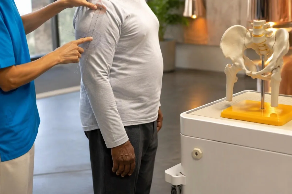
The 2012 criteria were developed to classify patients consistently for research, not to rule out every unusual clinical presentation (Dasgupta et al., 2012). Someone with normal ESR and CRP does not meet that framework's entry requirements, but this does not logically prove that PMR is impossible; it means the standard research classification cannot be applied in the usual way.
The same framework assigns weight to morning stiffness lasting more than 45 minutes, hip symptoms, absence of rheumatoid-factor or anti-CCP antibodies, and absence of other joint pain. Clinicians interpret those features rather than asking patients to diagnose themselves with a score, because early elderly-onset rheumatoid arthritis may resemble PMR before persistent hand or wrist swelling becomes obvious.
A pretreatment baseline is especially useful when the diagnosis is uncertain, provided urgent assessment is not delayed. Bring the symptom timeline and any previous ESR or CRP results, including a result obtained 2 weeks before steroids started; our အခြေခံစမ်းသပ်မှုအတွက် ကြိုတင်ပြင်ဆင်မှု explains how medication dates and the reason for testing make that first report more interpretable.
သွေးစစ်ချက်များ ပုံမှန်ဖြစ်နေသည့်အခါ မည်သည့်ရောဂါများက PMR နှင့် ဆင်တူနိုင်သနည်း။
Mechanical shoulder disorders, osteoarthritis, thyroid disease, medication-related muscle symptoms, and other rheumatic conditions can resemble PMR. Normal ESR and CRP make a careful review of these alternatives particularly valuable, especially when symptoms are one-sided, morning stiffness is brief, or the person is younger than 50.
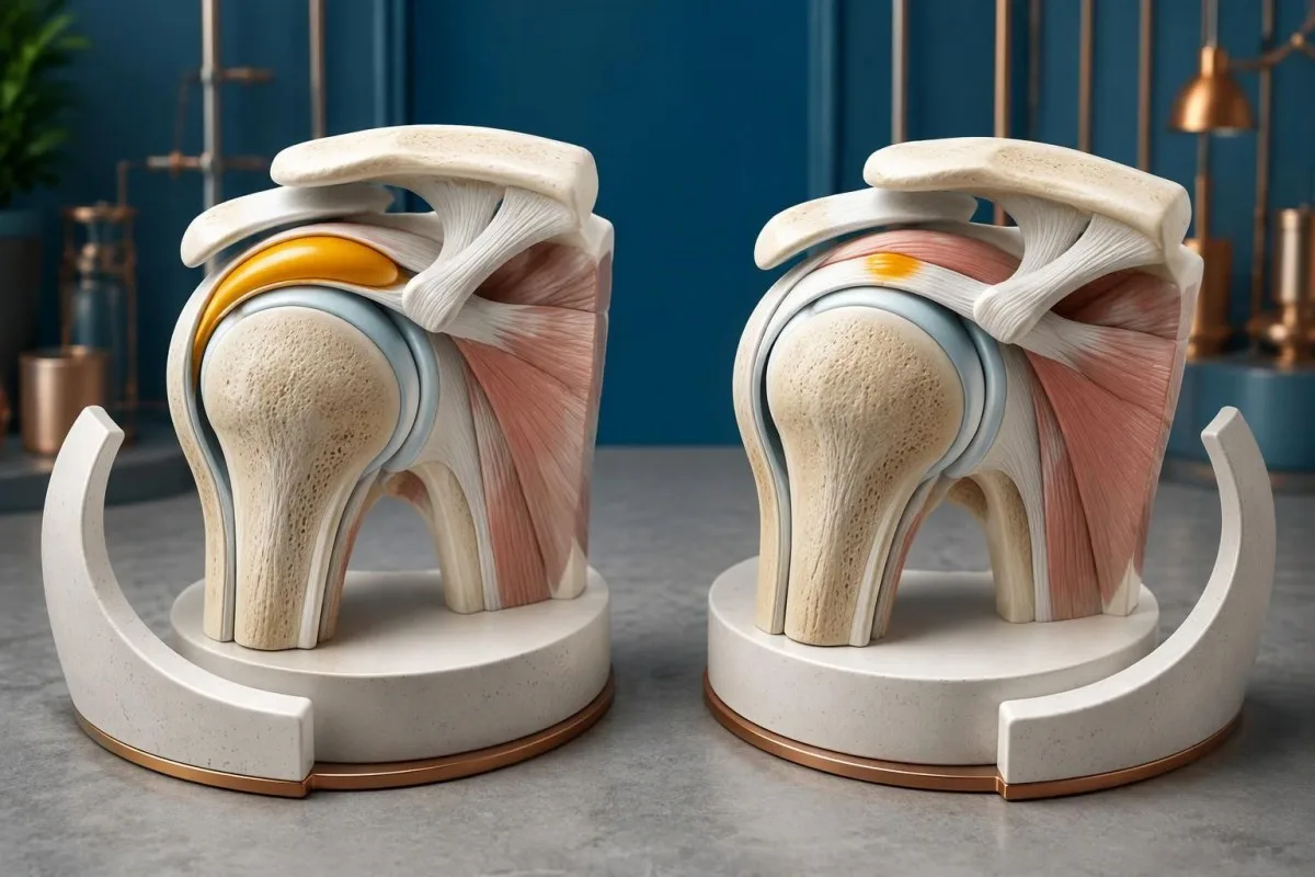
Rotator cuff disease and adhesive capsulitis are assessed through movement patterns, including whether passive movement is restricted and whether a specific arc provokes discomfort. A hypothetical 61-year-old with one stiff shoulder and 15 minutes of morning symptoms is less typical for PMR than someone with bilateral shoulder and hip restriction; coexistence is possible, so finding a tendon problem does not automatically explain everything.
Thyroid dysfunction can contribute to muscle aching, fatigue, and reduced exercise tolerance, but a mildly abnormal TSH does not prove that the thyroid explains the entire symptom picture. Clinicians interpret TSH with free T4 and the examination rather than anchoring on a single flag; our subclinical hypothyroidism guide explains why modest biochemical changes may or may not be clinically relevant.
Statin-associated muscle symptoms can occur with normal creatine kinase, making the medication timeline useful even when several tests look reassuring. Symptoms beginning 3 weeks after a dose increase deserve discussion rather than an unsupervised medication stop; our statin muscle-risk guide explains one susceptibility factor, but genetic testing is not a routine way to distinguish PMR from statin symptoms.
PMR နှင့် ဆင်တူသောရောဂါများကို ဖယ်ထုတ်ရန် အခြားမည်သည့်သွေးစစ်ချက်များက အထောက်အကူပြုသနည်း။
Additional tests look for alternative diagnoses and establish treatment safety; they do not create a confirmatory PMR panel. Depending on the presentation, clinicians may review a full blood count, kidney and liver function, calcium, glucose, thyroid tests, creatine kinase, and rheumatoid-factor or anti-CCP antibodies.
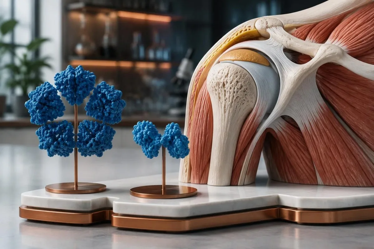
Creatine kinase is usually not elevated by uncomplicated PMR itself. True weakness with a hypothetical CK of 1,200 U/L, interpreted against the laboratory's interval and recent exercise, shifts attention toward muscle injury or myopathy; selected cases may also involve aldolase testing, discussed in our muscle enzyme follow-up guide, rather than assuming that every proximal symptom represents joint-girdle inflammation.
Anemia or unusual plasma protein results can help explain why ESR and CRP disagree, and they may identify a problem that needs its own evaluation. For example, hemoglobin of 95 g/L with a disproportionately high ESR should not simply be filed under PMR; our သွေးရည်ကြည် ပရိုတိန်း ညွှန်ကြားရေးသားချက် explains protein patterns without implying that every abnormal globulin result warrants the same investigation.
ကန်တက်စတီသည် AI biomarker အဓိပ္ပာယ်ဖော်ခြင်း ပလက်ဖောင်း that can help organize a report containing ESR, CRP, CK, and thyroid results, but those 4 measurements answer different clinical questions. We encourage targeted interpretation: ANA or complement testing may be appropriate when specific systemic features suggest connective-tissue disease, whereas ordering broad antibody panels solely because PMR markers are normal can produce distracting incidental positives.
ESR နှင့် CRP ပုံမှန်ဖြစ်နေသည့်အခါ အာထရာဆောင်းက အထောက်အကူပြုနိုင်ပါသလား။
Ultrasound can support suspected PMR by showing shoulder or hip-region bursitis, tendon-sheath changes, or joint inflammation. Bilateral subacromial-subdeltoid bursitis is a useful pattern, but imaging findings are not specific enough to confirm PMR independently, particularly in adults over 50 with common degenerative changes.
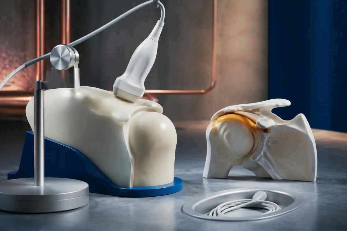
The 2012 EULAR/ACR study incorporated ultrasound as an optional classification aid, while recognizing overlap with other shoulder conditions (Dasgupta et al., 2012). Finding bursal changes on both sides can strengthen a coherent clinical picture, but a scan obtained after 2 weeks of treatment may not show the same degree of change as a pretreatment examination.
Ultrasound is most useful when it answers a specific question: is the main restriction related to bursitis, a tendon disorder, joint changes, or several processes together? A hypothetical 73-year-old with CRP of 3 mg/L and bilateral bursitis still needs assessment for rheumatoid arthritis and mechanical disease; the scan does not convert a nonspecific symptom pattern into PMR simply because a bursa contains fluid.
Negative rheumatoid factor does not rule out rheumatoid arthritis, and positive rheumatoid factor does not automatically exclude PMR. Persistent swelling of 2 or more small joints changes the differential more meaningfully than a low-positive antibody alone; our rheumatoid factor အဓိပ္ပာယ်ဖော်ပြချက် လမ်းညွှန် explains why antibody concentration, examination, and imaging must be interpreted together.
prednisone သောက်ပြီး လျင်မြန်စွာ သက်သာလာခြင်းက PMR ဖြစ်ကြောင်း သက်သေပြပါသလား။
A rapid response to glucocorticoids supports PMR but does not prove the diagnosis, because other inflammatory conditions also improve. The 2015 EULAR/ACR recommendations describe an initial prednisone-equivalent range of 12.5–25 mg daily for clinician-managed PMR, adjusted to disease activity and treatment risks.
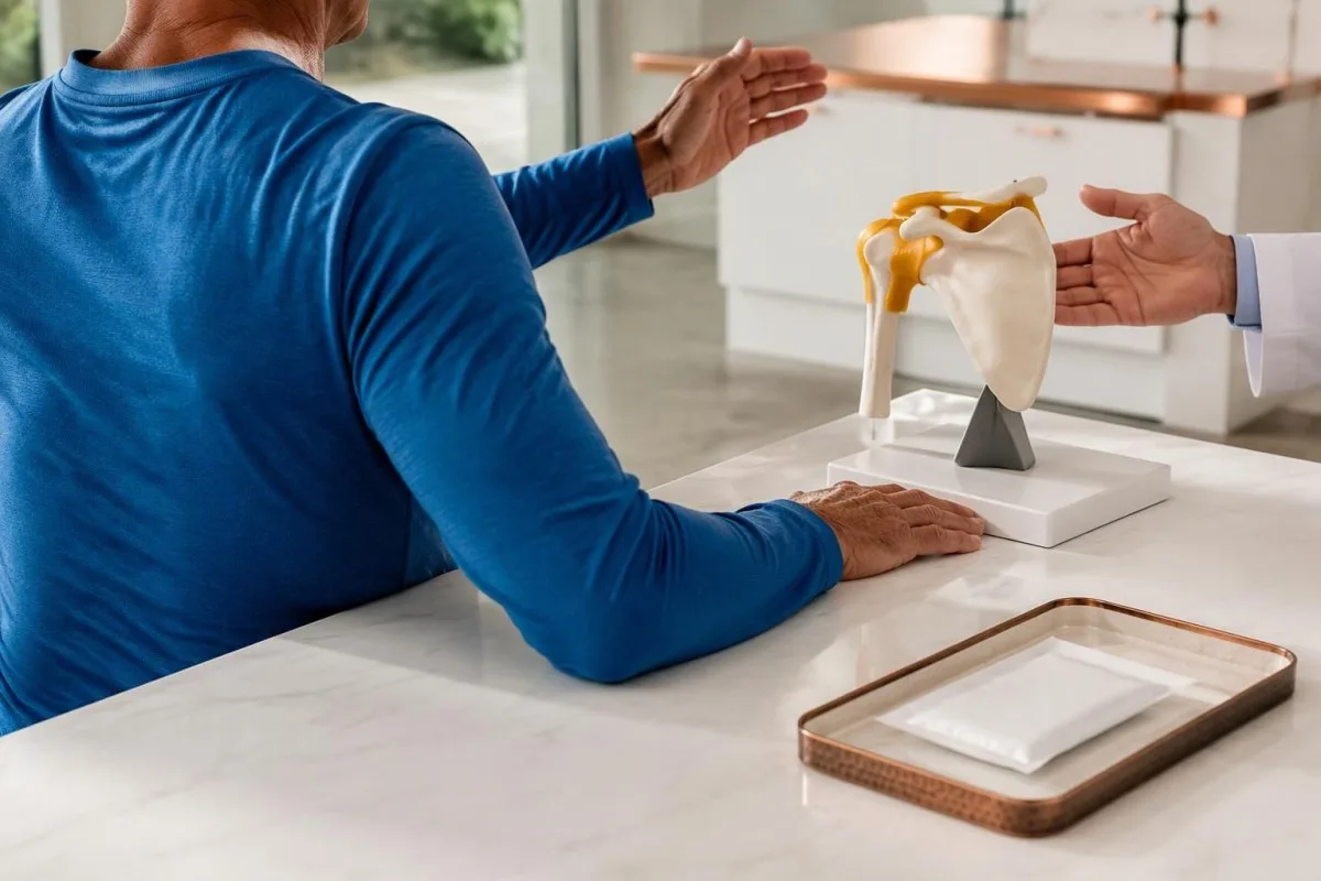
Prednisone should not be used as a self-directed diagnostic experiment. Starting 15 mg daily before evaluation may lower inflammatory markers and obscure an alternative illness, while exposing someone without PMR to adverse effects; the EULAR/ACR recommendations emphasize individualized dosing and reassessment rather than treating response as a binary diagnostic challenge (Dejaco et al., 2015).
Improvement should include the actual features that prompted concern, not just a vague increase in energy or better sleep. In a hypothetical case, morning stiffness falling from 90 minutes to 20 minutes and easier dressing provides useful follow-up information, but persistent one-sided shoulder restriction may indicate a coexisting tendon disorder; incomplete response should trigger clinical review rather than automatic dose escalation.
Glucocorticoid safety depends on pre-existing risks and other medicines, including diabetes, osteoporosis, glaucoma, and infection vulnerability. A glucose rise from 6 to 12 mmol/L after treatment needs a management plan even if shoulder symptoms improve; our older-adult medication effects guide explains why a changing laboratory result may reflect treatment rather than worsening of the original condition.
ဧရာမဆဲလ်သွေးကြောရောင်ရောဂါအတွက် မည်သည့်လက္ခဏာများရှိလျှင် အရေးပေါ်စစ်ဆေးမှုခံယူသင့်သနည်း။
New headache, scalp tenderness, jaw pain when chewing, or visual disturbance in someone with suspected PMR require urgent assessment for giant cell arteritis. Sudden visual loss, double vision, or a temporary curtain over vision needs immediate emergency evaluation—even if CRP is below 5 mg/L.

Normal ESR and CRP cannot safely exclude giant cell arteritis when the symptoms are concerning. Clinicians may start treatment before laboratory confirmation or vascular imaging because delay can threaten vision; a hypothetical 76-year-old with jaw fatigue during meals and a new temporal headache should not wait 1 week for a routine repeat blood test.
Jaw claudication means discomfort or fatigue triggered by chewing and relieved by resting, rather than simply constant dental pain. A new symptom appearing after 2 minutes of chewing, especially with headache or scalp tenderness, deserves same-day medical assessment; describe the trigger and relief pattern clearly, because “jaw pain” alone can conceal the feature that makes clinicians concerned.
Sudden speech difficulty, facial asymmetry, or weakness on one side is an emergency, whether PMR has been diagnosed or not. Follow the emergency actions in our stroke warning-sign guide rather than waiting for an inflammatory marker result; fever with severe illness or a new hot, swollen single joint also warrants urgent assessment for causes other than uncomplicated PMR.
ရောင်ရမ်းမှုအမှတ်အသားများ ပုံမှန်အတိုင်း ဆက်ရှိနေပါက PMR ကို မည်သို့ စောင့်ကြည့်သနည်း။
When ESR and CRP are uninformative, PMR monitoring relies more heavily on symptoms, function, examination, and treatment safety. Track morning stiffness in minutes and repeatable tasks such as dressing or chair rising; a CRP of 2 mg/L cannot independently establish remission or exclude relapse.
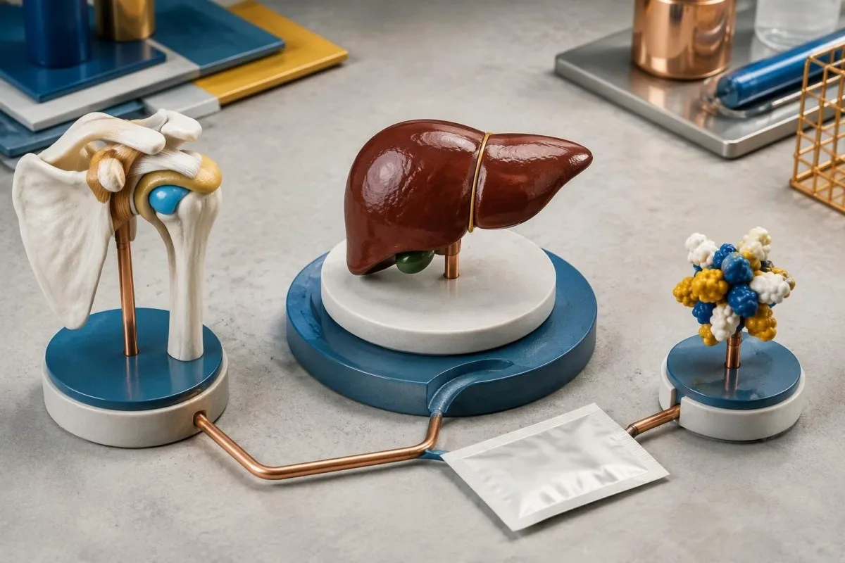
Glucocorticoids can normalize previously elevated markers, so a normal result after treatment is different from a normal result before any treatment. Some IL-6 receptor-blocking medicines also suppress CRP directly, making a value below 5 mg/L less useful for judging inflammatory activity; the medication name and timing belong beside the laboratory value during review.
A practical diary can record 3 repeatable observations: morning stiffness duration, ease of raising both arms, and ability to get up from the same chair. If stiffness changes from 15 to 60 minutes after a taper, contact the treating clinician, but do not assume relapse automatically; osteoarthritis, deconditioning, tendon disease, and glucocorticoid-related complications can also change how those tasks feel.
With Kantesti AI, laboratory trends can be considered alongside treatment dates rather than comparing 2 isolated “normal” labels. Our ဆေးဝါး-လုံခြုံရေး ခေတ်ရေစီးကြောင်းလမ်းညွှန် explains why glucose, kidney function, and other safety measurements may remain useful even when ESR and CRP provide little information; dose changes still require the prescribing clinician's judgment.
PMR စစ်ဆေးမှုခံယူရန် မည်သည့်အရာများ ယူဆောင်သွားသင့်သနည်း။
Bring the original laboratory report, a symptom timeline, and a complete medication list to a PMR assessment. Include whether both shoulders and hips are affected, how many minutes morning stiffness lasts, and whether ESR or CRP was measured before or after the first glucocorticoid dose.
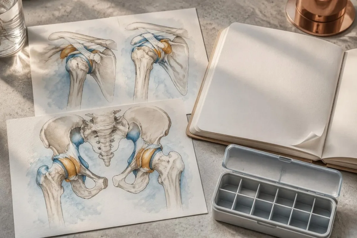
Exact dates are more useful than “recently.” Write down whether symptoms started 6 weeks ago, whether a statin dose changed 3 weeks ago, and whether the laboratory sample was collected 4 days after prednisone began; also list fever, weight change, persistent swollen joints, and any new headache or chewing-related jaw symptoms because these details can redirect the assessment.
Keep the original units and reference intervals visible when sharing a report, since ESR of 25 mm/hour may be flagged differently across laboratories. Before sending a PDF or photo to any digital service, review our report-sharing privacy steps; retaining clinically relevant dates and removing unnecessary identifiers are different tasks, and neither should alter the actual test values.
Kantesti is operated by Kantesti LTD, UK Company No. 17090423; information about ကျွန်တော်တို့ အဖွဲ့အစည်းမှာ helps distinguish the service provider from the clinician responsible for diagnosis. Our AI နည်းပညာ လမ်းညွှန် explains the report interpretation workflow, but even a clearly explained set of 2 inflammatory markers cannot replace a physical examination.
AI ၏ အဓိပ္ပာယ်ဖော်ပြမှုက မည်သည့်အရာများကို ကူညီပေးနိုင်ပြီး—မည်သည့်ရောဂါများကို မသတ်မှတ်နိုင်သနည်း။
AI interpretation can explain laboratory units, reference intervals, and trends, but it cannot independently diagnose PMR or exclude giant cell arteritis. ကန်တက်စတီသည် AI သွေးစစ်ဆေးမှု ရလဒ် ပလက်ဖောင်း that supports understanding of ESR and CRP reports without turning a CRP of 3 mg/L into a diagnostic verdict.
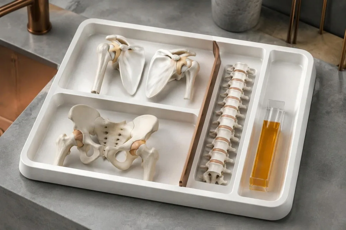
The safest output separates observation from conclusion. “CRP is 4 mg/L and within this laboratory's interval” is a report observation, while “PMR is excluded” is an unsupported clinical conclusion; similarly, a raised ESR should generate questions about symptoms and confounders rather than automatically label an inflammatory disease.
Kantesti AI cannot test passive shoulder movement, establish pain-independent muscle strength, or verify chewing-related jaw claudication from a laboratory PDF. Those limitations matter even when 10 other biomarkers appear reassuring; a concerning visual symptom requires urgent care directly, not completion of an upload or waiting for an interpretation result.
A technical benchmark is not the same as a PMR diagnostic accuracy study. ကျွန်တော်တို့ရဲ့ အတည်ပြုစိစစ်မှုနှင့် ဆေးဘက်ဆိုင်ရာ ကြီးကြပ်မှု information should be read with that distinction in mind: explaining a CRP result correctly and diagnosing a 70-year-old's shoulder stiffness are different tasks, and performance on one cannot be assumed to establish performance on the other.
PMR အဓိပ္ပာယ်ဖော်ပြမှုနောက်ခံရှိ သုတေသနစာတမ်းများနှင့် အထောက်အထားများ
The most relevant PMR evidence addresses clinical classification, treatment, and the limitations of normal inflammatory markers. The 2012 EULAR/ACR classification study, 2015 EULAR/ACR management recommendations, and 2018 review of normal ESR and CRP each answer a different question; none establishes a single confirmatory blood test.
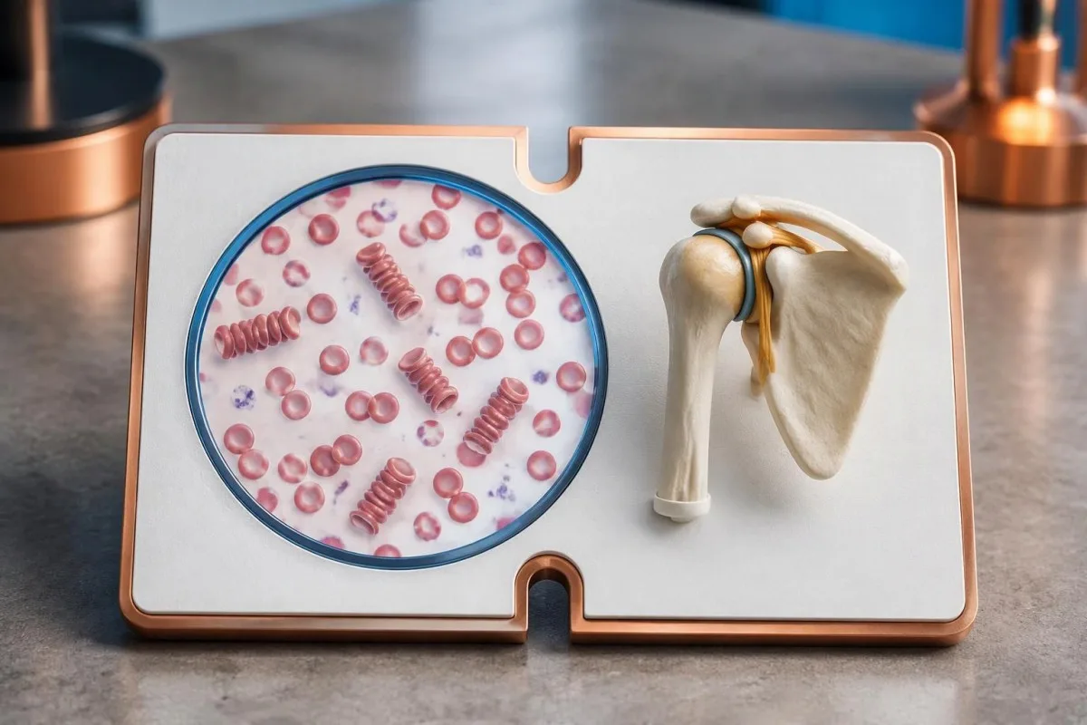
Evidence about normal-marker PMR is less certain than evidence about the usual presentation. The 7–22% normal-ESR range summarized by Manzo and Milchert (2018) comes from heterogeneous reports, while estimates for both markers being normal depend strongly on patient selection; these studies support keeping PMR possible, not bypassing the search for a better explanation.
The 2 DOI-linked background publications below cover serum proteins and autoimmune laboratory interpretation, not PMR diagnostic validation or treatment effectiveness. Our complement နှင့် ANA လမ်းညွှန် is relevant when a clinician suspects a different systemic autoimmune condition, but normal-marker shoulder stiffness alone does not justify assuming that ANA or C3/C4 will confirm PMR.
Kantesti's background guides should be distinguished from the external peer-reviewed PMR references listed separately, including the 2015 treatment recommendations. Our 0 ဆေးဘက်ဆိုင်ရာ အကြောင်းအရာစံနှုန်းများ describe the editorial framework; the practical decision remains clinical: assess the shoulder and hip pattern, review plausible mimics, and treat new visual or jaw symptoms as urgent rather than waiting for a laboratory flag.
ተደጋግሞ የሚጠየቁ ጥያቄዎች
0 သင့်တွင် ESR နှင့် CRP ပုံမှန်ဖြစ်နေသော်လည်း polymyalgia rheumatica ရှိနိုင်ပါသလား။
0 ဟုတ်ကဲ့၊ polymyalgia rheumatica သည် ESR နှင့် CRP ပုံမှန်ဖြစ်နေသော်လည်း ရံဖန်ရံခါ ဖြစ်ပွားနိုင်ပါသည်။ သို့သော် ထုတ်ဝေထားသော လူနာအုပ်စုလေ့လာမှုများစွာတွင် ကုသမှုမစမီ နှစ်မျိုးစလုံး ပုံမှန်ဖြစ်နေခြင်းမှာ ရှားပါးပါသည်။ ၂၀၁၈ ခုနှစ် သုံးသပ်ချက်တစ်ခုတွင် စုစည်းဖော်ပြထားသည့် ဖြစ်ရပ်များ၏ ခန့်မှန်းခြေ ၇–၂၂% တွင် ESR တစ်မျိုးတည်း ပုံမှန်ဖြစ်ကြောင်း အစီရင်ခံထားပြီး၊ များသောအားဖြင့် CRP မြင့်တက်နေပါသည်။ ဆရာဝန်များသည် ဓာတ်ခွဲခန်းရလဒ်တစ်ခုတည်းကို အခြေခံ၍ PMR ကို ဖယ်ထုတ်မည့်အစား ပခုံးနှစ်ဖက်တောင့်တင်းခြင်း၊ တင်ပါးဆုံဆိုင်ရာ လက္ခဏာများ၊ အသက် ၅၀ နှစ်နှင့်အထက်ဖြစ်ခြင်း၊ စမ်းသပ်စစ်ဆေးတွေ့ရှိချက်များနှင့် အခြားဖြစ်နိုင်သော ရောဂါရှာဖွေချက်များကို အကဲဖြတ်ကြပါသည်။ လက္ခဏာပုံစံက ယုံကြည်လက်ခံနိုင်ဖွယ်ရှိသော်လည်း အမှတ်အသားနှစ်မျိုးစလုံး ပုံမှန်ဖြစ်နေသည့်အခါ အထူးကု၏ ပြန်လည်သုံးသပ်မှုသည် အထူးအသုံးဝင်ပါသည်။.
polymyalgia rheumatica ကို အတည်ပြုပေးနိုင်သည့် သွေးစစ်ဆေးမှုက ဘာလဲ?
Polymyalgia rheumatica ကို အတည်ပြုနိုင်သည့် သွေးစစ်ဆေးမှုတစ်မျိုးတည်း မရှိပါ။ ESR နှင့် CRP တို့သည် ရောင်ရမ်းတုံ့ပြန်မှုရှိကြောင်း သီးခြားမသတ်မှတ်နိုင်သော အထောက်အထားများကို ပေးပြီး၊ သင့်လျော်သည့်အခါ CK၊ သိုင်းရွိုက်လုပ်ဆောင်ချက်၊ rheumatoid factor နှင့် anti-CCP ကဲ့သို့သော စစ်ဆေးမှုများက အခြားဖြစ်နိုင်ခြေများကို စုံစမ်းရာတွင် အထောက်အကူပြုသည်။ CRP 5 mg/L အောက်သည် ဓာတ်ခွဲခန်းများစွာတွင် ပုံမှန်အတိုင်းအတာအတွင်း ရှိသော်လည်း၊ ထိုတန်ဖိုးဖြစ်စေ ESR မြင့်တက်နေခြင်းဖြစ်စေ ရောဂါအမည်ကို အတည်မပြုနိုင်ပါ။ PMR ကို ဆေးဘက်ဆိုင်ရာ အကဲဖြတ်မှုနှင့် ပိုမိုဖြစ်နိုင်ခြေရှိသော အကြောင်းရင်းများကို ဖယ်ထုတ်ခြင်းတို့ဖြင့် ရောဂါသတ်မှတ်သည်။.
polymyalgia rheumatica ကို ညွှန်ပြသော ESR သို့မဟုတ် CRP အဆင့်မှာ မည်မျှဖြစ်သနည်း?
polymyalgia rheumatica ကို တစ်ခုတည်းဖြင့် ယုံကြည်စိတ်ချစွာ ခွဲခြားသတ်မှတ်ပေးနိုင်သည့် ESR သို့မဟုတ် CRP သတ်မှတ်ချက် မရှိပါ။ ရောဂါရှာဖွေသည့် ချဉ်းကပ်နည်းဟောင်းအချို့တွင် ESR 40 mm/hour ကို အလေးပေးဖော်ပြခဲ့သော်လည်း၊ ၎င်းသည် ဆေးခန်းဆိုင်ရာ ရောဂါရှာဖွေသတ်မှတ်မှုအတွက် အားလုံးနှင့်သက်ဆိုင်သော လိုအပ်ချက်တစ်ခု မဟုတ်ပါ။ ဓာတ်ခွဲခန်းများစွာတွင် CRP အပေါ်ဆုံးကန့်သတ်ချက်ကို 5 mg/L ဟု သတ်မှတ်ကြပြီး၊ အချို့တွင် 10 mg/L ဟု သတ်မှတ်ကြသည်။ ထိုကန့်သတ်ချက်များထက် မြင့်သောတန်ဖိုးများသည် ရောင်ရမ်းမှုဖြစ်စဉ်ရှိကြောင်း ထောက်ခံသော်လည်း PMR ကို ပိုးဝင်ခြင်း သို့မဟုတ် အခြားရောင်ရမ်းမှုအခြေအနေများနှင့် ခွဲခြားမပေးနိုင်ပါ။.
သွေးစစ်ဆေးမှုရလဒ်များ ပုံမှန်ဖြစ်နေသည့်အခါ polymyalgia rheumatica ကို မည်သို့ ရောဂါရှာဖွေသနည်း။
သွေးစစ်ဆေးမှုရလဒ်များ ပုံမှန်ဖြစ်သည့်အခါ ဆရာဝန်များသည် ရောဂါလက္ခဏာများ၏ ပုံစံကို အကဲဖြတ်ပြီး၊ ပခုံးနှင့် တင်ပါးဆုံ လှုပ်ရှားမှုကို စစ်ဆေးကာ polymyalgia rheumatica နှင့် ဆင်တူသော အခြားအခြေအနေများကို ရှာဖွေစစ်ဆေးကြသည်။ အသက် ၅၀ နှစ်နှင့်အထက်ဖြစ်ခြင်း၊ ပခုံးနှစ်ဖက်လုံးတွင် ရောဂါလက္ခဏာများရှိခြင်းနှင့် မနက်ခင်းတောင့်တင်းမှု မိနစ် ၄၅ ကျော်ကြာခြင်းတို့သည် ပုံမှန်ဖြစ်ပွားပုံကို ထောက်ခံသည်။ အာထရာဆောင်းဖြင့် ထောက်ခံနိုင်သည့် အရည်အိတ်ရောင်ခြင်း သို့မဟုတ် အရွတ်အိမ်ပြောင်းလဲမှုများကို တွေ့ရှိနိုင်သော်လည်း၊ ထိုတွေ့ရှိချက်များသည် ၎င်းတို့တစ်ခုတည်းဖြင့် ရောဂါကို အတည်မပြုနိုင်ပါ။ အချိန်ကြာလာသည်နှင့်အမျှ အခြားအဆစ်အမြစ်ရောဂါ သို့မဟုတ် စက်ပိုင်းဆိုင်ရာအခြေအနေတစ်ခု ပိုမိုရှင်းလင်းလာနိုင်သောကြောင့် နောက်ဆက်တွဲစစ်ဆေးမှုများ မကြာခဏလိုအပ်သည်။.
prednisone သောက်သုံးပြီး သက်သာလာတယ်ဆိုတာ ကျွန်ုပ်မှာ PMR ရှိတယ်လို့ သေချာပေါက် ဆိုလိုပါသလား။
Prednisone သုံးစွဲပြီး သက်သာလာခြင်းသည် polymyalgia rheumatica ဖြစ်ကြောင်း အတိအကျ သက်သေမပြနိုင်ပါ၊ အကြောင်းမှာ အခြားရောင်ရမ်းမှုဆိုင်ရာ အခြေအနေများလည်း glucocorticoids ကို တုံ့ပြန်နိုင်သောကြောင့်ဖြစ်သည်။ 2015 EULAR/ACR အကြံပြုချက်များတွင် ရောဂါလက္ခဏာများနှင့် ကုသမှုဆိုင်ရာ အန္တရာယ်များအပေါ် မူတည်၍ တစ်ဦးချင်းညှိနှိုင်းပေးရမည့်၊ ဆရာဝန်ကြီးကြပ်မှုဖြင့် စတင်ပေးသော prednisone-equivalent ပမာဏမှာ တစ်နေ့လျှင် 12.5–25 mg ဖြစ်ကြောင်း ဖော်ပြထားသည်။ တုံ့ပြန်မှုကို အားအင်တိုးတက်လာခြင်းတစ်ခုတည်းဖြင့် မဆုံးဖြတ်ဘဲ တောင့်တင်းမှုနှင့် လုပ်ဆောင်နိုင်စွမ်းတို့တွင် ဖြစ်ပေါ်လာသော တိကျသည့်ပြောင်းလဲမှုများဖြင့် အကဲဖြတ်သင့်သည်။ မိမိဘာသာ ရောဂါရှာဖွေရန် စမ်းသပ်သည့်အနေဖြင့် prednisone ကို မစတင်ပါနှင့်၊ ပမာဏကိုလည်း မပြောင်းလဲပါနှင့်။.
ရောင်ရမ်းမှုအမှတ်အသားများ ပုံမှန်ဖြစ်နေသော်လည်း ဧရာမဆဲလ် သွေးကြောရောင်ရမ်းခြင်း ဖြစ်နိုင်ပါသလား။
ဟုတ်ကဲ့၊ ရောင်ရမ်းမှုအမှတ်အသားများ ပုံမှန်ဖြစ်နေခြင်းက စိုးရိမ်ဖွယ်လက္ခဏာများရှိသည့်အခါ ဧရာမဆဲလ်သွေးလွှတ်ကြောရောင်ရမ်းခြင်းကို ဘေးကင်းစွာ ဖယ်ထုတ်နိုင်သည်ဟု မဆိုလိုပါ။ အသက် ၅၀ နှစ်နှင့်အထက်ရှိသူတစ်ဦးတွင် ခေါင်းကိုက်မှုအသစ်ဖြစ်ခြင်း၊ ဦးရေပြားနာကျင်ထိလွယ်ခြင်း သို့မဟုတ် ဝါးစားသည့်အခါ မေးရိုးမအီမသာဖြစ်ခြင်းရှိပါက အရေးပေါ်စစ်ဆေးမှုခံယူသင့်သည်။ CRP သည် 5 mg/L အောက်ဖြစ်နေသော်လည်း ရုတ်တရက် အမြင်အာရုံဆုံးရှုံးခြင်း၊ နှစ်ထပ်မြင်ခြင်း သို့မဟုတ် ယာယီအမြင်အာရုံဆုံးရှုံးခြင်းရှိပါက အရေးပေါ်ဆေးကုသမှုကို ချက်ချင်းခံယူရန် လိုအပ်သည်။ ကုသမှုနောက်ကျခြင်းသည် အမြင်အာရုံကို ခြိမ်းခြောက်နိုင်သောကြောင့် စမ်းသပ်ချက်ဖြင့် အတည်မပြုမီ ဆရာဝန်များက ကုသမှုစတင်နိုင်သည်။.
သင့်သွေးစစ်ဆေးမှု ရလဒ်ကို ရှင်းလင်းချက်ရယူပါ
သင့်ဓာတ်ခွဲခန်းအစီရင်ခံစာကို တင်ပါ၊ Kantesti ၏ AI က ဇီဝအမှတ်အသားတစ်ခုစီကို နားလည်လွယ်သော ဘာသာစကားဖြင့် စက္ကန့်ပိုင်းအတွင်း ရှင်းပြပေးမည်။ ကမ္ဘာတစ်ဝန်းရှိ လူ ၂ သန်းကျော် အသုံးပြုထားသည်။.
ကိုးကားချက်များ
- Klein, T., Mitchell, S., & Weber, H. (2026)။. သွေးရည်ကြည်ပရိုတင်း လမ်းညွှန်៖ Globulins၊ Albumin နှင့် A/G အချိုး သွေးစစ်ဆေးမှု။. Kantesti AI ဆေးဘက်ဆိုင်ရာသုတေသန။. DOI
- Klein, T., Mitchell, S., & Weber, H. (2026)။. C3 C4 ဖြည့်စွက်ပရိုတင်း သွေးစစ်ဆေးမှုနှင့် ANA တိုက်တာ လမ်းညွှန်။. Kantesti AI ဆေးဘက်ဆိုင်ရာသုတေသန။. DOI
- Dasgupta B et al. (2012). 2012 provisional classification criteria for polymyalgia rheumatica: a European League Against Rheumatism/American College of Rheumatology collaborative initiative. Annals of the Rheumatic Diseases. DOI
- Dejaco C et al. (2015). 2015 recommendations for the management of polymyalgia rheumatica: a European League Against Rheumatism/American College of Rheumatology collaborative initiative. Annals of the Rheumatic Diseases. DOI
- Manzo C, Milchert M. (2018). Polymyalgia rheumatica with normal values of both erythrocyte sedimentation rate and C-reactive protein concentration at the time of diagnosis: a four-point guidance. Reumatologia.
- 2M+ခွဲခြမ်းစိတ်ဖြာထားသော စစ်ဆေးမှုများ
- 127+နိုင်ငံတွေ
- 100+ဘာသာစကားများ
ဆေးဘက်ဆိုင်ရာအဖွဲ့အကြောင်း


E-E-A-T ယုံကြည်မှု အချက်ပြများ
အတွေ့အကြုံ
ဆရာဝန်ဦးဆောင်သည့် ဓာတ်ခွဲခန်း အဓိပ္ပာယ်ဖတ်ခြင်း လုပ်ငန်းစဉ်များကို ပြန်လည်သုံးသပ်ခြင်း။.
ကျွမ်းကျင်မှု
biomarker များသည် ဆေးဘက်ဆိုင်ရာ အခြေအနေတွင် မည်သို့ ပြုမူနေသည်ကို အာရုံစိုက်သည့် ဓာတ်ခွဲခန်း ဆေးပညာ။.
အခွင့်အာဏာရှိခြင်း
ဒေါက်တာ Sarah Mitchell နှင့် ပါမောက္ခ ဒေါက်တာ Hans Weber တို့၏ ပံ့ပိုးကူညီမှုဖြင့် ဒေါက်တာ Thomas Klein က ဆေးဘက်ဆိုင်ရာသုံးသပ်မှုပြုလုပ်ထားသည်။.
ယုံကြည်စိတ်ချရမှု
စိတ်ပူစရာများကို လျော့ချရန်အတွက် ရှင်းလင်းသော နောက်ဆက်တွဲ လမ်းကြောင်းများပါဝင်သည့် သက်သေအထောက်အထားအခြေပြု အဓိပ္ပာယ်ဖတ်ချက်။.


ဆေးဘက်ဆိုင်ရာ ငြင်းဆိုချက်
ဤဆောင်းပါးသည် ပညာရေးဆိုင်ရာ ရည်ရွယ်ချက်အတွက်သာ ဖြစ်ပြီး ဆေးဘက်ဆိုင်ရာ အကြံဉာဏ် မဟုတ်ပါ။ ရောဂါရှာဖွေခြင်းနှင့် ကုသမှု ဆုံးဖြတ်ချက်များအတွက် အရည်အချင်းပြည့်မီသော ကျန်းမာရေးစောင့်ရှောက်မှုပေးသူနှင့် အမြဲတမ်း တိုင်ပင်ပါ။.
ထုတ်ဝေသည်။ · အယ်ဒီတာအဖွဲ့ကို ဆက်သွယ်ပါ
Kantesti Ltd မှ ထုတ်ဝေသည်၊ အင်္ဂလန်နှင့် ဝေလနယ်တွင် မှတ်ပုံတင်ထားပြီး ကုမ္ပဏီအမှတ်. 17090423, ၊ လန်ဒန်၊ ယူနိုက်တက်ကင်းဒမ်း။.
