Yes—blood results can be normal in polymyalgia rheumatica, although normal ESR and CRP together are uncommon before treatment. The diagnosis depends on the symptom pattern, examination, and exclusion of other causes—not one laboratory flag.

ዋና ዋና ነጥቦች
- መደበኛ ውጤቶች do not completely exclude polymyalgia rheumatica; normal ESR and CRP together make an untreated diagnosis less typical and justify checking alternatives.
- Age 50 or older is a central diagnostic clue: polymyalgia rheumatica is very uncommon below 50.
- Morning stiffness lasting more than 45 minutes supports the diagnosis when both shoulders are affected, especially with hip-girdle symptoms.
- CRP ከ5 mg/L በታች is within range at many laboratories, but some laboratories use an upper limit of 10 mg/L; neither limit is a PMR diagnostic threshold.
- ESR and CRP disagreement requires context: anemia and plasma proteins can raise ESR without a matching CRP increase.
- Shoulder ultrasound can reveal bursitis or tendon-sheath changes, but these findings support rather than independently confirm PMR.
- Prednisone 12.5–25 mg daily is the initial range in the 2015 EULAR/ACR recommendations for clinician-managed PMR; response alone does not prove the diagnosis.
- New headache, jaw pain when chewing, or visual symptoms require urgent assessment for giant cell arteritis—even when inflammatory markers are normal.
Can a polymyalgia rheumatica blood test be normal?
Yes: ESR can be normal in polymyalgia rheumatica, and occasionally both ESR and CRP are normal before treatment. No single polymyalgia rheumatica blood test confirms or excludes the condition; clinicians combine symptoms, examination, and evidence against competing diagnoses, usually in adults aged 50 or older.
Normal inflammatory markers reduce diagnostic confidence rather than end the assessment. A 72-year-old with new stiffness in both shoulders and hips for 90 minutes each morning deserves a different evaluation from someone with one painful shoulder after lifting; both could have a CRP of 3 mg/L, but their symptom patterns carry very different diagnostic weight.
An elevated ESR or CRP also cannot identify PMR on its own, because these 2 tests measure nonspecific responses rather than a disease-specific antibody. Infection, rheumatoid arthritis, and some cancers can produce similar laboratory findings, while mechanical shoulder problems may coexist with a separate reason for elevated markers; our guide to ባልታወቀ ህመም ምርመራ explains why the clinical question comes first.
ካንቴስቲ እ.ኤ.አ. AI የደም ምርመራ ተንታኝ that can help explain ESR and CRP reports, including why a CRP of 4 mg/L does not settle a PMR diagnosis. Our role is to make laboratory information understandable—not to substitute for assessment of shoulder movement, hip function, or urgent vascular symptoms that a laboratory report cannot capture.
Which shoulder and hip symptoms make PMR more likely?
PMR typically causes new pain and prolonged stiffness in both shoulders, often accompanied by hip-girdle stiffness. Morning stiffness lasting more than 45 minutes supports the pattern, especially when dressing, lifting the arms, or getting up from a chair has become unexpectedly difficult.

Pain-limited movement is not the same as true muscle weakness. A hypothetical 68-year-old may struggle to lift a jumper overhead because the shoulders hurt, yet retain muscle strength when examined in a comfortable position; demonstrable weakness despite pain accommodation makes muscle, nerve, or endocrine disorders more relevant than uncomplicated PMR.
Hip-girdle symptoms can be felt around the buttocks, upper thighs, or lateral hips rather than directly inside the groin, and clinicians ask about stiffness after sitting as well as after sleep. Recording whether morning restriction lasts 10 minutes or 70 minutes is more useful than writing only “aching everywhere”; the duration should describe your actual experience, not a target you try to meet.
Tingling in specific fingers is a separate diagnostic clue, even when shoulder stiffness is also present. Numbness affecting the thumb, index, and middle fingers may suggest median nerve compression, described in our የካርፓል ቱነል ምልክቶች መመሪያ ውስጥ ከተብራራው የጣት ክፍል ጋር ተያያዥ ስርጭት የተለየ ነው; clinicians should assess such symptoms rather than assume that every new complaint belongs to 1 inflammatory diagnosis.
What do ESR and CRP actually measure in PMR?
ESR and CRP support PMR assessment by measuring different aspects of the inflammatory response, not PMR itself. Many laboratories use CRP below 5 mg/L as their reference limit, while ESR reference intervals depend on the method, age, sex, and local laboratory.
ESR measures how far cellular elements settle in a testing column over 1 hour, usually reported in mm/hour. Fibrinogen, immunoglobulins, anemia, and cellular characteristics affect that settling, so an ESR of 48 mm/hour with CRP of 2 mg/L is not automatically evidence of active PMR; it may prompt review of the full blood count and protein results.
CRP is a liver-produced acute-phase protein that generally changes faster than ESR when the inflammatory stimulus changes. Check the units before comparing reports: 1 mg/dL እኩል 10 mg/L ነው።, so CRP 0.6 mg/dL and 6 mg/L represent the same concentration; the distinction between cellular counts and acute-phase proteins is explored in WBC እና CRP አለመግባባት.
An ESR of 40 mm/hour is not a universal diagnostic boundary for PMR. Older diagnostic approaches often emphasized that value, but contemporary assessment considers the whole presentation and the laboratory's reference interval; conversely, ESR of 90 mm/hour does not establish PMR and should not distract from fever, focal symptoms, or unexplained abnormalities elsewhere in the report.
| ደረጃ | ክልል | ትርጉሙ |
|---|---|---|
| CRP within a common reference interval | <5 mg/L at many laboratories | Does not exclude PMR; some laboratories use an upper limit of 10 mg/L. |
| Illustrative adult ESR intervals | 0–20 or 0–30 mm/hour | These are examples, not universal limits; use the age- and method-appropriate interval on the report. |
| CRP above the local upper limit | >5 or >10 mg/L, depending on the laboratory | Supports an inflammatory process but does not distinguish PMR from infection or other inflammatory disorders. |
| ESR above the local upper limit | Above the report's stated interval, in mm/hour | Interpret with CRP, hemoglobin, plasma proteins, symptoms, and treatment timing; no ESR value alone confirms PMR. |
How common are normal ESR and normal CRP in PMR?
Normal ESR is reported more often than normal ESR and CRP together in untreated PMR. A 2018 review described normal ESR in approximately 7–22% of reported cases, but CRP was usually elevated in those patients; these figures are not an individual's diagnostic probability.
Both markers being normal was uncommon in several older PMR cohorts, with proportions around 1–2% in some series discussed by Manzo and Milchert in Reumatologia (2018). Selected specialist cohorts have reported higher proportions, so a single universal percentage would be misleading; referral patterns, diagnostic definitions, treatment exposure, and how thoroughly mimics were excluded all influence the denominator.
The phrase “polymyalgia rheumatica normal ESR” can hide 2 different situations: ESR is normal but CRP is raised, or both tests are within range. A hypothetical ESR of 18 mm/hour with CRP of 24 mg/L still provides biochemical evidence of an inflammatory response, whereas ESR of 18 with CRP of 2 mg/L requires more reliance on examination and alternative explanations.
Cardiovascular hs-CRP categories are not PMR diagnostic categories, even though hs-CRP measures the same protein with greater low-level sensitivity. A value of 3 mg/L may matter in a cardiovascular risk discussion without confirming musculoskeletal inflammation; our hs-CRP context guide separates those uses, preventing a heart-risk label from being mistaken for a rheumatology diagnosis.
How is polymyalgia rheumatica diagnosed clinically?
Polymyalgia rheumatica is diagnosed clinically after assessing the characteristic symptom pattern and excluding more plausible causes. The 2012 EULAR/ACR classification framework starts with age 50 or older, bilateral shoulder aching, and abnormal ESR or CRP, but classification criteria are not a stand-alone diagnostic test.
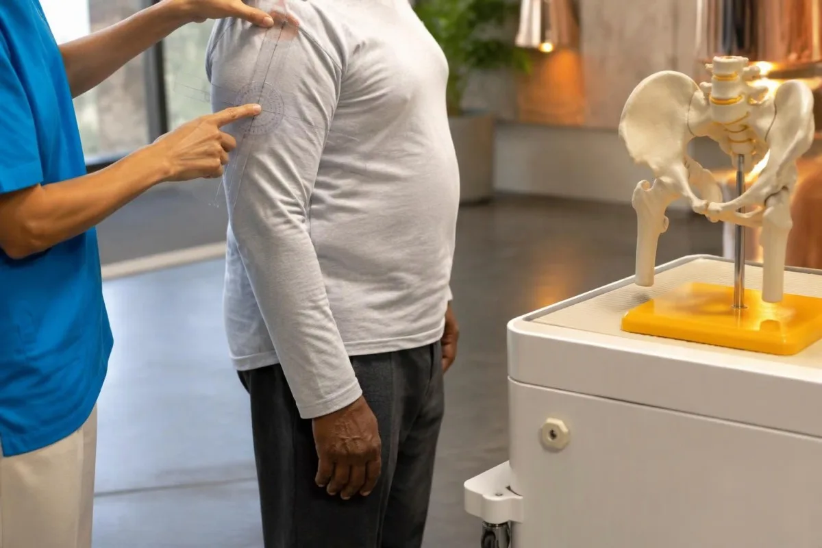
The 2012 criteria were developed to classify patients consistently for research, not to rule out every unusual clinical presentation (Dasgupta et al., 2012). Someone with normal ESR and CRP does not meet that framework's entry requirements, but this does not logically prove that PMR is impossible; it means the standard research classification cannot be applied in the usual way.
The same framework assigns weight to morning stiffness lasting more than 45 minutes, hip symptoms, absence of rheumatoid-factor or anti-CCP antibodies, and absence of other joint pain. Clinicians interpret those features rather than asking patients to diagnose themselves with a score, because early elderly-onset rheumatoid arthritis may resemble PMR before persistent hand or wrist swelling becomes obvious.
A pretreatment baseline is especially useful when the diagnosis is uncertain, provided urgent assessment is not delayed. Bring the symptom timeline and any previous ESR or CRP results, including a result obtained 2 weeks before steroids started; our የመነሻ ምርመራ ዝግጅት explains how medication dates and the reason for testing make that first report more interpretable.
Which conditions can mimic PMR with normal blood tests?
Mechanical shoulder disorders, osteoarthritis, thyroid disease, medication-related muscle symptoms, and other rheumatic conditions can resemble PMR. Normal ESR and CRP make a careful review of these alternatives particularly valuable, especially when symptoms are one-sided, morning stiffness is brief, or the person is younger than 50.
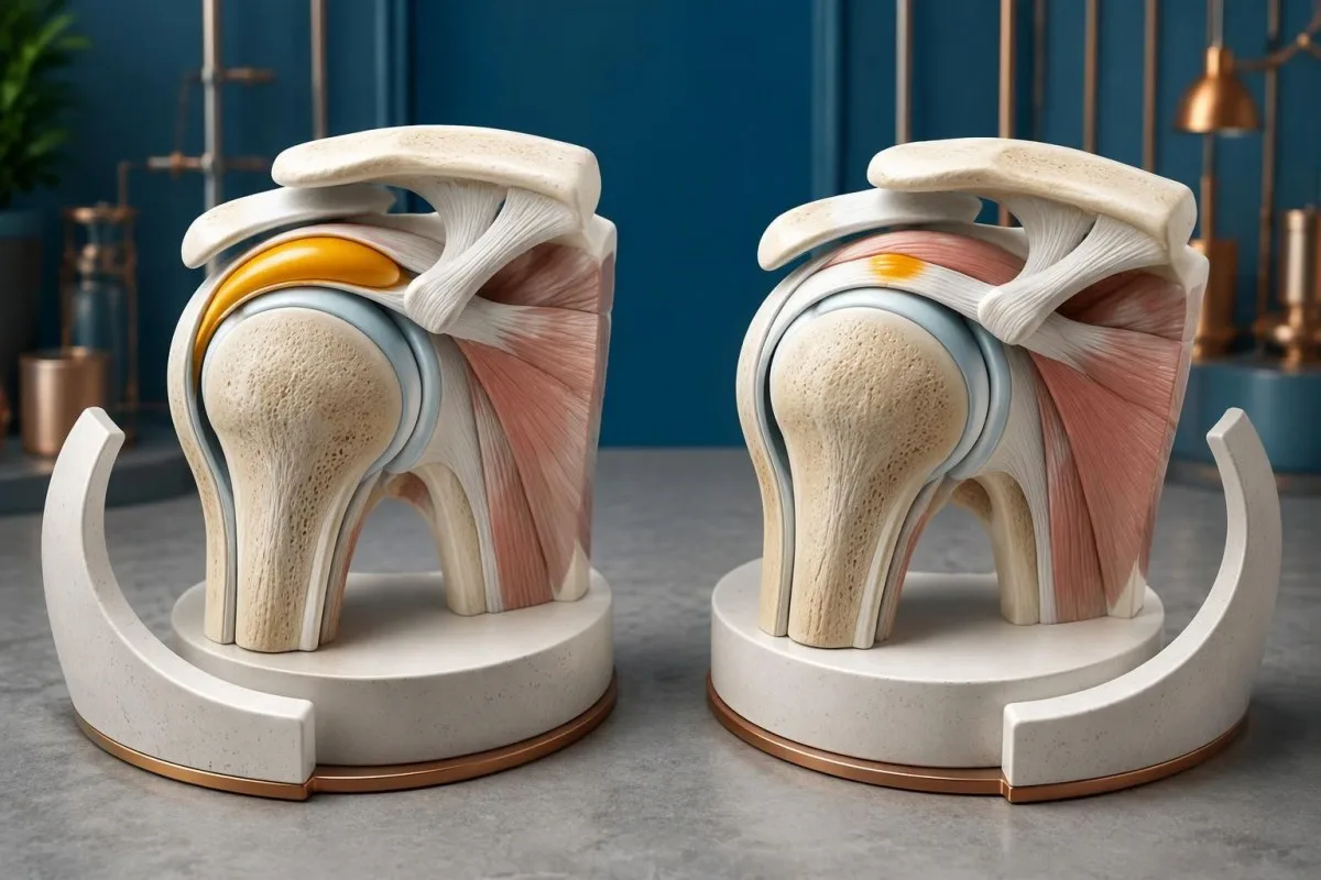
Rotator cuff disease and adhesive capsulitis are assessed through movement patterns, including whether passive movement is restricted and whether a specific arc provokes discomfort. A hypothetical 61-year-old with one stiff shoulder and 15 minutes of morning symptoms is less typical for PMR than someone with bilateral shoulder and hip restriction; coexistence is possible, so finding a tendon problem does not automatically explain everything.
Thyroid dysfunction can contribute to muscle aching, fatigue, and reduced exercise tolerance, but a mildly abnormal TSH does not prove that the thyroid explains the entire symptom picture. Clinicians interpret TSH with free T4 and the examination rather than anchoring on a single flag; our subclinical hypothyroidism guide explains why modest biochemical changes may or may not be clinically relevant.
Statin-associated muscle symptoms can occur with normal creatine kinase, making the medication timeline useful even when several tests look reassuring. Symptoms beginning 3 weeks after a dose increase deserve discussion rather than an unsupervised medication stop; our statin muscle-risk guide explains one susceptibility factor, but genetic testing is not a routine way to distinguish PMR from statin symptoms.
What other blood tests help exclude PMR mimics?
Additional tests look for alternative diagnoses and establish treatment safety; they do not create a confirmatory PMR panel. Depending on the presentation, clinicians may review a full blood count, kidney and liver function, calcium, glucose, thyroid tests, creatine kinase, and rheumatoid-factor or anti-CCP antibodies.

Creatine kinase is usually not elevated by uncomplicated PMR itself. True weakness with a hypothetical CK of 1,200 U/L, interpreted against the laboratory's interval and recent exercise, shifts attention toward muscle injury or myopathy; selected cases may also involve aldolase testing, discussed in our muscle enzyme follow-up guide, rather than assuming that every proximal symptom represents joint-girdle inflammation.
Anemia or unusual plasma protein results can help explain why ESR and CRP disagree, and they may identify a problem that needs its own evaluation. For example, hemoglobin of 95 g/L with a disproportionately high ESR should not simply be filed under PMR; our የሴረም ፕሮቲን ትርጓሜ መመሪያ explains protein patterns without implying that every abnormal globulin result warrants the same investigation.
ካንቴስቲ እ.ኤ.አ. የAI ባዮማርከር ትርጓሜ መድረክ that can help organize a report containing ESR, CRP, CK, and thyroid results, but those 4 measurements answer different clinical questions. We encourage targeted interpretation: ANA or complement testing may be appropriate when specific systemic features suggest connective-tissue disease, whereas ordering broad antibody panels solely because PMR markers are normal can produce distracting incidental positives.
Can ultrasound help when ESR and CRP are normal?
Ultrasound can support suspected PMR by showing shoulder or hip-region bursitis, tendon-sheath changes, or joint inflammation. Bilateral subacromial-subdeltoid bursitis is a useful pattern, but imaging findings are not specific enough to confirm PMR independently, particularly in adults over 50 with common degenerative changes.
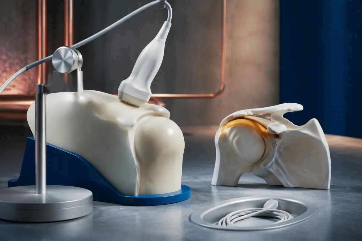
The 2012 EULAR/ACR study incorporated ultrasound as an optional classification aid, while recognizing overlap with other shoulder conditions (Dasgupta et al., 2012). Finding bursal changes on both sides can strengthen a coherent clinical picture, but a scan obtained after 2 weeks of treatment may not show the same degree of change as a pretreatment examination.
Ultrasound is most useful when it answers a specific question: is the main restriction related to bursitis, a tendon disorder, joint changes, or several processes together? A hypothetical 73-year-old with CRP of 3 mg/L and bilateral bursitis still needs assessment for rheumatoid arthritis and mechanical disease; the scan does not convert a nonspecific symptom pattern into PMR simply because a bursa contains fluid.
Negative rheumatoid factor does not rule out rheumatoid arthritis, and positive rheumatoid factor does not automatically exclude PMR. Persistent swelling of 2 or more small joints changes the differential more meaningfully than a low-positive antibody alone; our የሩማቶይድ ፋክተር ውጤት መተርጎሚያ መመሪያ explains why antibody concentration, examination, and imaging must be interpreted together.
Does a rapid response to prednisone prove PMR?
A rapid response to glucocorticoids supports PMR but does not prove the diagnosis, because other inflammatory conditions also improve. The 2015 EULAR/ACR recommendations describe an initial prednisone-equivalent range of 12.5–25 mg daily for clinician-managed PMR, adjusted to disease activity and treatment risks.
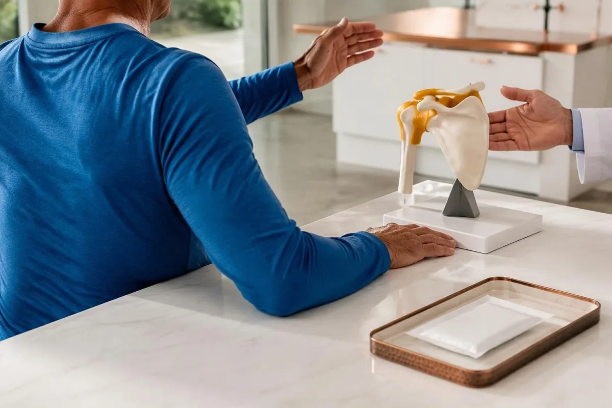
Prednisone should not be used as a self-directed diagnostic experiment. Starting 15 mg daily before evaluation may lower inflammatory markers and obscure an alternative illness, while exposing someone without PMR to adverse effects; the EULAR/ACR recommendations emphasize individualized dosing and reassessment rather than treating response as a binary diagnostic challenge (Dejaco et al., 2015).
Improvement should include the actual features that prompted concern, not just a vague increase in energy or better sleep. In a hypothetical case, morning stiffness falling from 90 minutes to 20 minutes and easier dressing provides useful follow-up information, but persistent one-sided shoulder restriction may indicate a coexisting tendon disorder; incomplete response should trigger clinical review rather than automatic dose escalation.
Glucocorticoid safety depends on pre-existing risks and other medicines, including diabetes, osteoporosis, glaucoma, and infection vulnerability. A glucose rise from 6 to 12 mmol/L after treatment needs a management plan even if shoulder symptoms improve; our older-adult medication effects guide explains why a changing laboratory result may reflect treatment rather than worsening of the original condition.
Which symptoms need urgent assessment for giant cell arteritis?
New headache, scalp tenderness, jaw pain when chewing, or visual disturbance in someone with suspected PMR require urgent assessment for giant cell arteritis. Sudden visual loss, double vision, or a temporary curtain over vision needs immediate emergency evaluation—even if CRP is below 5 mg/L.

Normal ESR and CRP cannot safely exclude giant cell arteritis when the symptoms are concerning. Clinicians may start treatment before laboratory confirmation or vascular imaging because delay can threaten vision; a hypothetical 76-year-old with jaw fatigue during meals and a new temporal headache should not wait 1 week for a routine repeat blood test.
Jaw claudication means discomfort or fatigue triggered by chewing and relieved by resting, rather than simply constant dental pain. A new symptom appearing after 2 minutes of chewing, especially with headache or scalp tenderness, deserves same-day medical assessment; describe the trigger and relief pattern clearly, because “jaw pain” alone can conceal the feature that makes clinicians concerned.
Sudden speech difficulty, facial asymmetry, or weakness on one side is an emergency, whether PMR has been diagnosed or not. Follow the emergency actions in our stroke warning-sign guide rather than waiting for an inflammatory marker result; fever with severe illness or a new hot, swollen single joint also warrants urgent assessment for causes other than uncomplicated PMR.
How is PMR monitored if inflammatory markers stay normal?
When ESR and CRP are uninformative, PMR monitoring relies more heavily on symptoms, function, examination, and treatment safety. Track morning stiffness in minutes and repeatable tasks such as dressing or chair rising; a CRP of 2 mg/L cannot independently establish remission or exclude relapse.
Glucocorticoids can normalize previously elevated markers, so a normal result after treatment is different from a normal result before any treatment. Some IL-6 receptor-blocking medicines also suppress CRP directly, making a value below 5 mg/L less useful for judging inflammatory activity; the medication name and timing belong beside the laboratory value during review.
A practical diary can record 3 repeatable observations: morning stiffness duration, ease of raising both arms, and ability to get up from the same chair. If stiffness changes from 15 to 60 minutes after a taper, contact the treating clinician, but do not assume relapse automatically; osteoarthritis, deconditioning, tendon disease, and glucocorticoid-related complications can also change how those tasks feel.
With Kantesti AI, laboratory trends can be considered alongside treatment dates rather than comparing 2 isolated “normal” labels. Our የመድኃኒት ደህንነት አዝማሚያ መመሪያ explains why glucose, kidney function, and other safety measurements may remain useful even when ESR and CRP provide little information; dose changes still require the prescribing clinician's judgment.
What should you bring to a PMR assessment?
Bring the original laboratory report, a symptom timeline, and a complete medication list to a PMR assessment. Include whether both shoulders and hips are affected, how many minutes morning stiffness lasts, and whether ESR or CRP was measured before or after the first glucocorticoid dose.
Exact dates are more useful than “recently.” Write down whether symptoms started 6 weeks ago, whether a statin dose changed 3 weeks ago, and whether the laboratory sample was collected 4 days after prednisone began; also list fever, weight change, persistent swollen joints, and any new headache or chewing-related jaw symptoms because these details can redirect the assessment.
Keep the original units and reference intervals visible when sharing a report, since ESR of 25 mm/hour may be flagged differently across laboratories. Before sending a PDF or photo to any digital service, review our report-sharing privacy steps; retaining clinically relevant dates and removing unnecessary identifiers are different tasks, and neither should alter the actual test values.
Kantesti is operated by Kantesti LTD, UK Company No. 17090423; information about ድርጅታችን helps distinguish the service provider from the clinician responsible for diagnosis. Our የAI ቴክኖሎጂ መመሪያ explains the report interpretation workflow, but even a clearly explained set of 2 inflammatory markers cannot replace a physical examination.
What can AI interpretation help with—and what can it not diagnose?
AI interpretation can explain laboratory units, reference intervals, and trends, but it cannot independently diagnose PMR or exclude giant cell arteritis. ካንቴስቲ እ.ኤ.አ. AI የደም ምርመራ ውጤት ትርጓሜ መድረክ that supports understanding of ESR and CRP reports without turning a CRP of 3 mg/L into a diagnostic verdict.
The safest output separates observation from conclusion. “CRP is 4 mg/L and within this laboratory's interval” is a report observation, while “PMR is excluded” is an unsupported clinical conclusion; similarly, a raised ESR should generate questions about symptoms and confounders rather than automatically label an inflammatory disease.
Kantesti AI cannot test passive shoulder movement, establish pain-independent muscle strength, or verify chewing-related jaw claudication from a laboratory PDF. Those limitations matter even when 10 other biomarkers appear reassuring; a concerning visual symptom requires urgent care directly, not completion of an upload or waiting for an interpretation result.
A technical benchmark is not the same as a PMR diagnostic accuracy study. የእኛ የማረጋገጫና የክሊኒካዊ ቁጥጥር information should be read with that distinction in mind: explaining a CRP result correctly and diagnosing a 70-year-old's shoulder stiffness are different tasks, and performance on one cannot be assumed to establish performance on the other.
Research publications and evidence behind PMR interpretation
The most relevant PMR evidence addresses clinical classification, treatment, and the limitations of normal inflammatory markers. The 2012 EULAR/ACR classification study, 2015 EULAR/ACR management recommendations, and 2018 review of normal ESR and CRP each answer a different question; none establishes a single confirmatory blood test.
Evidence about normal-marker PMR is less certain than evidence about the usual presentation. The 7–22% normal-ESR range summarized by Manzo and Milchert (2018) comes from heterogeneous reports, while estimates for both markers being normal depend strongly on patient selection; these studies support keeping PMR possible, not bypassing the search for a better explanation.
The 2 DOI-linked background publications below cover serum proteins and autoimmune laboratory interpretation, not PMR diagnostic validation or treatment effectiveness. Our complement እና ANA መመሪያ is relevant when a clinician suspects a different systemic autoimmune condition, but normal-marker shoulder stiffness alone does not justify assuming that ANA or C3/C4 will confirm PMR.
Kantesti's background guides should be distinguished from the external peer-reviewed PMR references listed separately, including the 2015 treatment recommendations. Our የሕክምና ይዘት መስፈርቶች describe the editorial framework; the practical decision remains clinical: assess the shoulder and hip pattern, review plausible mimics, and treat new visual or jaw symptoms as urgent rather than waiting for a laboratory flag.
ተደጋግመው የሚጠየቁ ጥያቄዎች
መደበኛ ESR እና CRP እያሉ ፖሊማያልጂያ ሪህማቲካ ሊኖር ይችላል?
0 Այո, ռևմատիկ պոլիմիալգիան երբեմն կարող է հանդիպել ESR-ի և CRP-ի նորմալ մակարդակների դեպքում, թեև մի շարք հրապարակված կոհորտներում բուժումից առաջ երկուսի նորմալ լինելն էլ հազվադեպ է։ 2018 թվականի ակնարկում ամփոփված դեպքերի մոտավորապես 7–22%-ի դեպքում արձանագրվել է միայն ESR-ի նորմալ մակարդակ, սովորաբար՝ CRP-ի բարձր մակարդակի հետ մեկտեղ։ Բժիշկները գնահատում են ուսերի երկկողմանի կարկամությունը, ազդրերի ախտանշանները, 50 և ավելի տարիքը, զննման արդյունքները և այլընտրանքային ախտորոշումները՝ փոխանակ PMR-ը բացառելու միայն լաբորատոր պատասխանի հիման վրա։ Մասնագետի գնահատումը հատկապես օգտակար է, երբ ախտանշանների պատկերը համոզիչ է, բայց երկու ցուցանիշներն էլ մնում են նորմալ։.
የደም ምርመራ ፖሊማይያልጂያ ሪዩማቲካን ያረጋግጣል?
የፖሊሚያልጂያ ሩማቲካ በአንድ የደም ምርመራ ብቻ አይረጋገጥም። ESR እና CRP ስለ እብጠት ምላሽ የተለየ ያልሆነ ማስረጃ ይሰጣሉ፤ CK፣ የታይሮይድ ተግባር፣ የሩማቶይድ ፋክተር እና ፀረ-CCP ያሉ ምርመራዎች ግን አስፈላጊ ሲሆን ሌሎች አማራጭ ምክንያቶችን ለመመርመር ይረዳሉ። ከ5 mg/L በታች የሆነ CRP በብዙ ላቦራቶሪዎች መደበኛ ክልል ውስጥ ነው፤ ሆኖም ያ እሴትም ሆነ ከፍ ያለ ESR ምርመራውን አያረጋግጡም። PMR የሚመረመረው በክሊኒካዊ ግምገማና ይበልጥ አሳማኝ የሆኑ ምክንያቶችን በማስወገድ ነው።.
የትኛው የESR ወይም CRP መጠን ፖሊሚያልጂያ ሪዩማቲካን ያመለክታል?
በራሱ የፖሊማያልጂያ ሪውማቲካን በአስተማማኝ ሁኔታ የሚለይ የESR ወይም CRP ወሰን የለም። በአንዳንድ የቆዩ የምርመራ ዘዴዎች የ40 mm/hour ESR አስፈላጊነት ተጠቅሷል፣ ነገር ግን ለክሊኒካዊ ምርመራ ሁሉን አቀፍ መስፈርት አይደለም። ብዙ ላቦራቶሪዎች የCRP ከፍተኛ ገደብ 5 mg/L ይጠቀማሉ፣ ሌሎች ደግሞ 10 mg/L ይጠቀማሉ። ከእነዚህ ገደቦች በላይ ያሉ እሴቶች የእብጠት ሂደትን ይደግፋሉ፣ ነገር ግን PMRን ከኢንፌክሽን ወይም ከሌሎች የእብጠት ሁኔታዎች አይለዩም።.
0. የደም ምርመራዎች መደበኛ ሲሆኑ ፖሊማያልጂያ ሪዩማቲካ እንዴት ይመረመራል?
የደም ምርመራዎች መደበኛ ሲሆኑ፣ ክሊኒኮች የምልክቶቹን አይነት ይገመግማሉ፣ የትከሻና የዳሌ እንቅስቃሴን ይመረምራሉ፣ እንዲሁም ፖሊማያልጂያ ሪዩማቲካን ሊመስሉ የሚችሉ ሁኔታዎችን ይመረምራሉ። ዕድሜ 50 ወይም ከዚያ በላይ መሆን፣ በሁለቱም ትከሻዎች ላይ ምልክቶች መኖር፣ እና ከ45 ደቂቃ በላይ የሚቆይ የጠዋት መገጣጠሚያ መድንዘዝ የተለመደውን አቀራረብ ይደግፋሉ። አልትራሳውንድ ድጋፍ ሰጪ የቡርሳ እብጠትን ወይም በጅማት ሽፋን ላይ ያሉ ለውጦችን ሊለይ ይችላል፣ ነገር ግን እነዚያ ግኝቶች ብቻቸውን ምርመራውን አያረጋግጡም። ሌላ የሩማቲክ ወይም የሜካኒካዊ ሁኔታ በጊዜ ሂደት ይበልጥ ግልጽ ሊሆን ስለሚችል ብዙውን ጊዜ ክትትል ያስፈልጋል።.
በፕሬድኒሶን ሲወስዱ መሻሻል ማሳየቴ PMR እንዳለብኝ በእርግጥ ያረጋግጣል?
በፕሬድኒሶን መሻሻል ፖሊሚያልጂያ ሩማቲካ መኖሩን በእርግጠኝነት አያረጋግጥም፤ ምክንያቱም ሌሎች የእብጠት ሁኔታዎችም ለግሉኮኮርቲኮይዶች ምላሽ ሊሰጡ ይችላሉ። የ2015 የEULAR/ACR ምክረ ሐሳቦች በሐኪም ክትትል የሚጀመር፣ እንደ ምልክቶችና የሕክምና አደጋዎች በግለሰብ የሚወሰን፣ በየቀኑ 12.5–25 mg የፕሬድኒሶን ተመጣጣኝ መጠን ይገልጻሉ። ምላሹ መገምገም ያለበት በጠንካራነትና በተግባራዊ ችሎታ ላይ በተወሰኑ ለውጦች መሠረት ነው፤ የኃይል መሻሻልን ብቻ በመመልከት አይደለም። በራስዎ ውሳኔ እንደ የምርመራ ሙከራ ፕሬድኒሶን አይጀምሩ ወይም መጠኑን አይቀይሩ።.
ግዙፍ ሴል አርቴራይተስ መደበኛ የእብጠት መለኪያዎች ሲኖሩ ሊከሰት ይችላል?
0] አዎ፣ የእብጠት መደበኛ መለኪያዎች ምልክቶቹ አሳሳቢ ሲሆኑ ግዙፍ ሴል አርተራይተስን በደህና ሊያስወግዱ አይችሉም። ዕድሜው 50 ወይም ከዚያ በላይ በሆነ ሰው ላይ አዲስ ራስ ምታት፣ የራስ ቆዳ ርኅራኄ ወይም በማኘክ የሚነሳ የመንጋጋ ምቾት ማጣት አስቸኳይ ግምገማ ይጠይቃሉ። ድንገተኛ የእይታ መጥፋት፣ ሁለት ማየት ወይም ጊዜያዊ የእይታ መጥፋት CRP ከ5 mg/L በታች ቢሆንም ወዲያውኑ የድንገተኛ ሕክምና ግምገማ ይፈልጋል። ሕክምናን ማዘግየት እይታን ሊያሰጋ ስለሚችል ሐኪሞች ምርመራው ከመረጋገጡ በፊት ሕክምና ሊጀምሩ ይችላሉ።.
የደም ምርመራ ውጤት እንዴት ማንበብ
የላቦራቶሪ ሪፖርትዎን ይስቀሉ፤ የKantesti AI እያንዳንዱን ባዮማርከር በቀላል ቋንቋ በጥቂት ሰከንዶች ውስጥ ያብራራል። በዓለም ዙሪያ ከ2 ሚሊዮን በላይ ሰዎች ይጠቀሙበታል።.
ማጣቀሻዎች
- ክላይን፣ ቲ.፣ ሚቸል፣ ኤስ.፣ እና ዌበር፣ ኤች. (2026)።. የሴረም ፕሮቲኖች መመሪያ፦ ግሎቡሊን፣ አልቡሚን እና የA/G ሬሾ የደም ምርመራ።. የKantesti AI የሕክምና ምርምር።. የዶአይ
- ክላይን፣ ቲ.፣ ሚቸል፣ ኤስ.፣ እና ዌበር፣ ኤች. (2026)።. C3 C4 የኮምፕለመንት የደም ምርመራ እና የANA ቲተር መመሪያ።. የKantesti AI የሕክምና ምርምር።. የዶአይ
- Dasgupta B et al. (2012). 2012 provisional classification criteria for polymyalgia rheumatica: a European League Against Rheumatism/American College of Rheumatology collaborative initiative. አናልስ ኦፍ ዘ ሩማቲክ ዲዚዝስ።. የዶአይ
- Dejaco C et al. (2015). 2015 recommendations for the management of polymyalgia rheumatica: a European League Against Rheumatism/American College of Rheumatology collaborative initiative. አናልስ ኦፍ ዘ ሩማቲክ ዲዚዝስ።. የዶአይ
- Manzo C, Milchert M. (2018). Polymyalgia rheumatica with normal values of both erythrocyte sedimentation rate and C-reactive protein concentration at the time of diagnosis: a four-point guidance. Reumatologia.
- 2ሚ+የተተነተኑ ምርመራዎች
- 127+አገሮች
- 100+ቋንቋዎች
ስለ የሕክምና ቡድኑ


የE-E-A-T እምነት ምልክቶች
ልምድ
በሐኪም መሪነት የላቦራቶሪ ትርጓሜ የስራ ፍሰቶች ክሊኒካዊ ግምገማ።.
ባለሙያነት
በክሊኒካዊ አውድ ውስጥ ባዮማርከሮች እንዴት እንደሚሰሩ ላይ የላቦራቶሪ ሕክምና ትኩረት።.
ስልጣን ያለው
በዶ/ር ቶማስ ክላይን በሕክምና የተገመገመ፣ ከዶ/ር ሳራ ሚቼል እና ከፕሮፌሰር ዶ/ር ሃንስ ዌበር አስተዋጽኦ ጋር።.
አስተማማኝነት
ለማስጠንቀቂያ ምላሽ መቀነስ ግልጽ የቀጣይ መንገዶች ያለው በማስረጃ የተደገፈ ትርጓሜ።.


የሕክምና ማስተባበያ
ይህ ጽሑፍ ለትምህርታዊ ዓላማ ብቻ ነው እና የሕክምና ምክር አይደለም። ለምርመራ እና ለሕክምና ውሳኔዎች ሁልጊዜ ብቁ የጤና ባለሙያን ያማክሩ።.
ታትሟል · የአርትዖት ቡድኑን ያነጋግሩ
በKantesti Ltd የታተመ፣ በእንግሊዝና ዌልስ የተመዘገበ፣ የኩባንያ ቁጥር. 17090423, ፣ ለንደን፣ ዩናይትድ ኪንግደም።.
