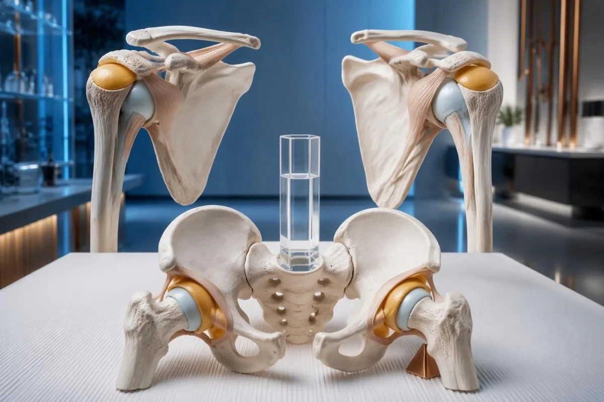
បាទ/ចាស—លទ្ធផលតេស្តឈាមអាចធម្មតាក្នុងជំងឺ polymyalgia rheumatica ទោះបីជា ESR និង CRP ធម្មតាទាំងពីររួមគ្នា គឺមិនសូវកើតមានមុនពេលព្យាបាលក៏ដោយ។ ការធ្វើរោគវិនិច្ឆ័យអាស្រ័យលើលំនាំរោគសញ្ញា ការពិនិត្យ និងការដកចេញនូវមូលហេតុផ្សេងៗ—មិនមែនសញ្ញាព្រមានមួយនៅក្នុងមន្ទីរពិសោធន៍ទេ។.

ចំណុចសំខាន់ៗដែលត្រូវចងចាំ
- លទ្ធផលធម្មតា មិនអាចដកចេញទាំងស្រុងនូវជំងឺ polymyalgia rheumatica បានទេ; ESR និង CRP ធម្មតាទាំងពីររួមគ្នា ធ្វើឱ្យការធ្វើរោគវិនិច្ឆ័យមុនព្យាបាលមិនសូវមានលក្ខណៈធម្មតា និងបង្ហាញថាគួរពិនិត្យមើលជម្រើសផ្សេងទៀត។.
- អាយុ 50 ឆ្នាំឡើងទៅ គឺជាតម្រុយសំខាន់សម្រាប់ការធ្វើរោគវិនិច្ឆ័យ៖ ជំងឺ polymyalgia rheumatica កម្រណាស់ចំពោះអ្នកអាយុក្រោម 50 ឆ្នាំ។.
- ការក្រិនរឹងនៅពេលព្រឹកដែលមានរយៈពេលលើសពី 45 នាទី ជួយគាំទ្រការធ្វើរោគវិនិច្ឆ័យ នៅពេលប៉ះពាល់ស្មាទាំងពីរ ជាពិសេសនៅពេលមានរោគសញ្ញានៅតំបន់ត្រគាកផងដែរ។.
- CRP ក្រោម 5 mg/L គឺស្ថិតក្នុងកម្រិតធម្មតានៅមន្ទីរពិសោធន៍ជាច្រើន ប៉ុន្តែមន្ទីរពិសោធន៍ខ្លះប្រើកម្រិតខាងលើ 10 mg/L; កម្រិតណាមួយក៏មិនមែនជាកម្រិតសម្រាប់ធ្វើរោគវិនិច្ឆ័យ PMR ដែរ។.
- ភាពមិនស្របគ្នារវាង ESR និង CRP ត្រូវការការបកស្រាយតាមបរិបទ៖ ភាពស្លេកស្លាំង និងប្រូតេអ៊ីនក្នុងប្លាស្មាអាចធ្វើឱ្យ ESR កើនឡើង ដោយមិនមានការកើនឡើងនៃ CRP ដែលស្របគ្នានោះទេ។.
- អេកូសាស្ត្រស្មា អាចបង្ហាញពីរលាកថង់សន្លាក់ ឬការប្រែប្រួលនៃស្រទាប់ស្រោមសរសៃពួរ ប៉ុន្តែការរកឃើញទាំងនេះគ្រាន់តែជួយគាំទ្រប៉ុណ្ណោះ មិនអាចបញ្ជាក់ PMR ដោយឯករាជ្យបានទេ។.
- Prednisone 12.5–25 mg ក្នុងមួយថ្ងៃ គឺជាកម្រិតដំបូងក្នុងអនុសាសន៍ EULAR/ACR ឆ្នាំ 2015 សម្រាប់ការគ្រប់គ្រង PMR ដោយគ្រូពេទ្យ; ការឆ្លើយតបតែឯងមិនអាចបញ្ជាក់រោគវិនិច្ឆ័យបានទេ។.
- ការឈឺក្បាលថ្មី ការឈឺថ្គាមពេលទំពារ ឬរោគសញ្ញាផ្នែកចក្ខុវិញ្ញាណ តម្រូវឱ្យមានការវាយតម្លៃជាបន្ទាន់រកជំងឺរលាកសរសៃឈាមកោសិកាយក្ស—ទោះបីសញ្ញាសម្គាល់ការរលាកមានកម្រិតធម្មតាក៏ដោយ។.
តើតេស្តឈាមរកជំងឺ polymyalgia rheumatica អាចមានលទ្ធផលធម្មតាដែរឬទេ?
បាទ/ចាស៖ ESR អាចមានកម្រិតធម្មតាក្នុងជំងឺ polymyalgia rheumatica ហើយជួនកាលទាំង ESR និង CRP សុទ្ធតែមានកម្រិតធម្មតាមុនពេលព្យាបាល។. មិនមានការធ្វើតេស្តឈាមតែមួយណាសម្រាប់ជំងឺ polymyalgia rheumatica ដែលអាចបញ្ជាក់ ឬបដិសេធជំងឺនេះបានទេ; គ្រូពេទ្យបូកបញ្ចូលរោគសញ្ញា ការពិនិត្យ និងភស្តុតាងប្រឆាំងនឹងរោគវិនិច្ឆ័យផ្សេងៗ ដែលជាទូទៅធ្វើលើមនុស្សពេញវ័យអាយុ 50 ឆ្នាំឡើងទៅ។.
សញ្ញាសម្គាល់ការរលាកដែលមានកម្រិតធម្មតា ធ្វើឱ្យទំនុកចិត្តលើរោគវិនិច្ឆ័យថយចុះ មិនមែនមានន័យថាបញ្ចប់ការវាយតម្លៃនោះទេ។. បុគ្គលអាយុ 72 ឆ្នាំម្នាក់ដែលទើបមានការរឹងសន្លាក់ស្មាទាំងពីរ និងត្រគាកទាំងពីររយៈពេល 90 នាទីរៀងរាល់ព្រឹក គួរទទួលការវាយតម្លៃខុសពីអ្នកដែលឈឺស្មាម្ខាងក្រោយពីលើករបស់ធ្ងន់; អ្នកទាំងពីរអាចមាន CRP 3 mg/L ប៉ុន្តែលំនាំរោគសញ្ញារបស់ពួកគេមានទម្ងន់រោគវិនិច្ឆ័យខុសគ្នាខ្លាំង។.
ការកើនឡើងនៃ ESR ឬ CRP ក៏មិនអាចកំណត់អត្តសញ្ញាណ PMR ដោយខ្លួនឯងបានដែរ ព្រោះតេស្តទាំង 2 នេះវាស់ការឆ្លើយតបមិនជាក់លាក់ ជាជាងអង់ទីករជាក់លាក់ចំពោះជំងឺ។ ការឆ្លងមេរោគ ជំងឺរលាកសន្លាក់រ៉ាំរ៉ៃ និងមហារីកមួយចំនួនអាចបង្កលទ្ធផលមន្ទីរពិសោធន៍ស្រដៀងគ្នា ខណៈបញ្ហាមេកានិចនៃស្មាអាចកើតរួមជាមួយមូលហេតុដាច់ដោយឡែកដែលធ្វើឱ្យសញ្ញាសម្គាល់កើនឡើង; មគ្គុទ្ទេសក៍របស់យើងអំពី ការធ្វើតេស្តការឈឺចាប់ដែលមិនអាចពន្យល់បាន ពន្យល់ពីមូលហេតុដែលសំណួរគ្លីនិកត្រូវមកមុន។.
កាន់តេស្ទី គឺជា ឧបករណ៍វិភាគឈាម AI ដែលអាចជួយពន្យល់របាយការណ៍ ESR និង CRP រួមទាំងមូលហេតុដែល CRP 4 mg/L មិនអាចសម្រេចរោគវិនិច្ឆ័យ PMR បាន។ តួនាទីរបស់យើងគឺធ្វើឱ្យព័ត៌មានមន្ទីរពិសោធន៍ងាយយល់—មិនមែនជំនួសការវាយតម្លៃចលនាស្មា មុខងារត្រគាក ឬរោគសញ្ញាសរសៃឈាមបន្ទាន់ដែលរបាយការណ៍មន្ទីរពិសោធន៍មិនអាចបង្ហាញបាននោះទេ។.
តើរោគសញ្ញានៅស្មា និងត្រគាកបែបណាដែលធ្វើឱ្យសង្ស័យថាជា PMR កាន់តែខ្លាំង?
ជាទូទៅ PMR បង្កការឈឺថ្មី និងការរឹងយូរនៅស្មាទាំងពីរ ដែលជាញឹកញាប់មានការរឹងនៅតំបន់ត្រគាករួមផងដែរ។. ការរឹងពេលព្រឹកដែលបន្តលើសពី 45 នាទី គាំទ្រលំនាំរោគសញ្ញានេះ ជាពិសេសនៅពេលការស្លៀកពាក់ ការលើកដៃ ឬការក្រោកពីកៅអីក្លាយជាការពិបាកដោយមិននឹកស្មានដល់។.

ចលនាដែលត្រូវបានកម្រិតដោយការឈឺ មិនដូចគ្នានឹងភាពខ្សោយសាច់ដុំពិតប្រាកដទេ។. បុគ្គលអាយុ 68 ឆ្នាំម្នាក់ជាឧទាហរណ៍ អាចពិបាកលើកអាវយឺតពីលើក្បាលដោយសារស្មាឈឺ ប៉ុន្តែនៅតែមានកម្លាំងសាច់ដុំល្អពេលពិនិត្យក្នុងទីតាំងដែលស្រួល; ភាពខ្សោយដែលអាចបង្ហាញឱ្យឃើញ ទោះបីបានសម្របតាមការឈឺក៏ដោយ ធ្វើឱ្យជំងឺសាច់ដុំ សរសៃប្រសាទ ឬប្រព័ន្ធអង់ដូគ្រីនមានភាពពាក់ព័ន្ធជាង PMR ដែលមិនមានភាពស្មុគស្មាញ។.
រោគសញ្ញាតំបន់ត្រគាកអាចមានអារម្មណ៍នៅជុំវិញគូទ ភ្លៅខាងលើ ឬផ្នែកចំហៀងនៃត្រគាក ជាជាងនៅខាងក្នុងក្រលៀនដោយផ្ទាល់ ហើយគ្រូពេទ្យសួរអំពីការរឹងក្រោយពេលអង្គុយ ក៏ដូចជាក្រោយពេលគេងផងដែរ។ ការកត់ត្រាថាការរឹតត្បិតនៅពេលព្រឹកមានរយៈពេល 10 នាទី ឬ 70 នាទី មានប្រយោជន៍ជាងការសរសេរត្រឹមតែថា “ឈឺចុកចាប់គ្រប់កន្លែង”; រយៈពេលគួរពិពណ៌នាបទពិសោធន៍ជាក់ស្តែងរបស់អ្នក មិនមែនជាគោលដៅដែលអ្នកព្យាយាមសម្រេចនោះទេ។.
ការស្ពឹករមួលនៅម្រាមដៃជាក់លាក់ គឺជាតម្រុយរោគវិនិច្ឆ័យដាច់ដោយឡែក, ទោះបីមានការរឹងស្មាក៏ដោយ។ ការស្ពឹកដែលប៉ះពាល់ដល់មេដៃ ម្រាមចង្អុល និងម្រាមកណ្តាល អាចបង្ហាញពីការសង្កត់លើសរសៃប្រសាទមេឌៀន ដែលបានរៀបរាប់ក្នុង រោគសញ្ញាផ្លូវរូងកាប៉ាល់កដៃ; របស់យើង; គ្រូពេទ្យគួរវាយតម្លៃរោគសញ្ញាទាំងនេះ ជាជាងសន្មតថារាល់រោគសញ្ញាថ្មីទាំងអស់សុទ្ធតែជាផ្នែកនៃរោគវិនិច្ឆ័យរលាកតែមួយ។.
តើ ESR និង CRP វាស់អ្វីខ្លះក្នុង PMR?
ESR និង CRP ជួយគាំទ្រការវាយតម្លៃ PMR ដោយវាស់ទិដ្ឋភាពផ្សេងៗគ្នានៃការឆ្លើយតបនៃការរលាក មិនមែនវាស់ PMR ផ្ទាល់ទេ។. មន្ទីរពិសោធន៍ជាច្រើនប្រើ CRP ក្រោម 5 mg/L ជាកម្រិតយោង ខណៈចន្លោះយោងរបស់ ESR អាស្រ័យលើវិធីសាស្ត្រ អាយុ ភេទ និងមន្ទីរពិសោធន៍ក្នុងតំបន់។.
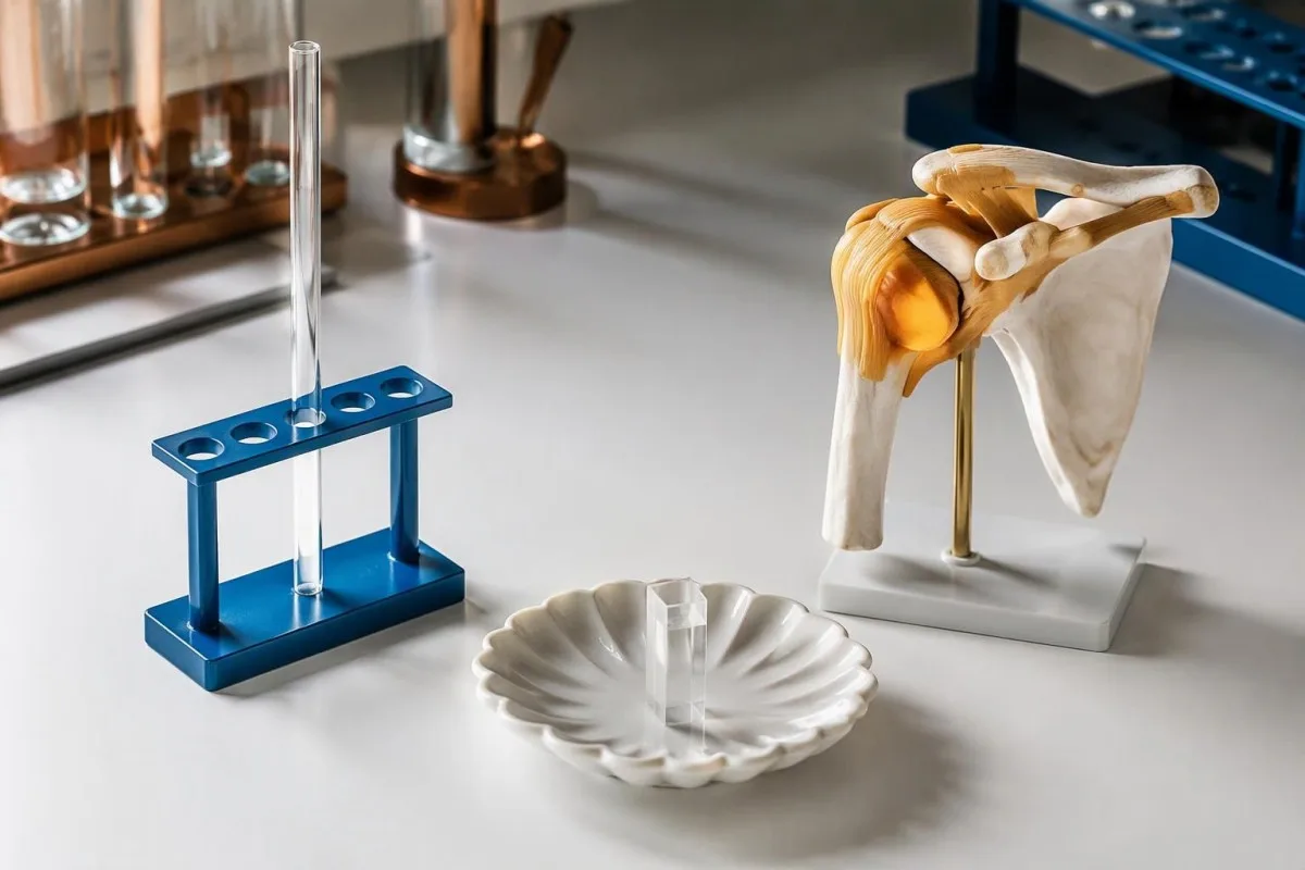
ESR measures how far cellular elements settle in a testing column over 1 hour, usually reported in mm/hour. Fibrinogen, immunoglobulins, anemia, and cellular characteristics affect that settling, so an ESR of 48 mm/hour with CRP of 2 mg/L is not automatically evidence of active PMR; it may prompt review of the full blood count and protein results.
CRP is a liver-produced acute-phase protein that generally changes faster than ESR when the inflammatory stimulus changes. Check the units before comparing reports: 1 mg/dL ស្មើនឹង 10 mg/L, so CRP 0.6 mg/dL and 6 mg/L represent the same concentration; the distinction between cellular counts and acute-phase proteins is explored in WBC និង CRP មិនស៊ីគ្នា.
An ESR of 40 mm/hour is not a universal diagnostic boundary for PMR. Older diagnostic approaches often emphasized that value, but contemporary assessment considers the whole presentation and the laboratory's reference interval; conversely, ESR of 90 mm/hour does not establish PMR and should not distract from fever, focal symptoms, or unexplained abnormalities elsewhere in the report.
| កម្រិត | ចន្លោះ | អត្ថន័យ |
|---|---|---|
| CRP within a common reference interval | <5 mg/L at many laboratories | Does not exclude PMR; some laboratories use an upper limit of 10 mg/L. |
| Illustrative adult ESR intervals | 0–20 or 0–30 mm/hour | These are examples, not universal limits; use the age- and method-appropriate interval on the report. |
| CRP above the local upper limit | >5 or >10 mg/L, depending on the laboratory | Supports an inflammatory process but does not distinguish PMR from infection or other inflammatory disorders. |
| ESR above the local upper limit | Above the report's stated interval, in mm/hour | Interpret with CRP, hemoglobin, plasma proteins, symptoms, and treatment timing; no ESR value alone confirms PMR. |
តើ ESR ធម្មតា និង CRP ធម្មតា កើតមានញឹកញាប់ប៉ុណ្ណាក្នុង PMR?
Normal ESR is reported more often than normal ESR and CRP together in untreated PMR. A 2018 review described normal ESR in approximately 7–22% of reported cases, but CRP was usually elevated in those patients; these figures are not an individual's diagnostic probability.
Both markers being normal was uncommon in several older PMR cohorts, with proportions around 1–2% in some series discussed by Manzo and Milchert in Reumatologia (2018). Selected specialist cohorts have reported higher proportions, so a single universal percentage would be misleading; referral patterns, diagnostic definitions, treatment exposure, and how thoroughly mimics were excluded all influence the denominator.
The phrase “polymyalgia rheumatica normal ESR” can hide 2 different situations: ESR is normal but CRP is raised, or both tests are within range. A hypothetical ESR of 18 mm/hour with CRP of 24 mg/L still provides biochemical evidence of an inflammatory response, whereas ESR of 18 with CRP of 2 mg/L requires more reliance on examination and alternative explanations.
Cardiovascular hs-CRP categories are not PMR diagnostic categories, even though hs-CRP measures the same protein with greater low-level sensitivity. A value of 3 mg/L may matter in a cardiovascular risk discussion without confirming musculoskeletal inflammation; our hs-CRP context guide separates those uses, preventing a heart-risk label from being mistaken for a rheumatology diagnosis.
តើគេធ្វើរោគវិនិច្ឆ័យ polymyalgia rheumatica តាមរោគសញ្ញា និងការពិនិត្យគ្លីនិកយ៉ាងដូចម្តេច?
Polymyalgia rheumatica is diagnosed clinically after assessing the characteristic symptom pattern and excluding more plausible causes. The 2012 EULAR/ACR classification framework starts with age 50 or older, bilateral shoulder aching, and abnormal ESR or CRP, but classification criteria are not a stand-alone diagnostic test.
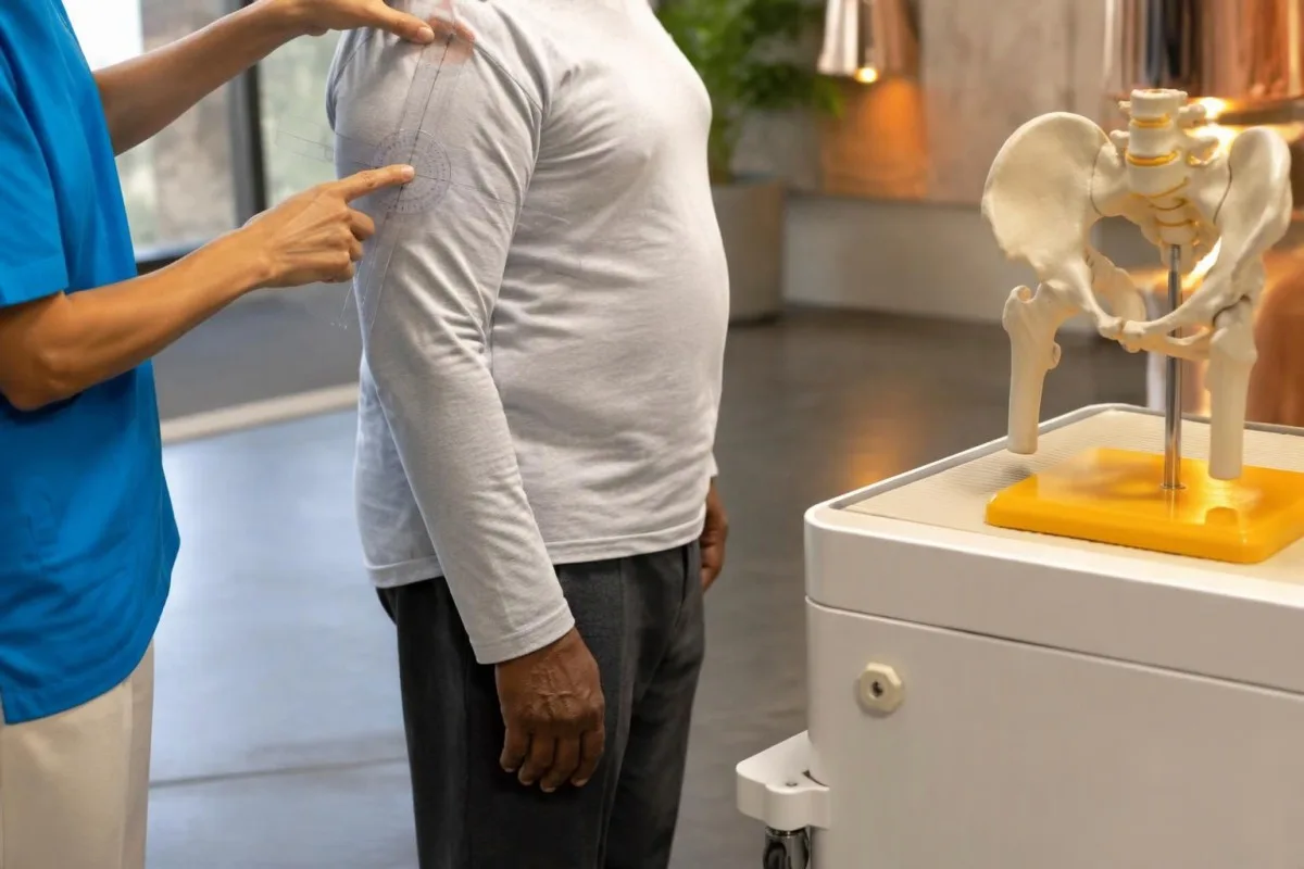
The 2012 criteria were developed to classify patients consistently for research, not to rule out every unusual clinical presentation (Dasgupta et al., 2012). Someone with normal ESR and CRP does not meet that framework's entry requirements, but this does not logically prove that PMR is impossible; it means the standard research classification cannot be applied in the usual way.
The same framework assigns weight to morning stiffness lasting more than 45 minutes, hip symptoms, absence of rheumatoid-factor or anti-CCP antibodies, and absence of other joint pain. Clinicians interpret those features rather than asking patients to diagnose themselves with a score, because early elderly-onset rheumatoid arthritis may resemble PMR before persistent hand or wrist swelling becomes obvious.
A pretreatment baseline is especially useful when the diagnosis is uncertain, provided urgent assessment is not delayed. Bring the symptom timeline and any previous ESR or CRP results, including a result obtained 2 weeks before steroids started; our ការត្រៀមខ្លួនសម្រាប់ការធ្វើតេស្តតម្លៃគោល explains how medication dates and the reason for testing make that first report more interpretable.
តើជំងឺណាខ្លះអាចមានរោគសញ្ញាស្រដៀង PMR ទោះបីលទ្ធផលតេស្តឈាមធម្មតាក៏ដោយ?
Mechanical shoulder disorders, osteoarthritis, thyroid disease, medication-related muscle symptoms, and other rheumatic conditions can resemble PMR. Normal ESR and CRP make a careful review of these alternatives particularly valuable, especially when symptoms are one-sided, morning stiffness is brief, or the person is younger than 50.
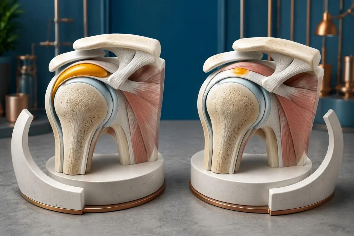
Rotator cuff disease and adhesive capsulitis are assessed through movement patterns, including whether passive movement is restricted and whether a specific arc provokes discomfort. A hypothetical 61-year-old with one stiff shoulder and 15 minutes of morning symptoms is less typical for PMR than someone with bilateral shoulder and hip restriction; coexistence is possible, so finding a tendon problem does not automatically explain everything.
Thyroid dysfunction can contribute to muscle aching, fatigue, and reduced exercise tolerance, but a mildly abnormal TSH does not prove that the thyroid explains the entire symptom picture. Clinicians interpret TSH with free T4 and the examination rather than anchoring on a single flag; our subclinical hypothyroidism guide explains why modest biochemical changes may or may not be clinically relevant.
Statin-associated muscle symptoms can occur with normal creatine kinase, making the medication timeline useful even when several tests look reassuring. Symptoms beginning 3 weeks after a dose increase deserve discussion rather than an unsupervised medication stop; our statin muscle-risk guide explains one susceptibility factor, but genetic testing is not a routine way to distinguish PMR from statin symptoms.
តើតេស្តឈាមអ្វីខ្លះទៀតជួយដកចេញនូវជំងឺដែលមានរោគសញ្ញាស្រដៀង PMR?
Additional tests look for alternative diagnoses and establish treatment safety; they do not create a confirmatory PMR panel. Depending on the presentation, clinicians may review a full blood count, kidney and liver function, calcium, glucose, thyroid tests, creatine kinase, and rheumatoid-factor or anti-CCP antibodies.
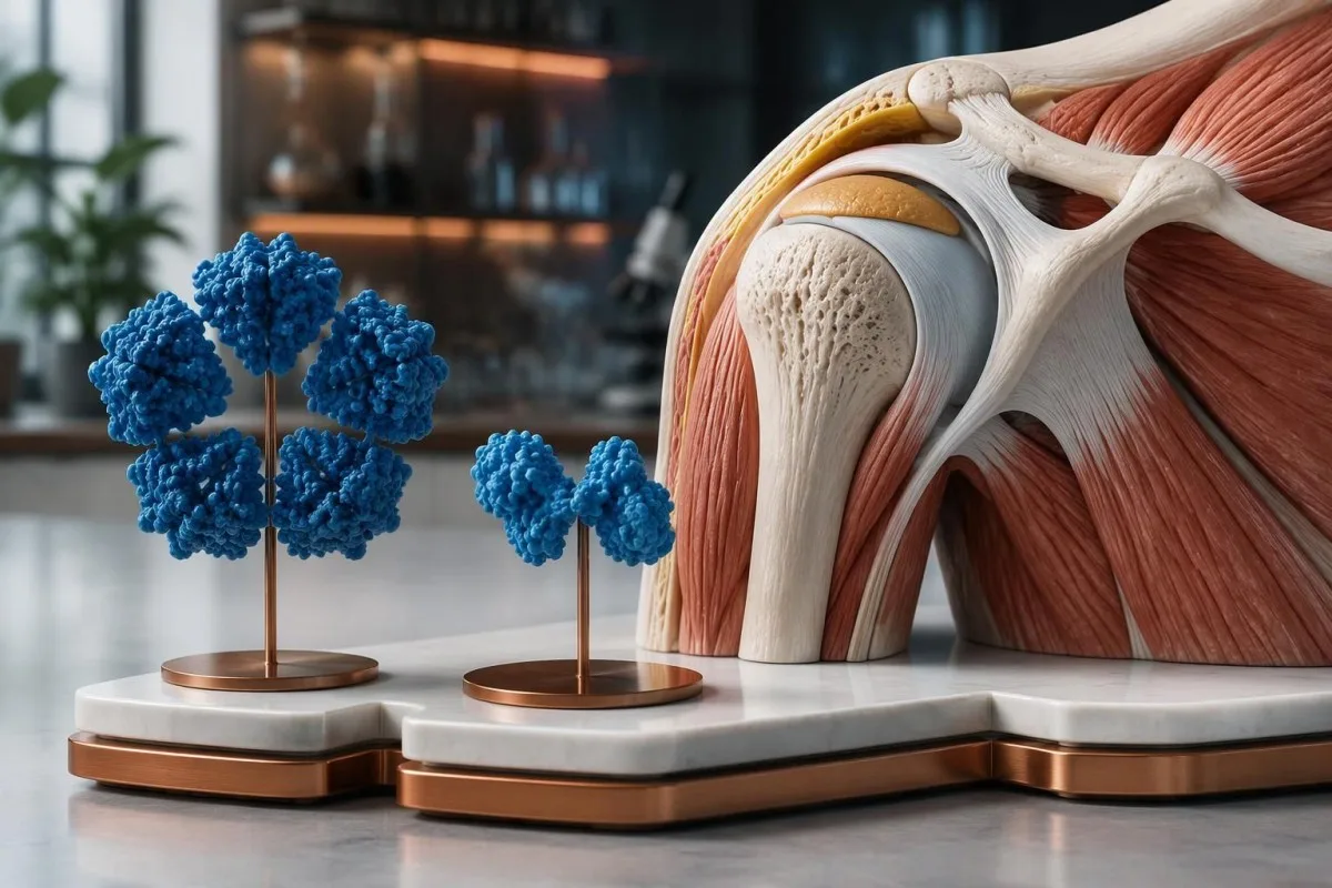
Creatine kinase is usually not elevated by uncomplicated PMR itself. True weakness with a hypothetical CK of 1,200 U/L, interpreted against the laboratory's interval and recent exercise, shifts attention toward muscle injury or myopathy; selected cases may also involve aldolase testing, discussed in our muscle enzyme follow-up guide, rather than assuming that every proximal symptom represents joint-girdle inflammation.
Anemia or unusual plasma protein results can help explain why ESR and CRP disagree, and they may identify a problem that needs its own evaluation. For example, hemoglobin of 95 g/L with a disproportionately high ESR should not simply be filed under PMR; our មគ្គុទ្ទេសក៍បកស្រាយប្រូតេអ៊ីនក្នុងឈាម explains protein patterns without implying that every abnormal globulin result warrants the same investigation.
កាន់តេស្ទី គឺជា វេទិកាបកស្រាយសូចនាករជីវសាស្ត្រដោយ AI that can help organize a report containing ESR, CRP, CK, and thyroid results, but those 4 measurements answer different clinical questions. We encourage targeted interpretation: ANA or complement testing may be appropriate when specific systemic features suggest connective-tissue disease, whereas ordering broad antibody panels solely because PMR markers are normal can produce distracting incidental positives.
តើអេកូសាស្ត្រអាចជួយបានដែរឬទេ នៅពេល ESR និង CRP ធម្មតា?
Ultrasound can support suspected PMR by showing shoulder or hip-region bursitis, tendon-sheath changes, or joint inflammation. Bilateral subacromial-subdeltoid bursitis is a useful pattern, but imaging findings are not specific enough to confirm PMR independently, particularly in adults over 50 with common degenerative changes.
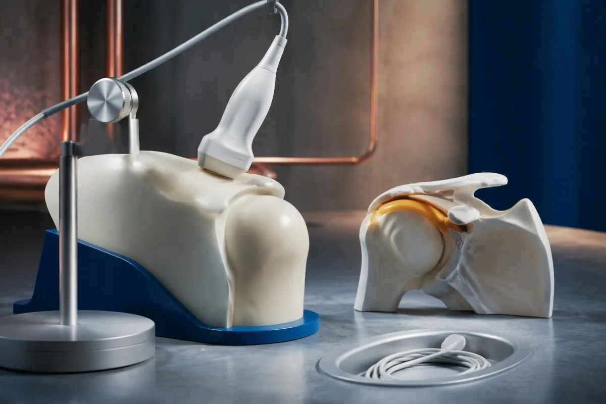
The 2012 EULAR/ACR study incorporated ultrasound as an optional classification aid, while recognizing overlap with other shoulder conditions (Dasgupta et al., 2012). Finding bursal changes on both sides can strengthen a coherent clinical picture, but a scan obtained after 2 weeks of treatment may not show the same degree of change as a pretreatment examination.
Ultrasound is most useful when it answers a specific question: is the main restriction related to bursitis, a tendon disorder, joint changes, or several processes together? A hypothetical 73-year-old with CRP of 3 mg/L and bilateral bursitis still needs assessment for rheumatoid arthritis and mechanical disease; the scan does not convert a nonspecific symptom pattern into PMR simply because a bursa contains fluid.
Negative rheumatoid factor does not rule out rheumatoid arthritis, and positive rheumatoid factor does not automatically exclude PMR. Persistent swelling of 2 or more small joints changes the differential more meaningfully than a low-positive antibody alone; our មគ្គុទ្ទេសក៍បកស្រាយ rheumatoid factor explains why antibody concentration, examination, and imaging must be interpreted together.
តើការឆ្លើយតបរហ័សចំពោះ prednisone បញ្ជាក់ថាជា PMR ដែរឬទេ?
A rapid response to glucocorticoids supports PMR but does not prove the diagnosis, because other inflammatory conditions also improve. The 2015 EULAR/ACR recommendations describe an initial prednisone-equivalent range of 12.5–25 mg daily for clinician-managed PMR, adjusted to disease activity and treatment risks.
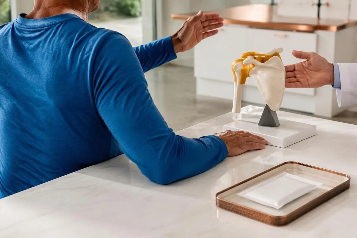
Prednisone should not be used as a self-directed diagnostic experiment. Starting 15 mg daily before evaluation may lower inflammatory markers and obscure an alternative illness, while exposing someone without PMR to adverse effects; the EULAR/ACR recommendations emphasize individualized dosing and reassessment rather than treating response as a binary diagnostic challenge (Dejaco et al., 2015).
Improvement should include the actual features that prompted concern, not just a vague increase in energy or better sleep. In a hypothetical case, morning stiffness falling from 90 minutes to 20 minutes and easier dressing provides useful follow-up information, but persistent one-sided shoulder restriction may indicate a coexisting tendon disorder; incomplete response should trigger clinical review rather than automatic dose escalation.
Glucocorticoid safety depends on pre-existing risks and other medicines, including diabetes, osteoporosis, glaucoma, and infection vulnerability. A glucose rise from 6 to 12 mmol/L after treatment needs a management plan even if shoulder symptoms improve; our older-adult medication effects guide explains why a changing laboratory result may reflect treatment rather than worsening of the original condition.
តើរោគសញ្ញាអ្វីខ្លះត្រូវការការវាយតម្លៃជាបន្ទាន់ចំពោះជំងឺរលាកសរសៃឈាមកោសិកាយក្ស?
New headache, scalp tenderness, jaw pain when chewing, or visual disturbance in someone with suspected PMR require urgent assessment for giant cell arteritis. Sudden visual loss, double vision, or a temporary curtain over vision needs immediate emergency evaluation—even if CRP is below 5 mg/L.

Normal ESR and CRP cannot safely exclude giant cell arteritis when the symptoms are concerning. Clinicians may start treatment before laboratory confirmation or vascular imaging because delay can threaten vision; a hypothetical 76-year-old with jaw fatigue during meals and a new temporal headache should not wait 1 week for a routine repeat blood test.
Jaw claudication means discomfort or fatigue triggered by chewing and relieved by resting, rather than simply constant dental pain. A new symptom appearing after 2 minutes of chewing, especially with headache or scalp tenderness, deserves same-day medical assessment; describe the trigger and relief pattern clearly, because “jaw pain” alone can conceal the feature that makes clinicians concerned.
Sudden speech difficulty, facial asymmetry, or weakness on one side is an emergency, whether PMR has been diagnosed or not. Follow the emergency actions in our stroke warning-sign guide rather than waiting for an inflammatory marker result; fever with severe illness or a new hot, swollen single joint also warrants urgent assessment for causes other than uncomplicated PMR.
តើតាមដាន PMR យ៉ាងដូចម្តេច ប្រសិនបើសញ្ញារលាកនៅតែធម្មតា?
When ESR and CRP are uninformative, PMR monitoring relies more heavily on symptoms, function, examination, and treatment safety. Track morning stiffness in minutes and repeatable tasks such as dressing or chair rising; a CRP of 2 mg/L cannot independently establish remission or exclude relapse.
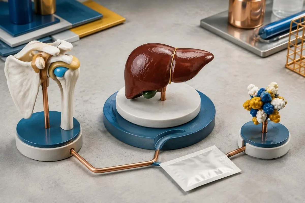
Glucocorticoids can normalize previously elevated markers, so a normal result after treatment is different from a normal result before any treatment. Some IL-6 receptor-blocking medicines also suppress CRP directly, making a value below 5 mg/L less useful for judging inflammatory activity; the medication name and timing belong beside the laboratory value during review.
A practical diary can record 3 repeatable observations: morning stiffness duration, ease of raising both arms, and ability to get up from the same chair. If stiffness changes from 15 to 60 minutes after a taper, contact the treating clinician, but do not assume relapse automatically; osteoarthritis, deconditioning, tendon disease, and glucocorticoid-related complications can also change how those tasks feel.
With Kantesti AI, laboratory trends can be considered alongside treatment dates rather than comparing 2 isolated “normal” labels. Our ការណែនាំអំពីនិន្នាការសុវត្ថិភាពថ្នាំ explains why glucose, kidney function, and other safety measurements may remain useful even when ESR and CRP provide little information; dose changes still require the prescribing clinician's judgment.
តើអ្នកគួរយកអ្វីទៅជាមួយសម្រាប់ការវាយតម្លៃ PMR?
Bring the original laboratory report, a symptom timeline, and a complete medication list to a PMR assessment. Include whether both shoulders and hips are affected, how many minutes morning stiffness lasts, and whether ESR or CRP was measured before or after the first glucocorticoid dose.
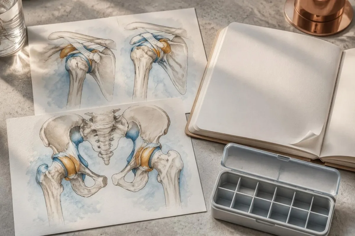
Exact dates are more useful than “recently.” Write down whether symptoms started 6 weeks ago, whether a statin dose changed 3 weeks ago, and whether the laboratory sample was collected 4 days after prednisone began; also list fever, weight change, persistent swollen joints, and any new headache or chewing-related jaw symptoms because these details can redirect the assessment.
Keep the original units and reference intervals visible when sharing a report, since ESR of 25 mm/hour may be flagged differently across laboratories. Before sending a PDF or photo to any digital service, review our report-sharing privacy steps; retaining clinically relevant dates and removing unnecessary identifiers are different tasks, and neither should alter the actual test values.
Kantesti is operated by Kantesti LTD, UK Company No. 17090423; information about អង្គការរបស់យើង helps distinguish the service provider from the clinician responsible for diagnosis. Our មគ្គុទេសក៍បច្ចេកវិទ្យា AI explains the report interpretation workflow, but even a clearly explained set of 2 inflammatory markers cannot replace a physical examination.
តើការបកស្រាយដោយ AI អាចជួយអ្វីខ្លះ—ហើយវាមិនអាចធ្វើរោគវិនិច្ឆ័យអ្វីបានខ្លះ?
AI interpretation can explain laboratory units, reference intervals, and trends, but it cannot independently diagnose PMR or exclude giant cell arteritis. កាន់តេស្ទី គឺជា វេទិកាបកស្រាយការធ្វើតេស្តឈាម AI that supports understanding of ESR and CRP reports without turning a CRP of 3 mg/L into a diagnostic verdict.
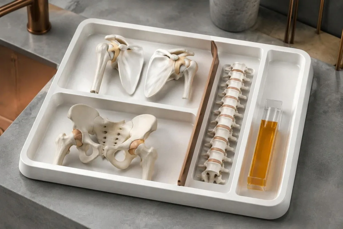
The safest output separates observation from conclusion. “CRP is 4 mg/L and within this laboratory's interval” is a report observation, while “PMR is excluded” is an unsupported clinical conclusion; similarly, a raised ESR should generate questions about symptoms and confounders rather than automatically label an inflammatory disease.
Kantesti AI cannot test passive shoulder movement, establish pain-independent muscle strength, or verify chewing-related jaw claudication from a laboratory PDF. Those limitations matter even when 10 other biomarkers appear reassuring; a concerning visual symptom requires urgent care directly, not completion of an upload or waiting for an interpretation result.
A technical benchmark is not the same as a PMR diagnostic accuracy study. របស់យើង។ ព័ត៌មានអំពីការផ្ទៀងផ្ទាត់សុពលភាព និងការត្រួតពិនិត្យផ្នែកគ្លីនិក information should be read with that distinction in mind: explaining a CRP result correctly and diagnosing a 70-year-old's shoulder stiffness are different tasks, and performance on one cannot be assumed to establish performance on the other.
ការបោះពុម្ពផ្សាយស្រាវជ្រាវ និងភស្តុតាងដែលនៅពីក្រោយការបកស្រាយ PMR
The most relevant PMR evidence addresses clinical classification, treatment, and the limitations of normal inflammatory markers. The 2012 EULAR/ACR classification study, 2015 EULAR/ACR management recommendations, and 2018 review of normal ESR and CRP each answer a different question; none establishes a single confirmatory blood test.
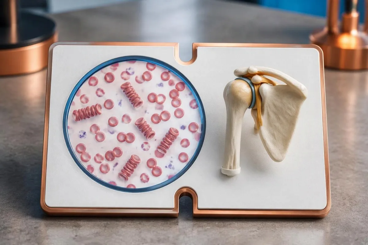
Evidence about normal-marker PMR is less certain than evidence about the usual presentation. The 7–22% normal-ESR range summarized by Manzo and Milchert (2018) comes from heterogeneous reports, while estimates for both markers being normal depend strongly on patient selection; these studies support keeping PMR possible, not bypassing the search for a better explanation.
The 2 DOI-linked background publications below cover serum proteins and autoimmune laboratory interpretation, not PMR diagnostic validation or treatment effectiveness. Our មគ្គុទេសក៍ complement និង ANA is relevant when a clinician suspects a different systemic autoimmune condition, but normal-marker shoulder stiffness alone does not justify assuming that ANA or C3/C4 will confirm PMR.
Kantesti's background guides should be distinguished from the external peer-reviewed PMR references listed separately, including the 2015 treatment recommendations. Our ស្តង់ដារខ្លឹមសារវេជ្ជសាស្ត្រ describe the editorial framework; the practical decision remains clinical: assess the shoulder and hip pattern, review plausible mimics, and treat new visual or jaw symptoms as urgent rather than waiting for a laboratory flag.
សំណួរដែលគេច្រើនសួរ
តើអ្នកអាចមានជំងឺរលាកសាច់ដុំ និងសន្លាក់ប្រភេទ polymyalgia rheumatica ដោយមាន ESR និង CRP ធម្មតាដែរឬទេ?
បាទ/ចាស ជំងឺរលាកសន្លាក់រ៉ាំរ៉ៃដែលបណ្តាលឱ្យឈឺសាច់ដុំ (polymyalgia rheumatica) អាចកើតមានម្តងម្កាលដោយ ESR និង CRP មានតម្លៃធម្មតា ទោះបីជាការដែលទាំងពីរមានតម្លៃធម្មតាមុនពេលព្យាបាល គឺមិនសូវកើតមានក្នុងក្រុមអ្នកជំងឺជាច្រើនដែលបានចុះផ្សាយក៏ដោយ។ ការសិក្សាបានរាយការណ៍ថា ESR មានតម្លៃធម្មតាតែឯងមាននៅប្រហែល 7–22% នៃករណីដែលបានសង្ខេបក្នុងការពិនិត្យឡើងវិញឆ្នាំ 2018 ហើយជាទូទៅ CRP កើនឡើង។ គ្រូពេទ្យវាយតម្លៃភាពរឹងស្មាទាំងសងខាង រោគសញ្ញាត្រគាក អាយុ 50 ឆ្នាំឡើងទៅ លទ្ធផលពិនិត្យរាងកាយ និងរោគវិនិច្ឆ័យជំនួសផ្សេងៗ ជាជាងដក PMR ចេញដោយផ្អែកតែលើរបាយការណ៍មន្ទីរពិសោធន៍។ ការពិនិត្យដោយអ្នកឯកទេសមានប្រយោជន៍ជាពិសេស នៅពេលលំនាំរោគសញ្ញាគួរឱ្យជឿជាក់ ប៉ុន្តែសញ្ញាសម្គាល់ទាំងពីរនៅតែមានតម្លៃធម្មតា។.
តើការធ្វើតេស្តឈាមមួយណាដែលបញ្ជាក់ពីជំងឺរលាកសន្លាក់និងសាច់ដុំដោយសារប្រព័ន្ធភាពស៊ាំ (polymyalgia rheumatica)?
មិនមានតេស្តឈាមតែមួយណាដែលអាចបញ្ជាក់ជំងឺរលាកសន្លាក់រ៉ាំរ៉ៃពហុសាច់ដុំបានទេ។ ESR និង CRP ផ្តល់ភស្តុតាងមិនជាក់លាក់អំពីប្រតិកម្មរលាក ខណៈតេស្តដូចជា CK មុខងារក្រពេញទីរ៉ូអ៊ីត កត្តារលាកសន្លាក់រ៉ាំរ៉ៃ និង anti-CCP ជួយស៊ើបអង្កេតមូលហេតុផ្សេងៗ នៅពេលសមស្រប។ CRP ក្រោម 5 mg/L ស្ថិតក្នុងកម្រិតធម្មតានៅមន្ទីរពិសោធន៍ជាច្រើន ប៉ុន្តែទាំងតម្លៃនោះ និង ESR កើនឡើង មិនអាចបញ្ជាក់ការធ្វើរោគវិនិច្ឆ័យបានទេ។ PMR ត្រូវបានធ្វើរោគវិនិច្ឆ័យតាមរយៈការវាយតម្លៃគ្លីនិក និងការបដិសេធមូលហេតុផ្សេងទៀតដែលអាចសមហេតុផលជាងនេះ។.
តើកម្រិត ESR ឬ CRP ប៉ុន្មានបង្ហាញពីជំងឺរលាកសន្លាក់រ៉ូម៉ាទិកពហុសាច់ដុំ?
0 មិនមានកម្រិត ESR ឬ CRP ណាមួយដែលអាចកំណត់អត្តសញ្ញាណជំងឺរលាកសន្លាក់រ៉ាំរ៉ៃប្រភេទ polymyalgia rheumatica ដោយខ្លួនឯងបានគួរឱ្យទុកចិត្តនោះទេ។ កម្រិត ESR 40 mm/hour ត្រូវបានលើកឡើងជាចំណុចសំខាន់ក្នុងវិធីសាស្ត្រធ្វើរោគវិនិច្ឆ័យចាស់ៗមួយចំនួន ប៉ុន្តែវាមិនមែនជាលក្ខខណ្ឌដែលតម្រូវជាសកលសម្រាប់ការធ្វើរោគវិនិច្ឆ័យតាមគ្លីនិកទេ។ មន្ទីរពិសោធន៍ជាច្រើនប្រើកម្រិតខាងលើធម្មតារបស់ CRP 5 mg/L ខណៈមន្ទីរពិសោធន៍ផ្សេងទៀតប្រើ 10 mg/L។ តម្លៃលើសពីកម្រិតទាំងនោះបង្ហាញគាំទ្រដល់ដំណើរការរលាក ប៉ុន្តែមិនអាចបែងចែក PMR ពីការឆ្លងមេរោគ ឬស្ថានភាពរលាកផ្សេងទៀតបានទេ។.
តើគេធ្វើរោគវិនិច្ឆ័យជំងឺរលាកសន្លាក់រ៉ាំរ៉ៃពហុសាច់ដុំ (polymyalgia rheumatica) យ៉ាងដូចម្តេច នៅពេលលទ្ធផលតេស្តឈាមធម្មតា?
នៅពេលលទ្ធផលតេស្តឈាមធម្មតា គ្រូពេទ្យវាយតម្លៃលំនាំនៃរោគសញ្ញា ពិនិត្យចលនាស្មា និងត្រគាក ហើយស៊ើបរកជំងឺដែលអាចមានរោគសញ្ញាស្រដៀងនឹងជំងឺរលាកសន្លាក់រ៉ូម៉ាទូអ៊ីតពហុសាច់ដុំ។ អាយុ ៥០ ឆ្នាំឡើងទៅ រោគសញ្ញានៅស្មាទាំងសងខាង និងការឡើងរឹងនៅពេលព្រឹកដែលមានរយៈពេលលើសពី ៤៥ នាទី គាំទ្រការបង្ហាញរោគសញ្ញាបែបធម្មតា។ អ៊ុលត្រាសោនអាចរកឃើញរលាកថង់សន្លាក់ ឬការប្រែប្រួលនៃស្រោមសរសៃពួរដែលគាំទ្រការធ្វើរោគវិនិច្ឆ័យ ប៉ុន្តែការរកឃើញទាំងនោះមិនអាចបញ្ជាក់រោគវិនិច្ឆ័យដោយខ្លួនឯងបានទេ។ ជារឿយៗត្រូវតាមដានបន្ត ព្រោះជំងឺរលាកសន្លាក់រ៉ូម៉ាទិច ឬជំងឺមេកានិចផ្សេងទៀតអាចកាន់តែច្បាស់ឡើងតាមពេលវេលា។.
តើការធូរស្រាលដោយប្រើថ្នាំ prednisone មានន័យថាខ្ញុំប្រាកដជាមាន PMR មែនទេ?
ការធូរស្រាលក្រោយប្រើ prednisone មិនអាចបញ្ជាក់ជាក់លាក់ថាជាជំងឺរលាកសន្លាក់រ៉ាំរ៉ៃ polymyalgia rheumatica បានទេ ព្រោះជំងឺរលាកផ្សេងទៀតអាចឆ្លើយតបនឹងថ្នាំ glucocorticoids។ អនុសាសន៍របស់ EULAR/ACR ឆ្នាំ 2015 ពិពណ៌នាអំពីកម្រិត prednisone-equivalent ដំបូងចន្លោះពី 12.5–25 mg ក្នុងមួយថ្ងៃ ដែលគ្រប់គ្រងដោយគ្រូពេទ្យ និងកំណត់តាមរោគសញ្ញា និងហានិភ័យនៃការព្យាបាល។ គួរវាយតម្លៃការឆ្លើយតបតាមរយៈការប្រែប្រួលជាក់លាក់នៃការរឹង និងមុខងារ មិនមែនគ្រាន់តែការមានកម្លាំងប្រសើរឡើងនោះទេ។ កុំចាប់ផ្តើម ឬកែប្រែការប្រើ prednisone ដោយខ្លួនឯង ដើម្បីធ្វើតេស្តវិនិច្ឆ័យ។.
តើជំងឺរលាកសរសៃឈាមកោសិកាយក្សអាចកើតមានដោយមានសូចនាកររលាកធម្មតាដែរឬទេ?
បាទ/ចាស សញ្ញាសម្គាល់ការរលាកដែលធម្មតា មិនអាចធានាបានថាបដិសេធជំងឺរលាកសរសៃឈាមកោសិកាយក្សបានទេ នៅពេលរោគសញ្ញាគួរឱ្យបារម្ភ។ ចំពោះអ្នកដែលមានអាយុ 50 ឆ្នាំ ឬច្រើនជាងនេះ ការឈឺក្បាលថ្មី ភាពឈឺចាប់នៅស្បែកក្បាល ឬភាពមិនស្រួលនៅថ្គាមដែលកើតឡើងពេលទំពារ តម្រូវឱ្យមានការវាយតម្លៃជាបន្ទាន់។ ការបាត់បង់ការមើលឃើញភ្លាមៗ ការមើលឃើញរូបភាពពីរជាន់ ឬការបាត់បង់ការមើលឃើញបណ្ដោះអាសន្ន តម្រូវឱ្យមានការវាយតម្លៃបន្ទាន់នៅមន្ទីរពេទ្យភ្លាមៗ ទោះបីជា CRP ទាបជាង 5 mg/L ក៏ដោយ។ គ្រូពេទ្យអាចចាប់ផ្តើមការព្យាបាលមុនពេលមានការបញ្ជាក់ពីលទ្ធផលតេស្ត ព្រោះការពន្យារពេលថែទាំអាចគំរាមកំហែងដល់ការមើលឃើញ។.
ទទួលបានការពន្យល់អំពីលទ្ធផលពិនិត្យឈាមរបស់អ្នក
បង្ហោះរបាយការណ៍មន្ទីរពិសោធន៍របស់អ្នក ហើយ AI របស់ Kantesti នឹងពន្យល់ជីវសញ្ញានីមួយៗជាភាសាសាមញ្ញ ក្នុងរយៈពេលត្រឹមតែប៉ុន្មានវិនាទី។ មនុស្សជាង 2 លាននាក់នៅទូទាំងពិភពលោកបានប្រើប្រាស់។.
ឯកសារយោង
- Klein, T., Mitchell, S., & Weber, H. (2026)។. មគ្គុទ្ទេសក៍អំពីប្រូតេអ៊ីនក្នុងសេរ៉ូម៖ ការធ្វើតេស្តឈាម Globulins, Albumin និងសមាមាត្រ A/G។. ការស្រាវជ្រាវវេជ្ជសាស្ត្រ AI របស់ Kantesti។. ក្រសួងអប់រំយុវជន និងកីឡា
- Klein, T., Mitchell, S., & Weber, H. (2026)។. ការណែនាំអំពីតេស្តឈាម C3 C4 Complement និងចំណងជើង ANA។. ការស្រាវជ្រាវវេជ្ជសាស្ត្រ AI របស់ Kantesti។. ក្រសួងអប់រំយុវជន និងកីឡា
- Dasgupta B et al. (2012). 2012 provisional classification criteria for polymyalgia rheumatica: a European League Against Rheumatism/American College of Rheumatology collaborative initiative. Annals of the Rheumatic Diseases។. ក្រសួងអប់រំយុវជន និងកីឡា
- Dejaco C et al. (2015). 2015 recommendations for the management of polymyalgia rheumatica: a European League Against Rheumatism/American College of Rheumatology collaborative initiative. Annals of the Rheumatic Diseases។. ក្រសួងអប់រំយុវជន និងកីឡា
- Manzo C, Milchert M. (2018). Polymyalgia rheumatica with normal values of both erythrocyte sedimentation rate and C-reactive protein concentration at the time of diagnosis: a four-point guidance. Reumatologia.
- 2M+ការធ្វើតេស្តដែលបានវិភាគ
- 127+ប្រទេស
- 100+ភាសា
អំពីក្រុមវេជ្ជសាស្ត្រ


សញ្ញាទុកចិត្ត E-E-A-T
បទពិសោធន៍
ការពិនិត្យព្យាបាលដោយវេជ្ជបណ្ឌិតលើដំណើរការការបកស្រាយលទ្ធផលមន្ទីរពិសោធន៍។.
ជំនាញ
ផ្តោតលើវិទ្យាសាស្ត្រមន្ទីរពិសោធន៍ថា សញ្ញាសម្គាល់ (biomarkers) មានឥរិយាបថយ៉ាងដូចម្តេចក្នុងបរិបទព្យាបាល។.
ភាពមានសិទ្ធិអំណាច
បានពិនិត្យផ្នែកវេជ្ជសាស្ត្រដោយវេជ្ជបណ្ឌិត Thomas Klein ដោយមានការចូលរួមចំណែកពីវេជ្ជបណ្ឌិត Sarah Mitchell និងសាស្ត្រាចារ្យបណ្ឌិត Hans Weber។.
ភាពគួរឱ្យទុកចិត្ត
ការបកស្រាយដោយផ្អែកលើភស្តុតាង ជាមួយផ្លូវបន្តតាមដានច្បាស់លាស់ ដើម្បីកាត់បន្ថយការភ័យខ្លាច។.


សេចក្តីប្រកាសបដិសេធការទទួលខុសត្រូវផ្នែកវេជ្ជសាស្ត្រ
អត្ថបទនេះមានគោលបំណងសម្រាប់ការអប់រំប៉ុណ្ណោះ ហើយមិនមែនជាការផ្តល់ដំបូន្មានផ្នែកវេជ្ជសាស្ត្រទេ។ សូមពិគ្រោះជាមួយអ្នកផ្តល់សេវាសុខភាពដែលមានសមត្ថភាពជានិច្ច សម្រាប់ការសម្រេចចិត្តអំពីការធ្វើរោគវិនិច្ឆ័យ និងការព្យាបាល។.
បានបោះពុម្ភ · ទាក់ទងក្រុមនិពន្ធ
បោះពុម្ពផ្សាយដោយ Kantesti Ltd ដែលបានចុះបញ្ជីនៅប្រទេសអង់គ្លេស និងវែលស៍ លេខក្រុមហ៊ុន. 17090423, ទីក្រុងឡុងដ៍ ចក្រភពអង់គ្លេស។.
