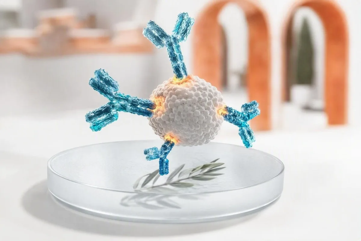
Et positivt antistoffresultat betyr at immunsystemet ditt gjenkjente et bestemt mål; det beviser ikke, i seg selv, en aktiv infeksjon eller sykdom. Antistoffklassen, testmetoden, tidspunktet, symptomer og medfølgende tester avgjør om du trenger beroligelse, en gjentatt test, bekreftelse eller umiddelbar klinisk behandling.


- Positivt antistoffresultat viser vanligvis immunerkjennelse, ikke nødvendigvis nåværende sykdom eller smittsomhet.
- IgM versus IgG er et tidssignal, ikke en klokke: IgM kan vedvare i måneder og IgG kan dukke opp tidlig.
- Vaksineantistoffer kan gi positive immunitetstester uten å indikere naturlig infeksjon.
- Allergen-spesifikk IgE over 0.35 kUA/L indikerer sensibilisering, men symptomer etter eksponering etablerer klinisk allergi.
- ANA-positivitet forekommer hos opptil 20% av friske voksne med lav titre, spesielt med alderen; symptomer og spesifikke antistoffer betyr mer.
- HIV-screening reaktivitet krever alltid laboratoriebekreftelsesalgoritmen før en diagnose stilles.
- Anti-tTG IgA er bare pålitelig for cøliakivurdering når personen spiser gluten og har tilstrekkelig total IgA.
- Akutte symptomer som pustevansker, forvirring, gulsott, brystsmerter eller et utbredt blemmerig utslett overskygger et rutinemessig antistoffresultat.
Hva et positivt antistoffresultat faktisk forteller deg
A positiv antistofftest betyr at analysen oppdaget proteiner i immunsystemet som binder et bestemt mål; det betyr ikke ikke automatisk at du har en aktiv infeksjon, en autoimmun sykdom eller en allergi som trenger behandling. Neste trinn avhenger av antistofftypen, resultatets styrke, symptomer, eksponeringstidspunkt og eventuelle parrede antigen-, PCR- eller organfunksjonstester.
Antistoffer er B-celleproteiner laget etter vaksinasjon, infeksjon, allergieksponering, autoimmunitet, eller av og til uten klinisk skadelig grunn. Et positivt resultat besvarer ett snevert analytisk spørsmål – “ble binding oppdaget over denne laboratorieavskjæringen?” – snarere enn det bredere kliniske spørsmålet om hva som skjer i kroppen din i dag.
I mine 15 år med klinisk praksis har den vanligste feilen vært å lese “positiv” som en diagnose. Dr. Thomas Kleins praktiske regel er å først identifisere målet– et virus, vaksineantigen, matprotein, thyreoideienzym, nukleært antigen eller medikament – deretter spørre om testen påviser tidligere eksponering, nåværende organisme, sensibilisering eller immunfeilretning.
Kantesti AI er en AI-blodprøveanalysator som leser antistoffflagg ved siden av datoer, immunoglobulinklasse og relaterte resultater heller enn å behandle en enkelt positiv linje som en dom. Den konteksten er spesielt nyttig når pasienter sammenligner rapporter fra forskjellige laboratorier; kvalitative og kvantitative resultater er ikke utskiftbare selv når begge sier positive.
Positiv IgG versus IgM: hvorfor tidspunkt er mindre enkelt enn det høres ut
IgM positiv kan tyde på en nylig immunrespons, mens IgG positiv ofte antyder tidligere eksponering eller vaksinerespons; ingen av mønstrene daterer en sykdom pålitelig på egen hånd. Noen infeksjoner produserer IgG innen 7–14 dager, og IgM kan forbli påviselig i 3–12 måneder eller lenger.
IgM er et stort antistoff med fem enheter som ofte er påviselig tidlig, men dets størrelse og brede binding gjør det mer utsatt for kryssreaksjoner. Epstein-Barr-virus, parvovirus B19, revmatoid faktor og noen autoimmune antistoffer kan forårsake misvisende IgM-reaksjoner; et isolert lavt IgM-resultat fortjener mer skepsis enn panikk.
IgG vedvarer vanligvis etter at immunminnet er utviklet, noen ganger i flere tiår. Et positivt IgG-resultat kan gjenspeile barndomsinfeksjon, en nylig vaksine, passivt antistoff fra intravenøs immunglobulin eller mütterlig overføring til et spedbarn; det kan ikke bevise beskyttelse med mindre den aktuelle analysen har en validert beskyttende terskel.
Parrede prøver kan være mer nyttige enn en enkelt blodprøve: en firedobling av antistofftiteren mellom prøver tatt med ca. 2–4 ukers mellomrom støtter nylig infeksjon ved utvalgte sykdommer. Hvis rapporten lister opp total IgG, IgA og IgM, gjennomgå mønsteret i stedet for ett tall med vårt guide for immunoglobulin interpretation.
Når infeksjonsantistoffer gjør og ikke betyr aktiv sykdom
Infeksjonsantistoffer viser vanligvis eksponering, mens PCR, antiggentesting, dyrking eller en stigende tittel er bedre verktøy for å påvise mange aktive infeksjoner. Et positivt antistoffresultat uten symptomer er ofte historisk, men unntaket avhenger av organismen og din immunstatus.
For hepatitt C betyr et reaktivt antistoff eksponering på et tidspunkt, men bare påviselig HCV RNA etablerer nåværende viremi. For hepatitt B betyr kombinasjonen noe: overflateantigen, overflateantistoff og kjerneantistoff skiller aktiv infeksjon, løst infeksjon og vaksineimmunitet mye bedre enn én positiv markør.
HIV er en annen setting hvor ordlyd betyr enormt mye. En reaktiv fjerdegenerasjons-screening er ikke en diagnose; laboratoriet følger opp med en HIV-1/HIV-2 differensieringsimmunoassay og, når det er nødvendig, nukleinsyretesting. En person som tar posteksponeringsprofylakse trenger tidsspesifikk testing, som forklart i vår guide for HIV-testing etter PEP.
Jeg har sett en frisk pasient skremt av et positivt treponema-syfilisantistoff etter behandling 18 år tidligere. Treponema-tester forblir ofte positive for livet, mens en RPR- eller VDRL-tittel bidrar til å vurdere aktuell aktivitet og respons; mønstre for syfilistesting gjør dette skillet klarere.
Hvordan vaksineantistoffer skiller seg fra antistoffer etter infeksjon
Vaksine-relaterte antistoffer viser at immunsystemet reagerte på et vaksineantigen; de betyr ikke at vaksinen forårsaket en aktiv infeksjon. Et positivt resultat kan fortsatt falle under en kjent beskyttende korrelat, fordi bindingsantistoffer og funksjonelle nøytraliserende antistoffer ikke er identiske.
Konsentrasjoner av hepatitt B-overflateantistoff på 10 mIU/mL eller høyere målt 1–2 måneder etter en dokumentert vaksineserie anses generelt som beskyttende hos immunkompetente personer. Anti-HBc, hepatitt B-kjerneantistoffet, produseres ikke av standard hepatitt B-vaksinasjon; dets tilstedeværelse peker mot naturlig eksponering og bør føre til fullstendig panel-tolkning.
Rubella IgG sjekkes vanligvis før graviditet fordi det indikerer tidligere immunitet, ikke en aktiv rubella-sykdom. Laboratorier setter assayspesifikke terskelverdier, og grenseverdier håndteres vanligvis som ikke-immune for planlegging før unnfangelse, heller enn tolkes som nylig infeksjon; tidspunkt er detaljert i vår guide for rubellatesting.
Kantesti AI er en AI-blodprøvetolkningsplattform som skiller vaksine-mønster-antistoffer fra infeksjons-mønster-antistoffer når rapporten inkluderer de relevante ledsagende markørene. Det praktiske spørsmålet er ofte ikke “er jeg positiv?”, men “hvilket antigen ble målt, når ble jeg vaksinert, og endrer dette resultatet dagens beskyttelsesplan?”.”
Hvorfor en positiv allergi-antistofftest kanskje ikke betyr en allergi
En positiv allergen-spesifikk IgE test indikerer sensibilisering, noe som betyr at IgE gjenkjenner et allergen; det diagnostiserer ikke klinisk allergi uten en kompatibel reaksjonshistorikk. En verdi over 0,35 kUA/L er en vanlig analytisk positivitetsterskel, ikke en universell terskel for å unngå en matvare eller medisin.
Historien før testen betyr mer enn mange tror. Hvis en person spiser peanøtter ukentlig uten elveblest, piping, oppkast eller kollaps, bør et lavt positivt peanøtt-IgE alene ikke føre til unngåelse; unødvendig matrestriksjon kan forårsake ernæringsmessige og sosiale skader.
NIAID-sponsede retningslinjer for matallergi sier at hudprikk- og spesifikk-IgE-tester identifiserer mulige triggermatvarer, men ikke kan diagnostisere matallergi uavhengig (Sampson et al., 2010). I utvalgte tilfeller bruker en allergolog komponent-oppløst testing eller en medisinsk overvåket oral matutfordring, som forblir referansestandarden for å løse usikkerhet.
Totalt IgE måles i IU/mL og kan være høyt ved eksem, parasittpåvirkning, røyking og flere immunsykdommer; det kan ikke identifisere et utløsende allergen. Komponenttesting kan skille et stabilt lagringsprotein fra et kryssreaktivt pollenrelatert protein, en forskjell som utforskes i vår artikkel om molekylær allergitesting.
Hvorfor autoimmune antistoffer krever symptomer og organsignaler
Autoimmune antistoffer kan gå foran sykdom, følge med sykdom, eller forekomme hos friske mennesker; et positivt resultat alene etablerer sjelden en autoimmun diagnose. Klinikere handler når antistoffmønsteret samsvarer med symptomer, undersøkelsesfunn, betennelsesmarkører eller objektive organskader.
Et antinukleært antistoff, eller ANA, er positivt ved lave titere hos omtrent 13% av befolkningen ved 1:80 og rundt 20% ved 1:40 i populasjonsstudier, med høyere rater hos kvinner og eldre voksne. Et homogent eller flekkete mønster diagnostiserer ikke lupus; ANA brukes best som en inngangstest når det kliniske bildet gjør bindevevssykdom sannsynlig.
2019 EULAR/ACR-klassifiseringsrammeverket for lupus bruker ANA ved en titer på minst 1:80 som et inngangskriterium, og krever deretter ytterligere vektede kliniske og immunologiske trekk (Aringer et al., 2019). Nytt protein i urin, lav C3/C4, cytopenier, inflammatorisk leddsmerte eller et overbevisende lysfølsomt utslett endrer alvorlighetsgraden mye mer enn en svak ANA alene.
Anti-dsDNA blir mer meningsfylt når det kombineres med lavt komplement og nyrefunn, selv om analysemetodene varierer betydelig. Vår veiledning for oppfølging av anti-dsDNA og oversikt over komplementtesting forklarer hvorfor en spesialist kan gjenta testing på en annen plattform.
Organ-spesifikke antistoffer: eksempler fra skjoldbruskkjertel, cøliaki og mage
Organspesifikke antistoffer identifiserer immunmålretting av et vev, men de viser ikke alltid om organet er skadet for øyeblikket eller trenger behandling. Tyreoperoksidaseantistoffer, vevstransglutaminaseantistoffer og iboende faktorantistoffer krever hver for seg ulike ledsagende tester.
TPO-antistoffer er vanlige ved Hashimoto tyreoiditt og kan være til stede i årevis mens TSH og fritt T4 forblir normale. Hos en voksen person som ikke er gravid med normal skjoldbruskkjertelfunksjon, støtter resultatet vanligvis periodisk TSH-overvåking snarere enn skjoldbruskkjertelhormonbehandling; graviditet, fertilitetsplaner og symptomer kan endre den tilnærmingen.
A positive tTG-IgA antyder cøliaki bare hvis personen fortsatt spiser gluten og total IgA er tilstrekkelig. Selektiv IgA-mangel kan gjøre tTG-IgA falsk negativ, så klinikere bruker IgG-baserte deamidert gliadinpeptid- eller tTG-tester når total IgA er lav; se vår forklaring av lav IgA-fallgruver.
Iboende faktorantistoff er relativt spesifikt, men ikke svært følsomt for perniciøs anemi. Når det er positivt sammen med lav B12, forhøyet metylmalonsyre og makrocytose, er mønsteret overbevisende; uten det mønsteret unngår jeg å behandle en laboratorieetikett heller enn pasienten. Test for perniciøs anemi viser den vanlige bekreftelsessekvensen.
Hva forårsaker et falskt positivt antistoffresultat
Falske-positive antistoffresultater oppstår når analysen oppdager utilsiktet binding, ikke når laboratoriet rett og slett “gjør feil”. Kryssreaktive antistoffer, revmatoid faktor, nylig immunstimulering, passiv immunglobulin og lav pre-test sannsynlighet er vanlige årsaker.
Spesifisitet er ikke perfeksjon. En test med 99% spesifisitet gir omtrent 10 falske-positive resultater for hver 1000 virkelig negative personer som testes; når tilstanden er sjelden, kan disse resultatene overgå sanne positive. Dette er grunnen til at bred screening hos noen uten symptomer ofte skaper tvetydighet heller enn trygghet.
Biotin er kjent for å forstyrre noen hormon- og hjerteanalyser, mens heterofile antistoffer og revmatoid faktor kan forstyrre visse immunoanalyser. Laboratoriet kan noen ganger fortynne prøven, bruke blokkerende reagenser, gjenta resultatet på en annen analysearkitektur, eller utføre en mer spesifikk bekreftelsesmetode.
Et positivt resultat som er i konflikt med et helt umerkelig klinisk bilde er ikke noe å ignorere; det er en grunn til å verifisere. Kantesti AI sammenligner inkonsekvente markører og testdatoer, og vår sjekkliste for nøyaktighet av laboratorieresultater kan hjelpe deg med å registrere kosttilskudd, nylige infusjoner og sykdommer før du snakker med rekvirerende lege.
Antistofftest positiv, men ingen symptomer: en praktisk triageplan
Når en antistofftest er positiv, men du føler deg frisk, trenger de fleste avklaring fremfor akuttbehandling. De tryggeste første stegene er å verifisere den nøyaktige analysen, sjekke eksponerings- og vaksinasjonsdatoer, og se etter den parede testen som bekrefter aktuell sykdom eller organinvolvering.
Start med selve rapporten: noter analytnavn, antistoffklasse, numerisk indeks eller titer, referanseintervall, laboratoriemetode og innsamlingsdato. “Reaktiv” kan bety et signal-til-avskjæringsforhold knapt over 1,0 eller et mye sterkere resultat; laboratorier bruker ikke identiske terskler, og størrelsen sporer bare noen ganger klinisk sannsynlighet.
Deretter, spør om det finnes en test for aktiv sykdom. Eksempler inkluderer PCR for hepatitt C, serielle RPR for syfilis, avførings- eller pustetesting for aktiv Helicobacter pylori, TSH/fritt T4 for skjoldbruskkjertelantistoffer, og urinprøve pluss kreatinin for lupusrelatert nyrebekymring.
Fra 8. september 2026 vil jeg ikke anbefale å starte antibiotika, antivirale midler, glutenfri diett, skjoldbruskkjertelmedisin eller matunnvikelse alene basert på et isolert antistoffresultat. Lagre den originale PDF-en, noter din baseline med longitudinell oppfølging av laboratorieprøver, og arranger rutinemessig kontroll med mindre det er røde flagg-symptomer til stede.
Bekreftende tester som endrer neste steg
Den beste bekreftelsestesten avhenger av hva antistoffet retter seg mot: molekylær påvisning bekrefter mange infeksjoner, funksjonelle analyser klargjør allergi, og organfunksjonstesting vurderer autoimmun påvirkning. Å gjenta samme antistofftest er bare nyttig når den forventede endringen over tid har klinisk betydning.
For et reaktivt HIV-screening, er den reflekterende bekreftelsesveien en differensieringsanalyse etterfulgt av HIV-nukleinsyretesting hvis resultatene er ubestemte. For et hepatitt C-antistoff, er HCV RNA den avgjørende oppfølgingen; for et positivt hepatitt B-kjerneantistoff, overflateantigen, overflateantistoff og noen ganger HBV DNA klargjør mønsteret.
For mistenkt cøliaki bør biopsisbeslutninger tas mens gluteneksponeringen er tilstrekkelig for testing. For mulig matallergi, vurderer en allergolog eksponeringshistorikken, spesifikt IgE-nivå, komponentprofil og utfordringsrisiko – ikke en panel av urelaterte lave positive resultater bestilt uten et klinisk spørsmål.
Kantesti AI er en Analyseverktøy for blodprøver drevet av KI som kan organisere disse sammenkoblede markørene til en kliniker-klar spørsmålsliste, men det erstatter ikke bekreftende testing eller diagnose. Hvis du er usikker på hvor mye du skal stole på et avvikende resultat, våre medisinske valideringsstandarder beskriver hvorfor verifisering og klinisk tilsyn er viktig.
Symptomer som gjør et positivt antistoffresultat mer presserende
Et positivt antistoffresultat berettiger akutt vurdering når det ledsages av pustevansker, ansikts- eller strupesvulst, besvimelse, forvirring, brystsmerter, gulsott, raskt spredende utslett, alvorlig dehydrering eller ny nevrologisk svakhet. Symptomer – ikke selve antall antistoff alene – bestemmer hastverket.
Mulig anafylaksi er en nødsituasjon: plutselige utbredte elveblest med piping i brystet, tetthet i halsen, vedvarende oppkast, svimmelhet eller lavt blodtrykk etter eksponering krever nødetater og foreskrevet adrenalin hvis tilgjengelig. Et positivt mat- eller gift-IgE fra måneder siden verken forutsier eller utelukker denne umiddelbare risikoen.
Ny gulsott med mørk urin, markert magesmerte, feber eller forvirring bør føre til evaluering samme dag, enten virale hepatittantistoffer er positive eller ikke. Leverenzymer, bilirubin, INR og direkte patogentesting bidrar til å skille immunhistorie fra aktiv hepatobiliær sykdom; leverpanel-komponenter er nyttig kontekst.
Autoimmun bekymring blir tidssensitiv med hoste blod, kortpustethet, raskt stigende kreatinin, blodig diaré, synsendring, anfall eller en ny ensidig svakhet. Jeg har lært å ikke berolige noen basert på en grensetittel for antistoffer når symptomforløpet deres tydelig forverres.
Betyr et høyere antall antistoffer mer alvorlig sykdom?
En høyere antistoffindeks eller titer øker noen ganger sannsynligheten for at et resultat er ekte, men det måler vanligvis ikke sykdommens alvorlighetsgrad. Analyseverdier er metodespesifikke, og et resultat på 80 AU/mL kan ikke sammenlignes direkte med 80 U/mL fra et annet laboratorium.
Ved autoimmune sykdommer kan anti-dsDNA-trender noen ganger følge lupusaktivitet, spesielt når de måles konsekvent med samme metode og tolkes med komplement og urinenprotein. ANA-titer er imidlertid et dårlig mål på lupus-alvorlighetsgrad og bør ikke jages gjentatte ganger hos en stabil person.
For infeksjoner kan et sterkt antistoffsignal støtte tidligere eksponering, men indikerer sjelden smitteevne. Nøytraliserende antistofftester er spesialiserte, og deres beskyttende terskler kan variere etter patogen, alder, vaksineprodukt og immunsuppresjon; en enkel kommersiell IgG-analyse er ikke et personlig immunitetspass.
Ved sammenligning av rapporter, hold laboratoriet, analysen og enhetene synlige. En reell biologisk endring bør overstige vanlig analytisk og daglig variasjon, og det er derfor vår guide for sammenligning av blodprøver fokuserer på datoer og metoder fremfor alene fargekodede flagg.
Graviditet, barn og immunsuppresjon endrer tolkningen
Graviditet, spedbarnsalder og immunsupprimerende behandling kan endre både antistoffproduksjon og sikkerheten ved å vente på bekreftelse. I disse gruppene kan et positivt resultat reflektere maternelle antistoffer, redusert vaksinerespons, reaktiveringsrisiko eller behov for tidligere spesialistråd.
Maternal IgG krysser placenta og kan forbli påviselig hos et spedbarn i flere måneder, mens maternal IgM generelt ikke krysser. Derfor reflekterer et spedbarns isolerte IgG-positivitet ofte maternal immunitet, mens organisms-spesifikk IgM eller direkte påvisning kan fortjene mer fokusert evaluering avhengig av infeksjonen og alderen.
Under graviditet tolkes rubella- eller parvovirus-antistoffresultater med eksponeringsdatoer, aviditytesting og gjentatte prøver heller enn en enkeltstående IgM alene. Falske positive IgM er et anerkjent problem, og unngåelig alarm kan føre til unødvendige invasive undersøkelser; obstetrisk eller infeksjonssykdomsinput er fornuftig når eksponering er troverdig.
Personer som mottar rituximab, høydose kortikosteroider, kjemoterapi eller transplantasjonsmedisiner kan ha svake eller manglende antistoffresponser til tross for infeksjon eller vaksinasjon. For familiebeslutninger, barns AI-tolkningsgrenser og den Kantesti medisinsk rådgivende styre forsterker behovet for kliniker-ledet gjennomgang.
Hvordan forberede seg til en nyttig klinisk gjennomgang av et positivt resultat
Ta med den fullstendige laboratorierapporten, grunnen til at testen ble bestilt, datoer for symptomer eller eksponering, vaksinehistorikk, medisiner og tidligere resultater. Denne informasjonen kan gjøre et tilsynelatende vagt positivt antistoffresultat til en klar neste handling innen en avtale.
Skriv ned fem fakta før besøket: første dag med symptomer, siste sannsynlige eksponering, vaksinasjonsdatoer, immunsupprimerende medisiner eller immunoglobulininfusjoner, og om symptomer blir bedre eller verre. En feber som startet 3 dager før prøvetaking tolkes veldig annerledes enn tretthet som startet for 6 måneder siden.
Still tre fokuserte spørsmål: “Hva påviser denne testen?”, “Hvilken test etablerer aktiv sykdom eller organpåvirkning?”, og “Hvilket resultat ville endre behandlingen?” Dr Thomas Klein finner at disse spørsmålene reduserer unødvendige gjentatte paneler og hjelper pasienter med å dra med en definert tidsramme – i dag, 1–2 uker, eller rutinemessig overvåking.
Kantesti AI støtter sikker rapportgjennomgang på over 75 språk og kan finne manglende ledsagende tester fra en fotografert eller opplastet PDF på omtrent 60 sekunder. Vår veiledning for AI-teknologi forklarer den kontekst-tilpassede tilnærmingen, mens vårt kliniske team gir den medisinske tilsynet bak pasientrettet tolkning.
Ofte stilte spørsmål
Hva betyr et positivt antistoffresultat hvis jeg ikke har noen symptomer?
Et positivt antistoffresultat uten symptomer indikerer oftest tidligere eksponering, vaksinasjon, sensibilisering eller et resultat som trenger bekreftelse, snarere enn aktiv sykdom. Neste trinn avhenger av målet: hepatitt C-antistoff trenger HCV RNA, et reaktivt HIV-screeningtest trenger laboratoriets bekreftelsesalgoritme, og thyreoideaantistoffer trenger TSH og fritt T4. Et isolert lavpositivt resultat er mindre overbevisende når det ikke er noen kompatibel eksponering eller symptomhistorie. Søk umiddelbar hjelp hvis ny gulsott, pustevansker, forvirring, brystsmerter eller et raskt forverrende utslett oppstår.
Betyr IgM positiv og IgG negativ alltid en ny infeksjon?
IgM positiv med IgG negativ kan forekomme tidlig i en infeksjon, men det beviser ikke alltid en ny infeksjon. IgM kan krysse-reagere med urelaterte immunproteiner, og noen analyser gir falsk-positive IgM-resultater oftere enn IgG-resultater. Klinikere kan gjenta serologi etter 1–2 uker, bestille PCR- eller antigentesting, eller bruke en mer spesifikk bekreftende analyse. Riktig tolkning avhenger også av sykdomsforløpet og den spesifikke organismen som testes.
Kan en vaksine gi positivt utslag på en antistofftest?
Ja, vaksiner gir vanligvis positive svar på antistofftester for antigenet i vaksinen. For hepatitt B indikerer en anti-HBs-konsentrasjon på minst 10 mIU/mL målt 1–2 måneder etter en fullført serie generelt en beskyttende vaksinerespons hos immunkompetente personer. Standard hepatitt B-vaksinasjon produserer ikke anti-HBc, så positivitet for kjerneproteinantistoffer tyder på naturlig eksponering snarere enn bare vaksinasjon. Vaksine-relatert positivitet betyr ikke at du har en aktiv infeksjon.
Kan man ha en positiv autoimmun antistofftest og være frisk?
Ja, friske mennesker kan ha positive autoimmune antistoffer, spesielt ANA-resultater med lav tittel. ANA er påvisbart hos ca. 13% av personer med en tittel på 1:80 og rundt 20% ved 1:40, så det er ikke en frittstående lupus-test. Symptomer som betennelsessvulst i ledd, munnsår, fotosensitiv utslett, lav komplement, protein i urin eller unormale blodverdier avgjør om det er behov for spesialistvurdering. Et positivt resultat bør tolkes i sammenheng med grunnen til at testen ble bestilt.
Betyr en positiv IgE-blodprøve at jeg må unngå maten?
Nei, et matspesifikt IgE-resultat betyr sensibilisering og beviser ikke at inntak av maten forårsaker en allergisk reaksjon. Mange laboratorier kaller resultater over 0,35 kUA/L positive, men denne grenseverdien er designet for påvisning heller enn en universell unngåelsesbeslutning. En person som regelmessig spiser maten uten elveblest, piping i brystet, gjentatt oppkast eller svimmelhet, bør ikke automatisk fjerne den fra kostholdet. En allergolog kan bruke reaksjonshistorikken, komponenttesting og noen ganger en overvåket oral matprovokasjon for å avklare risikoen.
Hvor lenge forblir antistoffer positive etter en infeksjon?
Antistoffvarighet varierer mye etter infeksjon og antistoffklasse. IgM kan forsvinne i løpet av uker, men kan vare i 3–12 måneder eller lenger, mens IgG kan forbli målbart i årevis eller livslangt. Treponemal syfilisantistoffer forblir ofte positive for livet etter vellykket behandling, mens RPR- eller VDRL-titre brukes til å følge aktivitet. Et positivt antistoffresultat kan derfor ikke pålitelig fortelle deg når en infeksjon oppsto uten annen klinisk og laboratorieinformasjon.
Få AI-drevet analyse av blodprøver i dag
Bli med over 2 millioner brukere over hele verden som stoler på Kantesti for øyeblikkelig, nøyaktig analyse av laboratorietester. Last opp blodprøveresultatene dine og få omfattende tolkning av 15,000+-biomarkører på sekunder.
📚 Refererte forskningspublikasjoner
Klein, T., Mitchell, S., & Weber, H. (2026). Flerspråklig AI-assistert klinisk beslutningsstøtte for tidlig hantavirus-triage: Design, ingeniørvalidering og reell distribusjon på tvers av 50 000 tolkede blodprøverapporter. Figshare. https://www.researchgate.net/. Kantesti KI medisinsk forskning.
Klein, T., Mitchell, S., & Weber, H. (2026). En forhåndsregistrert, rubrikkbasert automatisert teknisk referanse for Kantesti blodprøvetolkning på 100 000 syntetiske testtilfeller. Figshare. https://www.academia.edu/. Kantesti KI medisinsk forskning.
📖 Eksterne medisinske referanser
⚕️ Medisinsk ansvarsfraskrivelse
Denne artikkelen er kun for informasjonsformål og utgjør ikke medisinsk rådgivning. Rådfør deg alltid med en kvalifisert helsepersonell for beslutninger om diagnose og behandling.
E-E-A-T tillitssignaler
Experience
Klinisk gjennomgang ledet av lege av arbeidsflyter for laboratorietolkning.
Expertise
Fagområde laboratoriemedisin med fokus på hvordan biomarkører oppfører seg i klinisk kontekst.
Authoritativeness
Skrevet av Dr. Thomas Klein med gjennomgang av Dr. Sarah Mitchell og Prof. Dr. Hans Weber.
Trustworthiness
Evidensbasert tolkning med tydelige oppfølgingsveier for å redusere uro.