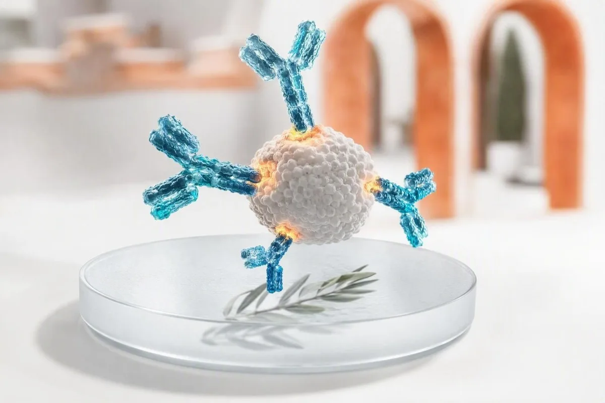
Положительный результат теста на антитела означает, что ваша иммунная система распознала определенную мишень; сам по себе он не подтверждает активную инфекцию или заболевание. Класс антител, метод тестирования, время проведения, симптомы и сопутствующие тесты определяют, требуется ли вам успокоение, повторный тест, подтверждение или срочное клиническое наблюдение.


- Положительный результат теста на антитела обычно указывает на распознавание иммунной системой, а не обязательно на текущее заболевание или заразность.
- IgM против IgG — это подсказка о времени, а не часы: IgM может сохраняться месяцами, а IgG может появиться раньше.
- Антитела к вакцине могут давать положительный результат теста на иммунитет, не указывая на естественную инфекцию.
- IgE, специфичные для аллергенов выше 0,35 kUA/L указывают на сенсибилизацию, но симптомы после воздействия подтверждают клиническую аллергию.
- Позитивность АНА встречается до 20% здоровых взрослых при низких титрах, особенно с возрастом; симптомы и специфические антитела имеют большее значение.
- Реактивность на ВИЧ-скрининг всегда требует алгоритма лабораторного подтверждения перед постановкой какого-либо диагноза.
- Anti-tTG IgA надежен для оценки целиакии только тогда, когда человек употребляет глютен и имеет достаточный уровень общего IgA.
- Срочные симптомы такие как затрудненное дыхание, спутанность сознания, желтуха, боль в груди или обширная буллезная сыпь перевешивают результат рутинного анализа на антитела.
Что на самом деле означает положительный результат теста на антитела
A положительный тест на антитела означает, что анализ обнаружил иммунные белки, которые связываются с определенной мишенью; он не нет автоматически означает, что у вас есть активная инфекция, аутоиммунное заболевание или аллергия, требующая лечения. Следующий шаг зависит от типа антител, силы результата, симптомов, времени воздействия и наличия парного антигена, ПЦР или тестов на функцию органов.
Антитела — это белки B-клеток, вырабатываемые после вакцинации, инфекции, воздействия аллергенов, аутоиммунитета или иногда по неопасной для здоровья причине. Положительный результат отвечает на один узкий аналитический вопрос — “было ли обнаружено связывание выше этого лабораторного порога?” — а не на более широкий клинический вопрос о том, что происходит в вашем организме сегодня.
За 15 лет моей клинической практики самой распространенной ошибкой было чтение “положительного” как диагноза. Практическое правило доктора Томаса Кляйна заключается в том, чтобы сначала определить мишень—вирус, антиген вакцины, пищевой белок, тиреоидный фермент, ядерный антиген или лекарство—затем спросить, выявляет ли тест прошлый контакт, присутствующий организм, сенсибилизацию или иммунную дезориентацию.
Kantesti AI — это Анализатор анализа крови на основе искусственного интеллекта который читает флаги антител рядом с датами, классом иммуноглобулина и связанными результатами, а не рассматривает одну положительную линию как вердикт. Этот контекст особенно полезен, когда пациенты сравнивают отчеты из разных лабораторий; качественные и количественные результаты не взаимозаменяемы, даже если оба показывают положительный результат.
Положительные результаты IgG против IgM: почему время проведения менее очевидно, чем кажется
Положительный IgM может указывать на недавний иммунный ответ, в то время как IgG положительный часто указывает на прошлый контакт или ответ на вакцину; ни один из паттернов не позволяет точно датировать заболевание сам по себе. Некоторые инфекции производят IgG в течение 7–14 дней, а IgM может оставаться обнаруживаемым в течение 3–12 месяцев или дольше.
IgM — это большой пятизвенный антитело, которое часто обнаруживается на ранних стадиях, но его размер и широкое связывание делают его более склонным к перекрестным реакциям. Вирус Эпштейна-Барр, парвовирус B19, ревматоидный фактор и некоторые аутоиммунные антитела могут вызывать ложные реакции IgM; изолированный результат IgM низкого уровня заслуживает большего скептицизма, чем паники.
IgG обычно сохраняется после развития иммунной памяти, иногда десятилетиями. Положительный результат IgG может отражать детскую инфекцию, недавнюю вакцинацию, пассивные антитела от внутривенного введения иммуноглобулина или материнскую передачу младенцу; он не может доказать защиту, если только данный анализ не имеет валидированного защитного порога.
Парные образцы могут быть более полезны, чем однократный забор: четырехкратное повышение титра антител между образцами, взятыми примерно с интервалом 2–4 недели, свидетельствует о недавней инфекции при определенных заболеваниях. Если в отчете указаны общий IgG, IgA и IgM, просмотрите паттерн, а не одно число с нашим руководство по интерпретации иммуноглобулинов.
Когда антитела при инфекции означают активное заболевание, а когда нет
Антитела к инфекциям обычно показывают воздействие, тогда как ПЦР, антигенное тестирование, посев или повышение титра являются лучшими инструментами для выявления многих активных инфекций. Положительный результат на антитела без симптомов часто является историческим, но исключение зависит от возбудителя и вашего иммунного статуса.
При гепатите C реактивные антитела означают воздействие в какой-то момент, но только обнаруживаемый РНК ВГС устанавливает текущую виремию. Для гепатита B важна комбинация: поверхностный антиген, поверхностное антитело и антитело к ядерному антигену лучше различают активную инфекцию, перенесенную инфекцию и вакцинальный иммунитет, чем один положительный маркер.
ВИЧ — это еще одна область, где формулировка имеет огромное значение. Реактивный скрининг четвертого поколения на ВИЧ не является диагнозом; лаборатория проводит дифференциальный иммуноанализ на ВИЧ-1/ВИЧ-2 и, при необходимости, тестирование нуклеиновых кислот. Человек, принимающий постэкспозиционную профилактику, нуждается в тестировании, специфичном для времени, как описано в нашем руководстве по тестированию на ВИЧ после PEP.
Я видел здорового пациента, испуганного положительным тестом на антитела к трепонеме при сифилисе после лечения 18 лет назад. Тесты на трепонему часто остаются положительными на всю жизнь, тогда как титр RPR или VDRL помогает оценить текущую активность и ответ; схемы тестирования на сифилис делают это различие более ясным.
Чем антитела после вакцинации отличаются от антител после инфекции
Антитела, связанные с вакцинами, показывают, что иммунная система отреагировала на антиген вакцины; они не означают, что вакцина вызвала активную инфекцию. Положительный результат может по-прежнему находиться ниже известного защитного коррелята, поскольку связывающие антитела и функциональные нейтрализующие антитела не идентичны.
Концентрации поверхностного антитела к гепатиту B 10 мМЕ/мл или выше измеренные через 1–2 месяца после документированной серии вакцинаций, обычно считаются защитными у иммунокомпетентных лиц. Анти-HBc, антитело к ядерному антигену гепатита B, не вырабатывается стандартной вакцинацией против гепатита B; его наличие указывает на естественное воздействие и должно потребовать полной интерпретации панели.
IgG к краснухе обычно проверяют перед беременностью, поскольку он указывает на предшествующий иммунитет, а не на активное заболевание краснухой. Лаборатории устанавливают пороговые значения для каждого анализа, и пограничные результаты обычно обрабатываются как отсутствие иммунитета для планирования перед зачатием, а не интерпретируются как недавняя инфекция; сроки подробно описаны в нашем руководстве по тестированию на краснуху.
Kantesti AI — это Платформа для расшифровки анализа крови AI которое разделяет антитела вакцинного типа и антитела инфекционного типа, когда отчет включает соответствующие сопутствующие маркеры. Практический вопрос часто не “положительный ли я?”, а “какой антиген был измерен, когда я был вакцинирован, и изменяет ли этот результат сегодняшний план защиты?”
Почему положительный результат теста на аллергические антитела может не означать аллергию
Положительный специфический аллерген IgE тест указывает на сенсибилизацию, означающую, что IgE распознает аллерген; он не диагностирует клиническую аллергию без совместимой истории реакции. Значение выше 0.35 кЕдА/л является общим аналитическим порогом положительности, а не универсальным порогом для избегания употребления пищи или лекарств.
Претестовая история имеет большее значение, чем многие люди осознают. Если человек ест арахис еженедельно без крапивницы, хрипов, рвоты или коллапса, один низкий положительный результат IgE к арахису не должен приводить к избеганию; ненужные пищевые ограничения могут нанести питательный и социальный вред.
Руководство NIAID по аллергии на пищевые продукты гласит, что кожные тесты и тесты на специфические IgE выявляют возможные триггеры пищевой аллергии, но не могут самостоятельно диагностировать пищевую аллергию (Sampson et al., 2010). В отдельных случаях аллерголог использует компонентно-резольвирующее тестирование или медицински контролируемую пероральную пищевую провокацию, которая остается эталонным стандартом для разрешения неопределенности.
Общий IgE измеряется в МЕ/мл и может быть высоким при экземе, паразитарном заражении, курении и нескольких иммунных состояниях; он не может идентифицировать вызывающий аллерген. Компонентное тестирование может отличить стабильный запасной белок от перекрестно-реактивного белка, связанного с пыльцой, различие, рассмотренное в нашем статья о молекулярной аллергодиагностике.
Почему аутоиммунные антитела требуют симптомов и признаков поражения органов
Аутоиммунные антитела могут предшествовать заболеванию, сопровождать его или присутствовать у здоровых людей; положительный результат сам по себе редко подтверждает диагноз аутоиммунного заболевания. Клиницисты действуют, когда картина антител соответствует симптомам, данным осмотра, маркерам воспаления или объективным изменениям органов.
Антинуклеарное антитело, или АНА, положительно при низких титрах примерно у 13% людей при разведении 1:80 и примерно у 20% при разведении 1:40 в популяционных исследованиях, с более высокими показателями у женщин и пожилых людей. Однородный или пятнистый паттерн не является диагностикой волчанки; ANA лучше всего использовать как начальный тест, когда клиническая картина делает заболевание соединительной ткани вероятным.
Классификационные критерии EULAR/ACR по волчанке 2019 года используют ANA в титре не менее 1:80 в качестве критерия включения, а затем требуют дополнительных взвешенных клинических и иммунологических признаков (Aringer et al., 2019). Появление белка в моче, низкие уровни C3/C4, цитопении, воспалительный отек суставов или убедительная сыпь, вызванная фотосенсибилизацией, меняют срочность гораздо больше, чем слабый ANA сам по себе.
Anti-dsDNA приобретает большее значение при сочетании с низким комплементом и поражением почек, хотя методы анализа существенно различаются. Наше руководство по мониторингу anti-dsDNA и обзор тестирования комплемента объясняет, почему специалист может повторить тестирование на другой платформе.
Органоспецифичные антитела: примеры щитовидной железы, целиакии и желудка
Орган-специфичные антитела определяют иммунное нацеливание на ткань, но не всегда показывают, поврежден ли орган в данный момент или нуждается в лечении. Антитела к тиреоидной пероксидазе, антитела к тканевой трансглутаминазе и антитела к внутреннему фактору требуют различных сопутствующих тестов.
Антитела к TPO распространены при тиреоидите Хашимото и могут присутствовать годами, в то время как TSH и свободный T4 остаются в норме. У взрослых небеременных женщин с нормальной функцией щитовидной железы результат обычно поддерживает периодический мониторинг TSH, а не заместительную гормональную терапию; беременность, планы по зачатию и симптомы могут изменить этот подход.
Положительный tTG-IgA предполагает целиакию только в том случае, если человек продолжает употреблять глютен, а общий уровень IgA достаточен. Селективный дефицит IgA может привести к ложноотрицательному результату tTG-IgA, поэтому клиницисты используют тесты на пептид деамидированных глиадинов на основе IgG или тесты tTG, когда общий уровень IgA низкий; см. наше объяснение ложных результатов по IgA.
Антитело к внутреннему фактору относительно специфично, но не очень чувствительно для пернициозной анемии. Когда оно положительно вместе с низким уровнем B12, повышенным уровнем метилмалоновой кислоты и макроцитозом, картина является убедительной; без этой картины я избегаю лечения лабораторной метки, а не пациента. тестирование на пернициозную анемию показывает обычную последовательность подтверждения.
Что вызывает ложноположительный результат теста на антитела
Ложноположительные результаты на антитела возникают, когда анализ обнаруживает непреднамеренное связывание, а не когда лаборатория просто “ошиблась”. Распространенными причинами являются перекрестно-реактивные антитела, ревматоидный фактор, недавняя иммунная стимуляция, пассивный иммуноглобулин и низкая вероятность до тестирования.
Специфичность — это не совершенство. Тест с 99% специфичностью дает около 10 ложноположительных результатов на каждые 1000 действительно отрицательных людей; когда состояние встречается редко, эти результаты могут превосходить истинные положительные. Вот почему широкое скрининговое тестирование у человека без симптомов часто создает неопределенность, а не успокоение.
Биотин известен тем, что мешает некоторым гормональным и кардиологическим анализам, в то время как гетерофильные антитела и ревматоидный фактор могут мешать определенным иммуноанализам. Лаборатория иногда может развести образец, использовать блокирующие реагенты, повторить результат на другом анализаторе или провести более специфичный подтверждающий метод.
Положительный результат, противоречащий совершенно невыразительной клинической картине, не следует игнорировать; это причина для проверки. Kantesti AI сравнивает дискордантные маркеры и даты тестов, а наш чек‑лист точности лабораторных результатов может помочь вам записать добавки, недавние инфузии и заболевания перед разговором с направляющим клиницистом.
Положительный результат теста на антитела, но без симптомов: практический план сортировки
Когда тест на антитела положительный, но вы чувствуете себя хорошо, большинству людей требуется разъяснение, а не неотложная помощь. Самые безопасные первые шаги — точно определить используемый анализ, проверить даты воздействия и вакцинации, а также найти парный тест, подтверждающий текущее заболевание или поражение органов.
Начните с самого отчета: запишите название аналита, класс антитела, числовой индекс или титр, референсный интервал, лабораторный метод и дату сбора. “Реактивный” может означать соотношение сигнал/порог, лишь немного превышающее 1,0, или гораздо более сильный результат; лаборатории не используют одинаковые пороговые значения, и величина лишь иногда коррелирует с клинической вероятностью.
Затем выясните, существует ли тест на активное заболевание. Примеры включают ПЦР на гепатит С, серийные RPR на сифилис, анализ кала или дыхания на активный Helicobacter pylori, TSH/свободный T4 на аутоантитела щитовидной железы, а также анализ мочи плюс креатинин для выявления проблем с почками, связанных с волчанкой.
По состоянию на 8 сентября 2026 года я бы не рекомендовал начинать прием антибиотиков, противовирусных препаратов, безглютеновой диеты, препаратов для щитовидной железы или избегать продуктов питания самостоятельно, основываясь только на изолированном результате анализа на антитела. Сохраните исходный PDF-файл, отметьте свой базовый уровень с длительном лабораторном наблюдении, и запланируйте рутинный осмотр, если нет тревожных симптомов.
Подтверждающие тесты, которые меняют следующий шаг
Лучший подтверждающий тест зависит от того, что является мишенью антитела: молекулярное обнаружение подтверждает многие инфекции, функциональные анализы уточняют аллергию, а тестирование функции органов оценивает аутоиммунное воздействие. Повторение того же анализа на антитела полезно только тогда, когда ожидаемое изменение со временем имеет клиническое значение.
Для реактивного скрининга на ВИЧ путь подтверждения включает дифференциальный анализ, за которым следует тестирование нуклеиновых кислот ВИЧ, если результаты неопределенные. Для антитела к гепатиту С РНК ВГС является решающим последующим шагом; для положительного антитела к ядерному антигену гепатита В, поверхностного антигена, поверхностного антитела и иногда ДНК ВГВ уточняется картина.
Для подозрения на целиакию решения о биопсии следует принимать, пока потребление глютена достаточно для тестирования. При возможной пищевой аллергии аллерголог учитывает историю воздействия, уровень специфического IgE, профиль компонента и риск провокации — а не панель несвязанных слабых положительных результатов, заказанных без клинического вопроса.
Kantesti AI — это Инструмент анализа результатов анализа крови с ИИ которые могут организовать эти связанные маркеры в список вопросов, готовый для врача, но это не заменяет подтверждающее тестирование или диагностику. Если вы не уверены, насколько можно доверять неоднозначному результату, наши стандарты медицинской валидации описывают, почему проверка и клинический надзор имеют значение.
Симптомы, которые делают положительный результат теста на антитела более срочным
Положительный результат анализа на антитела требует срочной оценки, когда он сопровождается затрудненным дыханием, отеком лица или горла, потерей сознания, спутанностью сознания, болью в груди, желтухой, быстро распространяющейся сыпью, сильным обезвоживанием или новой неврологической слабостью. Срочность определяют симптомы, а не только число антител.
Возможный анафилактический шок — это чрезвычайная ситуация: внезапная распространенная крапивница с хрипами, стеснением в горле, стойкой рвотой, слабостью или низким артериальным давлением после воздействия требует вызова экстренных служб и введения адреналина, если он есть. Положительный результат анализа на пищевой IgE или IgE к яду, полученный несколько месяцев назад, не предсказывает и не исключает этого немедленного риска.
Новая желтуха с темной мочой, выраженная боль в животе, лихорадка или спутанность сознания должны вызвать оценку в тот же день, независимо от того, положительны ли тесты на антитела к вирусным гепатитам. Ферменты печени, билирубин, INR и прямое тестирование патогенов помогают отличить иммунную историю от активного гепатобилиарного заболевания; компонентам печеночной панели являются полезным контекстом.
Аутоиммунное заболевание становится срочным при кашле с кровью, одышке, быстром повышении креатинина, кровавой диарее, изменениях зрения, судорогах или новой односторонней слабости. Я научился не успокаивать человека на основании пограничного титра антител, когда траектория его симптомов явно ухудшается.
Означает ли более высокое число антител более тяжелое заболевание?
Более высокий индекс антител или титр иногда увеличивает вероятность того, что результат является подлинным, но он обычно не измеряет тяжесть заболевания. Значения анализов специфичны для метода, и результат 80 АЕ/мл нельзя напрямую сравнивать с 80 Ед/мл из другой лаборатории.
При аутоиммунных заболеваниях тенденции анти-дсДНК иногда могут следовать за активностью волчанки, особенно при последовательном измерении одним и тем же методом и интерпретации с учетом комплемента и белка в моче. Однако титр ANA является плохим показателем тяжести волчанки и не должен повторно отслеживаться у стабильного пациента.
Для инфекций сильный сигнал антител может указывать на предшествующее воздействие, но редко указывает на заразность. Тесты на нейтрализующие антитела являются специализированными, и их защитные пороговые значения могут различаться в зависимости от патогена, возраста, типа вакцины и иммуносупрессии; простой коммерческий тест IgG не является личным паспортом иммунитета.
При сравнении отчетов держите в поле зрения лабораторию, анализ и единицы измерения. Реальное биологическое изменение должно превышать обычные аналитические и ежедневные вариации, поэтому наши руководство по сравнению анализов крови фокусируются на датах и методах, а не только на цветных флажках.
Беременность, дети и иммуносупрессия меняют интерпретацию
Беременность, младенчество и иммуносупрессивная терапия могут влиять как на выработку антител, так и на безопасность ожидания подтверждения. В этих группах положительный результат может отражать материнские антитела, сниженный ответ на вакцину, риск реактивации или необходимость более ранней консультации специалиста.
Материнские IgG пересекают плаценту и могут оставаться обнаруживаемыми у младенца в течение нескольких месяцев, тогда как материнские IgM обычно не пересекают. Следовательно, изолированная положительная реакция на IgG у младенца часто отражает материнский иммунитет, в то время как специфические для возбудителя IgM или прямое обнаружение могут потребовать более пристального изучения в зависимости от инфекции и возраста.
Во время беременности результаты анализа на антитела к вирусу краснухи или парвовирусу интерпретируются с учетом дат воздействия, тестирования авидности и повторных образцов, а не только IgM, определенного однократно. Ложноположительный IgM является признанной проблемой, и ненужное беспокойство может привести к необоснованным инвазивным исследованиям; консультация акушера или инфекциониста целесообразна при достоверном контакте.
Люди, получающие ритуксимаб, высокие дозы кортикостероидов, химиотерапию или трансплантационные препараты, могут иметь слабый или отсутствующий иммунный ответ на антитела, несмотря на инфекцию или вакцинацию. Для семейных решений, пределы интерпретации детских AI и Медицинский консультативный совет Kantesti подчеркивают необходимость рассмотрения врачом.
Как подготовиться к полезному рассмотрению положительного результата клиницистом
Возьмите полный лабораторный отчет, причину назначения анализа, даты симптомов или контакта, историю вакцинации, принимаемые лекарства и предыдущие результаты. Эта информация может превратить aparentemente неясный положительный результат анализа на антитела в четкий следующий шаг в течение одного приема.
Запишите пять фактов перед визитом: первый день симптомов, последний вероятный контакт, даты вакцинации, иммуносупрессивные препараты или инфузии иммуноглобулина, а также улучшаются или ухудшаются симптомы. Лихорадка, начавшаяся за 3 дня до взятия образца, интерпретируется совершенно иначе, чем усталость, начавшаяся 6 месяцев назад.
Задайте три сфокусированных вопроса: “Что обнаруживает этот тест?”, “Какой тест подтверждает активное заболевание или поражение органа?” и “Какой результат изменит лечение?” Доктор Томас Кляйн считает, что эти вопросы сокращают ненужные повторные панели и помогают пациентам уйти с определенным сроком — сегодня, через 1–2 недели или плановый мониторинг.
Kantesti AI поддерживает безопасный просмотр отчетов на более чем 75 языках и может выявлять отсутствующие сопутствующие тесты из сфотографированного или загруженного PDF-файла примерно за 60 секунд. Наша Руководство по технологии ИИ объясняет подход сопоставления с контекстом, в то время как наша клиническая команда обеспечивает медицинский надзор за интерпретацией, ориентированной на пациента.
Часто задаваемые вопросы
Что означает положительный результат анализа на антитела, если у меня нет симптомов?
Положительный результат на антитела при отсутствии симптомов чаще всего указывает на предыдущее воздействие, вакцинацию, сенсибилизацию или результат, требующий подтверждения, а не на активное заболевание. Следующий шаг зависит от мишени: антитела к гепатиту C требуют РНК ВГС, реактивный скрининг на ВИЧ требует алгоритма лабораторного подтверждения, а тиреоидные антитела требуют TSH и свободного Т4. Изолированный низкоположительный результат менее убедителен при отсутствии соответствующей истории воздействия или симптомов. Обратитесь за неотложной помощью при появлении новой желтухи, затрудненного дыхания, спутанности сознания, боли в груди или быстро ухудшающейся сыпи.
Означает ли IgM положительный и IgG отрицательный всегда новую инфекцию?
IgM положительный при IgG отрицательном может наблюдаться на ранней стадии инфекции, но это не всегда доказывает новую инфекцию. IgM может давать перекрестную реакцию с несвязанными иммунными белками, и некоторые анализы дают ложноположительные результаты IgM чаще, чем результаты IgG. Клиницисты могут повторить серологию через 1–2 недели, запросить ПЦР или антигенное тестирование, или использовать более специфический подтверждающий анализ. Правильная интерпретация также зависит от временной шкалы заболевания и конкретного исследуемого организма.
Может ли вакцина сделать тест на антитела положительным?
Да, вакцины обычно делают тесты на антитела положительными на антиген, содержащийся в вакцине. Для гепатита В концентрация anti-HBs не менее 10 мМЕ/мл, измеренная через 1–2 месяца после завершения курса, обычно указывает на защитный ответ на вакцину у иммунокомпетентных людей. Стандартная вакцинация против гепатита В не вызывает anti-HBc, поэтому положительный результат по коревым антителам предполагает естественное заражение, а не только вакцинацию. Положительный результат, связанный с вакциной, не означает наличия активной инфекции.
Может ли быть положительный тест на аутоиммунные антитела и при этом быть здоровым?
Да, у здоровых людей могут быть положительные аутоиммунные антитела, особенно результаты АНА с низким титром. АНА обнаруживаются примерно у 13% людей с титром 1:80 и примерно у 20% с титром 1:40, поэтому это не самостоятельный тест на волчанку. Такие симптомы, как воспалительный отек суставов, язвы во рту, сыпь, чувствительная к солнцу, низкий уровень комплемента, белок в моче или аномальные показатели крови, определяют, требуется ли консультация специалиста. Положительный результат следует интерпретировать в контексте причины назначения анализа.
Означает ли положительный анализ крови на IgE, что я должен избегать этого продукта?
Нет, положительный результат пищеспецифического IgE означает сенсибилизацию и не доказывает, что употребление пищи вызывает аллергическую реакцию. Многие лаборатории называют результаты выше 0,35 kUA/L положительными, но этот пороговый уровень предназначен для обнаружения, а не для принятия универсального решения об избегании. Человек, который регулярно употребляет пищу без крапивницы, хрипов, повторяющейся рвоты или обмороков, не должен автоматически исключать ее из рациона. Аллерголог может использовать историю реакции, тестирование на компоненты и иногда контролируемое оральное пищевое провокационное исследование для уточнения риска.
Как долго антитела остаются положительными после инфекции?
Длительность присутствия антител сильно варьируется в зависимости от инфекции и класса антител. IgM могут исчезать в течение нескольких недель, но могут сохраняться в течение 3–12 месяцев или дольше, тогда как IgG могут оставаться измеримыми в течение многих лет или пожизненно. Антитела к трепонемному сифилису часто остаются положительными пожизненно после успешного лечения, в то время как титры RPR или VDRL используются для отслеживания активности. Таким образом, положительный результат анализа на антитела не может достоверно определить, когда произошла инфекция, без другой клинической и лабораторной информации.
Получите анализ крови с помощью ИИ уже сегодня
Присоединяйтесь к более чем 2 миллионам пользователей по всему миру, которые доверяют Kantesti для мгновенного и точного анализа лабораторных тестов. Загрузите результаты анализа крови и получите комплексную интерпретацию биомаркеров 15,000+ за считанные секунды.
📚 Ссылки на научные публикации
Кляйн, Т., Митчелл, С., и Вебер, Х. (2026). Многоязычная система поддержки принятия клинических решений с помощью ИИ для ранней сортировки хантавирусных инфекций: дизайн, инженерная валидация и реальное развертывание на основе 50 000 интерпретированных отчетов о анализах крови. Figshare. https://www.researchgate.net/. Kantesti AI Medical Research.
Кляйн, Т., Митчелл, С., и Вебер, Х. (2026). Предварительно зарегистрированный автоматизированный технический бенчмарк на основе рубрик для движка интерпретации анализов крови Kantesti на 100 000 синтетических тестовых случаев. Figshare. https://www.academia.edu/. Kantesti AI Medical Research.
📖 Внешние медицинские источники
⚕️ Медицинское предупреждение
Эта статья предназначена только для образовательных целей и не является медицинской консультацией. Всегда консультируйтесь с квалифицированным специалистом здравоохранения по вопросам диагностики и решений по лечению.
Сигналы доверия E-E-A-T
Опыт
Клинический обзор рабочих процессов интерпретации лабораторных данных под руководством врача.
Экспертиза
Акцент на лабораторной медицине: как биомаркеры ведут себя в клиническом контексте.
Авторитетность
Написано доктором Томасом Кляйном при рецензировании доктором Сарой Митчелл и профессором доктором Хансом Вебером.
Доверие
Интерпретация на основе доказательств с понятными маршрутами дальнейших действий, чтобы снизить тревожность.