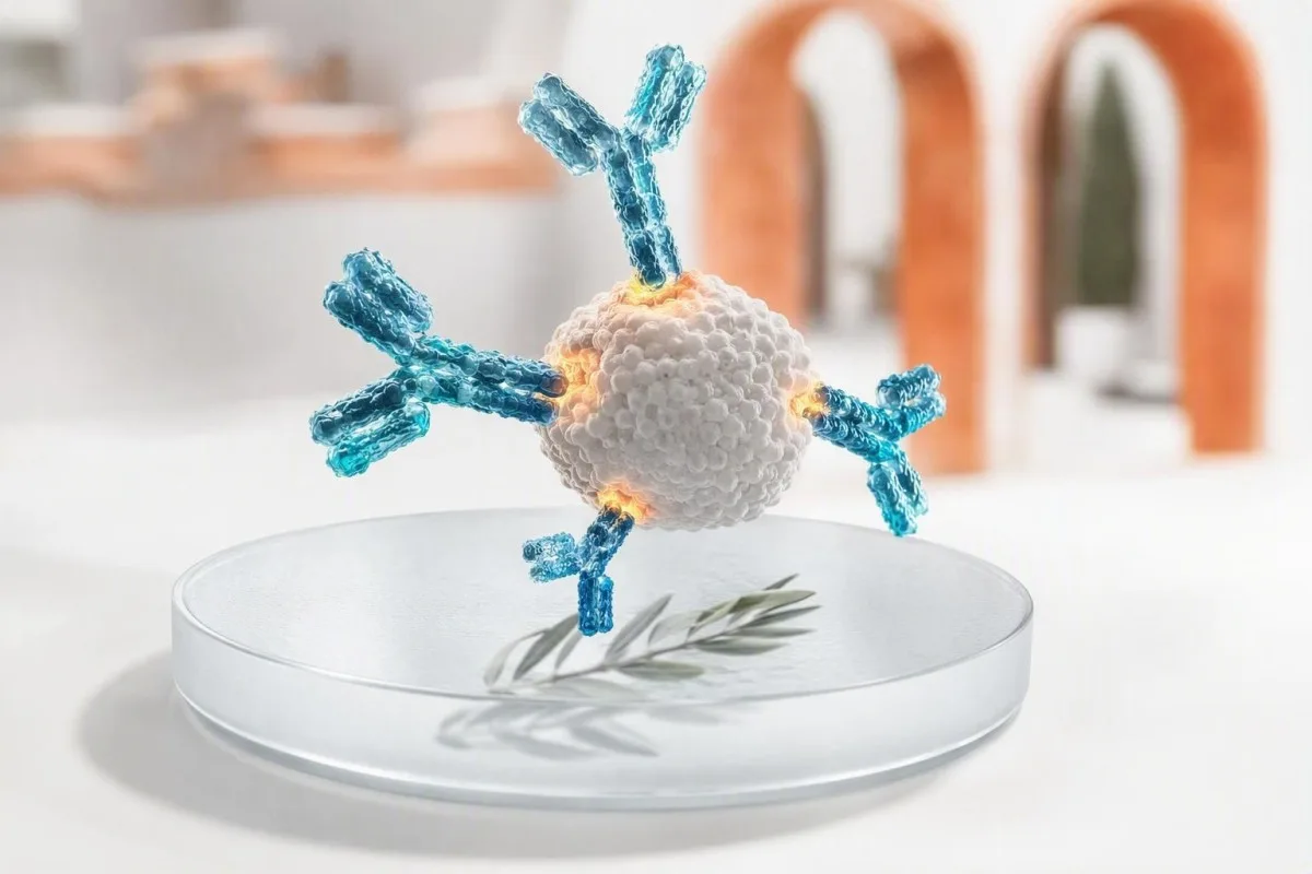
Un resultado positivo de anticuerpos significa que su sistema inmunológico reconoció un objetivo particular; no prueba, por sí solo, una infección activa o una enfermedad. La clase de anticuerpos, el método de prueba, el momento, los síntomas y las pruebas complementarias determinan si necesita tranquilidad, una prueba repetida, confirmación o atención clínica inmediata.


- Resultado positivo de anticuerpos generalmente muestra reconocimiento inmune, no necesariamente enfermedad actual o contagiosidad.
- IgM frente a IgG es una pista de tiempo, no un reloj: la IgM puede persistir durante meses y la IgG puede aparecer temprano.
- Anticuerpos de vacunas pueden hacer que las pruebas de inmunidad den positivo sin indicar infección natural.
- IgE específica de alérgeno por encima de 0.35 kUA/L indica sensibilización, pero los síntomas después de la exposición establecen la alergia clínica.
- positividad del ANA ocurre en hasta 1 de cada 4 adultos sanos con títulos bajos, especialmente con la edad; los síntomas y los anticuerpos específicos son más importantes.
- reactividad de la detección del VIH siempre requiere el algoritmo de confirmación de laboratorio antes de que se establezca cualquier diagnóstico.
- IgA anti-tTG es fiable para la evaluación celíaca solo cuando la persona está comiendo gluten y tiene IgA total adecuada.
- Síntomas urgentes como dificultad para respirar, confusión, ictericia, dolor en el pecho o una erupción ampollosa generalizada, tienen más peso que el resultado de un anticuerpo de rutina.
Lo que realmente le dice un resultado positivo de anticuerpos
A resultado positivo de anticuerpo significa que el ensayo detectó proteínas inmunes que se unen a un objetivo particular; no no significa automáticamente que tiene una infección activa, una enfermedad autoinmune o una alergia que requiere tratamiento. El siguiente paso depende del tipo de anticuerpo, la intensidad del resultado, los síntomas, el momento de la exposición y cualquier antígeno, PCR o prueba de función orgánica asociada.
Los anticuerpos son proteínas de las células B producidas después de la vacunación, infección, exposición a alérgenos, autoinmunidad o, ocasionalmente, sin una razón clínicamente perjudicial. Un resultado positivo responde a una pregunta analítica limitada —¿se detectó unión por encima de este umbral de laboratorio?— en lugar de la pregunta clínica más amplia de qué está sucediendo en su cuerpo hoy.
En mis 15 años de práctica clínica, el error más común ha sido interpretar “positivo” como un diagnóstico. La regla práctica del Dr. Thomas Klein es identificar primero el objetivo—un virus, antígeno de vacuna, proteína alimentaria, enzima tiroidea, antígeno nuclear o fármaco— y luego preguntar si la prueba detecta exposición pasada, organismo presente, sensibilización o desorientación inmune.
Kantesti AI es un Analizador de sangre con inteligencia artificial que lee banderas de anticuerpos junto a fechas, clase de inmunoglobulina y resultados relacionados en lugar de tratar una sola línea positiva como un veredicto. Ese contexto es particularmente útil cuando los pacientes comparan informes de diferentes laboratorios; resultados cualitativos y cuantitativos no son intercambiables incluso cuando ambos dicen positivo.
IgG frente a IgM positivo: por qué el momento es menos simple de lo que parece
IgM positiva puede sugerir una respuesta inmune reciente, mientras que IgG positiva a menudo sugiere exposición pasada o respuesta a la vacuna; ningún patrón data de manera fiable una enfermedad por sí solo. Algunas infecciones producen IgG en 7-14 días, y la IgM puede permanecer detectable durante 3-12 meses o más.
La IgM es un anticuerpo grande de cinco unidades que a menudo es detectable al principio, pero su tamaño y su unión amplia la hacen más propensa a reacciones cruzadas. El virus de Epstein-Barr, el parvovirus B19, el factor reumatoide y algunos anticuerpos autoinmunes pueden causar reacciones de IgM engañosas; un resultado aislado de IgM de bajo nivel merece más escepticismo que pánico.
La IgG generalmente persiste después de que se desarrolla la memoria inmune, a veces durante décadas. Un resultado positivo de IgG puede reflejar una infección infantil, una vacuna reciente, anticuerpos pasivos de inmunoglobulina intravenosa o transferencia materna a un bebé; no puede probar protección a menos que ese ensayo en particular tenga un umbral protector validado.
Los especímenes emparejados pueden ser más útiles que una sola extracción: un aumento de cuatro veces en el título de anticuerpos entre muestras tomadas aproximadamente 2-4 semanas de diferencia apoya la infección reciente en enfermedades seleccionadas. Si el informe enumera IgG, IgA e IgM totales, revise el patrón en lugar de un número con nuestro guía de interpretación de inmunoglobulinas.
Cuándo los anticuerpos de infección significan o no enfermedad activa
Los anticuerpos de infección suelen indicar exposición, mientras que la PCR, las pruebas de antígenos, el cultivo o un título creciente son mejores herramientas para detectar muchas infecciones activas. Un resultado positivo de anticuerpos sin síntomas suele ser histórico, pero la excepción depende del organismo y de su estado inmunológico.
En la hepatitis C, un anticuerpo reactivo significa exposición en algún momento, pero solo el ARN del VHC detectable establece la viremia actual. En la hepatitis B, la combinación es importante: el antígeno de superficie, el anticuerpo de superficie y el anticuerpo central separan la infección activa, la infección resuelta y la inmunidad por vacunación mucho mejor que un solo marcador positivo.
El VIH es otro entorno en el que la redacción importa enormemente. Un cribado reactivo de cuarta generación no es un diagnóstico; el laboratorio realiza una inmunoensayo de diferenciación de VIH-1/VIH-2 y, cuando es necesario, una prueba de ácido nucleico. Una persona que recibe profilaxis postexposición necesita pruebas específicas del momento, como se explica en nuestra guía de pruebas de VIH después de PEP.
He visto a un paciente bien asustado por un anticuerpo treponémico de sífilis positivo después de un tratamiento 18 años antes. Las pruebas treponémicas a menudo permanecen positivas de por vida, mientras que un título de RPR o VDRL ayuda a juzgar la actividad actual y la respuesta; patrones de pruebas de sífilis hacen esta distinción más clara.
Cómo los anticuerpos de las vacunas difieren de los anticuerpos después de la infección
Los anticuerpos relacionados con vacunas muestran que el sistema inmunológico respondió a un antígeno de vacuna; no significan que la vacuna causó una infección activa. Un resultado positivo aún puede caer por debajo de un correlato protector conocido, porque los anticuerpos de unión y los anticuerpos neutralizantes funcionales no son idénticos.
Las concentraciones de anticuerpos de superficie de la hepatitis B 10 mUI/mL o más medidas 1-2 meses después de una serie de vacunación documentada se consideran generalmente protectoras en personas inmunocompetentes. El anti-HBc, el anticuerpo central de la hepatitis B, no es producido por la vacunación estándar contra la hepatitis B; su presencia apunta a una exposición natural y debe indicar una interpretación de panel completo.
La IgG de rubéola se suele comprobar antes del embarazo porque indica inmunidad previa, no una enfermedad de rubéola activa. Los laboratorios establecen umbrales específicos para cada ensayo, y los resultados límite suelen tratarse como no inmunes para la planificación preconcepcional en lugar de interpretarse como una infección reciente; el momento se detalla en nuestra guía de pruebas de rubéola.
Kantesti AI es un plataforma de interpretación análisis de sangre de AI que separa los anticuerpos del patrón de vacuna de los anticuerpos del patrón de infección cuando el informe incluye los marcadores complementarios relevantes. La pregunta práctica a menudo no es “¿soy positivo?”, sino “¿qué antígeno se midió, cuándo me vacuné y este resultado cambia el plan de protección de hoy?”.”
Por qué un resultado positivo en una prueba de anticuerpos contra alergias puede no significar una alergia
Un alérgeno específico positivo IgE la prueba indica sensibilización, lo que significa que la IgE reconoce un alérgeno; no diagnostica una alergia clínica sin un historial de reacción compatible. Un valor superior a 0.35 kUA/L es un umbral de positividad analítica común, no un umbral universal para evitar un alimento o medicamento.
La historia pre-prueba es más importante de lo que mucha gente cree. Si una persona come cacahuete semanalmente sin urticaria, sibilancias, vómitos o colapso, una IgE de cacahuete positiva baja por sí sola no debería llevar a la evitación; la restricción alimentaria innecesaria puede causar daños nutricionales y sociales.
La guía de alergias alimentarias patrocinada por el NIAID establece que las pruebas cutáneas y las pruebas de IgE específicas identifican posibles alimentos desencadenantes pero no pueden diagnosticar de forma independiente la alergia alimentaria (Sampson et al., 2010). En casos seleccionados, un alergólogo utiliza pruebas dirigidas por componentes o un desafío alimentario oral médicamente supervisado, que sigue siendo el estándar de referencia para resolver la incertidumbre.
La IgE total se mide en UI/mL y puede ser alta en eczema, exposición parasitaria, tabaquismo y varias afecciones inmunitarias; no puede identificar un alérgeno causante. Las pruebas de componentes pueden distinguir una proteína de almacenamiento estable de una proteína relacionada con el polen y de reactividad cruzada, una diferencia explorada en nuestra artículo sobre pruebas de alergia molecular.
Por qué los anticuerpos autoinmunes requieren síntomas y pistas orgánicas
Los autoanticuerpos pueden preceder a la enfermedad, acompañarla o presentarse en personas sanas; un resultado positivo por sí solo rara vez establece un diagnóstico autoinmune. Los médicos actúan cuando el patrón de anticuerpos coincide con síntomas, hallazgos del examen, marcadores de inflamación o cambios orgánicos objetivos.
Un anticuerpo antinuclear, o ANA, es positivo a títulos bajos en aproximadamente 1 de cada 4 personas a 1:80 y alrededor de 1 de cada 4 a 1:40 en estudios poblacionales, con tasas más altas en mujeres y adultos mayores. Un patrón homogéneo o moteado no diagnostica el lupus; los ANA se utilizan mejor como prueba de detección inicial cuando el cuadro clínico hace que la enfermedad del tejido conectivo sea plausible.
El marco de clasificación de lupus EULAR/ACR de 2019 utiliza ANA a un título de al menos 1:80 como criterio de entrada, y luego requiere características clínicas e inmunológicas adicionales ponderadas (Aringer et al., 2019). La aparición de proteínas en la orina, niveles bajos de C3/C4, citopenias, hinchazón articular inflamatoria o una erupción fotosensible convincente cambian la urgencia mucho más que un ANA débil por sí solo.
Los anti-ADNdc cobran mayor significado cuando se combinan con un complemento bajo y hallazgos renales, aunque los métodos de ensayo difieren sustancialmente. Nuestra guía de seguimiento de anti-ADNdc y descripción general de las pruebas del complemento explica por qué un especialista puede repetir las pruebas en una plataforma diferente.
Anticuerpos específicos de órganos: ejemplos de tiroides, CE y estómago
Los anticuerpos órgano-específicos identifican el ataque inmune a un tejido, pero no siempre muestran si el órgano está dañado actualmente o necesita tratamiento. Los anticuerpos contra la peroxidasa tiroidea, los anticuerpos contra la transglutaminasa tisular y los anticuerpos contra el factor intrínseco requieren cada uno pruebas complementarias diferentes.
Los anticuerpos anti-TPO son comunes en la tiroiditis de Hashimoto y pueden estar presentes durante años mientras la TSH y la T4 libre permanecen normales. En un adulto no embarazado con función tiroidea normal, el resultado generalmente apoya el monitoreo periódico de la TSH en lugar del tratamiento con hormona tiroidea; el embarazo, los planes de fertilidad y los síntomas pueden alterar ese enfoque.
Una prueba tTG-IgA sugiere la enfermedad celíaca solo si la persona sigue consumiendo gluten y la IgA total es adecuada. La deficiencia selectiva de IgA puede hacer que la tTG-IgA sea falsamente negativa, por lo que los médicos utilizan pruebas de péptidos de gliadina desaminados basadas en IgG o tTG cuando la IgA total es baja; consulte nuestra explicación de trampas de IgA baja.
El anticuerpo contra el factor intrínseco es relativamente específico pero no muy sensible para la anemia perniciosa. Cuando es positivo junto con niveles bajos de B12, aumento del ácido metilmalónico y macrocitosis, el patrón es convincente; sin ese patrón, evito tratar una etiqueta de laboratorio en lugar del paciente. pruebas de anemia perniciosa muestra la secuencia de confirmación habitual.
Qué causa un resultado de anticuerpos falsos positivos
Los resultados de anticuerpos falsos positivos surgen cuando el ensayo detecta unión no deseada, no cuando el laboratorio simplemente “se equivoca”. Anticuerpos cruzados, factor reumatoide, estimulación inmune reciente, inmunoglobulina pasiva y baja probabilidad pre-prueba son razones comunes.
La especificidad no es perfección. Una prueba con 99% de especificidad produce aproximadamente 10 resultados falsos positivos por cada 1000 personas verdaderamente negativas analizadas; cuando la condición es rara, esos resultados pueden superar a los positivos verdaderos. Por eso el cribado amplio en alguien sin síntomas a menudo crea ambigüedad en lugar de tranquilidad.
La biotina es famosa por interferir con algunos ensayos hormonales y cardíacos, mientras que los anticuerpos heterófilos y el factor reumatoide pueden interferir con ciertos inmunoensayos. El laboratorio a veces puede diluir la muestra, usar reactivos bloqueantes, repetir el resultado en otra arquitectura de ensayo o realizar un método de confirmación más específico.
Un resultado positivo que entra en conflicto con un cuadro clínico completamente normal no es algo que deba ignorarse; es una razón para verificar. 1% AI compara marcadores discordantes y fechas de prueba, y nuestro lista de verificación de la precisión del resultado de laboratorio puede ayudarle a registrar suplementos, infusiones recientes y enfermedades antes de hablar con el médico que solicitó la prueba.
Resultado positivo de anticuerpos pero sin síntomas: un plan de triaje práctico
Cuando una prueba de anticuerpos es positiva pero usted se siente bien, la mayoría de las personas necesitan aclaración en lugar de atención de emergencia. Los primeros pasos más seguros son verificar el ensayo exacto, comprobar las fechas de exposición y vacunación, y buscar la prueba complementaria que confirme la enfermedad o la afectación orgánica actual.
Comience con el informe en sí: registre el nombre del analito, la clase de anticuerpo, el índice numérico o el título, el intervalo de referencia, el método de laboratorio y la fecha de recolección. “Reactivo” puede significar una relación señal-corte apenas superior a 1.0 o un resultado mucho más fuerte; los laboratorios no utilizan umbrales idénticos, y la magnitud solo a veces sigue la probabilidad clínica.
A continuación, pregunte si existe una prueba de enfermedad activa. Los ejemplos incluyen PCR para hepatitis C, RPR en serie para sífilis, pruebas de heces o aliento para Helicobacter pylori activo, TSH/T4 libre para anticuerpos tiroideos y análisis de orina más creatinina para preocupación renal relacionada con el lupus.
A partir del 8 de septiembre de 2026, no recomendaría iniciar antibióticos, antivirales, dietas sin gluten, medicamentos para la tiroides o evitar alimentos basándome únicamente en un resultado aislado de anticuerpos. Guarde el PDF original, anote su línea base con el seguimiento longitudinal de laboratorio, y programe una revisión de rutina a menos que haya síntomas de advertencia.
Pruebas confirmatorias que cambian el siguiente paso
La mejor prueba confirmatoria depende de lo que tenga como objetivo el anticuerpo: la detección molecular confirma muchas infecciones, los ensayos funcionales aclaran alergias y las pruebas de función orgánica evalúan el impacto autoinmune. Repetir la misma prueba de anticuerpos solo es útil cuando el cambio esperado con el tiempo tiene significado clínico.
Para una prueba de detección de VIH reactiva, la vía de confirmación de reflejo es un ensayo de diferenciación seguido de pruebas de ácido nucleico del VIH si los resultados son indeterminados. Para un anticuerpo de hepatitis C, el ARN del VHC es el seguimiento decisivo; para un anticuerpo central de hepatitis B positivo, el antígeno de superficie, el anticuerpo de superficie y a veces el ADN del VHB aclaran el patrón.
Para la sospecha de enfermedad celíaca, las decisiones de biopsia deben tomarse mientras la exposición al gluten sea suficiente para la prueba. Para una posible alergia alimentaria, un alergólogo considera el historial de exposición, el nivel de IgE específico, el perfil de componentes y el riesgo de provocación, no un panel de resultados positivos bajos y no relacionados pedidos sin una pregunta clínica.
Kantesti AI es un Herramienta de análisis de pruebas de sangre con IA que puede organizar estos marcadores vinculados en una lista de preguntas lista para el médico, pero no reemplaza las pruebas de confirmación o el diagnóstico. Si no está seguro de cuánto confiar en un resultado atípico, nuestro normas de validación médica describe por qué la verificación y la supervisión clínica son importantes.
Síntomas que hacen que un resultado positivo de anticuerpos sea más urgente
Un resultado positivo de anticuerpos justifica una evaluación urgente cuando se acompaña de dificultad para respirar, hinchazón facial o de garganta, desmayos, confusión, dolor en el pecho, ictericia, erupción cutánea que se propaga rápidamente, deshidratación severa o debilidad neurológica nueva. Los síntomas, no el número de anticuerpos solo, establecen la urgencia.
La posible anafilaxia es una emergencia: urticaria generalizada repentina con sibilancias, opresión en la garganta, vómitos persistentes, desvanecimiento o presión arterial baja después de la exposición requiere servicios de emergencia y epinefrina recetada si está disponible. Una IgE alimentaria o de veneno positiva de hace meses no predice ni descarta ese riesgo inmediato.
La ictericia nueva con orina oscura, dolor abdominal marcado, fiebre o confusión debe provocar una evaluación el mismo día, ya sean positivos los anticuerpos de hepatitis viral o no. Las enzimas hepáticas, la bilirrubina, el INR y las pruebas directas de patógenos ayudan a distinguir el historial inmune de la enfermedad hepatobiliar activa; componentes del panel hepático son un contexto útil.
La preocupación autoinmune se vuelve sensible al tiempo con tos con sangre, dificultad para respirar, creatinina en rápido aumento, diarrea con sangre, cambios visuales, convulsiones o una nueva debilidad unilateral. He aprendido a no tranquilizar a alguien basándome en un título de anticuerpo límite cuando su trayectoria sintomática se está deteriorando claramente.
¿Un número de anticuerpos más alto significa una enfermedad más grave?
Un índice o título de anticuerpo más alto a veces aumenta la probabilidad de que un resultado sea genuino, pero generalmente no mide la gravedad de la enfermedad. Los valores de los ensayos son específicos del método, y un resultado de 80 AU/mL no se puede comparar directamente con 80 U/mL de otro laboratorio.
En la enfermedad autoinmune, las tendencias de los anti-ADN de cadena doble a veces pueden seguir la actividad del lupus, particularmente cuando se miden consistentemente por el mismo método y se interpretan con el complemento y la proteína en la orina. El título de ANA, sin embargo, es una medida deficiente de la gravedad del lupus y no debe repetirse en una persona estable.
En las infecciones, una fuerte señal de anticuerpos puede respaldar la exposición previa, pero rara vez indica infecciosidad. Las pruebas de anticuerpos neutralizantes son especializadas y sus niveles de corte protectores pueden variar según el patógeno, la edad, el producto de la vacuna y la inmunosupresión; un simple ensayo comercial de IgG no es un pasaporte de inmunidad personal.
Al comparar informes, mantenga visible el laboratorio, el ensayo y las unidades. Un cambio biológico real debe exceder la variación analítica y diaria ordinaria, razón por la cual nuestro guía de comparación de análisis de sangre se centra en las fechas y los métodos en lugar de solo en las banderas codificadas por colores.
El embarazo, los niños y la inmunosupresión cambian la interpretación
El embarazo, la infancia y el tratamiento inmunosupresor pueden alterar tanto la producción de anticuerpos como la seguridad de esperar la confirmación. En estos grupos, un resultado positivo puede reflejar anticuerpos maternos, respuesta reducida a la vacuna, riesgo de reactivación o la necesidad de asesoramiento especializado más temprano.
La IgG materna cruza la placenta y puede permanecer detectable en un bebé durante varios meses, mientras que la IgM materna generalmente no cruza. Por lo tanto, la positividad aislada de IgG en un bebé a menudo refleja la inmunidad materna, mientras que la IgM específica del organismo o la detección directa pueden merecer una evaluación más específica según la infección y la edad.
Durante el embarazo, los resultados de anticuerpos contra la rubéola o el parvovirus se interpretan con las fechas de exposición, pruebas de avidez y muestras repetidas en lugar de una única IgM. La IgM falsamente positiva es un problema reconocido, y la alarma evitable puede conducir a investigaciones invasivas innecesarias; la opinión obstétrica o de enfermedades infecciosas es sensata cuando la exposición es creíble.
Las personas que reciben rituximab, corticosteroides en dosis altas, quimioterapia o medicación para trasplantes pueden tener respuestas de anticuerpos débiles o ausentes a pesar de la infección o la vacunación. Para las decisiones familiares, los límites de interpretación de la IA para niños y el junta asesora médica refuerzan la necesidad de una revisión dirigida por el médico.
Cómo prepararse para una revisión clínica útil de un resultado positivo
Traiga el informe de laboratorio completo, el motivo por el que se solicitó la prueba, las fechas de los síntomas o la exposición, el historial de vacunas, los medicamentos y los resultados previos. Esta información puede convertir un resultado de anticuerpo positivo aparentemente vago en una acción clara en una sola cita.
Anote cinco hechos antes de la visita: el primer día de los síntomas, la última exposición plausible, las fechas de vacunación, los medicamentos inmunosupresores o las infusiones de inmunoglobulina, y si los síntomas están mejorando o empeorando. Una fiebre que comenzó 3 días antes del muestreo se interpreta de manera muy diferente a la fatiga que comenzó hace 6 meses.
Haga tres preguntas específicas: “¿Qué detecta esta prueba?”, “¿Qué prueba establece la enfermedad activa o el efecto orgánico?” y “¿Qué resultado cambiaría el tratamiento?” El Dr. Thomas Klein considera que estas preguntas reducen los paneles de repetición innecesarios y ayudan a los pacientes a irse con un marco de tiempo definido: hoy, 1-2 semanas o seguimiento de rutina.
La IA Kantesti admite la revisión segura de informes en más de 75 idiomas y puede detectar pruebas complementarias faltantes de un PDF fotografiado o cargado en aproximadamente 60 segundos. Nuestro Guía de tecnología de IA explica el enfoque de coincidencia de contexto, mientras que nuestro equipo clínico proporciona la supervisión médica detrás de la interpretación para el paciente.
Preguntas frecuentes
¿Qué significa un resultado positivo de anticuerpos si no tengo síntomas?
Un resultado de anticuerpos positivo sin síntomas indica la mayoría de las veces exposición previa, vacunación, sensibilización o un resultado que necesita confirmación en lugar de enfermedad activa. El siguiente paso depende del objetivo: el anticuerpo de la hepatitis C necesita ARN del VHC, una prueba de detección reactiva de VIH necesita el algoritmo de confirmación de laboratorio, y los anticuerpos tiroideos necesitan TSH y T4 libre. Un resultado aislado de bajo positivo es menos convincente cuando no hay antecedentes de exposición o síntomas compatibles. Busque atención inmediata si desarrolla ictericia nueva, dificultad para respirar, confusión, dolor en el pecho o una erupción que empeora rápidamente.
¿Significa IgM positivo y IgG negativo siempre una infección nueva?
IgM positivo con IgG negativo puede ocurrir temprano en una infección, pero no siempre prueba una nueva infección. IgM puede reaccionar de forma cruzada con proteínas inmunes no relacionadas, y algunos ensayos producen resultados falsos positivos de IgM con más frecuencia que los resultados de IgG. Los médicos pueden repetir la serología después de 1-2 semanas, solicitar pruebas de PCR o antígenos, o usar un ensayo de confirmación más específico. La interpretación correcta también depende de la línea de tiempo de la enfermedad y del organismo específico analizado.
¿Puede una vacuna hacer que una prueba de anticuerpos dé positivo?
Sí, las vacunas suelen dar positivo en las pruebas de anticuerpos para el antígeno contenido en la vacuna. Para la hepatitis B, una concentración de anti-HBs de al menos 10 mIU/mL medida 1-2 meses después de completar la serie generalmente indica una respuesta protectora de la vacuna en personas inmunocompetentes. La vacunación estándar contra la hepatitis B no produce anti-HBc, por lo que la positividad de los anticuerpos del núcleo sugiere una exposición natural en lugar de la vacunación sola. La positividad relacionada con la vacuna no significa que tenga una infección activa.
¿Se puede tener un análisis de anticuerpos autoinmunes positivo y estar sano?
Sí, las personas sanas pueden tener anticuerpos autoinmunes positivos, especialmente resultados ANA de bajo título. El ANA es detectable en aproximadamente el 13% de las personas con un título de 1:80 y alrededor del 20% con un título de 1:40, por lo que no es una prueba independiente para el lupus. Síntomas como hinchazón inflamatoria de las articulaciones, úlceras bucales, erupción fotosensible, complemento bajo, proteínas en la orina o recuentos sanguíneos anormales determinan si se necesita una evaluación especializada. Un resultado positivo debe interpretarse en el contexto del motivo por el que se solicitó la prueba.
¿Significa un análisis de sangre IgE positivo que debo evitar ese alimento?
No, un resultado positivo de IgE específico para alimentos significa sensibilización y no demuestra que comer el alimento cause una reacción alérgica. Muchos laboratorios llaman positivos a los resultados superiores a 0,35 kUA/L, pero ese umbral está diseñado para la detección en lugar de una decisión universal de evitación. Una persona que consume regularmente el alimento sin urticaria, sibilancias, vómitos repetitivos o desmayos no debe eliminarlo automáticamente de la dieta. Un alergólogo puede utilizar el historial de reacciones, las pruebas de componentes y, a veces, un desafío alimentario oral supervisado para aclarar el riesgo.
¿Cuánto tiempo permanecen positivos los anticuerpos después de una infección?
La duración de los anticuerpos varía ampliamente según la infección y la clase de anticuerpo. La IgM puede desaparecer en pocas semanas, pero puede persistir durante 3 a 12 meses o más, mientras que la IgG puede permanecer medible durante años o de por vida. Los anticuerpos de la sífilis treponémica a menudo permanecen positivos de por vida después de un tratamiento exitoso, mientras que los títulos de RPR o VDRL se utilizan para seguir la actividad. Por lo tanto, un resultado de anticuerpo positivo no puede indicar de manera confiable cuándo ocurrió una infección sin otra información clínica y de laboratorio.
Obtén hoy un análisis de sangre con IA
Únete a más de 2 millones de usuarios en todo el mundo que confían en Kantesti para el análisis instantáneo y preciso de pruebas de laboratorio. Sube tus resultados de análisis de sangre y recibe una interpretación completa de los biomarcadores de 15,000+ en segundos.
📚 Publicaciones de investigación citadas
Klein, T., Mitchell, S., & Weber, H. (2026). Soporte de Decisión Clínica Asistido por IA Multilingüe para el Triaje Temprano de Hantavirus: Diseño, Validación de Ingeniería y Despliegue en el Mundo Real en 50.000 Informes de Análisis de Sangre Interpretados. Figshare. https://www.researchgate.net/. Investigación médica con IA de Kantesti.
Klein, T., Mitchell, S., & Weber, H. (2026). Una evaluación comparativa técnica automática pre-registrada y basada en rúbricas del motor de interpretación de análisis de sangre Kantesti en 100.000 casos de prueba sintéticos. Figshare. https://www.academia.edu/. Investigación médica con IA de Kantesti.
📖 Referencias médicas externas
⚕️ Descargo de responsabilidad médica
Este artículo es solo con fines educativos y no constituye asesoramiento médico. Consulta siempre a un profesional sanitario cualificado para decisiones de diagnóstico y tratamiento.
Señales de confianza E-E-A-T
Experiencia
Revisión clínica dirigida por un médico de los flujos de interpretación de análisis.
Pericia
Enfoque en medicina de laboratorio sobre cómo se comportan los biomarcadores en el contexto clínico.
Autoridad
Escrito por el Dr. Thomas Klein, con revisión de la Dra. Sarah Mitchell y el Prof. Dr. Hans Weber.
Integridad
Interpretación basada en la evidencia con vías de seguimiento claras para reducir la alarma.