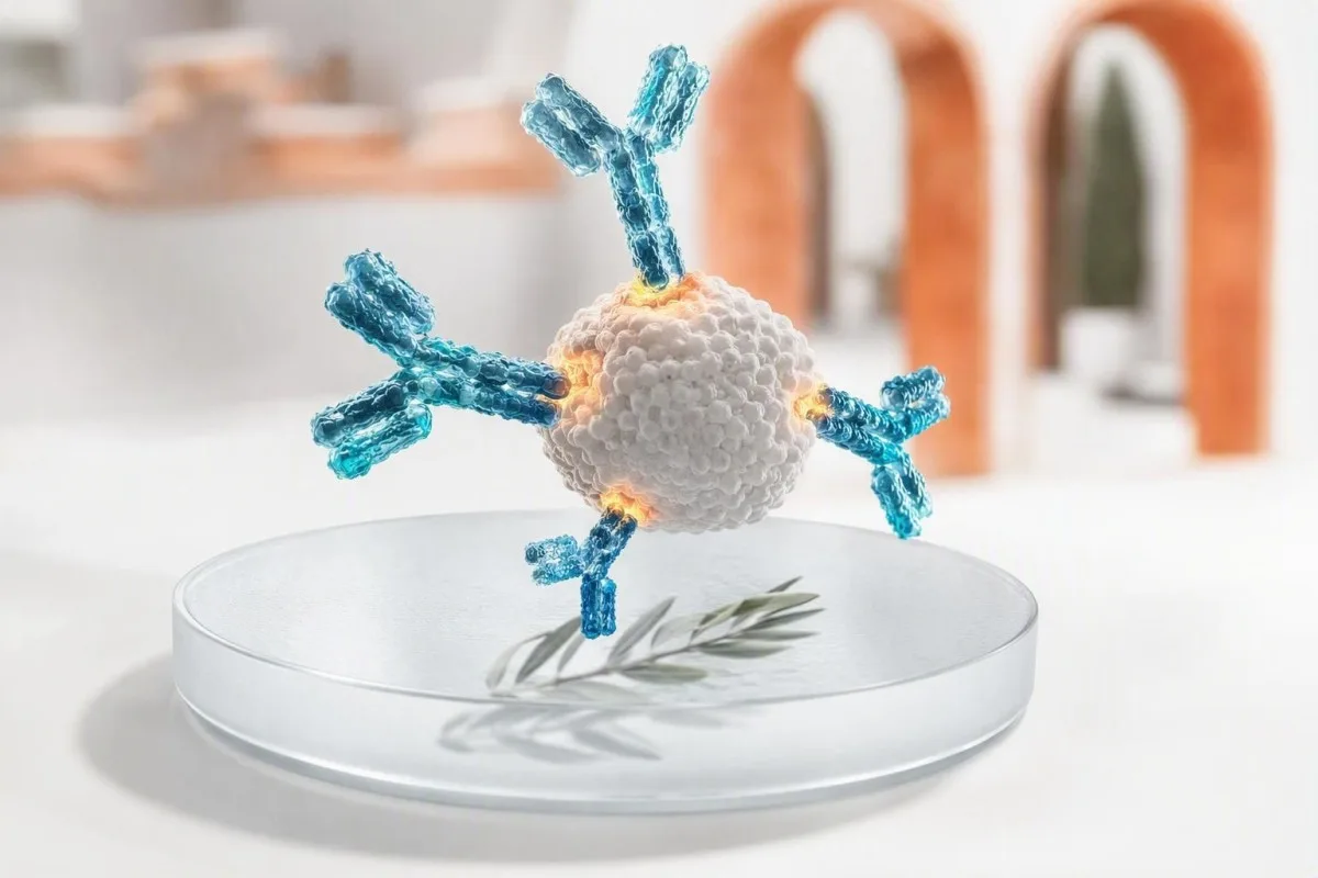
A positive antibody result means your immune system recognised a particular target; it does not, by itself, prove an active infection or a disease. The antibody class, test method, timing, symptoms and companion tests determine whether you need reassurance, a repeat test, confirmation or prompt clinical care.


- Positive antibody result usually shows immune recognition, not necessarily current illness or contagiousness.
- IgM versus IgG is a timing clue, not a clock: IgM may persist for months and IgG can appear early.
- Vaccine antibodies can make immunity tests positive without indicating natural infection.
- Allergen-specific IgE above 0.35 kUA/L indicates sensitisation, but symptoms after exposure establish clinical allergy.
- ANA positivity occurs in up to 20% of healthy adults at low titres, especially with age; symptoms and specific antibodies matter more.
- HIV screening reactivity always requires the laboratory confirmation algorithm before any diagnosis is made.
- Anti-tTG IgA is reliable for coeliac assessment only when the person is eating gluten and has adequate total IgA.
- Urĝaj simptomoj such as breathing difficulty, confusion, jaundice, chest pain or a widespread blistering rash outweigh a routine antibody result.
What a positive antibody result actually tells you
A positive antibody test means the assay detected immune proteins that bind a particular target; it does ne automatically mean you have an active infection, an autoimmune disease, or an allergy that needs treatment. The next step depends on the antibody type, result strength, symptoms, exposure timing and any paired antigen, PCR or organ-function tests.
Antibodies are B-cell proteins made after vaccination, infection, allergy exposure, autoimmunity, or occasionally for no clinically harmful reason. A positive result answers one narrow analytical question—“was binding detected above this laboratory cutoff?”—rather than the broader clinical question of what is happening in your body today.
In my 15 years of clinical practice, the commonest mistake has been reading “positive” as a diagnosis. Dr Thomas Klein’s practical rule is to first identify the target—a virus, vaccine antigen, food protein, thyroid enzyme, nuclear antigen or drug—then ask whether the test detects past exposure, present organism, sensitisation, or immune misdirection.
Kantesti AI estas AI-sangotesta analizilo that reads antibody flags beside dates, immunoglobulin class and related results rather than treating a single positive line as a verdict. That context is particularly useful when patients compare reports from different laboratories; qualitative and quantitative results are not interchangeable even when both say positive.
IgG versus IgM positive: why timing is less simple than it sounds
IgM positive can suggest a recent immune response, while IgG pozitiva often suggests past exposure or vaccine response; neither pattern reliably dates an illness on its own. Some infections produce IgG within 7–14 days, and IgM may remain detectable for 3–12 months or longer.
IgM is a large five-unit antibody that is often detectable early, but its size and broad binding make it more prone to cross-reactions. Epstein–Barr virus, parvovirus B19, rheumatoid factor and some autoimmune antibodies can cause misleading IgM reactions; an isolated low-level IgM result deserves more skepticism than panic.
IgG usually persists after immune memory develops, sometimes for decades. A positive IgG result may reflect childhood infection, a recent vaccine, passive antibody from intravenous immunoglobulin, or maternal transfer to an infant; it cannot prove protection unless that particular assay has a validated protective threshold.
Paired specimens can be more useful than a single draw: a fourfold rise in antibody titre between samples taken roughly 2–4 weeks apart supports recent infection in selected diseases. If the report lists total IgG, IgA and IgM, review the pattern rather than one number with our immunoglobulin interpretation guide.
When infection antibodies do and do not mean active disease
Infection antibodies usually show exposure, whereas PCR, antigen testing, culture, or a rising titre are better tools for detecting many active infections. A positive antibody result without symptoms is often historical, but the exception depends on the organism and your immune status.
For hepatitis C, a reactive antibody means exposure at some point, but only detectable HCV RNA establishes current viraemia. For hepatitis B, the combination matters: surface antigen, surface antibody and core antibody separate active infection, resolved infection and vaccine immunity far better than one positive marker.
HIV is another setting where wording matters enormously. A reactive fourth-generation screen is not a diagnosis; the laboratory follows with an HIV-1/HIV-2 differentiation immunoassay and, when needed, nucleic-acid testing. A person taking post-exposure prophylaxis needs time-specific testing, as explained in our HIV testing after PEP guide.
I have seen a well patient frightened by a positive treponemal syphilis antibody after treatment 18 years earlier. Treponemal tests commonly stay positive for life, while an RPR or VDRL titre helps judge current activity and response; syphilis test patterns make this distinction clearer.
How vaccine antibodies differ from antibodies after infection
Vaccine-related antibodies show that the immune system responded to a vaccine antigen; they do not mean the vaccine caused an active infection. A positive result may still fall below a known protective correlate, because binding antibodies and functional neutralising antibodies are not identical.
Hepatitis B surface antibody concentrations of 10 mIU/mL aŭ pli alte measured 1–2 months after a documented vaccine series are generally considered protective in immunocompetent people. Anti-HBc, the hepatitis B core antibody, is not produced by standard hepatitis B vaccination; its presence points toward natural exposure and should prompt full-panel interpretation.
Rubella IgG is commonly checked before pregnancy because it indicates prior immunity, not an active rubella illness. Laboratories set assay-specific thresholds, and borderline results are usually handled as non-immune for preconception planning rather than interpreted as recent infection; timing is detailed in our rubella testing guide.
Kantesti AI estas AI sangotesta interpretada platformo that separates vaccine-pattern antibodies from infection-pattern antibodies when the report includes the relevant companion markers. The practical question is often not “am I positive?” but “which antigen was measured, when was I vaccinated, and does this result change today’s protection plan?”
Why a positive allergy antibody test may not mean an allergy
A positive allergen-specific IgE test indicates sensitisation, meaning IgE recognises an allergen; it does not diagnose clinical allergy without a compatible reaction history. A value above 0.35 kUA/L is a common analytical positivity threshold, not a universal threshold for avoiding a food or medicine.
The pre-test story matters more than many people realise. If a person eats peanut weekly without hives, wheeze, vomiting or collapse, a low positive peanut IgE alone should not lead to avoidance; unnecessary food restriction can create nutritional and social harm.
The NIAID-sponsored food allergy guideline states that skin-prick and specific-IgE tests identify possible trigger foods but cannot independently diagnose food allergy (Sampson et al., 2010). In selected cases, an allergist uses component-resolved testing or a medically supervised oral food challenge, which remains the reference standard for resolving uncertainty.
Total IgE is measured in IU/mL and can be high in eczema, parasitic exposure, smoking and several immune conditions; it cannot identify a culprit allergen. Component testing can distinguish a stable storage protein from a cross-reactive pollen-related protein, a difference explored in our molecular allergy testing article.
Why autoimmune antibodies require symptoms and organ clues
Autoimmune antibodies can precede disease, accompany disease, or occur in healthy people; a positive result alone rarely establishes an autoimmune diagnosis. Clinicians act when the antibody pattern matches symptoms, examination findings, inflammation markers or objective organ changes.
An antinuclear antibody, or ANA, is positive at low titres in roughly 13% of people at 1:80 and around 20% at 1:40 in population studies, with higher rates in women and older adults. A homogeneous or speckled pattern does not diagnose lupus; ANA is best used as a gateway test when the clinical picture makes connective-tissue disease plausible.
The 2019 EULAR/ACR lupus classification framework uses ANA at a titre of at least 1:80 as an entry criterion, then requires additional weighted clinical and immunological features (Aringer et al., 2019). New protein in urine, low C3/C4, cytopenias, inflammatory joint swelling or a convincing photosensitive rash change the urgency much more than a weak ANA alone.
Anti-dsDNA becomes more meaningful when paired with low complement and kidney findings, although assay methods differ substantially. Our anti-dsDNA follow-up guide kaj complement testing overview explain why a specialist may repeat testing on a different platform.
Organ-specific antibodies: thyroid, coeliac and stomach examples
Organ-specific antibodies identify immune targeting of a tissue, but they do not always show whether the organ is currently damaged or needs treatment. Thyroid peroxidase antibodies, tissue transglutaminase antibodies and intrinsic-factor antibodies each require different companion tests.
TPO antibodies are common in Hashimoto thyroiditis and can be present for years while TSH and free T4 remain normal. In a non-pregnant adult with normal thyroid function, the result usually supports periodic TSH monitoring rather than thyroid hormone treatment; pregnancy, fertility plans and symptoms can alter that approach.
Pozitiva tTG-IgA suggests coeliac disease only if the person is still consuming gluten and total IgA is adequate. Selective IgA deficiency can make tTG-IgA falsely negative, so clinicians use IgG-based deamidated gliadin peptide or tTG tests when total IgA is low; see our explanation of low IgA pitfalls.
Intrinsic-factor antibody is relatively specific but not highly sensitive for pernicious anaemia. When it is positive alongside low B12, raised methylmalonic acid and macrocytosis, the pattern is compelling; without that pattern, I avoid treating a laboratory label rather than the patient. Pernicious anaemia testing shows the usual confirmation sequence.
What causes a false-positive antibody result
False-positive antibody results arise when the assay detects unintended binding, not when the laboratory simply “gets it wrong.” Cross-reactive antibodies, rheumatoid factor, recent immune stimulation, passive immunoglobulin and low pre-test probability are common reasons.
Specificity is not perfection. A test with 99% specificity produces about 10 false-positive results for every 1,000 truly negative people tested; when the condition is rare, those results can outnumber true positives. This is why broad screening in someone with no symptoms often creates ambiguity rather than reassurance.
Biotin is famous for interfering with some hormone and cardiac assays, while heterophile antibodies and rheumatoid factor can interfere with certain immunoassays. The laboratory can sometimes dilute the sample, use blocking reagents, repeat the result on another assay architecture, or perform a more specific confirmatory method.
A positive result that conflicts with a completely unremarkable clinical picture is not something to ignore; it is a reason to verify. Kantesti AI compares discordant markers and test dates, and our kontrol-listo pri precizeco de labortestrezultoj can help you record supplements, recent infusions and illnesses before speaking with the ordering clinician.
Antibody test positive but no symptoms: a practical triage plan
When an antibody test is positive but you feel well, most people need clarification rather than emergency care. The safest first steps are to verify the exact assay, check exposure and vaccination dates, and look for the paired test that confirms current disease or organ involvement.
Start with the report itself: record the analyte name, antibody class, numeric index or titre, reference interval, laboratory method and collection date. “Reactive” can mean a signal-to-cutoff ratio barely above 1.0 or a much stronger result; laboratories do not use identical thresholds, and the magnitude only sometimes tracks clinical probability.
Next, ask whether an active-disease test exists. Examples include PCR for hepatitis C, serial RPR for syphilis, stool or breath testing for active Helicobacter pylori, TSH/free T4 for thyroid antibodies, and urinalysis plus creatinine for lupus-related kidney concern.
As of September 8, 2026, I would not recommend self-starting antibiotics, antivirals, gluten-free diets, thyroid medication or food avoidance solely from an isolated antibody result. Save the original PDF, note your baseline with longitudaj laboratoriaj sekvadoj, and arrange routine review unless red-flag symptoms are present.
Confirmatory tests that change the next step
The best confirmatory test depends on what the antibody targets: molecular detection confirms many infections, functional assays clarify allergy, and organ-function testing assesses autoimmune impact. Repeating the same antibody test is useful only when the expected change over time has clinical meaning.
For a reactive HIV screen, the reflex confirmation pathway is a differentiation assay followed by HIV nucleic-acid testing if results are indeterminate. For a hepatitis C antibody, HCV RNA is the decisive follow-up; for a positive hepatitis B core antibody, surface antigen, surface antibody and sometimes HBV DNA clarify the pattern.
For suspected coeliac disease, biopsy decisions should be made while gluten exposure is sufficient for testing. For possible food allergy, an allergist considers the exposure history, specific-IgE level, component profile and challenge risk—not a panel of unrelated low positives ordered without a clinical question.
Kantesti AI estas AI-movita ilo por analizo de sangotestoj that can organise these linked markers into a clinician-ready question list, but it does not replace confirmatory testing or diagnosis. If you are unsure how much to trust an outlying result, our medicinan validigan normaron describe why verification and clinical oversight matter.
Symptoms that make a positive antibody result more urgent
A positive antibody result warrants urgent assessment when it accompanies breathing difficulty, facial or throat swelling, fainting, confusion, chest pain, jaundice, rapidly spreading rash, severe dehydration or new neurological weakness. Symptoms—not the antibody number alone—set the urgency.
Possible anaphylaxis is an emergency: sudden widespread hives with wheeze, throat tightness, persistent vomiting, faintness or low blood pressure after exposure needs emergency services and prescribed adrenaline if available. A positive food or venom IgE from months ago neither predicts nor rules out that immediate risk.
New jaundice with dark urine, marked abdominal pain, fever or confusion should prompt same-day evaluation whether viral hepatitis antibodies are positive or not. Liver enzymes, bilirubin, INR and direct pathogen testing help distinguish immune history from active hepatobiliary illness; hepata-panelaj komponantoj are useful context.
Autoimmune concern becomes time-sensitive with coughing blood, shortness of breath, rapidly rising creatinine, bloody diarrhoea, visual change, seizure or a new one-sided weakness. I have learned not to reassure someone based on a borderline antibody titre when their symptom trajectory is clearly deteriorating.
Does a higher antibody number mean more severe disease?
A higher antibody index or titre sometimes increases the likelihood that a result is genuine, but it usually does not measure disease severity. Assay values are method-specific, and a result of 80 AU/mL cannot be compared directly with 80 U/mL from another laboratory.
In autoimmune disease, anti-dsDNA trends can sometimes follow lupus activity, particularly when measured consistently by the same method and interpreted with complement and urine protein. ANA titre, however, is a poor measure of lupus severity and should not be chased repeatedly in a stable person.
For infections, a strong antibody signal may support prior exposure but rarely indicates infectiousness. Neutralising antibody tests are specialised and their protective cutoffs may vary by pathogen, age, vaccine product and immune suppression; a simple commercial IgG assay is not a personal immunity passport.
When comparing reports, keep the laboratory, assay and units visible. A real biological change should exceed ordinary analytical and day-to-day variation, which is why our sang-testa kompara gvidilo focuses on dates and methods rather than colour-coded flags alone.
Pregnancy, children and immune suppression change interpretation
Pregnancy, infancy and immune-suppressing treatment can alter both antibody production and the safety of waiting for confirmation. In these groups, a positive result may reflect maternal antibodies, reduced vaccine response, reactivation risk or a need for earlier specialist advice.
Maternal IgG crosses the placenta and can remain detectable in an infant for several months, whereas maternal IgM generally does not cross. Therefore, an infant’s isolated IgG positivity often reflects maternal immunity, while organism-specific IgM or direct detection may deserve more focused evaluation depending on the infection and age.
During pregnancy, rubella or parvovirus antibody results are interpreted with exposure dates, avidity testing and repeat samples rather than a one-off IgM alone. False-positive IgM is a recognised problem, and avoidable alarm can lead to unnecessary invasive investigations; obstetric or infectious-disease input is sensible when exposure is credible.
People receiving rituximab, high-dose corticosteroids, chemotherapy or transplant medication may have weak or absent antibody responses despite infection or vaccination. For family decisions, children’s AI interpretation limits kaj la medicina konsila komitato reinforce the need for clinician-led review.
How to prepare for a useful clinician review of a positive result
Bring the complete laboratory report, the reason the test was ordered, dates of symptoms or exposure, vaccine history, medicines and prior results. This information can turn an apparently vague positive antibody result into a clear next action within one appointment.
Write down five facts before the visit: the first day of symptoms, the last plausible exposure, vaccine dates, immune-suppressing medicines or immunoglobulin infusions, and whether symptoms are improving or worsening. A fever that began 3 days before sampling is interpreted very differently from fatigue that started 6 months ago.
Ask three focused questions: “What does this test detect?”, “What test establishes active disease or organ effect?”, and “What result would change treatment?” Dr Thomas Klein finds these questions reduce unnecessary repeat panels and help patients leave with a defined time frame—today, 1–2 weeks, or routine monitoring.
Kantesti AI supports secure report review across more than 75 languages and can surface missing companion tests from a photographed or uploaded PDF in about 60 seconds. Our Gvidilo de AI-teknologio explains the context-matching approach, while nia klinika teamo provides the medical oversight behind patient-facing interpretation.
Oftaj Demandoj
Kion signifas pozitiva antikorpa rezulto se mi havas neniujn simptomojn?
Pozitiva antikorpa rezulto sen simptomoj plejofte indikas antaŭan ekspozicion, vakcinadon, sensivigon aŭ rezulton kiu bezonas konfirmon prefere ol aktivan malsanon. La sekva paŝo dependas de la celo: hepatito C antikorpo bezonas HCV RNA, reaktiva HIV ekzameno bezonas la laboratorian konfirman algoritmon, kaj tiroidaj antikorpoj bezonas TSH kaj liberan T4. Izolita malalta-pozitiva rezulto estas malpli konvinka kiam ne ekzistas kongrua ekspozicio aŭ simptoma historio. Serĉu rapidan zorgon se nova iktero, spiradproblemo, konfuzeco, brustodoloro aŭ rapide plimalboniĝanta ekzemo evoluas.
Ĉu IgM pozitiva kaj IgG negativa ĉiam signifas novan infekton?
IgM pozitiva kun IgG negativa povas okazi frue en infekto, sed ĝi ne ĉiam pruvas novan infekton. IgM povas transreagi kun nerelataj imuno-proteinoj, kaj kelkaj analizoj produktas falsajn pozitivajn IgM rezultojn pli ofte ol IgG rezultojn. Kuracistoj povas ripeti serologion post 1–2 semajnoj, peti PCR aŭ antigenteston, aŭ uzi pli specifan konfirmantan analizilon. La ĝusta interpreto ankaŭ dependas de la malsana temposkemo kaj la specifa testita organismo.
Ĉu vakcino povas igi teston pri antikorpoj pozitiva?
Jes, vakcinoj ofte igas antikorpo-testojn pozitivaj por la antigentipo enhavita en la vakcino. Por hepatito B, anti-HBs-koncentriĝo de almenaŭ 10 mIU/mL mezurita 1–2 monatojn post kompleta serio ĝenerale indikas protektan vakcinan respondon ĉe imunokompetentaj homoj. Norma hepatito B-vakcinado ne produktas anti-HBc, do pozitiveco de kern-antikorpo sugestas naturan eksponiĝon prefere ol sole vakcinadon. Vakcin-rilata pozitiveco ne signifas, ke vi havas aktivan infekton.
Ĉu oni povas havi pozitivan aŭtoimunan antikorpo-teston kaj esti sana?
Yes, healthy people can have positive autoimmune antibodies, especially low-titre ANA results. ANA is detectable in approximately 13% of people at a 1:80 titre and around 20% at 1:40, so it is not a stand-alone lupus test. Symptoms such as inflammatory joint swelling, mouth ulcers, photosensitive rash, low complement, protein in urine or abnormal blood counts determine whether specialist assessment is needed. A positive result should be interpreted in the context of the reason the test was ordered.
Does a positive IgE blood test mean I must avoid that food?
No, a positive food-specific IgE result means sensitisation and does not prove that eating the food causes an allergic reaction. Many laboratories call results above 0.35 kUA/L positive, but that cutoff is designed for detection rather than a universal avoidance decision. A person who regularly eats the food without hives, wheeze, repetitive vomiting or faintness should not automatically remove it from the diet. An allergist can use the reaction history, component testing and sometimes a supervised oral food challenge to clarify risk.
How long do antibodies stay positive after an infection?
Antibody duration varies widely by infection and antibody class. IgM may disappear within weeks but can persist for 3–12 months or longer, while IgG can remain measurable for years or lifelong. Treponemal syphilis antibodies often remain positive for life after successful treatment, whereas RPR or VDRL titres are used to follow activity. A positive antibody result therefore cannot reliably tell you when an infection occurred without other clinical and laboratory information.
Akiru hodiaŭ AI-movitan analizon de sangoanalizo
Aliĝu al pli ol 2 milionoj da uzantoj tutmonde, kiuj fidas je Kantesti por tuja, preciza analizo de laboratoriaj testoj. Alŝutu viajn rezultojn de sangoanalizo kaj ricevu ampleksan interpretadon de 15,000+-biomarkiloj en sekundoj.
📚 Referencitaj esplorpublikaĵoj
Klein, T., Mitchell, S., & Weber, H. (2026). Multilingual AI Assisted Clinical Decision Support for Early Hantavirus Triage: Design, Engineering Validation, and Real-World Deployment Across 50,000 Interpreted Blood Test Reports. Figshare. https://www.researchgate.net/. Kantesti AI Medicina Esploro.
Klein, T., Mitchell, S., & Weber, H. (2026). A Pre-Registered, Rubric-Based Automated Technical Benchmark of the Kantesti Blood-Test Interpretation Engine on 100,000 Synthetic Test Cases. Figshare. https://www.academia.edu/. Kantesti AI Medicina Esploro.
📖 Eksteraj medicinaj referencoj
⚕️ Medicina Deklaro
Ĉi tiu artikolo estas nur por edukaj celoj kaj ne konsistigas medicinan konsilon. Ĉiam konsultu kvalifikitan sanprovizanton por diagnozaj kaj traktadaj decidoj.
Signaloj de fido E-E-A-T
Sperto
Kuracista gvidata klinika revizio de laboratoriaj interpretaj laborfluoj.
Kompetenteco
Laboratoria medicino fokusiĝas pri kiel biomarkiloj kondutas en klinika kunteksto.
Aŭtoritateco
Skribita de d-ro Thomas Klein kun revizio de d-ro Sarah Mitchell kaj prof. d-ro Hans Weber.
Fidindeco
Evident-bazita interpretado kun klaraj sekvaj vojoj por redukti alarmon.