Нізкая актыўнасць антытрамбіну не абавязкова азначае спадчынны дэфіцыт: востры тромб, гепарын, цяжкае захворванне або страта бялку могуць часова яе знізіць. Клініцысты пацвярджаюць устойлівыя адхіленні пры дапамозе аналізу актыўнасці, выкананага ў належны час, часам — аналізу на антыген, а таксама з улікам прыёму лекаў складаюць план лабараторных даследаванняў — часта пасля першых 3 месяцаў лячэння тромба.

Асноўныя высновы
- Faixa de referência: У дарослых актыўнасць антытрамбіну звычайна знаходзіцца ў дыяпазоне 80–120%; арыентуйцеся на інтэрвал, указаны ў вашым заключэнні.
- Адзін нізкі вынік: Актыўнасць 65% пры новым тромбе або лячэнні гепарынам не дазваляе ўстанавіць спадчынны дэфіцыт антытрамбіну.
- Тэрміны паўтору: Брытанскае таварыства гематалогіі рэкамендуе праводзіць аналіз на дэфіцыт прыродных антыкаагулянтаў толькі пасля 3 месяцаў антыкаагулянтнай тэрапіі пры вострым трамбозе.
- Актыўнасць і антыген: Пры дэфіцыце тыпу I звычайна зніжаны абодва паказчыкі; пры дэфіцыце тыпу II зніжаецца функцыянальная актыўнасць, а ўзровень антыгена можа заставацца нармальным.
- Уплыў лекаў: Апіксабан, рывараксабан і эдаксабан могуць ілжыва павышаць актыўнасць у аналізах на аснове фактару Xa; дабігатран можа даваць такі эфект у аналізах на аснове трамбіну.
- Страта бялку: Дарослая пратэінурыя нефратычнага дыяпазону традыцыйна складае не менш за 3.5 g на працягу 24 гадзін і можа спрыяць набытага зніжэння ўзроўню антытрамбіну.
- Абследаванне сям’і: Пацверджаны гетэразіготны спадчынны дэфіцыт звычайна азначае, што кожнае дзіця мае 50% верагоднасць успадкаваць адпаведны варыянт, а не 50% верагоднасць утварэння тромба.
- Бяспека лекавых сродкаў: Ніколі не спыняйце антыкаагулянтную тэрапію, каб атрымаць больш дакладны вынік лабараторнага аналізу, без індывідуальнага плана ад лекара, які прызначыў лячэнне.
Што насамрэч азначае нізкая актыўнасць антытрамбіну?
Нізкая актыўнасць антытрамбіну азначае, што лабараторны аналіз выявіў меншую, чым чакалася, антыкаагулянтную функцыю; само па сабе гэта не вызначае прычыну. Вынік ніжэйшы за ніжнюю мяжу нормы для дарослых у лабараторыі — часта каля 80% — можа адлюстроўваць спадчынны дэфіцыт, часовае спажыванне, зніжэнне выпрацоўкі, страту бялку або праблему з аналізам.

Антытрамбін абмяжоўвае згусанне крыві, інгібіруючы ферменты, у тым ліку трамбін, таксама званы фактарам IIa, і фактар Xa. Вынік актыўнасці 60% апісвае паказчык адносна калібровачнага стандарту аналізу; гэта не азначае, што ваша сістэма кровазвароту абаронена на 60% або што верагоднасць тромба складае 40%.
Нармальная стандартная каагулаграма не выключае дэфіцыту антытрамбіну, бо ПЧ і АЧТЧ не з’яўляюцца скрынінгавымі аналізамі на ўсе спадчынныя схільнасці да тромбаўтварэння. Наша тлумачэнне абмежаванняў нармальнай каагулаграмы дапамагае адрозніць гэтыя пытанні: ці падоўжаны час згусання і ці нармальна функцыянуе пэўны прыродны антыкаагулянт — гэта 2 розныя ацэнкі.
Kantesti — гэта аналізатар аналізаў крыві са штучным інтэлектам, які дапамагае разглядаць працэнт актыўнасці разам з гісторыяй прыёму лекаў і звязанымі лабараторнымі вынікамі, а не лічыць 1 адхіленне ад нормы дыягназам. Наша гісторыя і мэта кампаніі тлумачаць гэтую ролю; для пацвярджэння спадчыннага дэфіцыту па-ранейшаму патрэбна абследаванне пад кіраўніцтвам лекара, а неадкладныя сімптомы неабходна ацэньваць незалежна ад любой інтэрпрэтацыі ШІ.
Які дыяпазон актыўнасці антытрамбіну лічыцца нізкім?
Актыўнасць антытрамбіну ў дарослых звычайна мае рэферэнтны інтэрвал прыблізна 80–120%, хоць некаторыя лабараторыі выкарыстоўваюць 80–130% або іншы валідзіраваны інтэрвал. Таму актыўнасць 75% часта знаходзіцца на памежна нізкім узроўні, але ні 75%, ні любая іншая асобная мяжа не пацвярджае спадчынны дэфіцыт і не вызначае неабходнасць неадкладнага лячэння.

У многіх людзей з гетэразіготным спадчынным дэфіцытам антытрамбіну актыўнасць знаходзіцца прыкладна ў межах 40–70%, але набытыя станы могуць даваць такія ж значэнні. І наадварот, некаторыя спадчынныя варыянты выклікаюць менш выражанае зніжэнне або не выяўляюцца пэўнымі аналізамі; клініцысты інтэрпрэтуюць ступень зніжэння разам з паўторнымі вынікамі, а не як самастойны генетычны адбітак.
Узрост істотна змяняе інтэрпрэтацыю: у здаровых доношаных нованароджаных актыўнасць антытрамбіну можа складаць каля 40–60% ад узроўню дарослых, а ў перыяд немаўлячага ўзросту яна павышаецца. Неданошанасць стварае дадатковую варыябельнасць, а позняя цяжарнасць або ранні пасляродавы перыяд таксама могуць уплываць на вынікі; без разбору прымяняючы ніжнюю мяжу нормы для дарослых 80%, можна памылкова паставіць такі дыягназ.
Для відавочна гіпатэтычнага прыкладу: здаровы ў астатнім дарослы чалавек з актыўнасцю 78% і без ранейшых тромбаў патрабуе іншай ацэнкі, чым дарослы чалавек з 42% падчас цяжкага сепсісу. У абодвух выніках можа быць пазначана аднолькавае адхіленне ўніз, але наша кіраўніцтва па пазадыяпазонныя лабараторныя вынікі тлумачыць, чаму рэферэнтныя межы нельга падмяняць дыягнастычнымі парогамі або парогамі лячэння.
| Узровень | Дыяпазон | Што гэта значыць |
|---|---|---|
| Звычайны інтэрвал для дарослых | Прыблізна 80–120% | Звычайна ў межах нормы, але перавагу маюць уласны інтэрвал лабараторыі і адчувальнасць аналізу. |
| Памежнае зніжэнне | 70–79% | Often warrants context review and appropriately timed confirmation; not a diagnosis. |
| Clear reduction | 50–69% | Compatible with inherited or acquired deficiency; activity alone cannot distinguish them. |
| Значнае зніжэнне | Below 50% | Requires clinical review, especially during illness or thrombosis; the percentage alone does not establish an emergency. |
Антыген антытрамбіну і яго актыўнасць: у чым розніца?
Ан antithrombin activity test measures how effectively the protein inhibits a coagulation enzyme, while antithrombin antigen measures how much protein is present. Type I inherited deficiency generally lowers both; type II generally lowers activity despite a relatively normal antigen concentration, making these 2 tests complementary rather than interchangeable.

Most routine activity methods are chromogenic assays: the laboratory adds an enzyme and measures how much remains after antithrombin has inhibited it. The ISTH laboratory recommendations favor activity testing first, followed by antigen measurement when activity is low, with exclusion of acquired causes and assay interference before classification (Van Cott et al., 2020).
Consider 2 hypothetical repeat profiles obtained under suitable conditions: activity 52% with antigen 49% supports a quantitative pattern, whereas activity 52% with antigen 101% supports a qualitative pattern. These examples are not diagnostic formulas, because acquired illness can lower both measurements and antigen assays do not explain whether a functional result was distorted by medication.
Type II deficiency includes reactive-site, heparin-binding-site and other functional abnormalities; the choice of assay can change how readily a variant is detected. Kantesti’s explanations therefore distinguish quantity from performance, a distinction also illustrated by protein level versus function in C1 esterase inhibitor testing—but those are different proteins with different clinical consequences.
Ці можа востры тромб часова знізіць узровень антытрамбіну?
Acute thrombosis can temporarily reduce antithrombin activity through consumption, particularly when accompanied by illness, procedures or treatment effects. A value such as 58% during a newly diagnosed deep vein thrombosis therefore cannot reliably distinguish an inherited baseline deficiency from a reduction caused by the current event.

The British Society for Haematology recommends testing deficiencies of natural anticoagulants only after 3 months of anticoagulation for acute thrombosis, because early results are unreliable and rarely change immediate management (Arachchillage et al., 2022). This is a recommendation about diagnostic timing, not an instruction to delay treating a suspected clot or to stop anticoagulants at month 3.
A hypothetical patient has activity of 61% during a pulmonary embolism and 96% after recovery using an appropriate assay. That change supports a temporary reduction, but comparing 2 percentages remains incomplete unless the clinician knows whether heparin, a direct oral anticoagulant, replacement products or different laboratory methods affected either measurement.
D-dimer answers a different question: it reflects fibrin formation and breakdown, not antithrombin quantity or inherited status. Our clotting tests and D-dimer guide explains why an elevated D-dimer and low activity can occur together during 1 acute event without proving that the low antithrombin caused the clot.
Чаму лячэнне гепарынам можа знізіць актыўнасць антытрамбіну?
Heparin can lower circulating antithrombin during treatment, so a low result obtained while receiving unfractionated or low-molecular-weight heparin may reflect an acquired reduction. Heparin also depends on antithrombin for much of its anticoagulant effect; activity of 50% during treatment can therefore raise both diagnostic and medication-monitoring questions.
Reduced antithrombin can contribute to apparent heparin resistance, but an unexpectedly high dose requirement does not prove inherited deficiency. During 1 episode of severe illness, increased factor VIII or fibrinogen may shorten aPTT-based impressions of anticoagulation even when heparin exposure is adequate, so clinicians may reassess the monitoring method rather than simply escalate treatment.
Some heparin anti-Xa assays add exogenous antithrombin and others do not; this difference can affect what the result says about heparin concentration versus its activity in the patient. Ask the laboratory about 2 separate methods—the antithrombin activity assay and the heparin monitoring assay—because their handling of antithrombin and heparin is not necessarily identical.
A platelet fall of more than 50% during heparin exposure raises a separate possibility, heparin-induced thrombocytopenia, which is not diagnosed by low antithrombin. Our explanation of PF4 testing and HIT assessment describes that distinction; decisions about changing heparin, using replacement antithrombin or scheduling a repeat require the treating team, not a self-directed medication pause.
Як цяжкае захворванне і ДВС-сіндром зніжаюць узровень антытрамбіну?
Sepsis, disseminated intravascular coagulation and major physiological stress can reduce antithrombin through consumption, impaired production and altered distribution. A markedly low result—for example, 35%—during critical illness is often an acquired finding; it cannot be used alone to label someone with lifelong hereditary antithrombin deficiency.

DIC is assessed from a pattern, not 1 abnormal protein: falling platelets, prolonged PT, increased fibrin-related markers and sometimes reduced fibrinogen provide the relevant context. Fibrinogen may initially remain normal or elevated because it is an acute-phase protein, so a preserved fibrinogen concentration does not automatically rule out evolving consumption.
For a hypothetical example, activity of 38%, platelets of 72 × 10^9/L and worsening PT during a major systemic illness point toward an acquired coagulation disturbance rather than an isolated inherited finding. Our DIC laboratory pattern explanation shows why trends across several measurements are more informative than repeatedly checking antithrombin while the illness remains active.
Recent antithrombin concentrate or plasma treatment can raise measured activity and obscure the underlying baseline, adding a second source of uncertainty during the same hospital stay. A result of 105% after replacement does not exclude inherited deficiency, just as 35% before recovery does not prove it; document the product and timing before interpreting either percentage.
Ці можа страта бялку праз ныркі выклікаць нізкую актыўнасць антытрамбіну?
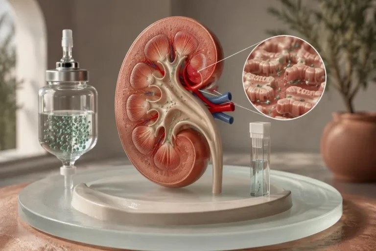
Heavy urinary protein loss can contribute to acquired antithrombin reduction, particularly in nephrotic syndrome. Adult nephrotic-range proteinuria is conventionally at least 3.5 g per 24 hours; clinicians interpret antithrombin alongside urine protein, serum albumin and the underlying kidney disorder, rather than relying on creatinine or eGFR alone.
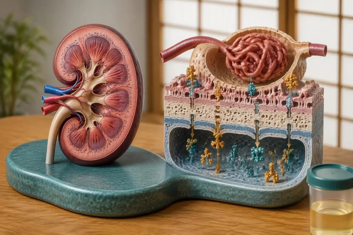
A normal eGFR does not exclude substantial protein leakage through the glomerular filtration barrier. For example, an adult with eGFR 95 mL/min/1.73 m², albumin 22 g/L and urine protein 6 g per day may have a strong acquired explanation for low activity despite apparently reassuring filtration results; persistent пенную мачу і аналіз на бялок deserve assessment in that context.
The relationship between nephrotic syndrome, antithrombin levels and clot risk is not uniform: not every person with nephrotic syndrome has low antithrombin, and the thrombotic tendency involves more than urinary loss of 1 protein. Underlying diagnosis matters, including membranous nephropathy, where PLA2R results and kidney monitoring help explain the kidney process rather than prove an inherited clotting disorder.
Kantesti is an AI blood test interpretation platform that can organize an antithrombin percentage alongside albumin and kidney markers, but it cannot infer the amount of urinary protein from eGFR alone. If activity remains 62% while heavy protein loss continues, repeat testing may still describe the acquired condition; the cleaner inherited assessment usually follows clinical stabilization and improvement of that competing explanation.
Ці могуць захворванні печані або іншыя метады лячэння знізіць узровень антытрамбіну?
Reduced liver synthesis can lower antithrombin, especially in advanced liver disease, while certain treatments such as L-asparaginase can also cause acquired reductions. An activity of 55% alongside other abnormalities requires review of protein production, protein loss and treatment history before clinicians consider a genetic explanation.
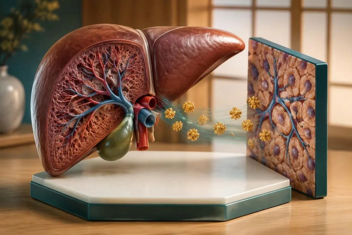
ALT and AST reflect injury more than protein-making capacity, so normal or mildly elevated enzymes do not guarantee normal antithrombin production. If activity is 57%, albumin is 25 g/L and INR is 1.6, impaired synthesis becomes a possibility—but warfarin, nutrition and protein loss can confound that pattern, as our кіраўніцтва па расшыфроўцы аналізу печані .
Albumin cannot serve as a substitute antithrombin test because the 2 proteins have different turnover and can respond differently to illness or therapy. Our albumin and serum protein guide helps interpret the surrounding panel; a normal albumin does not exclude inherited antithrombin deficiency, and a low albumin does not prove that the liver caused it.
L-asparaginase can reduce anticoagulant proteins during cancer treatment, creating an acquired thrombosis risk that may need active management rather than merely a future repeat test. Major procedures, extracorporeal support and recent replacement products can also complicate 1 measurement; eating more protein or taking a vitamin supplement does not correct a SERPINC1-related defect or replace treatment of the underlying acquired cause.
Якія антыкаагулянты ўплываюць на вынікі аналізу на антытрамбін?
Direct oral anticoagulants can falsely increase antithrombin activity, potentially masking deficiency rather than causing a low flag. Apixaban, rivaroxaban and edoxaban interfere with factor Xa-based activity assays, while dabigatran can interfere with thrombin-based assays; the same patient’s 2 results may therefore differ because of assay design.
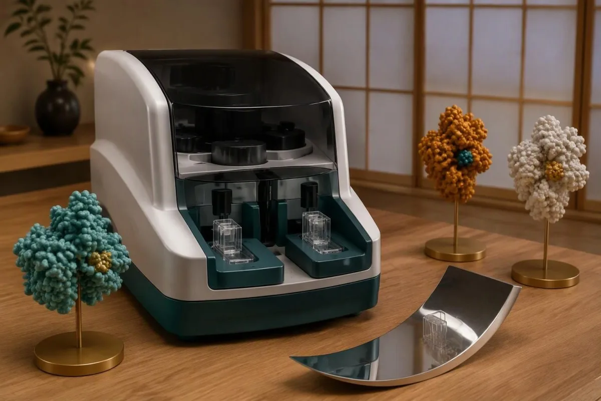
The mechanism is straightforward: the medication inhibits the assay’s added enzyme, and the instrument can misread that inhibition as stronger antithrombin function. The ISTH communication explicitly identifies these drug–assay combinations, including effects from direct thrombin inhibitors such as argatroban and bivalirudin (Van Cott et al., 2020); antigen testing usually avoids this particular functional interference, but does not replace an activity assessment.
A hypothetical activity of 108% on a factor Xa-based assay while taking rivaroxaban may conceal an underlying reduction; activity of 66% on that same drug still cannot be mathematically corrected into a true baseline. Anticoagulants also affect lupus anticoagulant test interpretation, but the direction and mechanism of interference are test-specific, not a universal rule for all thrombophilia assays.
Kantesti AI should support medication-aware interpretation rather than claim that 1 uploaded report identifies the laboratory’s reagent or removes a drug effect. Depending on the situation, the laboratory may use a less-affected assay, measure drug exposure or apply a validated removal procedure; our технічний валідаційний контроль і нагляд describes the broader standards behind interpretation, not a guarantee that AI can resolve missing assay information.
Ці можа неадпаведная апрацоўка ўзору растлумачыць нечакана нізкі вынік?
Sample quality and assay differences can make antithrombin results unreliable, especially when the laboratory flags collection or processing problems. Coagulation samples typically require a 9:1 ratio of sample to citrate anticoagulant; underfilling, inappropriate handling or an unsuitable specimen should be considered before interpreting an isolated abnormal percentage.

A hematocrit above approximately 55% may require citrate adjustment for coagulation testing because the amount of plasma relative to citrate is reduced. This is a laboratory preparation issue, not evidence of hereditary deficiency; if activity is 73% in a flagged specimen, ask whether the laboratory accepted the sample as valid and whether recollection is preferable to repeating the same material.
Hemolysis, lipemia and marked discoloration can interfere with optical methods, although the effect depends on the instrument and reagent rather than having 1 predictable direction. Our guides to hemolysis-related result distortion і lipemic specimens and redraws explain why a specimen-quality warning needs a method-specific answer instead of an automatic diagnosis.
Changing laboratories can create a second problem: an activity of 76% by one method and 84% by another may reflect analytical differences rather than biological recovery. Using the same laboratory helps trend comparison, but persistent clinical suspicion may justify a specialist laboratory using an alternative assay that is more sensitive to certain inherited variants; consistency and diagnostic sensitivity are 2 different goals.
Калі варта паўтарыць аналіз на нізкую актыўнасць антытрамбіну?
Repeat antithrombin testing should occur after temporary causes have settled and medication effects have been addressed. Following an acute clot, the BSH recommendation is to wait until after 3 months of anticoagulation before testing natural anticoagulant deficiencies; the safest collection date then depends on the drug, assay and clinical situation.

An incidental borderline result in a well adult without a recent clot does not automatically require the same 3-month wait, because that interval addresses acute thrombosis. The clinician may instead arrange a separate repeat once collection issues and acquired causes are reviewed, adding antigen testing if activity remains low; there is no universally validated waiting interval that fits every outpatient finding.
Heparin-related reductions may recover after treatment ends, but laboratory policies and ongoing illness affect when a repeat becomes meaningful. For a DOAC, a fixed 48-hour pause is not universally adequate or safe: kidney function, dose timing, drug accumulation and assay sensitivity matter, and choosing a compatible method may avoid interrupting treatment entirely.
Kantesti’s trend tools can help organize 2 measurements with their dates, but a timeline should also record clot onset, heparin exposure, the last relevant medication dose and any replacement products. Our explanation of выніках пасля выпіскі з бальніцы supports that workflow; warfarin does not usually lower antithrombin as it lowers vitamin K-dependent proteins, so protein C or S stopping rules should not be copied automatically.
Якія вынікі пацвярджаюць спадчынны дэфіцыт антытрамбіну?
Inherited antithrombin deficiency becomes more likely when activity remains low on separate, interpretable samples, acquired causes are excluded and the personal or family history fits. Two concordant results provide stronger evidence than 1 result during illness, but activity, antigen and sometimes SERPINC1 testing must still be considered together.

A clearly hypothetical profile of activity 54% and 56% on 2 suitable occasions, antigen 51%, no major protein loss and a first-degree relative with confirmed deficiency would support a quantitative inherited pattern. Early or recurrent venous thrombosis increases suspicion, but age alone is not diagnostic, and absence of a family history does not exclude a new variant or previously unrecognized disease.
SERPINC1 testing can help establish a molecular explanation when the laboratory phenotype is convincing and the result could affect management or family counseling. A variant of uncertain significance is not equivalent to a diagnosis, while a negative genetic test does not necessarily exclude every relevant change; the ISTH recommendations also note that molecular testing may identify defects missed by activity assays (Van Cott et al., 2020).
Antithrombin and protein S deficiency require different interpretive rules, even when they appear on the same thrombophilia panel. Our guide to low protein S activity explains several contrasting confounders; a type II antithrombin heparin-binding-site variant can also have a different risk profile from type I deficiency, so 1 generic risk estimate should not be applied to all confirmed variants.
Калі варта абследаваць членаў сям’і?
Family evaluation is usually considered after the original patient’s inherited deficiency is convincingly confirmed, especially when the result could change prevention, hormone choices or pregnancy planning. Most inherited antithrombin deficiency is autosomal dominant, giving each child of a heterozygous carrier a 50% chance of inheriting the relevant variant—not a certainty of thrombosis.
The BSH guideline discourages routine thrombophilia testing of all first-degree relatives but supports selective evaluation for relatives of people with antithrombin, protein C or protein S deficiency when results could influence management (Arachchillage et al., 2022). Our Кіраўніцтва па сямейным тэсціраванні на аснове рызыкі explains why testing 1 relevant inherited condition can be more useful than ordering a broad panel without a clinical question.
ASH’s 2023 thrombophilia guideline similarly supports selective testing in certain families with known high-risk deficiencies, including decisions about estrogen exposure and prophylaxis during particular risk situations (Middeldorp et al., 2023). Many recommendations are conditional and based on very low-certainty evidence; antithrombin deficiency should not be treated as interchangeable with factor V Leiden results, which often carry a different risk profile.
Kantesti’s Family Health Risk feature can help organize information shared with consent, but it cannot determine whether a relative inherited a SERPINC1 variant from 1 family member’s activity percentage. Testing children requires age-aware laboratory interpretation and an assessment of whether the result would change care now; predictive testing also deserves counseling about privacy, uncertainty and the difference between inherited susceptibility and an actual clot.
Што павінны ўключаць ваш план наступнага назірання і праверкі крыніц?
Bring the original percentage, reference interval, medication list and sampling date to follow-up, along with details of the clot, illness or protein loss that could explain the result. Two decisions come first: whether the current finding needs clinical action and whether repeat testing would genuinely change treatment or family counseling.

A low activity percentage alone does not automatically require lifelong anticoagulation or antithrombin concentrate; treatment depends on previous thrombosis, the provoking circumstances, bleeding risk and the confirmed diagnosis. New one-sided leg swelling needs prompt assessment, while sudden shortness of breath, chest pain or fainting requires emergency care even if the last activity was 100%—do not wait for a repeat thrombophilia panel.
Kantesti is an AI lab test interpretation service that can help organize the 2-stage workflow of context review followed by clinician-directed confirmation, not replace hematology advice or authorize medication interruption. Our прынцыпы адказнага выкарыстання AI describe that boundary; use AI explanations to prepare precise questions, including which enzyme the assay uses and whether activity and antigen should be collected together.
The clinical recommendations here draw on 3 directly relevant external sources: the ISTH laboratory communication, the BSH testing guideline and the ASH thrombophilia guideline. The 2 Zenodo publications below provide background on serum proteins and immune-related laboratory context—not clinical trials validating antithrombin diagnosis; our стандарты медыцынскага кантэнту і даведнік па даследаванні камлементу і аўтаімунных захворванняў help readers distinguish supporting education from condition-specific evidence.
Часта задаваныя пытанні
Ці заўсёды нізкая актыўнасць антытрамбіну сведчыць пра спадчынны дэфіцыт?
Нізкая актыўнасць антытрамбіну не заўсёды азначае спадчынны дэфіцыт. Актыўнасць 60% можа назірацца пры вострым трамбозе, лячэнні гепарынам, цяжкім захворванні, запушчанай хваробе печані або значнай страце бялку з мачой. Клініцысты шукаюць стойкае зніжэнне ў асобных пробах, узятых у належны час, пасля ацэнкі гэтых набытых прычын. Даследаванне антыгена, а часам і даследаванне SERPINC1 дапамагаюць класіфікаваць пацверджанае адхіленне ад нормы.
Калі трэба паўтарыць аналіз на актыўнасць антытрамбіну пасля ўтварэння тромба?
Пасля вострага тромбозу Брытанскае таварыства гематалогіі рэкамендуе правяраць дэфіцыт прыродных антыкаагулянтаў толькі пасля 3 месяцаў антыкаагулянтнай тэрапіі. Пры складанні плана забору проб таксама трэба ўлічваць аднаўленне, антыкаагулянт, які ўжываецца, і лабараторны аналіз. Гэтая рэкамендацыя не азначае, што ўсім трэба спыніць прыём прэпаратаў праз 3 месяцы. Ніколі не перапыняйце антыкаагулянтную тэрапію для правядзення аналізаў без указанняў лекара, які прызначыў лячэнне.
У чым розніца паміж антыгенам і актыўнасцю антытрамбіну?
0 Актыўнасць антытрамбіну паказвае яго функцыю, а антыген антытрамбіну — колькасць бялку. Пры дэфіцыце I тыпу звычайна зніжаюцца абодва паказчыкі; пры дэфіцыце II тыпу актыўнасць звычайна зніжаецца, нягледзячы на адносна нармальны ўзровень антыгена. Напрыклад, паўторны вынік актыўнасці 52% пры ўзроўні антыгена 101% указвае на якасны тып парушэння, але не пацвярджае спадчынную прычыну без праверкі набытых станаў і ўплыву лекавых сродкаў. Аналіз на антыген не можа замяніць належную ацэнку функцыі.
Ці могуць апіксабан або рывараксабан паўплываць на вынікі аналізу на антытрамбін?
Апіксабан і рывараксабан могуць ілжыва павышаць актыўнасць антытрамбіну ў аналізах на аснове фактару Xa, патэнцыйна маскіруючы дэфіцыт. Дабігатран можа аналагічна павышаць уяўную актыўнасць у аналізах на аснове трамбіну. Таму вынік 100% падчас прыёму прэпарату, які ўплывае на аналіз, можа быць менш абнадзейлівым, чым здаецца. Лабараторыя можа выбраць менш адчувальны да такога ўплыву метад або іншую валідзіраваную стратэгію; самастойна спыняць прыём прэпарату небяспечна.
Ці небяспечная актыўнасць антытрамбіну 70%?
Актыўнасць антытрамбіну 70% ніжэйшая за значную частку дарослых лабараторных рэферэнтных інтэрвалаў, але сам па сабе гэты працэнт не сведчыць пра неадкладны стан і не дазваляе ацаніць асабістую рызыку тромбаўтварэння. Значэнне выніку залежыць ад метаду аналізу, прыёму лекаў, наяўнасці тромбаў у мінулым, сямейнага анамнезу і бягучага захворвання. Ізаляваны вынік звычайна патрабуе клінічнай ацэнкі і плана адпаведнага пацвярджэння. Раптоўная дыхавіца, боль у грудзях, непрытомнасць або новы аднабаковы ацёк нагі патрабуюць абследавання незалежна ад гэтага працэнта.
Ці трэба маёй сям’і прайсці абследаванне, калі ў мяне дэфіцыт антытрамбіну?
Выбарачнае абследаванне членаў сям’і можа быць мэтазгодным пасля пераканаўчага пацвярджэння спадчыннага дэфіцыту антытрамбіну, асабліва калі вынікі паўплываюць на прафілактыку, выбар гарманальных прэпаратаў або планаванне цяжарнасці. Гетэразіготны носьбіт звычайна мае 50% верагоднасць перадаць адпаведны варыянт кожнаму дзіцяці, але гэта не 50% гарантыя таго, што ў дзіцяці разаўецца трамбоз. Рэкамендацыі BSH і ASH падтрымліваюць абследаванне з улікам таго, ці паўплываюць вынікі на прыняцце рашэнняў, а не бессістэмнае тэсціраванне ўсіх сваякоў. Вынікі для дзяцей трэба інтэрпрэтаваць з улікам узросту і абмяркоўваць, ці зменіць абследаванне лячэнне цяпер.
Атрымайце тлумачэнне вынікаў аналізу крыві
Загрузіце свой лабараторны справаздачу, і AI Kantesti за некалькі секунд простай мовай растлумачыць кожны біямаркер. Ім карыстаюцца больш за 2 мільёны чалавек па ўсім свеце.
Спасылкі
- Кляйн, Т., Мітчэл, С., і Вебер, Х. (2026). Кіраўніцтва па бялках сыроваткі: глабуліны, альбумін і суадносіны A/G у аналізе крыві. Kantesti AI Medical Research. DOI
- Кляйн, Т., Мітчэл, С., і Вебер, Х. (2026). Дапаможнік па аналізах крыві на C3, C4, камплемент і тытр ANA. Kantesti AI Medical Research. DOI
- Van Cott EM et al. (2020). Recommendations for clinical laboratory testing for antithrombin deficiency; Communication from the SSC of the ISTH. Journal of Thrombosis and Haemostasis. DOI
- Arachchillage DJ і інш. (2022). Тэставанне на тромбафілію: рэкамендацыі Брытанскага таварыства гематалогіі. Брытанскі часопіс гематалогіі. DOI
- Middeldorp S і інш. (2023). Рэкамендацыі Амерыканскага таварыства гематалогіі за 2023 год па вядзенні вянознай тромбаэмбаліі: тэставанне на тромбафілію. Blood Advances. DOI
- 2 млн+Прааналізаваныя тэсты
- 127+Краіны
- 100+Мовы
Пра медыцынскую каманду


Сігналы даверу E-E-A-T
Вопыт
Клінічны агляд, які вядзе лекар, працэсаў інтэрпрэтацыі лабараторных паказчыкаў.
Экспертыза
Фокус лабараторнай медыцыны на тым, як біямаркеры паводзяць сябе ў клінічным кантэксце.
Аўтарытэтнасць
Медыцынскую рэцэнзію правёў доктар Томас Кляйн пры ўдзеле доктара Сары Мітчэл і прафесара, доктара Ганса Вэбера.
Надзейнасць
Інтэрпрэтацыя на аснове доказаў з выразнымі шляхамі далейшых дзеянняў, каб паменшыць трывогу.




Медыцынская адмова ад адказнасці
Гэты артыкул прызначаны толькі для адукацыйных мэт і не з’яўляецца медычнай парадай. Заўсёды кансультуйцеся з кваліфікаваным спецыялістам аховы здароўя для прыняцця рашэнняў па дыягностыцы і лячэнні.
Апублікавана · Звязацца з рэдакцыйнай камандай
Апублікавана кампаніяй Kantesti Ltd, зарэгістраванай у Англіі і Уэльсе, нумар кампаніі. 17090423, Лондан, Злучанае Каралеўства.
