Low protein S activity does not by itself prove inherited protein S deficiency. Pregnancy, estrogen, warfarin, acute illness and assay effects can explain the result; paired activity and free antigen testing, repeated under clinician-selected conditions, help clarify whether the reduction persists.

Асноўныя высновы
- Адзін нізкі вынік cannot establish inherited protein S deficiency, particularly during pregnancy, acute illness or anticoagulant treatment.
- Activity percentages describe performance relative to the assay's calibrator; an activity of 45% does not mean a 45% probability of developing a clot.
- Free protein S antigen measures the unbound protein pool, normally approximately 40% of circulating protein S, rather than its functional performance.
- тэставанне на цяжарнасць can show physiological reductions; diagnostic reassessment is commonly deferred until at least 6 weeks postpartum, sometimes longer.
- Варфарын genuinely lowers protein S because protein S is vitamin K dependent; even free antigen testing cannot reliably establish the untreated baseline during treatment.
- Памылкі вызначэння аналізу (assay interference) can falsely lower or raise activity. Direct oral anticoagulants commonly cause falsely increased results in susceptible clot-based assays, potentially concealing deficiency.
- Repeat testing after a clot is generally deferred until after 3 months of anticoagulation when testing is indicated, with medication and assay arrangements determined by clinicians.
- Бяспека лекаў comes first: never stop anticoagulants, estrogen treatment or other prescribed medicines independently to obtain a cleaner laboratory result.
Does low protein S activity mean inherited deficiency?
Low protein S activity can be inherited, acquired or falsely low because of the testing method. A single abnormal result cannot distinguish these possibilities: pregnancy, estrogen exposure, warfarin, recent thrombosis and severe illness must be considered before assigning a lifelong diagnosis, and activity usually needs interpretation alongside free antigen.
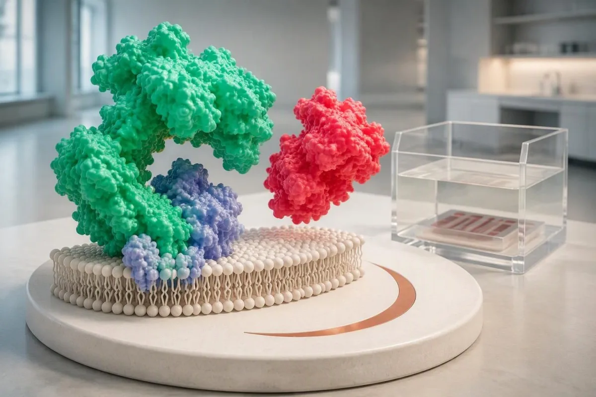
Protein S helps activated protein C limit coagulation by supporting the inactivation of factors Va and VIIIa. Its concentration is not the same as the total protein measurement on a chemistry panel; a total protein of 7.0 g/dL provides no useful estimate of protein S function, because the tests measure entirely different biological quantities.
An isolated flag is a starting point, not a verdict. For a hypothetical person whose activity is 48% against a laboratory lower limit of 60%, the next question is whether the sample was collected during pregnancy, medication treatment or illness—not whether the number automatically proves a genetic disorder; our даведачны гід па выніках па-за межамі тлумачыць гэтую розніцу.
Kantesti is an AI blood test analyzer that helps organize protein S results with their reported ranges and relevant context rather than treating 1 flagged value as a diagnosis. Our інфармацыю пра кампанію explains the organization behind this educational service; confirmation of inherited deficiency still requires clinician-directed laboratory assessment and, occasionally, specialist genetic evaluation.
How should you read the activity percentage and range?
Protein S activity is usually reported as a percentage of the assay's reference activity, not as a percentage chance of thrombosis. Reference intervals depend on the laboratory, method, age and hormonal context; an adult interval such as 60–150% is an illustrative example, not a universal diagnostic cutoff.

A protein S activity of 45% means reduced functional activity in that assay, not that 45% of your circulation is blocked. Percentages also differ from concentration units such as mg/L, so an activity result cannot be directly compared numerically with an antigen concentration; the distinction resembles other колькасных лабараторных вымярэнняў that quantify different properties.
Borderline values need especially careful handling. In a hypothetical laboratory using 60% as its lower limit, results of 58% and 61% fall on opposite sides of the flag despite being only 3 percentage points apart; analytical variation, biological fluctuation and different assay reagents may explain that difference without a meaningful change in clotting risk.
There is no single protein S percentage that independently determines whether anticoagulation is required. A result of 25% deserves more attention than a minimally reduced value, but it can still occur during an acquired reduction; decisions depend on confirmation, clot history, provoking factors and bleeding risk, rather than treating 1 numerical threshold as a medication trigger.
| Узровень | Дыяпазон | Што гэта значыць |
|---|---|---|
| Within an illustrative adult interval | 60–150% activity | Example only. Use the reporting laboratory's age- and context-appropriate interval; an interfering anticoagulant can sometimes mask deficiency. |
| Below that illustrative interval | <60% activity | Requires context review and, when appropriate, confirmation. This does not establish inherited deficiency. |
| Free antigen result | Laboratory-specific % or concentration units | Measures the unbound protein pool. Do not apply an activity reference interval to an antigen result. |
| Pregnancy or childhood result | Population- and method-specific | A nonpregnant adult interval may be inappropriate. Diagnostic interpretation may need specialist advice. |
What do activity and free protein S antigen show together?
Activity testing measures functional performance, while free protein S antigen measures the available unbound protein pool. Approximately 40% of circulating protein S is free and the remainder is bound mainly to C4b-binding protein; comparing these measurements helps identify a quantitative reduction, a possible functional defect or a misleading activity result.
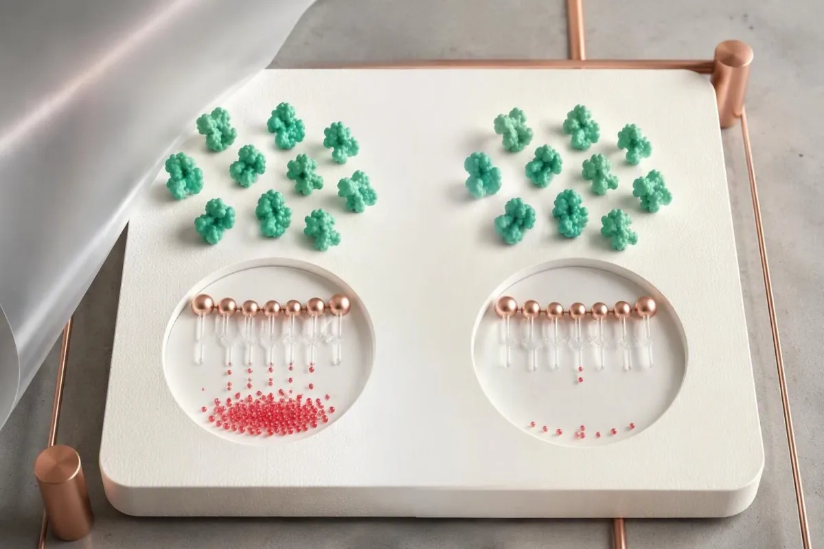
Type I protein S deficiency classically produces low total antigen, low free antigen and low activity; type III produces normal total antigen with low free antigen and activity. These 2 patterns can occur within the same family and are not necessarily separate biological diseases; total antigen alone can therefore miss a clinically relevant reduction in the free pool.
Type II protein S deficiency classically shows reduced activity despite normal free and total antigen. This uncommon pattern is not diagnosed from 1 discordant sample, because factor V Leiden and other assay problems can imitate functional deficiency; laboratories may repeat the activity using another method before considering a genuinely dysfunctional protein.
Paired results clarify the next step but do not identify the cause by themselves. For example, activity of 42% with free antigen of 38% could reflect either inherited quantitative deficiency or an acquired reduction, while activity of 42% with normal free antigen raises a different set of questions; our coagulation marker overview explains why complementary tests are interpreted together.
Why does pregnancy lower protein S results?
Pregnancy physiologically lowers free protein S and often protein S activity, so a low pregnancy result cannot reliably establish inherited deficiency. Changes can begin early and become more pronounced later; the British Society for Haematology identifies pregnancy and the puerperium as confounders of protein S testing (Arachchillage et al., 2022).

A nonpregnant reference interval can create a misleading diagnosis during pregnancy. In a hypothetical second-trimester sample, activity of 43% and free antigen of 36% may both be flagged, but their agreement does not remove the pregnancy effect; both tests can reflect the same physiological reduction, just as інтэрпрэтацыя феритину пры цяжарнасці requires pregnancy-specific context.
Reassessment for inherited protein S deficiency is commonly postponed until at least 6 weeks after delivery, and some services prefer 12 weeks or longer. Six weeks is not a guarantee of an unaffected baseline: recovery, continuing illness, a recent clot and newly started estrogen contraception can all influence whether a postpartum sample is suitable.
Protein S testing does not decide pregnancy anticoagulation by itself. When reviewing 2 pregnancy measurements, Kantesti AI can help distinguish activity from antigen on the report, but pregnancy prophylaxis requires obstetric and thrombosis assessment based on prior venous thromboembolism, family history and other risks; a low pregnancy value alone also does not establish the cause of a miscarriage.
Can estrogen contraception or HRT cause a low result?
Estrogen-containing contraception and oral estrogen therapy can lower free protein S and protein S activity without an inherited defect. The change is biological rather than merely a laboratory artifact; a result collected during estrogen use may therefore describe the treatment-associated state, not the person's untreated baseline.

The exact preparation matters more than the word contraception. A combined pill containing 30 micrograms of ethinylestradiol is not equivalent to a progestogen-only pill, and a contraceptive patch is not equivalent to a transdermal menopausal estrogen patch; hormone results during contraception require the active ingredients and route, not just the brand name.
Transdermal menopausal estrogen generally has less effect on hepatic coagulation pathways than oral estrogen, but this does not make every transdermal hormonal product risk-free. Evidence about clot risk and evidence about a specific protein S assay are different questions; an activity of 52% during treatment cannot be corrected mathematically by subtracting a standard estrogen effect.
There is no universally reliable 1-week estrogen washout for protein S diagnosis. If confirmation would change care, the prescribing clinician can discuss timing, alternative contraception and whether treatment should change at all; our pre-HRT testing considerations support that conversation, but never stop prescribed hormones independently just to remove a laboratory flag.
Why does warfarin lower protein S activity?
Warfarin lowers protein S because protein S is a vitamin K-dependent anticoagulant protein. This is an expected treatment effect rather than proof of inherited deficiency; free antigen can also be reduced, so replacing an activity test with antigen testing does not reliably reveal the untreated baseline during warfarin therapy.

A therapeutic INR does not make protein S testing diagnostically reliable during warfarin treatment. For a hypothetical patient taking 5 mg daily with an INR of 2.4 and activity of 31%, the low activity may be medication related; the meaning of an INR concerns anticoagulant monitoring, not confirmation of inherited protein S deficiency.
When an off-warfarin measurement is genuinely needed, laboratories commonly require at least 2 weeks after discontinuation, sometimes longer according to local policy and the clinical situation. That interval is not an instruction to stop treatment: a hematologist or anticoagulation clinician must decide whether interruption is safe, whether an alternative is needed and which assay remains interpretable.
Vitamin K deficiency can also reduce protein S, but supplements should not be used to self-correct an unexplained result. Taking even 1 new vitamin K-containing supplement can alter warfarin management; keep dietary intake reasonably consistent and discuss changes with the anticoagulation team, using our кіраўніцтве па прадуктах вітаміна K for food context rather than as a deficiency treatment plan.
Which illnesses can temporarily reduce protein S?
Severe infection, disseminated intravascular coagulation, liver disease, nephrotic syndrome and recent thrombosis can reduce protein S measurements. Some reductions resolve with recovery, while others persist because the underlying condition persists; a low result during hospitalization therefore cannot be classified as inherited from 1 sample.
Disseminated intravascular coagulation can reduce natural anticoagulants while other coagulation markers change simultaneously. A hypothetical combination of protein S activity at 35%, platelets at 65 × 10^9/L and a prolonged prothrombin time calls for assessment of the acute illness, not immediate genetic labeling; our DIC laboratory pattern guide explains why multiple abnormal findings matter together.
The liver contributes to protein S production, but an isolated ALT of 75 IU/L does not prove that liver dysfunction caused a low protein S result. Clinicians look for impaired synthesis and the broader clinical picture, including albumin, bilirubin and coagulation results; modest enzyme elevation and advanced synthetic failure are different processes, with very different implications for interpretation.
Nephrotic-range protein loss can produce acquired protein S abnormalities, especially in the free fraction. An albumin of 2.2 g/dL with substantial urinary protein loss changes the interpretation of activity at 44%; persistent foamy urine assessment may help identify a reason to investigate renal protein loss, although bubbles alone neither diagnose nephrotic syndrome nor explain protein S.
How can assay interference make protein S misleading?
Clot-based protein S activity assays can report falsely low or falsely high results because medications and other coagulation abnormalities alter the endpoint. Direct oral anticoagulants often falsely increase activity in susceptible assays, whereas factor V Leiden can falsely decrease it; the direction and magnitude depend on the assay design.
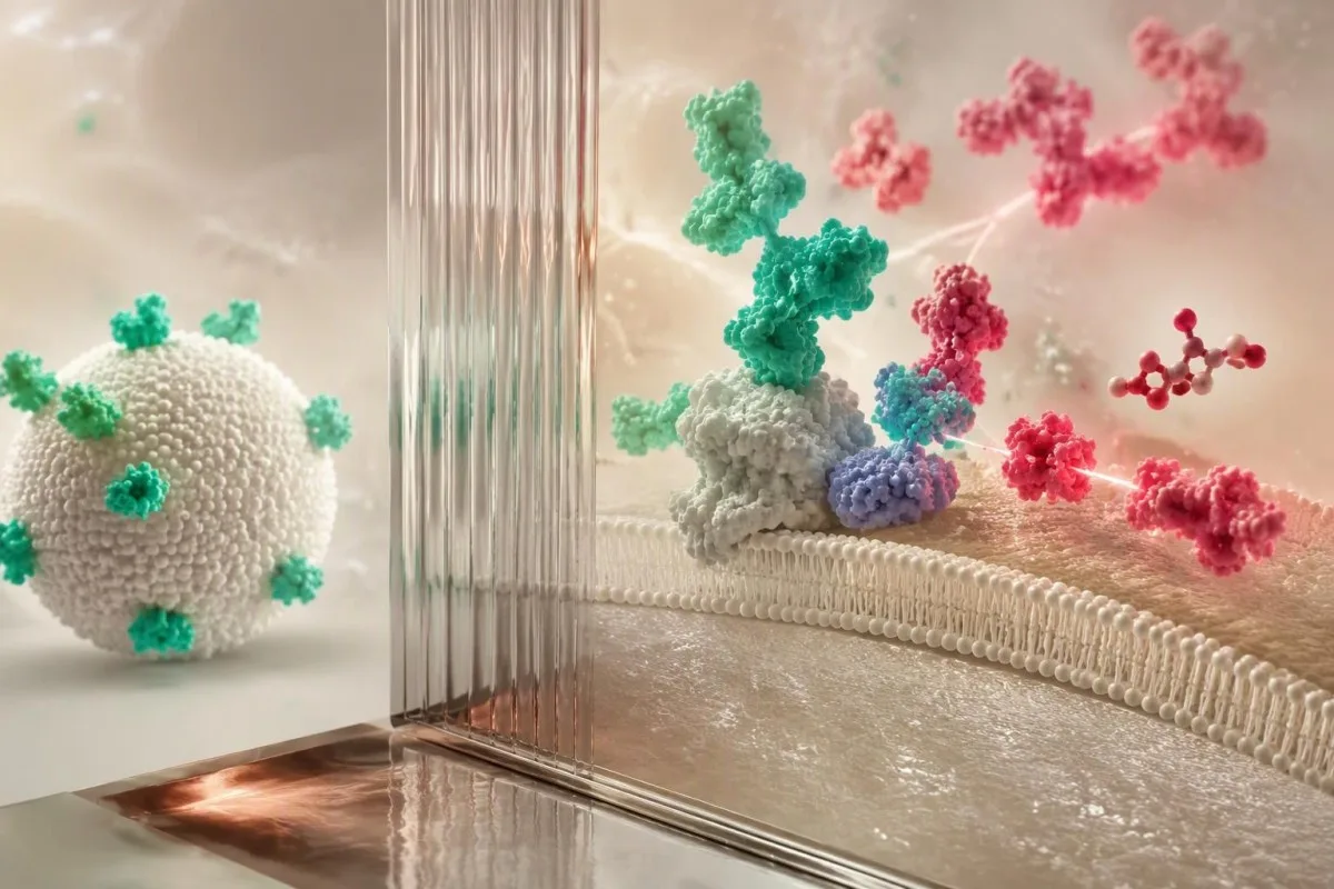
Factor V Leiden can interfere with some protein S activity assays by altering the activated protein C-dependent reaction. A hypothetical activity of 46% with normal free antigen could therefore mimic type II deficiency; the factor V Leiden result and the laboratory's assay characteristics should be reviewed before concluding that 2 inherited disorders coexist.
Direct oral anticoagulants can conceal deficiency rather than explain a low number. For example, an apparently normal activity of 110% during rivaroxaban treatment may be falsely increased in a susceptible clot-based method; lupus anticoagulant, high factor VIII and heparin can also complicate interpretation, so lupus anticoagulant testing and protein S testing need coordinated laboratory advice.
Kantesti is an AI lab test interpretation service that can place 2 discordant measurements—activity and free antigen—side by side for discussion, but cannot determine reagent interference from the numbers alone. Free antigen immunoassays generally avoid clot-endpoint interference, yet they still cannot remove warfarin's biological effect; the BSH guideline specifically lists medication- and assay-related confounders (Arachchillage et al., 2022).
Can collection or sample handling create a low result?
Collection and handling problems can distort protein S testing, particularly functional assays that depend on correctly prepared citrated plasma. The laboratory should check tube filling, citrate balance, processing and sample quality before a discordant result becomes a diagnosis; repeating a technically unsuitable sample answers a different question from confirming inherited deficiency.
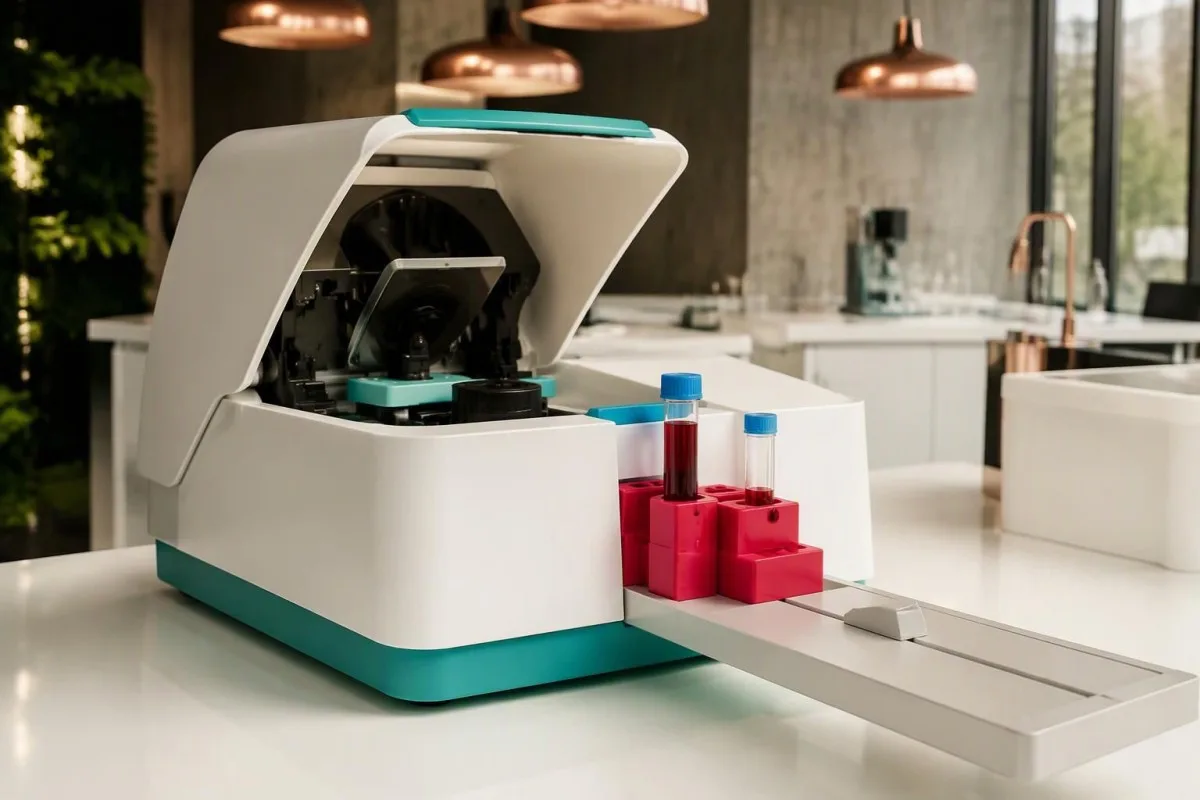
Standard coagulation collection uses approximately 9 parts sample to 1 part citrate anticoagulant. Underfilling changes that ratio and can distort clot-based measurements; tube-cap colors vary internationally, so the fill line and laboratory instructions matter more than recognizing a particular color, and a rejected specimen should not be interpreted as if its numerical result were trustworthy.
A hematocrit above 55% generally requires laboratory-directed adjustment of citrate volume for coagulation testing. A hypothetical patient with a hematocrit of 58% needs that issue addressed before a low functional result is taken at face value; otherwise, excess citrate relative to plasma can affect the assay even when the collection tube appears adequately filled.
Hemolysis, lipemia and delayed processing can affect methods differently, so there is no universal correction factor for a protein S result of 49%. Ask whether the laboratory accepted the specimen and whether it recommends recollection; our hemolysis interference guide explains why a sample-quality warning may affect some reported analytes but not others.
When is clinician-guided repeat testing appropriate?
Repeat protein S testing is appropriate when the first result was confounded, borderline or unexpected and confirmation could change care. After acute thrombosis, the British Society for Haematology recommends testing natural anticoagulant deficiencies only after 3 months of anticoagulation when indicated, with medication-related interference addressed separately (Arachchillage et al., 2022).

Three months of anticoagulation is a testing-timing recommendation, not permission to stop anticoagulants at 3 months. Treatment duration depends on the clot and the patient's risks, while assay suitability depends on the medication; clinicians may defer testing, choose an antigen assay or seek specialist laboratory methods without interrupting necessary protection.
Pregnancy, warfarin and acute illness each require a different reassessment plan. Common examples include at least 6 weeks postpartum, at least 2 weeks after clinician-approved warfarin discontinuation, or testing after recovery from significant illness; none is a universal washout schedule, and an unconfounded repeat may need to be postponed further if several influences overlap.
Kantesti's trend analysis can help display 2 measurements, but comparison is strongest when the method, laboratory and clinical conditions are comparable. Record pregnancy stage, illness dates, medicines and the exact timing of the last anticoagulant dose for the clinician; our кіраўніцтва па лабараторным раскладзе explains why elapsed time alone does not establish that a change is biological.
What makes inherited protein S deficiency more likely?
Inherited protein S deficiency becomes more likely when low results persist under suitable testing conditions and fit a personal or family history of venous thrombosis. A pathogenic PROS1 variant can support the diagnosis, but genetic testing is selective; 1 low activity result, especially during treatment or pregnancy, is insufficient.

Most inherited protein S deficiency associated with a single pathogenic PROS1 variant follows an autosomal dominant inheritance pattern. Each child has a 50% chance of inheriting that variant, not a 50% certainty of developing a clot; penetrance varies, and relatives with the same variant can have different protein measurements and clinical histories.
A first venous clot at age 28 with several affected close relatives raises more suspicion than an incidental borderline result at age 72 during pneumonia. Age is not a diagnostic cutoff, however: absence of family history does not exclude inheritance, and the BSH guideline recommends genetic testing for phenotypically identified deficiencies when the result would influence management (Arachchillage et al., 2022).
Family screening should be selective rather than a broad panel for every relative. Testing an unaffected first-degree relative may be useful if the result would change estrogen decisions or prophylaxis during high-risk periods; our Кіраўніцтва па сямейным тэсціраванні на аснове рызыкі explains why testing plans should follow a confirmed family diagnosis instead of expanding from 1 uncertain laboratory flag.
Does a low result mean you have a clot now?
Low protein S activity does not diagnose an existing deep vein thrombosis or pulmonary embolism. Confirmed inherited deficiency increases susceptibility to venous thrombosis, but current symptoms require the usual urgent assessment; the BSH guideline describes an approximately 5–7-fold increased first-event risk for heterozygous protein C or protein S deficiency (Arachchillage et al., 2022).
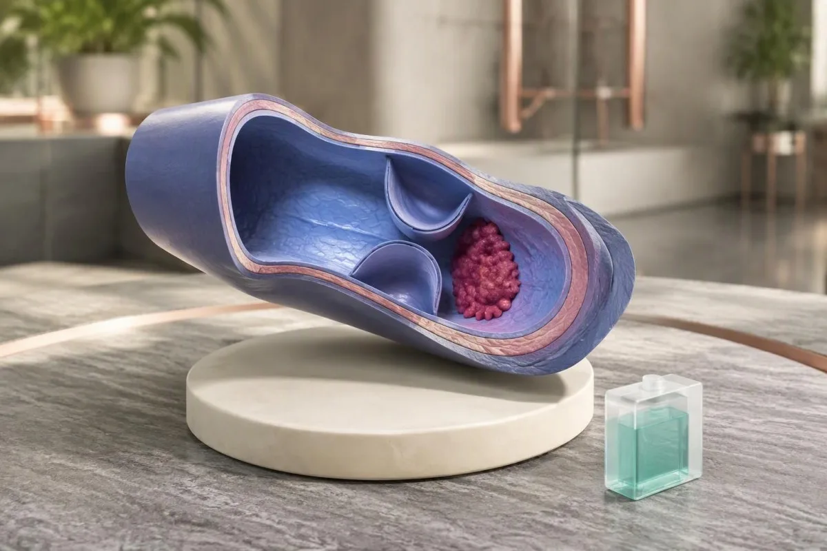
A 5–7-fold relative risk is not a 50–70% absolute probability of thrombosis. Absolute risk varies with age, family ascertainment, prior events and exposures such as surgery or estrogen; a person without symptoms should not interpret a low protein S value as evidence that a clot is silently present or start an anticoagulant independently.
Sudden unexplained breathlessness, chest pain, coughing blood or collapse requires emergency evaluation; new one-sided leg swelling or pain needs urgent assessment for deep vein thrombosis. A normal activity of 95% does not rule out either condition, and normal PT or aPTT also cannot exclude thrombophilia; our абмежаванні каагулаграмы explain that common misconception.
A confirmed protein S deficiency does not automatically require lifelong anticoagulation in someone who has never had thrombosis. The ASH 2023 guideline discourages testing unprovoked VTE solely to determine duration but conditionally supports testing in selected hormonal or nonsurgical provoked scenarios; those latter recommendations rely on very low-certainty evidence, so 1 label cannot replace individualized decisions (Middeldorp et al., 2023).
What should you bring to the next clinical appointment?
Bring the complete report, medication list, collection date and relevant clot or pregnancy history—not just the low protein S activity number. The most useful appointment establishes whether the result is interpretable, whether free antigen was measured and whether confirmation would change treatment; 3 focused questions often prevent unnecessary retesting.

Ask: Was the activity method vulnerable to my medication; was free antigen measured; and what would a confirmed deficiency change? For a hypothetical person with activity of 47% and free antigen of 41% while taking warfarin, both results may remain treatment-confounded; ordering a larger thrombophilia panel immediately does not solve the underlying baseline problem.
A different hypothetical pattern—activity of 47%, normal free antigen and known factor V Leiden—first raises an assay question rather than establishing type II deficiency. A third pattern, with activity of 39% and free antigen of 35% persisting on suitably timed samples without an acquired explanation, makes specialist evaluation more reasonable; these examples are illustrations, not diagnostic algorithms.
Kantesti can help organize these 3 patterns for discussion, but the clinician and testing laboratory must decide whether another assay or repeat specimen is needed. Our клінічную рамку валідацыі describes the role of evaluation and oversight; bring actual reference intervals and units, and do not rely on a cropped screenshot that omits the method or sample comments.
Research evidence and limits of AI interpretation
Clinical guidelines support context-sensitive protein S testing, but neither a laboratory flag nor an AI explanation confirms inherited deficiency. Kantesti is an AI blood test interpretation platform that helps explain activity and antigen results together; the diagnosis still depends on appropriate testing conditions, reproducibility and clinical assessment rather than 1 automated interpretation.
The 2022 BSH and 2023 ASH recommendations address different testing decisions, and neither recommends indiscriminate thrombophilia screening. BSH emphasizes validity and whether findings would change management, while ASH conditionally supports testing in some selected VTE scenarios using very low-certainty evidence; their differences reflect clinical questions and modeling assumptions, not proof that every low result needs genetic testing.
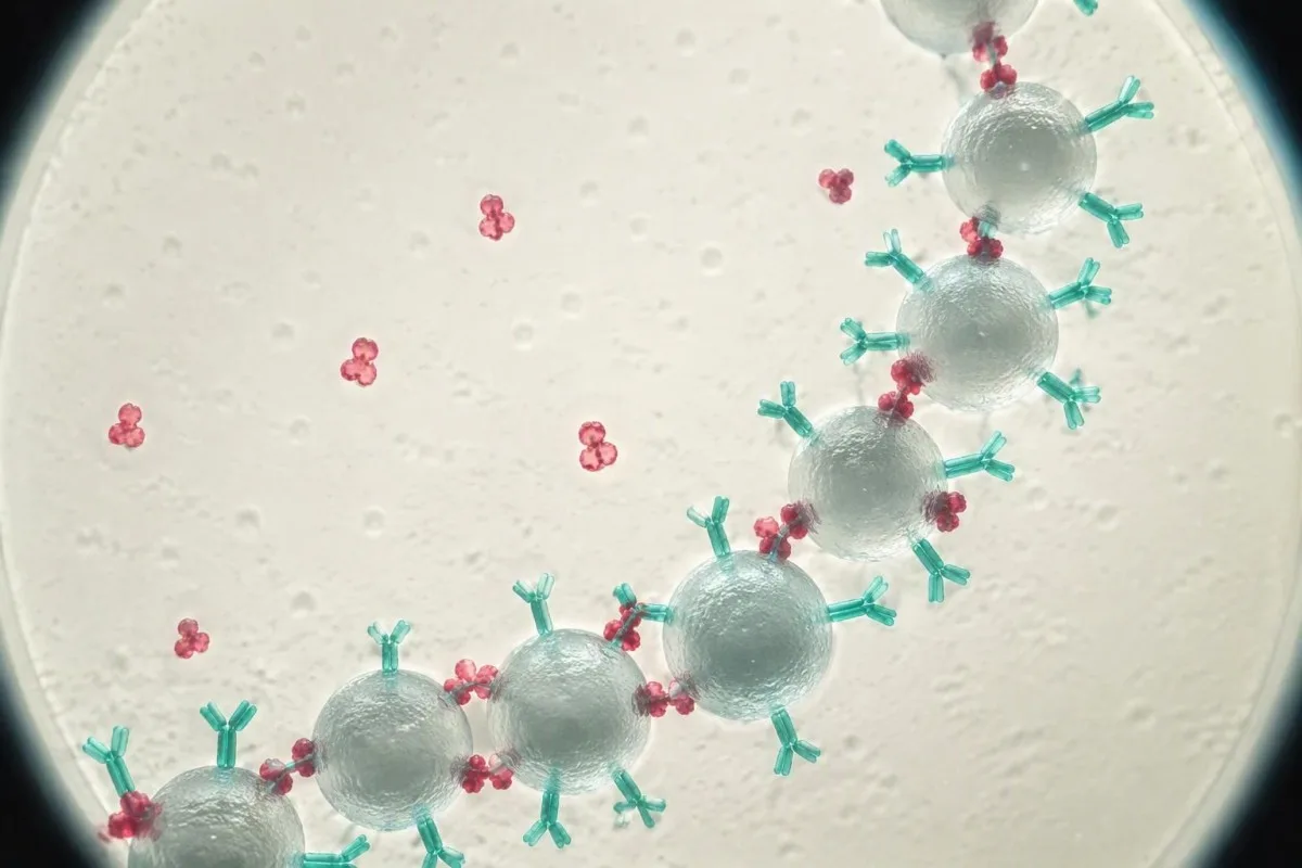
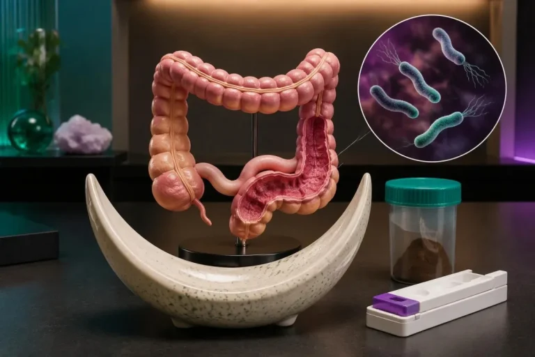
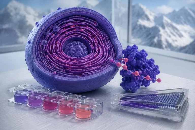
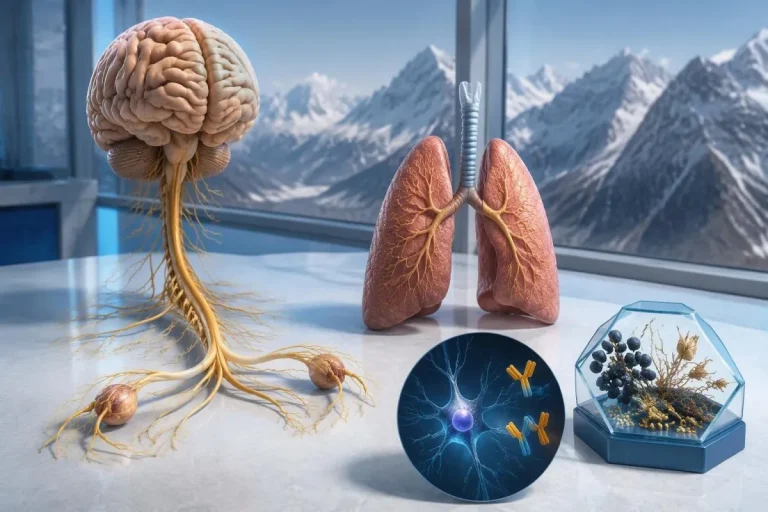
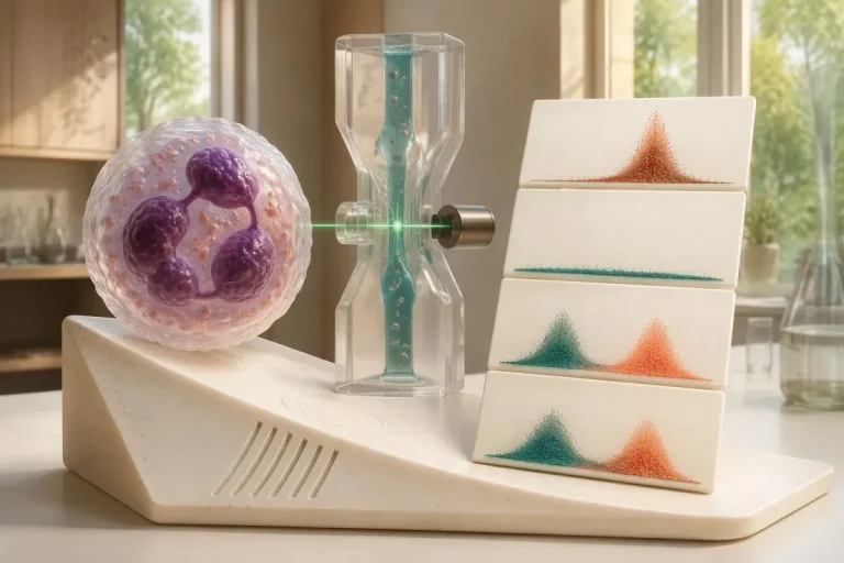
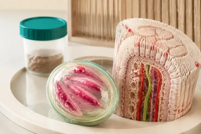
Protein S is not diagnosed by routine tissue microscopy, and the molecular illustrations here are educational models rather than images of an individual's deficiency. Our тлумачэнне тэхналогіі ШІ describes the interpretation workflow; the hormonal health research overview supplies broader context but does not provide a validated protein S cutoff or a medication washout protocol.
The 2 Kantesti research publications listed below are Figshare-hosted materials covering hormonal health and multilingual clinical decision-support engineering; neither establishes diagnostic accuracy for inherited protein S deficiency. Repository hosting and DOI assignment do not establish peer review or protein S-specific clinical validation; our стандарты медыцынскага кантэнту explain why educational claims should remain separate from disease-specific evidence.
Часта задаваныя пытанні
Ці можа паніжаная актыўнасць бялку S быць часовай?
Нізкая актыўнасць бялку S можа быць часовай падчас цяжарнасці, пры ўздзеянні эстрагенаў, лячэнні варфарынам, цяжкім захворванні або аднаўленні пасля трамбозу. Па адным выніку, напрыклад 45%, немагчыма адрозніць часовае зніжэнне ад спадчыннага дэфіцыту. Актыўнасць і ўзровень свабоднага антыгена бялку S трэба інтэрпрэтаваць разам, паўтараючы аналіз, калі клініцыст палічыць умовы прыдатнымі. Не спыняйце самастойна прызначаныя лекі, каб атрымаць вынік без лячэння.
У чым розніца паміж актыўнасцю бялку S і свабодным антыгенам бялку S?
Тэст актыўнасці бялку S ацэньвае функцыянальную актыўнасць, а аналіз на свабодны антыген бялку S — незвязаны пул бялку. Прыкладна 40% бялку S, які цыркулюе ў крыві, знаходзіцца ў свабоднай форме, а астатняя частка звязана пераважна з C4b-звязвальным бялком. Нізкая актыўнасць пры нізкім узроўні свабоднага антыгена можа назірацца як пры спадчынным, так і пры набытым колькасным зніжэнні. Нізкая актыўнасць пры нармальным узроўні свабоднага антыгена можа сведчыць пра функцыянальны дэфект або інтэрферэнцыю аналізу, таму такую карціну неабходна пацвердзіць.
Ці нармальная нізкая актыўнасць пратэіну S падчас цяжарнасці?
0 Нармальная фізіялогія цяжарнасці зніжае ўзровень вольнага бялку S і часта актыўнасць бялку S, таму нізкі вынік падчас цяжарнасці не дазваляе надзейна дыягнаставаць спадчынны дэфіцыт. Значэнне 40% варта ацэньваць з улікам тэрміну цяжарнасці, аналізу і клінічнай прычыны прызначэння тэсту, а не толькі па рэферэнтным інтэрвале для дарослых, якія не цяжарныя. Паўторную дыягнастычную ацэнку звычайна адкладаюць як мінімум да 6 тыдняў пасля родаў, часам да 12 тыдняў або пазней. Рашэнні наконт антыкаагуляцыі падчас цяжарнасці залежаць ад анамнезу тромбаўтварэння і іншых фактараў рызыкі, а не толькі ад узроўню бялку S.
Праз колькі часу пасля варфарыну можна правесці аналіз на бялок S?
Многія лабараторыі патрабуюць пачакаць не менш за 2 тыдні пасля адмены варфарыну, перш чым ацэньваць зыходны ўзровень пратэіну S без лячэння, хоць мясцовыя патрабаванні і клінічныя абставіны адрозніваюцца. Варфарын можа зніжаць як актыўнасць, так і ўзровень антыгена, бо пратэін S залежыць ад вітаміну K. Гэты перыяд чакання не азначае дазволу перапыніць антыкаагуляцыю. Лекар, які прызначыў лячэнне, павінен вырашыць, ці бяспечна спыніць яго, ці патрэбна абследаванне і ці варта аддаць перавагу іншаму падыходу.
Ці могуць апіксабан або рывараксабан паўплываць на вынік аналізу актыўнасці пратэіну S?
Апіксабан і рывараксабан могуць уплываць на вынікі аналізаў актыўнасці пратэіну S, заснаваных на згусанні, калі да іх адчувальныя, і часта выклікаць ілжыва павышаную актыўнасць, а не сапраўдны дэфіцыт. Таму знешне нармальны вынік, напрыклад 100%, можа не выключаць нізкую фактычную актыўнасць пратэіну S. Імунааналізы на свабодны антыген звычайна не залежаць ад уплыву на канчатковы паказчык згусання, але лабараторыя павінна паведаміць, які метад падыходзіць. Не прапускайце і не спыняйце прыём доз антыкаагулянта для падрыхтоўкі да аналізу, калі толькі лекар, які прызначыў прэпарат, прама не даручыў прытрымлівацца бяспечнага плана.
When should low protein S activity be repeated after a blood clot?
When testing is indicated after acute thrombosis, the British Society for Haematology recommends waiting until after 3 months of anticoagulation before testing natural anticoagulant deficiencies. Medication interference still has to be addressed separately, so reaching 3 months does not make every activity assay reliable. Repeat testing is most useful when the result could change management and the initial sample was confounded or unexpected. Treatment must not be stopped independently for the repeat test.
Does protein S deficiency mean I need lifelong blood thinners?
Confirmed protein S deficiency does not automatically mean lifelong anticoagulation, especially in someone who has never had venous thrombosis. The approximately 5–7-fold relative first-event risk described for heterozygous protein C or protein S deficiency is not an individual's absolute probability of a clot. Decisions depend on previous events, provoking factors, bleeding risk and whether prophylaxis is needed during high-risk periods. A single low activity result should not be used to start, stop or extend anticoagulant treatment independently.
Атрымайце тлумачэнне вынікаў аналізу крыві
Загрузіце свой лабараторны справаздачу, і AI Kantesti за некалькі секунд простай мовай растлумачыць кожны біямаркер. Ім карыстаюцца больш за 2 мільёны чалавек па ўсім свеце.
Спасылкі
- Кляйн, Т., Мітчэл, С., і Вебер, Х. (2026). Women's Health Guide: Ovulation, Menopause & Hormonal Symptoms. Kantesti AI Medical Research. DOI
- Кляйн, Т., Мітчэл, С., і Вебер, Х. (2026). Multilingual AI Assisted Clinical Decision Support for Early Hantavirus Triage: Design, Engineering Validation, and Real-World Deployment Across 50,000 Interpreted Blood Test Reports. Kantesti AI Medical Research. DOI
- Arachchillage DJ et al. (2022). Thrombophilia testing: A British Society for Haematology guideline. British Journal of Haematology. DOI
- Middeldorp S et al. (2023). American Society of Hematology 2023 guidelines for management of venous thromboembolism: thrombophilia testing. Blood Advances. DOI
- 2 млн+Прааналізаваныя тэсты
- 127+Краіны
- 100+Мовы
Пра медыцынскую каманду


Сігналы даверу E-E-A-T
Вопыт
Клінічны агляд, які вядзе лекар, працэсаў інтэрпрэтацыі лабараторных паказчыкаў.
Экспертыза
Фокус лабараторнай медыцыны на тым, як біямаркеры паводзяць сябе ў клінічным кантэксце.
Аўтарытэтнасць
Медыцынскую рэцэнзію правёў доктар Томас Кляйн пры ўдзеле доктара Сары Мітчэл і прафесара, доктара Ганса Вэбера.
Надзейнасць
Інтэрпрэтацыя на аснове доказаў з выразнымі шляхамі далейшых дзеянняў, каб паменшыць трывогу.
Медыцынская адмова ад адказнасці
Гэты артыкул прызначаны толькі для адукацыйных мэт і не з’яўляецца медычнай парадай. Заўсёды кансультуйцеся з кваліфікаваным спецыялістам аховы здароўя для прыняцця рашэнняў па дыягностыцы і лячэнні.
Апублікавана · Звязацца з рэдакцыйнай камандай
Апублікавана кампаніяй Kantesti Ltd, зарэгістраванай у Англіі і Уэльсе, нумар кампаніі. 17090423, Лондан, Злучанае Каралеўства.
