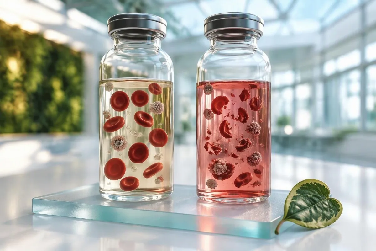
Высокі гемалітычны індэкс звычайна апісвае пашкоджанне клетак пасля збору, а не дыягназ у вас. Карысным пытаннем з'яўляецца тое, якія паведамленыя вынікі застаюцца надзейнымі, а якія патрабуюць паўтарэння.


- Высокі гемалітычны індэкс азначае, што аналізатар выявіў свабодны гемаглабін у сыроватцы або плазме, звычайна з-за разбурэння клетак падчас збору або транспарціроўкі.
- Калій асабліва ўразлівы, таму што чырвоныя клеткавыя элементы ўтрымліваюць прыкладна 100–120 ммоль/л калію ў параўнанні з плазмай 3,5–5,0 ммоль/л.
- ЛДГ і AST можа значна павышацца ў гемалізаваным узоры; ізаляванае нечаканае павышэнне не павінна прыпісвацца захворванням печані або цягліц да паўторнага тэставання.
- Працэнтныя кропкі аналізатара з'яўляюцца спецыфічнымі для метаду: гемалітычны індэкс 100 на адным аналізатары не з'яўляецца ўніверсальнай мерай цяжкасці або дыягназам гемалітычнай анеміі.
- Рашэнні аб паўторным заборы залежыць ад індывідуальнага аналізу, ступені ўмяшання і клінічнай неадкладнасці — а не толькі ад знешняга выгляду ўзору.
- Тэрміновыя рашэнні па калію патрабуюць хуткага пацвярджэння негемалізаваным плазмавым узорам, аналізам газу крыві ў цэльнай крыві або ЭКГ, калі вынік высокі.
- Гемолиз in vivo выклікае клінічную карціну, такую як анемія, ретикулоцитоз, нізкі гаптаглабін і павышэнне непрамога білірубіну; толькі высокі індэкс не можа яго ўстанавіць.
- Па стане на 27 верасня 2026 г., пацыенты павінны пытацца, ці быў вынік лабараторыяй адхілены, кваліфікаваны або аналітычна выпраўлены перад прыняццем мер.
Што насамрэч вымярае высокі гемалітычны індэкс
A высокі індэкс гемолізу азначае, што аналізатар выявіў лішак свабоднага гемаглабіну ў вадкай частцы лабараторнага ўзору. У большасці амбулаторных мазкоў гэта адлюстроўвае разбурэнне клетак пасля збору, а не разбурэнне эрытрацытаў, якое адбываецца ўнутры арганізма.
Індэкс звычайна генеруецца спектрофотометрией: аналізатар вымярае паглынанне пры даўжынях хваль, дзе гемаглабін паглынае святло, затым прымяняе правілы ўмяшання, спецыфічныя для аналізу. Ружовая або чырвоная сыроватка можа мець індэкс, пазначаны як 50, 100 або 300, але гэтыя лічбы не стандартызаваны сярод вытворцаў. падыход да медыцынскай валідацыі мы выкарыстоўваем, разглядаем каментар лабараторыі і маркеры аналізу як першапачатковае доказа.
Цяжкі збор, занадта энергічнае страсенне прабіркі, затрымка аддзялення ад клетак, экстрэмальная тэмпература або транспарціроўка па пнеўматычнай трубе могуць прывесці да гемалізаванага ўзору крыві. У маёй клінічнай практыцы найбольш частай прычынай з'яўляецца не вены пацыента; гэта тэхнічна складаны мазок у спалучэнні з вельмі маленькай прыладай для збору або прымусовай перадачай. Нават узор, які выглядае нармальным для вока, можа мець дастаткова свабоднага гемаглабіну, каб паўплываць на адчувальны аналіз.
Кантэсці — гэта Аналізатар крыві са штучным інтэлектам які чытае каментар гемолізу як сігнал якасці ўзору, а не як доказ хваробы. Практычнае правіла доктара Томаса Кляйна простае: інтэрпрэтаваць закрануты аналіт толькі пасля праверкі, ці пазначыла яго лабараторыя як ненадійны, адхіліла яго або выпусціла з папярэджаннем.
Чаму колер мае значэнне, але не вырашае справу
Відавочная ружовая сыроватка часта адпавядае значнаму колькасці свабоднага гемаглабіну, аднак візуальны агляд прапускае лёгкі гемолиз і з'яўляецца суб'ектыўным. Сучасныя індэксы больш паслядоўныя, чым візуальная ацэнка, але іх клінічнае значэнне па-ранейшаму вагаецца ў залежнасці ад аналізатара, рэагента і метаду тэсціравання.
Гемаліз узору супраць гемалізу, які адбываецца ў арганізме
Высокі індэкс звычайна паказвае гемолиз in vitro, што азначае, што пашкоджанне клетак адбылося ў прабірцы або падчас апрацоўкі. Гемолиз in vivo - гэта медыцынскі працэс, які патрабуе пацвярджэння сімптомаў, вынікаў CBC і мэтавых аналізаў крыві.
Гемолиз in vivo часта дае зладжаную карціну: зніжэнне гемаглабіну, ретикулоцитоз, павышэнне непрамога білірубіну, павышэнне ЛДГ і нізкі гаптаглабін. Можа з'явіцца цёмная моча, жаўтуха, стомленасць або боль у спіне, хоць хранічны лёгкі гемолиз можа быць незаўважным. Адзіны высокі індэкс гемолізу пры нармальным CBC значна часцей з'яўляецца перад аналітычным, чым клінічным.
Гэта адрозненне важна, таму што гемалізаваны ўзор можа памылкова імітаваць тую самую карціну, якая выкарыстоўваецца для дыягностыкі гемолізу — асабліва павышаны калій, ЛДГ, AST і фасфат. Ліпі і інш. апісалі гемолиз як асноўную прычыну непрыдатных узораў у клінічных лабараторыях, менавіта таму, што ўнутрыклетачнае змесціва выліваецца ва ўзор, а спектральнае ўмяшанне ўплывае на фотаметрычныя метады (Lippi et al., 2008).
Калі анемія або жаўтуха сапраўды прысутнічаюць, лекары могуць паўтарыць узор і дадаць ретикулацыты, фракцыі білірубіну, гаптаглабін і інтэрпрэтацыю ретикулацытаў rather than relying on the index. Kantesti’s сервісі інтерпретації тестів ШІ compares these linked results and flags when the pattern deserves clinician review rather than a routine redraw.
A useful clue from timing
True hemolysis persists across carefully collected samples; collection-related hemolysis usually disappears on repeat testing. That contrast is often more informative than one isolated laboratory flag.
Якія вынікі аналізу крыві найбольш змяняюцца пры гемалізе
Hemolysis most often creates falsely high potassium, LDH, AST, phosphate, magnesium, iron, and sometimes total protein results. The magnitude is assay-dependent, so a laboratory’s interference comment outweighs any universal correction formula.
Potassium is the safety-critical example. Red cellular elements contain approximately 100–120 mmol/L potassium, while normal plasma potassium is 3.5–5.0 mmol/L; even modest cellular leakage can push a normal result above 5.5 mmol/L. A reported potassium of 6.2 mmol/L with a high hemolysis index may be pseudohyperkalemia, but it still needs rapid clinical triage if symptoms, kidney disease, ECG changes, or potassium-raising medicines are present.
LDH is highly concentrated within cellular elements, and AST is also more abundant inside them than in serum. In a 52-year-old runner with AST 89 IU/L, a hemolysis warning plus normal ALT and CK changes my first step from diagnosing liver disease to repeating the panel. See our practical guide to high AST results for the non-hemolysis causes that remain relevant.
Phosphate, magnesium, and iron may rise because they are released from disrupted cells; bilirubin and some enzyme assays can be biased in either direction by optical interference. This is why a numerical result may be released but not clinically usable. A тлумач панэлі печані helps separate a real enzyme pattern from one compromised result.
Tests that can be falsely lower
Hemoglobin absorbance can produce negative interference in some colorimetric or enzymatic assays, including selected lipase, alkaline phosphatase, and bilirubin methods. There is no safe assumption that hemolysis only makes values high.
Высокі калій пры гемалізе: калі гэта неадкладна?
A potassium result of 6.0 mmol/L or higher with a hemolysis flag should be confirmed promptly, but it must not be dismissed automatically. Immediate assessment is appropriate when potassium is 6.5 mmol/L or higher, an ECG is abnormal, or weakness, palpitations, chest symptoms, or significant kidney impairment are present.
Pseudohyperkalemia is common when cellular elements rupture during collection, but genuine hyperkalemia can coexist with a difficult draw. A fresh lithium-heparin plasma specimen, carefully collected whole-blood analysis, and ECG are common confirmation routes. Do not try to lower potassium through diet or supplements before speaking with the clinician who ordered the test; that could delay necessary care.
The risk rises when a patient has eGFR below 30 mL/min/1.73 m², takes an ACE inhibitor, ARB, spironolactone, trimethoprim, or potassium supplement, or has metabolic acidosis. In those settings, a result of 5.8 mmol/L deserves more attention than the same result in a well person with normal renal function and an obviously compromised tube. Our detailed даведнік па памылках забору калію explains this decision point.
A clenched fist during collection can raise potassium slightly even without visible sample hemolysis, while prolonged tourniquet use adds another source of pre-analytical variation. The thing is, neither detail can prove the result is false. Repeat testing is the safe answer when the number could alter urgent treatment.
Symptoms that override the laboratory caveat
Severe muscle weakness, fainting, new palpitations, chest discomfort, or shortness of breath need urgent assessment regardless of a hemolysis comment. Laboratory uncertainty should never be used to explain away concerning symptoms.
ЛДГ, AST і КФК: чытанне ферментаў у гемалізаваным узоры
LDH and AST are among the least reliable results in a markedly hemolyzed specimen because both are released from disrupted cellular elements. CK is often less directly affected by red-cell leakage but can still be analytically affected depending on the laboratory method.
An isolated LDH elevation should not trigger a cancer, liver, or hemolytic-anemia work-up until specimen quality is addressed. LDH has five isoenzymes and is found in many tissues, so even a pristine high value is nonspecific. Our explanation of LDH test meaning shows why trends and companion tests matter more than a single result.
AST can rise after strenuous exercise, alcohol exposure, muscle injury, or liver injury, while ALT is more liver-focused. When AST is 90 IU/L but ALT is 22 IU/L and the sample is hemolyzed, repeating AST, ALT, CK, and bilirubin after 48–72 hours without unusual exercise often clarifies the picture. This timing is clinical convention, not a hard evidence-based rule for every patient.
Kantesti AI uses pattern logic: AST plus LDH plus a hemolysis flag is labelled potentially compromised, whereas AST plus ALT plus bilirubin elevation in a clean sample carries different weight. That distinction prevents false alarms while preserving follow-up for genuine liver or muscle disease.
Why an AST:ALT ratio may be unusable
A hemolyzed AST value can artificially inflate the AST:ALT ratio, making it unsuitable for alcohol-related liver pattern interpretation or fibrosis scores. Repeat the panel before calculating ratios that may influence a referral.
Якія вынікі могуць заставацца прыдатнымі пасля гемалізу?
Many results can remain usable despite a hemolyzed sample if the hemolysis level is below that assay’s validated interference limit. Sodium, chloride, urea, creatinine, glucose, and many immunoassays are often less affected, but “often” is not a guarantee.
Laboratories validate each test by measuring bias at increasing free-hemoglobin concentrations, then set suppression or comment thresholds. A sodium result may be reliable in a moderately hemolyzed specimen while potassium from the same tube is not. This analyte-by-analyte decision is why a blanket request to repeat an entire panel can be unnecessary.
Cardiac troponin, thyroid hormones, vitamin B12, and ferritin have assay-specific susceptibility to hemoglobin interference; no result should be assumed valid or invalid solely from the test name. If chest pain is present, troponin testing follows urgent clinical pathways regardless of a specimen flag. Read our guide to troponin result differences for why assay identity matters.
A laboratory may release a result with an HIL comment, suppress it entirely, or report that interference is not significant at the measured index. Kantesti’s neural network preserves that distinction when extracting PDF reports rather than treating every number as equally precise. The Kantesti AI-бенчмарк describes technical evaluation of this report-reading workflow.
The serum-versus-plasma detail
Serum waits for clotting before separation, whereas plasma is separated from anticoagulated whole blood. Delay before separation can increase contact between cells and liquid, but both specimen types can hemolyze during collection or transport.
Чаму парогі гемалітычнага індэксу адрозніваюцца ў розных лабараторыях
There is no universal normal range for a hemolysis index because instruments, wavelengths, sample types, and assay reagents differ. A value of 150 may be a minor flag in one laboratory and a rejection threshold in another.
The HIL system—hemolysis, icterus, and lipemia indices—usually estimates optical interference before selected chemistry assays run. It is not the same as plasma free hemoglobin measured for suspected intravascular hemolysis, which may be reported in mg/dL. Confusing these two measures is a surprisingly common source of unnecessary worry.
Laboratories choose action limits based on acceptable analytical bias, frequently aligned with a change no greater than 10% or a fraction of biological variation. Yet a 10% error is trivial for one test and consequential for potassium near 6.0 mmol/L. Clinicians disagree on the most practical cutoff because the correct threshold depends on what decision the result will drive.
Кантэсці — гэта Платформа AI для расшыфроўкі аналізу крыві that displays the exact printed laboratory flag and reference context instead of translating an index into a made-up universal severity grade. Readers comparing reports from different countries can use our даведнік на 15 000 маркераў to check units and assay names before comparing trends.
Why you should not compare index values over time
A hemolysis index of 80 in one laboratory and 80 in another may not represent the same free-hemoglobin concentration. Compare clinical analytes across laboratories cautiously unless the assay method and specimen handling are consistent.
Калі паўторны забор неабходны — а калі не
A redraw is usually needed when hemolysis could change a result that guides an urgent or major clinical decision. It may not be needed when the laboratory confirms negligible interference for the released tests and the result fits the clinical picture.
Redraw promptly for a high potassium, unexpectedly high LDH or AST, discrepant phosphate or magnesium, or any result the laboratory has withheld. Repeat testing also makes sense when a result conflicts sharply with a prior baseline—such as creatinine 0.8 to 2.1 mg/dL without illness, dehydration, or medication change. A тлумачэнне delta-check can help frame that conversation.
A repeat may be unnecessary for a stable HbA1c, TSH, or creatinine when the lab has explicitly released it without an interference warning. Still, repeat if the number would trigger a medication change, imaging, or referral. In my experience, patients are reassured by asking one specific question: “Which individual results did the lab consider affected?”
Do not assume fasting fixes hemolysis. Better prevention is a relaxed collection, appropriate tube filling, gentle inversions rather than shaking, rapid transport, and timely centrifugation. Our repeat-test guide after hemolysis gives patients a practical checklist.
Hospital and emergency settings
In emergency care, clinicians may use a rapid whole-blood result while simultaneously sending a carefully collected confirmatory specimen. Treatment decisions are based on symptoms, ECG findings, kidney function, and repeat values—not the hemolysis index alone.
Чаму крывяныя ўзоры гемалізуюцца падчас збору і транспарціроўкі
Most hemolyzed blood samples arise from mechanical stress, delayed processing, or temperature extremes rather than any fault in the patient. Prevention begins before the tube reaches the analyzer.
Drawing through an undersized device, pulling too forcefully with a vacuum device, forcing a sample through a connector, or vigorous shaking can damage cellular elements. Underfilling additive tubes creates a wrong blood-to-additive ratio, which can introduce separate errors even if hemolysis is absent. The details sound fussy, but they change real results.
Transportation matters after collection. Extended heat exposure, freezing, rough transit, and a long delay before separating serum or plasma can increase hemolysis. Samples from lines can also be misleading if collection follows infusion or if technique is difficult; a clean peripheral collection may be preferable when the result is pivotal.
Patients can help by arriving hydrated unless fluid restriction applies, keeping their arm warm, and avoiding repeated fist pumping. None of these steps guarantees a clean sample, particularly in fragile veins. A baseline draw preparation guide covers preparation without turning routine testing into an ordeal.
Exercise is a separate issue
A hard workout can genuinely raise CK, AST, and creatinine for 24–72 hours even when the specimen is pristine. Exercise may coexist with collection hemolysis, so a repeat result should ideally follow an ordinary rest day when medically appropriate.
Што могуць паведаміць CBC і мазок крыві
A complete blood count can help distinguish a poor specimen from possible true hemolysis, but it cannot do so by itself. Falling hemoglobin, rising reticulocytes, and smear changes carry more diagnostic weight than the chemistry hemolysis index.
A true hemolytic process commonly causes reticulocytosis as marrow responds to shortened red-cell survival, although this response may be absent with iron deficiency, marrow disease, or very early onset. A reticulocyte count above the laboratory’s upper limit—often roughly 2.0%—must be converted to an absolute count and interpreted alongside hemoglobin. Percentage alone can mislead in anemia.
A peripheral cell sample slide may show schistocytes, spherocytes, bite cells, or polychromasia depending on cause. Schistocytes at 1% or more on a standardized review can support thrombotic microangiopathy in the right setting, but this is a specialist interpretation and urgent symptoms matter. Our guide to тэрміновых знаходках са шызтацытамі explains why this is not a home diagnosis.
Dr. Thomas Klein advises looking for internal consistency: high LDH plus low haptoglobin plus anemia on a clean repeat sample is meaningful; high LDH alone in a hemolyzed tube is not. The hematocrit and hemoglobin comparison is useful when CBC values seem contradictory.
A falsely high MCHC can be a clue
Markedly high MCHC can occur with sample hemolysis, lipemia, cold agglutinins, or spherocytosis. It should prompt laboratory review rather than an immediate conclusion about red-cell disease.
Асаблівыя сітуацыі: дзеці, цяжарнасць і цяжкі доступ
Hemolysis is more common when collection is technically difficult, including neonatal, pediatric, emergency, and critical-care testing. The consequences may also be greater because small electrolyte shifts can affect urgent decisions.
In newborns and young children, capillary collection and low sample volumes can increase cellular disruption, while potassium reference intervals differ by age. A potassium value that is high for an adult may be less abnormal in a newborn, yet any suspected hyperkalemia still requires prompt clinician-led confirmation. Never apply adult cutoffs to a child’s report without the laboratory’s pediatric range.
Pregnancy adds another layer: anemia is common from dilution or iron deficiency, while HELLP syndrome and other obstetric emergencies can involve real hemolysis. New headache, visual symptoms, right-upper abdominal pain, high blood pressure, or reduced fetal movement need urgent maternity assessment—not an assumption that the tube was damaged. Our HELLP laboratory pattern guide details the red flags.
For people with dialysis access, implanted lines, burns, or very difficult peripheral access, a higher rate of unsuitable samples may be unavoidable. The care team can often arrange an approach that minimizes redraws, but the test’s clinical urgency determines whether another sample is required.
Home collection kits
Finger-prick samples are not interchangeable with venous samples for every analyte, and inadequate handling can create interference. Follow kit-specific instructions and ask the provider whether a venous redraw is needed after a high hemolysis flag.
Як чытаць каментар гемалізу ў вашым лабараторным справаздачы
A lab report may say “hemolyzed,” “hemolysis index elevated,” “interference possible,” or “result not reported.” These phrases have different implications: a suppressed result is unusable, while a released result may remain valid if interference is below the assay’s limit.
Look for a comment attached to the individual analyte, not merely a general specimen note. For example, potassium may say “not reported due to hemolysis,” while creatinine is reported without qualification. This reflects the laboratory’s validation work and is more useful than judging the tube’s color from a photograph.
If a number is marked with an H for high and also a hemolysis caveat, it can be both analytically elevated and clinically uncertain. Ask the ordering clinician whether the result fits your medications, symptoms, earlier values, and other tests. Our guide to позамежових позначок explains why a flag is a prompt for context, not a diagnosis.
Kantesti AI can organize a photographed or PDF report in about 60 seconds, but it cannot inspect the physical specimen or replace a laboratory’s interference decision. Our гід па тэхналогіі explains how we preserve qualifiers, units, and reference intervals for clinician discussion.
A question worth writing down
Ask: “Was this specific test suppressed, corrected, or released as valid despite the hemolysis index?” That single question usually gets a clearer answer than asking whether the whole panel was “bad.”
Што нельга рабіць пасля атрымання гемалізаванага выніку
Do not self-treat a possibly false result, ignore a potentially dangerous one, or assume every result in the panel is invalid. The right response is targeted confirmation based on the analyte and your clinical context.
Do not stop prescribed ACE inhibitors, diuretics, thyroid medicine, or supplements solely because one hemolyzed sample was abnormal. Potassium, thyroid, and kidney results must be interpreted against the medication list and a repeat value where needed. Sudden medication changes can be riskier than waiting a few hours for confirmation.
Avoid searching for a diagnosis from a high LDH or AST result before checking sample quality. Serious disease can cause these elevations, of course, but an unreliable first sample does not justify a frightening conclusion. For muscle-specific concerns, our high CK safety guide sets out symptoms that do need prompt care.
Likewise, do not repeatedly redraw yourself through commercial testing without sharing the original report with a clinician. Recurrent hemolysis may be a collection issue, but recurrent abnormal results in properly handled samples deserve a structured evaluation.
When to seek same-day help
Seek urgent advice for high potassium with weakness or palpitations, jaundice with dark urine, severe fatigue with breathlessness, chest pain, fainting, or rapidly worsening symptoms. A specimen-quality note cannot lower the urgency of these clinical signs.
Як бяспечна параўноўваць вынікі пасля паўторнага забору
A clean repeat sample is most useful when collected under comparable conditions and interpreted as a trend, not as a verdict on the first result. A large change after a hemolyzed draw often identifies interference, but biological variation still exists.
Record the draw date, fasting status, exercise in the previous 48 hours, illness, supplements, and medications. Creatinine can shift with hydration and exercise; AST and CK can shift after training; potassium may shift with kidney function and medicines. These ordinary variables matter even when the second sample has no hemolysis flag.
For potassium, a change from 6.1 mmol/L in a hemolyzed sample to 4.3 mmol/L in a clean sample strongly supports pseudohyperkalemia. For AST, a decline from 88 to 29 IU/L may reflect removal of interference, recovery after exercise, or both; timing and ALT help adjudicate. Our даведнік па зменах аналізу крыві explains reference change value more fully.
Кантэсці — гэта Інструмент аналізу вынікаў аналізу крыві з падтрымкай AI that can place repeat values beside prior results while retaining specimen-quality comments. Trend displays are useful, but they must never obscure a redrawn critical result that needs immediate clinician action.
Захавайце арыгінальны справаздачу
Save the first report, including the hemolysis comment, rather than deleting it after the repeat. It documents why a value changed and prevents future clinicians from mistaking a collection artifact for sudden disease resolution.
Пытанні, якія трэба задаць вашаму лекару або лабараторыі
The best question after a high hemolysis index is not “Do I have hemolysis?” but “Which analytes were affected, how urgently should they be repeated, and what symptoms would change the plan?” A short, specific discussion avoids both false reassurance and unnecessary alarm.
Ask whether the abnormal value was suppressed, whether the lab has a validated interference threshold for that method, and whether a fresh plasma or whole-blood sample is preferable. For potassium, ask whether an ECG or same-day repeat is needed. For enzyme results, ask whether exercise, alcohol, infection, or medicines may also be contributing.
Ask whether you need a full panel or only selected tests repeated. A targeted redraw often includes potassium, AST, LDH, phosphate, magnesium, or bilirubin, whereas a CBC or HbA1c may not need repeating. This can reduce cost, discomfort, and confusing duplicate results without compromising care.
Нашы лекары на Медыцынская кансультатыўная рада review clinical-safety principles used in Kantesti content, but personal treatment decisions belong with the clinician who knows your history and can assess you. Most patients find that a precise repeat plan is far calmer than trying to interpret a colored sample image online.
Кароткі вынік
A high hemolysis index is usually a laboratory-quality warning, not a diagnosis. Respect the warning, identify the affected tests, and repeat only what could change clinical decisions.
Як Kantesti апрацоўвае сцяжкі гемалізу ў справаздачах аб крыві
Kantesti identifies hemolysis flags and links them to the results most likely to be affected, but it does not replace laboratory validation or urgent clinical assessment. A flagged potassium, troponin, or critical electrolyte result always needs the laboratory and treating team’s protocol.
Kantesti AI reads the test name, value, unit, reference interval, collection context, and printed laboratory comments together. That matters because “high potassium” without the accompanying hemolysis statement is an incomplete clinical fact. Our system is designed to surface uncertainty plainly rather than conceal it behind a confident-looking score.
Кантэсці — гэта платформа інтэрпрэтацыі біямаркераў AI used across 127+ countries, so it also avoids assuming that identical labels or index values mean identical methods. Different laboratories may use serum, plasma, direct or indirect assays, and different interference thresholds. International reports need the source laboratory’s own interpretive note preserved.
As of September 27, 2026, our safety approach is to encourage a clinician discussion for results that are critical, internally inconsistent, newly abnormal, or clearly compromised by blood test hemolysis. If you want to understand how health data are handled, our старонцы About Us describes the organization and its privacy-focused approach.
The limit of any report interpretation
No report-reading system can know whether you have palpitations, jaundice, dehydration, or a new medication unless you provide that context to a clinician. The specimen flag is one piece of evidence, not a substitute for examination or urgent care.
Часта задаваныя пытанні
Што азначае высокі індэкс гемолізу ў аналізе крыві?
Высокі паказчык гемалізу азначае, што лабараторны аналізатар выявіў свабодны гемаглабін са зруйнаваных эрытрацытаў у сыроватцы або плазме. Большасць выпадкаў узнікае падчас збору, транспарціроўкі або апрацоўкі, а не з-за медыцынскага стану пацыента. Паказчык можа зрабіць вынікі калію, ЛДГ, AST, фасфатаў, магнію і жалеза ненадзейнымі, але эфект залежыць ад аналізу. Паказчык гемалізу не з'яўляецца дыягнастычным тэстам на гемалітычную анемію.
Ці можа высокі паказчык гемалізу выклікаць высокі ўзровень калію?
Так, высокі паказчык гемолізу можа выклікаць псеўдагіперкаліемію, паколькі эрытрацыты змяшчаюць каля 100–120 ммоль/л калію, а нармальны ўзровень калію ў плазме складае 3,5–5,0 ммоль/л. Нават невялікі разбурэнне клетак можа ілжыва павысіць вымераны ўзровень калію вышэй за 5,5 ммоль/л. Вынік калію 6,0 ммоль/л або вышэй варта неадкладна пацвердзіць, асабліва пры захворваннях нырак, прыёме лекаў, якія павышаюць узровень калію, слабасці або пры пачашчаным сэрцабіцці. Можа спатрэбіцца ЭКГ і старанна сабраны паўторны ўзор.
Ці трэба паўтарыць усе аналізы крыві пасля гемалізаванага ўзору?
Не, гемалізаваны ўзор не абавязкова робіць несапраўдным кожны аналіз крыві. Лабараторыі ацэньваюць умяшанне асобна для кожнага аналізу, і натрый, хларыд, крэатынін, глюкоза і некаторыя імунааналізы могуць заставацца прыдатнымі пры зададзеным узроўні гемалізу. Паўтарыце тэсты, якія лабараторыя прыпыніла, абмежавала або якія маглі б змяніць тэрміновае рашэнне, такія як калій або нечакана высокі ўзровень ЛДГ і AST. Спытайце, на якія менавіта аналіты былі ўплывы, перш чым арганізоўваць поўны адбор проб.
Ці можа гемалітычны ўзор азначаць, што ў мяне гемалітычная анемія?
Гемолізна проба сама па сабе не азначае, што ў вас гемалітычная анемія. Гемалітычная анемія пацвярджаецца сукупнасцю прыкмет, якія могуць уключаць зніжэнне гемаглабіну, высокі ўзровень рэтыкулацытаў, павышаны ўскосны білірубін і ЛДГ, а таксама нізкі гаптаглабін у старанна сабранай пробе. Такія сімптомы, як жаўтуха, цёмная мача, дыхавіца або значная стомленасць, павышаюць занепакоенасць, але ў некаторых пацыентаў сімптомы лёгкія. Паўторная чыстая проба звычайна з'яўляецца першым спосабам адрозніць гемоліз, звязаны са зборам, ад сапраўднага разбурэння эрытрацытаў.
Колькі часу трэба чакаць перад паўторным аналізам крыві пасля гемалізу?
Паўторны аналіз часта можа быць праведзены ў той жа дзень, калі справа тычыцца калію, крытычнага электраліта, трапаніну або іншага тэрміновага выніку. Для неэкстраных аналізаў AST, CK або LDH пасля інтэнсіўных фізічных нагрузак клініцысты часам паўтараюць аналіз праз 48–72 гадзіны пасля адмовы ад незвычайных практыкаванняў, паколькі самі практыкаванні могуць павышаць узровень гэтых ферментаў. Універсальнага перыяду чакання для высокага індэкса гемолізу не існуе; адпаведны тэст і прычына тэсціравання вызначаюць час. Лабараторыя або які прызначыў тэст клініцыст павінны прадставіць канкрэтны план.
Ці можа абязводжванне выклікаць высокі індэкс гемолізу?
Абязводжванне не выклікае наўпрост высокі індэкс гемолізу, але можа ўскладніць збор і павялічыць верагоднасць тэхнічна складанага забору. Уздзеянне цяпла або дрэннае захоўванне пасля збору таксама можа пашкодзіць клеткавыя элементы і павысіць індэкс. Гідратацыя можа дапамагчы ў доступе да вены для людзей, якім не абмежавана вадкасць, але яна не можа выправіць гемалізаваны ўзор пасля яго збору. Свежы правільна апрацаваны ўзор з'яўляецца належным пацвярджэннем.
Атрымаць аналіз крыві з AI сёння
Далучайцеся больш чым да 2 мільёнаў карыстальнікаў па ўсім свеце, якія давяраюць Kantesti для імгненнага, дакладнага аналізу лабараторных тэстаў. Загрузіце вынікі аналізу крыві і атрымайце поўную інтэрпрэтацыю біямаркераў 15,000+ за лічаныя секунды.
📚 Публікацыі даследаванняў са спасылкамі
Кляйн, Т., Мітчэл, С., і Вебер, Г. (2026). Klein, T. (2026). A Pre-Registered, Rubric-Based Automated Technical Benchmark of the Kantesti Blood-Test Interpretation Engine on 100,000 Synthetic Test Cases. Figshare. https://doi.org/10.6084/m9.figshare.32095435. Kantesti AI Medical Research.
Кляйн, Т., Мітчэл, С., і Вебер, Г. (2026). Kantesti LTD. (2026). Каркас клінічнай валідацыі v2.0 (Старонка медыцынскай валідацыі). Zenodo. https://doi.org/10.5281/zenodo.17993721. Kantesti AI Medical Research.
📖 Знешнія медыцынскія спасылкі
⚕️ Медыцынская адмова ад адказнасці
Гэты артыкул прызначаны толькі для адукацыйных мэт і не з’яўляецца медычнай парадай. Заўсёды кансультуйцеся з кваліфікаваным спецыялістам аховы здароўя для прыняцця рашэнняў па дыягностыцы і лячэнні.
Сігналы даверу E-E-A-T
Вопыт
Клінічны агляд, які вядзе лекар, працэсаў інтэрпрэтацыі лабараторных паказчыкаў.
Экспертыза
Фокус лабараторнай медыцыны на тым, як біямаркеры паводзяць сябе ў клінічным кантэксце.
Аўтарытэтнасць
Напісана доктарам Томасам Кляйнам, з аглядам доктара Сара Мітчэл і праф. доктара Ганса Вебера.
Надзейнасць
Інтэрпрэтацыя на аснове доказаў з выразнымі шляхамі далейшых дзеянняў, каб паменшыць трывогу.