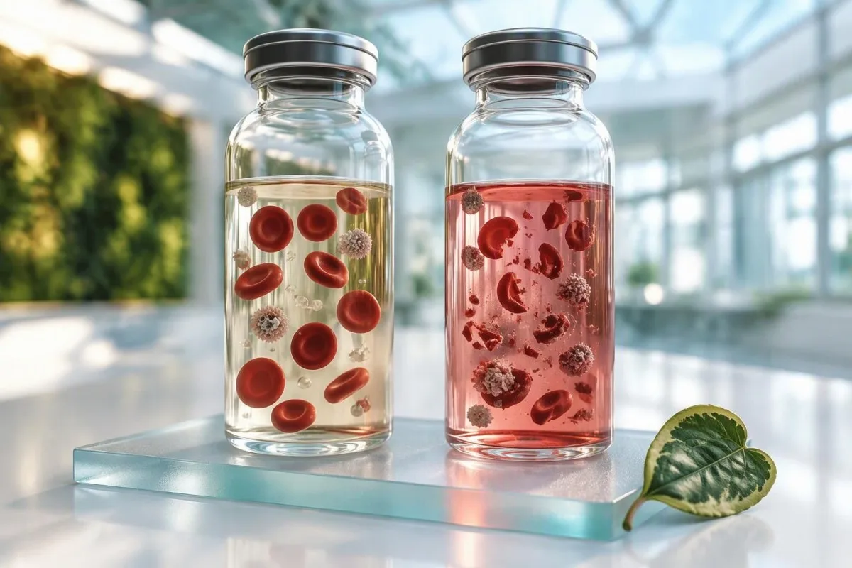
د وینې ازموینې پایلې.


- د انحال لوړ شاخص پدې معنی چې شنونکی د سیرم یا پلازما دننه وړیا هیموګلوبین کشف کوي، معمولا د راټولولو یا لیږدولو پرمهال د حجرو د ماتیدو له امله.
- پوټاشیم په ځانګړي ډول زیان منونکی دی ځکه چې سور حجروي عناصر نږدې 100-120 mmol/L پوتاشیم د 3.5-5.0 mmol/L پلازما حد په پرتله لري.
- LDH او AST په انحال شوې نمونه کې په څرګنده توګه لوړ کیدی شي؛ یوه جلا ناڅاپي لوړوالی باید د تکراري ازموینې دمخه د جگر یا عضلاتو ناروغۍ لیبل نه شي.
- د شنونکي حدونه میتود ځانګړي دي: په یوه شنونکي کې د 100 انحال شاخص د شدت نړیوال اندازه یا د انحلالي انیمیا تشخیص ندی.
- د بیا رسمولو پریکړې په ازموینې، د لاسوهنې کچې، او کلینیکي اضطرار پورې اړه لري — نه یوازې د نمونې په ظاهري بڼه.
- د پوتاشیم عاجل پریکړې د لوړې پایلې په صورت کې د غیر محلول شوي پلازما نمونې، بشپړ وینې د وینې ګاز اندازه کولو، یا ECG لخوا چټک تایید ته اړتیا لري.
- په بدن کې د وینې محلولیدل د انیمیا، ریټیکولوسایټوسس، ټیټ هاپتوګلوبین او لوړ شوي غیر مستقیم بلیروبین په څیر یو کلینیکي انځور رامینځته کوي؛ یوازې یو لوړ شاخص نشي دا ثابتولی.
- د سپتمبر 27، 2026 پورې, ، ناروغان باید پوښتنه وکړي چې ایا لابراتوار د دې په اړه عمل کولو دمخه هر اغیزمن پایله پټ کړې، وړتیا ورکړې، یا په تحلیلي توګه سم کړې.
د وینې ازموینې پایلې
A د محلول لوړ شاخص پدې معنی چې یو تحلیل کونکي د لابراتوار نمونې په مایع برخه کې اضافي وړیا هیموګلوبین کشف کړی. په ډیرو بهرنیو ناروغانو کې، دا د بدن دننه د سره وینې حجرو د تخریب په پرتله د راټولولو وروسته د حجرو ماتیدل منعکس کوي.
دا شاخص معمولا د سپیکټروفتومتري لخوا رامینځته کیږي: تحلیل کونکی په څپو کې جذب اندازه کوي چیرې چې هیموګلوبین رڼا جذبوي، بیا د ازموینې ځانګړي مداخلي قواعد پلي کوي. ګلابي تر سور سیرم نمونې ممکن 50، 100، یا 300 په توګه فلګ شوي شاخص ولري، مګر دا شمیرې د تولید کونکو ترمنځ معیاري ندي. د طبي تایید (validation) طریقه موږ د لابراتوار خپل نظر او د ازموینې فلګونه لومړني شواهد په توګه کاروو.
یوه سخته راټولونه، یوه ټیوب چې ډیره زور سره وخوځول شي، له حجرو څخه ځنډول شوی جلا کول، خورا لوړه تودوخه، یا د هوایی نل له لارې لیږدول ټول کولی شي د محلول شوي وینې نمونه رامینځته کړي. زما په کلینیکي کار کې، تر ټولو عام مجرم د ناروغ رګونه ندي؛ دا د تخنیکي پلوه ننګونې سره یوځای د ډیر کوچني راټولولو وسیله یا زور سره لیږدول دي. حتی یوه نمونه چې سترګو ته عادي ښکاري ممکن دومره وړیا هیموګلوبین ولري چې یو حساس ازموینې اغیزه وکړي.
کانټیسټي یو دی د AI د وینې معاینې شنونکی چې د محلول تبصره د نمونې کیفیت سیګنال په توګه لولي، نه د ناروغۍ ثبوت په توګه. ډاکټر توماس کلین regras عملي قاعده ساده ده: یوازې د اغیزمن انالیټ تفسیر کړئ وروسته له دې چې وګورئ چې ایا لابراتوار یې د غیر معتبر په توګه نښه کړی، پټ کړی، یا یې په اخطار سره خپور کړی.
ولې رنګ مهم دی خو قضیه نه پریکړه کوي
څرګند ګلابي سیرم اکثرا د پام وړ وړیا هیموګلوبین سره مطابقت لري، بیا هم لیدلوری تفتیش د محلول د کمولو یا ذهنيت له لاسه ورکوي. عصري شاخصونه د بصري درجې په پرتله ډیر ثابت دي، مګر د دوی کلینیکي معنی د تحلیل کونکي، ریجنټ، او ازموینې میتود پورې اړه لري.
د نمونې انحلال د بدن دننه د انحال کیدو په پرتله
یو لوړ شاخص معمولا په ګوته کوي په بدن کې د وینې محلولیدل, ، پدې معنی چې حجرو ته زیان په ټیوب کې یا د لاسرسي پرمهال پیښ شو. په بدن کې د وینې محلولیدل یو طبي پروسه ده چې د تایید شوي علایمو، CBC موندنو، او هدف لرونکي وینې ازموینې ته اړتیا لري.
د بدن دننه د وینې محلولیدل اکثرا یو همغږي انځور رامینځته کوي: د هیموګلوبین کمیدل، ریټیکولوسایټوسس، د غیر مستقیم بلیروبین لوړیدل، د LDH زیاتوالی، او ټیټ هاپتوګلوبین. تیاره پیشاب، ژیړی، ستړیا، یا د ملا درد ممکن پیښ شي، که څه هم مزمن کم محلول کیدای شي خاموشه وي. یو واحد لوړ محلول شاخص د بل هرڅه سره مستحکم CBC سره تل له تحلیلي څخه کلینیکي وي.
دا توپیر مهم دی ځکه چې یوه محلول شوې نمونه کولی شي په غلطۍ سره هغه انځور تقلید کړي چې د محلول تشخیص لپاره کارول کیږي - په ځانګړي توګه لوړ پوتاشیم، LDH، AST، او فاسفیٹ. لیپي او نورو محلول د کلینیکي لابراتوارونو کې د نا مناسب نمونو مخکښ لامل په توګه بیان کړی، په دقیق ډول ځکه چې د حجرو دننه مینځپانګې په نمونه کې تویږي او د سپیکټرل مداخله د فوتومیتریک میتودونو اغیزه کوي (Lippi et al., 2008).
که انیمیا یا ژیړی په ریښتیا شتون ولري، کلینیکي ډاکټران ممکن نمونه بیا تکرار کړي او ریټیکولوسایټس، بلیروبین برخې، هاپتوګلوبین، او یو ریټیکولوسایټ تفسیر اضافه کړي rather than relying on the index. Kantesti’s د AI لاب ټېسټ د تفسیر خدمت compares these linked results and flags when the pattern deserves clinician review rather than a routine redraw.
A useful clue from timing
True hemolysis persists across carefully collected samples; collection-related hemolysis usually disappears on repeat testing. That contrast is often more informative than one isolated laboratory flag.
کومې وینې ازموینې پایلې د انحال کیدو سره خورا بدلیږي
Hemolysis most often creates falsely high potassium, LDH, AST, phosphate, magnesium, iron, and sometimes total protein results. The magnitude is assay-dependent, so a laboratory’s interference comment outweighs any universal correction formula.
Potassium is the safety-critical example. Red cellular elements contain approximately 100–120 mmol/L potassium, while normal plasma potassium is 3.5–5.0 mmol/L; even modest cellular leakage can push a normal result above 5.5 mmol/L. A reported potassium of 6.2 mmol/L with a high hemolysis index may be pseudohyperkalemia, but it still needs rapid clinical triage if symptoms, kidney disease, ECG changes, or potassium-raising medicines are present.
LDH is highly concentrated within cellular elements, and AST is also more abundant inside them than in serum. In a 52-year-old runner with AST 89 IU/L, a hemolysis warning plus normal ALT and CK changes my first step from diagnosing liver disease to repeating the panel. See our practical guide to high AST results for the non-hemolysis causes that remain relevant.
Phosphate, magnesium, and iron may rise because they are released from disrupted cells; bilirubin and some enzyme assays can be biased in either direction by optical interference. This is why a numerical result may be released but not clinically usable. A د ینې دندې ازموینه helps separate a real enzyme pattern from one compromised result.
Tests that can be falsely lower
Hemoglobin absorbance can produce negative interference in some colorimetric or enzymatic assays, including selected lipase, alkaline phosphatase, and bilirubin methods. There is no safe assumption that hemolysis only makes values high.
د انحال کیدو سره لوړ پوتاشیم: کله بیړنی دی؟
A potassium result of 6.0 mmol/L or higher with a hemolysis flag should be confirmed promptly, but it must not be dismissed automatically. Immediate assessment is appropriate when potassium is 6.5 mmol/L or higher, an ECG is abnormal, or weakness, palpitations, chest symptoms, or significant kidney impairment are present.
Pseudohyperkalemia is common when cellular elements rupture during collection, but genuine hyperkalemia can coexist with a difficult draw. A fresh lithium-heparin plasma specimen, carefully collected whole-blood analysis, and ECG are common confirmation routes. Do not try to lower potassium through diet or supplements before speaking with the clinician who ordered the test; that could delay necessary care.
The risk rises when a patient has eGFR below 30 mL/min/1.73 m², takes an ACE inhibitor, ARB, spironolactone, trimethoprim, or potassium supplement, or has metabolic acidosis. In those settings, a result of 5.8 mmol/L deserves more attention than the same result in a well person with normal renal function and an obviously compromised tube. Our detailed د پوتاشیم راټولولو غلطي لارښود explains this decision point.
A clenched fist during collection can raise potassium slightly even without visible sample hemolysis, while prolonged tourniquet use adds another source of pre-analytical variation. The thing is, neither detail can prove the result is false. Repeat testing is the safe answer when the number could alter urgent treatment.
Symptoms that override the laboratory caveat
Severe muscle weakness, fainting, new palpitations, chest discomfort, or shortness of breath need urgent assessment regardless of a hemolysis comment. Laboratory uncertainty should never be used to explain away concerning symptoms.
LDH، AST او CK: په انحال شوې نمونه کې انزایمونه لوستل
LDH and AST are among the least reliable results in a markedly hemolyzed specimen because both are released from disrupted cellular elements. CK is often less directly affected by red-cell leakage but can still be analytically affected depending on the laboratory method.
An isolated LDH elevation should not trigger a cancer, liver, or hemolytic-anemia work-up until specimen quality is addressed. LDH has five isoenzymes and is found in many tissues, so even a pristine high value is nonspecific. Our explanation of LDH test meaning shows why trends and companion tests matter more than a single result.
AST can rise after strenuous exercise, alcohol exposure, muscle injury, or liver injury, while ALT is more liver-focused. When AST is 90 IU/L but ALT is 22 IU/L and the sample is hemolyzed, repeating AST, ALT, CK, and bilirubin after 48–72 hours without unusual exercise often clarifies the picture. This timing is clinical convention, not a hard evidence-based rule for every patient.
Kantesti AI uses pattern logic: AST plus LDH plus a hemolysis flag is labelled potentially compromised, whereas AST plus ALT plus bilirubin elevation in a clean sample carries different weight. That distinction prevents false alarms while preserving follow-up for genuine liver or muscle disease.
Why an AST:ALT ratio may be unusable
A hemolyzed AST value can artificially inflate the AST:ALT ratio, making it unsuitable for alcohol-related liver pattern interpretation or fibrosis scores. Repeat the panel before calculating ratios that may influence a referral.
کومې پایلې د انحال کیدو وروسته هم د کارونې وړ کیدی شي؟
Many results can remain usable despite a hemolyzed sample if the hemolysis level is below that assay’s validated interference limit. Sodium, chloride, urea, creatinine, glucose, and many immunoassays are often less affected, but “often” is not a guarantee.
Laboratories validate each test by measuring bias at increasing free-hemoglobin concentrations, then set suppression or comment thresholds. A sodium result may be reliable in a moderately hemolyzed specimen while potassium from the same tube is not. This analyte-by-analyte decision is why a blanket request to repeat an entire panel can be unnecessary.
Cardiac troponin, thyroid hormones, vitamin B12, and ferritin have assay-specific susceptibility to hemoglobin interference; no result should be assumed valid or invalid solely from the test name. If chest pain is present, troponin testing follows urgent clinical pathways regardless of a specimen flag. Read our guide to troponin result differences for why assay identity matters.
A laboratory may release a result with an HIL comment, suppress it entirely, or report that interference is not significant at the measured index. Kantesti’s neural network preserves that distinction when extracting PDF reports rather than treating every number as equally precise. The د Kantesti AI بنچمارک describes technical evaluation of this report-reading workflow.
The serum-versus-plasma detail
Serum waits for clotting before separation, whereas plasma is separated from anticoagulated whole blood. Delay before separation can increase contact between cells and liquid, but both specimen types can hemolyze during collection or transport.
ولې د انحال کیدو شاخص حدونه د لابراتوارونو ترمنځ توپیر لري
There is no universal normal range for a hemolysis index because instruments, wavelengths, sample types, and assay reagents differ. A value of 150 may be a minor flag in one laboratory and a rejection threshold in another.
The HIL system—hemolysis, icterus, and lipemia indices—usually estimates optical interference before selected chemistry assays run. It is not the same as plasma free hemoglobin measured for suspected intravascular hemolysis, which may be reported in mg/dL. Confusing these two measures is a surprisingly common source of unnecessary worry.
Laboratories choose action limits based on acceptable analytical bias, frequently aligned with a change no greater than 10% or a fraction of biological variation. Yet a 10% error is trivial for one test and consequential for potassium near 6.0 mmol/L. Clinicians disagree on the most practical cutoff because the correct threshold depends on what decision the result will drive.
کانټیسټي یو دی AI د وینې ازموینې تشریح پلیټفارم that displays the exact printed laboratory flag and reference context instead of translating an index into a made-up universal severity grade. Readers comparing reports from different countries can use our د 15,000-مارکر لارښود to check units and assay names before comparing trends.
Why you should not compare index values over time
A hemolysis index of 80 in one laboratory and 80 in another may not represent the same free-hemoglobin concentration. Compare clinical analytes across laboratories cautiously unless the assay method and specimen handling are consistent.
کله چې بیا رسمولو ته اړتیا وي - او کله نه
A redraw is usually needed when hemolysis could change a result that guides an urgent or major clinical decision. It may not be needed when the laboratory confirms negligible interference for the released tests and the result fits the clinical picture.
Redraw promptly for a high potassium, unexpectedly high LDH or AST, discrepant phosphate or magnesium, or any result the laboratory has withheld. Repeat testing also makes sense when a result conflicts sharply with a prior baseline—such as creatinine 0.8 to 2.1 mg/dL without illness, dehydration, or medication change. A د delta-check تشریح can help frame that conversation.
A repeat may be unnecessary for a stable HbA1c, TSH, or creatinine when the lab has explicitly released it without an interference warning. Still, repeat if the number would trigger a medication change, imaging, or referral. In my experience, patients are reassured by asking one specific question: “Which individual results did the lab consider affected?”
Do not assume fasting fixes hemolysis. Better prevention is a relaxed collection, appropriate tube filling, gentle inversions rather than shaking, rapid transport, and timely centrifugation. Our repeat-test guide after hemolysis gives patients a practical checklist.
Hospital and emergency settings
In emergency care, clinicians may use a rapid whole-blood result while simultaneously sending a carefully collected confirmatory specimen. Treatment decisions are based on symptoms, ECG findings, kidney function, and repeat values—not the hemolysis index alone.
ولې د وینې نمونې د راټولولو او لیږدولو پرمهال انحلال کیږي
Most hemolyzed blood samples arise from mechanical stress, delayed processing, or temperature extremes rather than any fault in the patient. Prevention begins before the tube reaches the analyzer.
Drawing through an undersized device, pulling too forcefully with a vacuum device, forcing a sample through a connector, or vigorous shaking can damage cellular elements. Underfilling additive tubes creates a wrong blood-to-additive ratio, which can introduce separate errors even if hemolysis is absent. The details sound fussy, but they change real results.
Transportation matters after collection. Extended heat exposure, freezing, rough transit, and a long delay before separating serum or plasma can increase hemolysis. Samples from lines can also be misleading if collection follows infusion or if technique is difficult; a clean peripheral collection may be preferable when the result is pivotal.
Patients can help by arriving hydrated unless fluid restriction applies, keeping their arm warm, and avoiding repeated fist pumping. None of these steps guarantees a clean sample, particularly in fragile veins. A baseline draw preparation guide covers preparation without turning routine testing into an ordeal.
Exercise is a separate issue
A hard workout can genuinely raise CK, AST, and creatinine for 24–72 hours even when the specimen is pristine. Exercise may coexist with collection hemolysis, so a repeat result should ideally follow an ordinary rest day when medically appropriate.
د CBC او د وینې مسح کولی شي تاسو ته څه ووایی
A complete blood count can help distinguish a poor specimen from possible true hemolysis, but it cannot do so by itself. Falling hemoglobin, rising reticulocytes, and smear changes carry more diagnostic weight than the chemistry hemolysis index.
A true hemolytic process commonly causes reticulocytosis as marrow responds to shortened red-cell survival, although this response may be absent with iron deficiency, marrow disease, or very early onset. A reticulocyte count above the laboratory’s upper limit—often roughly 2.0%—must be converted to an absolute count and interpreted alongside hemoglobin. Percentage alone can mislead in anemia.
A peripheral cell sample slide may show schistocytes, spherocytes, bite cells, or polychromasia depending on cause. Schistocytes at 1% or more on a standardized review can support thrombotic microangiopathy in the right setting, but this is a specialist interpretation and urgent symptoms matter. Our guide to د عاجلو schistocyte موندنو په اړه explains why this is not a home diagnosis.
Dr. Thomas Klein advises looking for internal consistency: high LDH plus low haptoglobin plus anemia on a clean repeat sample is meaningful; high LDH alone in a hemolyzed tube is not. The hematocrit and hemoglobin comparison is useful when CBC values seem contradictory.
A falsely high MCHC can be a clue
Markedly high MCHC can occur with sample hemolysis, lipemia, cold agglutinins, or spherocytosis. It should prompt laboratory review rather than an immediate conclusion about red-cell disease.
ځانګړي شرایط: ماشومان، امیندوارۍ او ستونزمن لاسرسی
Hemolysis is more common when collection is technically difficult, including neonatal, pediatric, emergency, and critical-care testing. The consequences may also be greater because small electrolyte shifts can affect urgent decisions.
In newborns and young children, capillary collection and low sample volumes can increase cellular disruption, while potassium reference intervals differ by age. A potassium value that is high for an adult may be less abnormal in a newborn, yet any suspected hyperkalemia still requires prompt clinician-led confirmation. Never apply adult cutoffs to a child’s report without the laboratory’s pediatric range.
Pregnancy adds another layer: anemia is common from dilution or iron deficiency, while HELLP syndrome and other obstetric emergencies can involve real hemolysis. New headache, visual symptoms, right-upper abdominal pain, high blood pressure, or reduced fetal movement need urgent maternity assessment—not an assumption that the tube was damaged. Our HELLP laboratory pattern guide details the red flags.
For people with dialysis access, implanted lines, burns, or very difficult peripheral access, a higher rate of unsuitable samples may be unavoidable. The care team can often arrange an approach that minimizes redraws, but the test’s clinical urgency determines whether another sample is required.
Home collection kits
Finger-prick samples are not interchangeable with venous samples for every analyte, and inadequate handling can create interference. Follow kit-specific instructions and ask the provider whether a venous redraw is needed after a high hemolysis flag.
په خپل لابراتوار راپور کې د انحال کیدو تبصرې لوستل
A lab report may say “hemolyzed,” “hemolysis index elevated,” “interference possible,” or “result not reported.” These phrases have different implications: a suppressed result is unusable, while a released result may remain valid if interference is below the assay’s limit.
Look for a comment attached to the individual analyte, not merely a general specimen note. For example, potassium may say “not reported due to hemolysis,” while creatinine is reported without qualification. This reflects the laboratory’s validation work and is more useful than judging the tube’s color from a photograph.
If a number is marked with an H for high and also a hemolysis caveat, it can be both analytically elevated and clinically uncertain. Ask the ordering clinician whether the result fits your medications, symptoms, earlier values, and other tests. Our guide to له حد څخه د وتلو د نښو explains why a flag is a prompt for context, not a diagnosis.
Kantesti AI can organize a photographed or PDF report in about 60 seconds, but it cannot inspect the physical specimen or replace a laboratory’s interference decision. Our د ټکنالوژۍ لارښود explains how we preserve qualifiers, units, and reference intervals for clinician discussion.
A question worth writing down
Ask: “Was this specific test suppressed, corrected, or released as valid despite the hemolysis index?” That single question usually gets a clearer answer than asking whether the whole panel was “bad.”
د انحال شوې پایلې وروسته څه نه باید وکړي
Do not self-treat a possibly false result, ignore a potentially dangerous one, or assume every result in the panel is invalid. The right response is targeted confirmation based on the analyte and your clinical context.
Do not stop prescribed ACE inhibitors, diuretics, thyroid medicine, or supplements solely because one hemolyzed sample was abnormal. Potassium, thyroid, and kidney results must be interpreted against the medication list and a repeat value where needed. Sudden medication changes can be riskier than waiting a few hours for confirmation.
Avoid searching for a diagnosis from a high LDH or AST result before checking sample quality. Serious disease can cause these elevations, of course, but an unreliable first sample does not justify a frightening conclusion. For muscle-specific concerns, our high CK safety guide sets out symptoms that do need prompt care.
Likewise, do not repeatedly redraw yourself through commercial testing without sharing the original report with a clinician. Recurrent hemolysis may be a collection issue, but recurrent abnormal results in properly handled samples deserve a structured evaluation.
When to seek same-day help
Seek urgent advice for high potassium with weakness or palpitations, jaundice with dark urine, severe fatigue with breathlessness, chest pain, fainting, or rapidly worsening symptoms. A specimen-quality note cannot lower the urgency of these clinical signs.
د تکراري رسمولو وروسته پایلې خوندي پرتله کولو څرنګوالی
A clean repeat sample is most useful when collected under comparable conditions and interpreted as a trend, not as a verdict on the first result. A large change after a hemolyzed draw often identifies interference, but biological variation still exists.
Record the draw date, fasting status, exercise in the previous 48 hours, illness, supplements, and medications. Creatinine can shift with hydration and exercise; AST and CK can shift after training; potassium may shift with kidney function and medicines. These ordinary variables matter even when the second sample has no hemolysis flag.
For potassium, a change from 6.1 mmol/L in a hemolyzed sample to 4.3 mmol/L in a clean sample strongly supports pseudohyperkalemia. For AST, a decline from 88 to 29 IU/L may reflect removal of interference, recovery after exercise, or both; timing and ALT help adjudicate. Our د وینې-ازموینې بدلون لارښود explains reference change value more fully.
کانټیسټي یو دی د AI پر بنسټ د وینې ازموینې تحلیل وسیله that can place repeat values beside prior results while retaining specimen-quality comments. Trend displays are useful, but they must never obscure a redrawn critical result that needs immediate clinician action.
اصلي راپور وساتئ
Save the first report, including the hemolysis comment, rather than deleting it after the repeat. It documents why a value changed and prevents future clinicians from mistaking a collection artifact for sudden disease resolution.
له خپل کلینیک یا لابراتوار څخه پوښتنې
The best question after a high hemolysis index is not “Do I have hemolysis?” but “Which analytes were affected, how urgently should they be repeated, and what symptoms would change the plan?” A short, specific discussion avoids both false reassurance and unnecessary alarm.
Ask whether the abnormal value was suppressed, whether the lab has a validated interference threshold for that method, and whether a fresh plasma or whole-blood sample is preferable. For potassium, ask whether an ECG or same-day repeat is needed. For enzyme results, ask whether exercise, alcohol, infection, or medicines may also be contributing.
Ask whether you need a full panel or only selected tests repeated. A targeted redraw often includes potassium, AST, LDH, phosphate, magnesium, or bilirubin, whereas a CBC or HbA1c may not need repeating. This can reduce cost, discomfort, and confusing duplicate results without compromising care.
زموږ ډاکټران په د طبي مشورتي بورډ review clinical-safety principles used in Kantesti content, but personal treatment decisions belong with the clinician who knows your history and can assess you. Most patients find that a precise repeat plan is far calmer than trying to interpret a colored sample image online.
لنډه پایله
A high hemolysis index is usually a laboratory-quality warning, not a diagnosis. Respect the warning, identify the affected tests, and repeat only what could change clinical decisions.
Kantesti څنګه په وینې راپورونو کې د انحال کیدو فلګونه اداره کوي
Kantesti identifies hemolysis flags and links them to the results most likely to be affected, but it does not replace laboratory validation or urgent clinical assessment. A flagged potassium, troponin, or critical electrolyte result always needs the laboratory and treating team’s protocol.
Kantesti AI reads the test name, value, unit, reference interval, collection context, and printed laboratory comments together. That matters because “high potassium” without the accompanying hemolysis statement is an incomplete clinical fact. Our system is designed to surface uncertainty plainly rather than conceal it behind a confident-looking score.
کانټیسټي یو دی د AI بایومارکر تفسیر پلیټفارم used across 127+ countries, so it also avoids assuming that identical labels or index values mean identical methods. Different laboratories may use serum, plasma, direct or indirect assays, and different interference thresholds. International reports need the source laboratory’s own interpretive note preserved.
As of September 27, 2026, our safety approach is to encourage a clinician discussion for results that are critical, internally inconsistent, newly abnormal, or clearly compromised by blood test hemolysis. If you want to understand how health data are handled, our د About Us پاڼه describes the organization and its privacy-focused approach.
The limit of any report interpretation
No report-reading system can know whether you have palpitations, jaundice, dehydration, or a new medication unless you provide that context to a clinician. The specimen flag is one piece of evidence, not a substitute for examination or urgent care.
پوښتل شوې پوښتنې
په وینې ازموینه کې د لوړ هیمولیسس شاخص څه معنی لري؟
د لوړ همولیسس شاخص پدې معنی دی چې د لابراتوار تحلیل کونکي د سیرم یا پلازما څخه د تخریب شوي سره وینې حجرو عناصرو څخه وړیا هیموګلوبین کشف کړی. ډیری قضیې د راټولولو، لیږدولو، یا پروسس کولو پرمهال رامینځته کیږي، د ناروغ د طبي حالت په پرتله. دا شاخص کولی شي د پوتاشیم، LDH، AST، فاسفیت، مګنیزیم، او اوسپنې پایلې غیر معتبرې کړي، مګر اغیز یې د ازموینې پورې اړه لري. د همولیسس شاخص د همولیتیک انیمیا لپاره تشخیصي ازموینه نده.
ایا د لوړ هیمولیسس شاخص کولی شي لوړ پوتاشیم رامینځته کړي؟
هو، د لوړ همولیسس شاخص کولی شي جعلي لوړ پوتاشیم رامینځته کړي ځکه چې د وینې سره حجرې شاوخوا 100–120 mmol/L پوتاشیم لري پداسې حال کې چې د عادي پلازما پوتاشیم 3.5–5.0 mmol/L دی. حتی محدود حجروي اختلال کولی شي د اندازه شوي پوتاشیم غلط لوړوالی تر 5.5 mmol/L پورې لوړ کړي. د 6.0 mmol/L یا لوړ پوتاشیم پایله باید په چټکۍ سره تایید شي، په ځانګړي توګه د پښتورګو په ناروغیو، د پوتاشیم لوړولو درملو، ضعف، یا زړه بدوالی سره. د ECG او په دقت سره راټول شوي تکراري نمونې ته اړتیا کیدی شي.
ایا زه باید د هیمولیز شوي نمونې وروسته ټول د وینې ازموینې تکرار کړم؟
نه، د وینې نمونې په بشپړه توګه په اتوماتيک ډول د وینې ټولو ازموینو اعتبار لغوه نه کوي. لابراتوارونه د هر ازموینې لپاره په جلا توګه د مداخلې ارزونه کوي، او سوډیم، کلوراید، کریټینین، ګلوکوز، او ځینې امون assays ممکن د هیمولیسس په یوه ټاکلې کچه کې د کارونې وړ پاتې شي. هغه ازموینې بیا ترسره کړئ چې لابراتوار یې وځنډولې، وړ بلل شوې، یا هغه چې اضطراري پریکړه به بدله کړي، لکه پوتاشیم یا په غیر متوقع ډول لوړ LDH او AST. د بشپړ پینل له بیا رسمولو دمخه پوښتنه وکړئ چې کوم ځانګړي تحلیلونه اغیزمن شوي وو.
ایا د وینې تويېدل (hemolyzed sample) د دې معنی لري چې زه د وینې سرې حجرو د تخریب ناروغي (hemolytic anemia) لرم؟
لهولایز شوی نمونه پخپله پدې معنی ندي چې تاسو هیمولیتیک انیمیا لرئ. هیمولیتیک انیمیا د یوې نمونې لخوا ملاتړ کیږي چې پکې د هیموګلوبین کمیدل، لوړ ریټیکولوسایټس، لوړ شوی غیر مستقیم بilirوبین او LDH، او د احتیاطي راټول شوي نمونې پرمهال د هاپټوګلوبین کمیدل شامل کیدی شي. علایم لکه یرقان، تیاره پیشاب، ساه لنډۍ، یا خورا ستړتیا اندیښنه زیاتوي، مګر ځینې ناروغان لږې علایم لري. د راټولولو پورې اړوند هیمولیسس د ریښتینې سره د وینې حجرو له ویجاړیدو څخه جلا کولو لومړنۍ لار معمولا پاکه تکراري نمونه ده.
د هیمولیسز وروسته د وینې معاینې تکرارولو دمخه څومره وخت باید انتظار وکړم؟
کله چې د پوتاشیم، یو مهم الکترولیت، ټراپونین، یا بل عاجل پایلې شتون ولري، تکراري ازموینه اکثرا په ورته ورځ ترسره کیدی شي. د غیر عاجل AST، CK، یا LDH غیر معمولي پایلو لپاره وروسته له سخت تمرین څخه، کلینیکونه کله ناکله د 48-72 ساعتونو تمرین څخه ډډه کولو وروسته تکرار کوي ځکه چې تمرین پخپله دا انزایمونه لوړولی شي. د لوړ هیمولیسس انډیکس لپاره د انتظار کوم نړیواله موده نشته؛ اړونده ازموینه او د ازموینې دلیل وخت ټاکي. لابراتوار یا امر کونکی کلینیک باید مشخص پلان چمتو کړي.
ایا کم اوبه کیدنه کولی شي د وینې لوړ د وینې وژلو لامل شي؟
وچیدل په مستقیمه توګه د وینې د وینې د اندازې لوړېدو لامل نه کیږي، مګر دا د وینې راټولول ستونزمن کولی شي او د تخنیکي پلوه ننګونکي راټولولو چانس زیاتولی شي. د راټولولو وروسته د تودوخې سره مخ کیدل یا ضعیف ذخیره کول هم کولی شي حجروي عناصر زیانمن کړي او شاخص لوړ کړي. د هغو خلکو لپاره چې د مایعاتو محدودیت نلري، هایډریشن کولی شي د رګونو لاسرسي کې مرسته وکړي، مګر دا نشي کولی د نمونې له راټولولو وروسته د وینې درملنه سمه کړي. تازه او په سمه توګه اداره شوې نمونه د تایید لپاره مناسبه ده.
همدا نن د AI په مرسته د وینې ازموینې تحلیل ترلاسه کړئ
له 2M+ څخه زیات کاروونکي په ټوله نړۍ کې زموږ په Kantesti باور لري چې د لابراتوار ازموینو تحلیل په فوري او دقیق ډول کوي. خپل د وینې ازموینې پایلې اپلوډ کړئ او په ثانیو کې د 15,000+ بایومارکرونو بشپړه تشریح ترلاسه کړئ.
📚 د څېړنې خپرونې چې حواله شوې دي
کلین، ټي، مېچېل، ایس، او وېبر، ایچ. (2026). Klein, T. (2026). A Pre-Registered, Rubric-Based Automated Technical Benchmark of the Kantesti Blood-Test Interpretation Engine on 100,000 Synthetic Test Cases. Figshare. https://doi.org/10.6084/m9.figshare.32095435. Kantesti د AI طبي څېړنه.
کلین، ټي، مېچېل، ایس، او وېبر، ایچ. (2026). Kantesti LTD. (2026). د کلینیکي تایید چوکاټ v2.0 (د طبي تایید پاڼه). Zenodo. https://doi.org/10.5281/zenodo.17993721. Kantesti د AI طبي څېړنه.
📖 بهرني طبي مراجع
لیپی جی او نور (2008). Haemolysis: د کلینیکي لابراتوارونو په نامناسبو نمونو کې د مخکښ لامل عمومي کتنه. Clinical Chemistry and Laboratory Medicine.
لیپی جی او نور. (2006). Influence of haemolysis on routine clinical chemistry testing. Clinical Chemistry and Laboratory Medicine.
⚕️ طبي ردونه
دا مقاله یوازې د زده کړې لپاره ده او طبي مشوره نه جوړوي. د تشخیص او درملنې د پرېکړو لپاره تل د وړ روغتیايي خدمت وړاندې کوونکي سره سلا وکړئ.
د E-E-A-T باور نښې
تجربه
د ډاکټر تر مشرۍ لاندې کلینیکي بیاکتنه د لابراتواري تفسیر د کاري بهیرونو لپاره.
تخصص
د لابراتواري طب تمرکز پر دې چې بایومارکرونه په کلینیکي شرایطو کې څنګه چلند کوي.
واک ورکول
د ډاکټر توماس کلاین له خوا لیکل شوی، د ډاکټر سارا میچل او پروف. ډاکټر هانس ویبر له خوا بیاکتنه.
اعتبار
د شواهدو پر بنسټ تفسیر د روښانه تعقیبي لارو چارو سره، تر څو اندیښنه کمه شي.