Una bassa attività della proteina S non dimostra di per sé una carenza ereditaria di proteina S. Gravidanza, estrogeni, warfarin, malattie acute ed effetti del test possono spiegare il risultato; l’analisi abbinata dell’attività e dell’antigene libero, ripetuta in condizioni stabilite dal medico, aiuta a chiarire se la riduzione persiste.

Punti chiave
- Un singolo risultato basso non possono stabilire una carenza ereditaria di proteina S, soprattutto durante la gravidanza, una malattia acuta o una terapia anticoagulante.
- Percentuali di attività descrivono la prestazione rispetto al calibratore del test; un’attività pari a 45% non significa una probabilità di sviluppare un coagulo pari a 45%.
- Antigene libero della proteina S misura la frazione di proteina non legata, normalmente circa 40% della proteina S circolante, anziché la sua attività funzionale.
- Test di gravidanza possono mostrare riduzioni fisiologiche; la rivalutazione diagnostica viene comunemente rimandata ad almeno 6 settimane dal parto, talvolta più a lungo.
- Warfarin riduce effettivamente la proteina S, poiché la proteina S dipende dalla vitamina K; neanche l’analisi dell’antigene libero può stabilire in modo affidabile i valori basali senza trattamento durante la terapia.
- Interferenza dell’analisi può ridurre o aumentare falsamente l’attività. Gli anticoagulanti orali diretti causano comunemente risultati falsamente aumentati nei test di coagulazione sensibili a questi farmaci, potendo così nascondere una carenza.
- Ripetere il test dopo un coagulo viene generalmente rimandato a dopo 3 mesi di terapia anticoagulante quando il test è indicato; la gestione dei farmaci e le modalità del test sono stabilite dai medici.
- Sicurezza dei farmaci viene prima di tutto: non interrompere mai anticoagulanti, terapia estrogenica o altri farmaci prescritti di propria iniziativa per ottenere un risultato di laboratorio più pulito.
Una bassa attività della proteina S indica una carenza ereditaria?
Una bassa attività della proteina S può essere ereditaria, acquisita o falsamente bassa a causa del metodo di analisi. Un singolo risultato anomalo non consente di distinguere queste possibilità: prima di formulare una diagnosi permanente occorre considerare la gravidanza, l’esposizione agli estrogeni, il warfarin, una trombosi recente e una malattia grave, e di solito l’attività va interpretata insieme all’antigene libero.
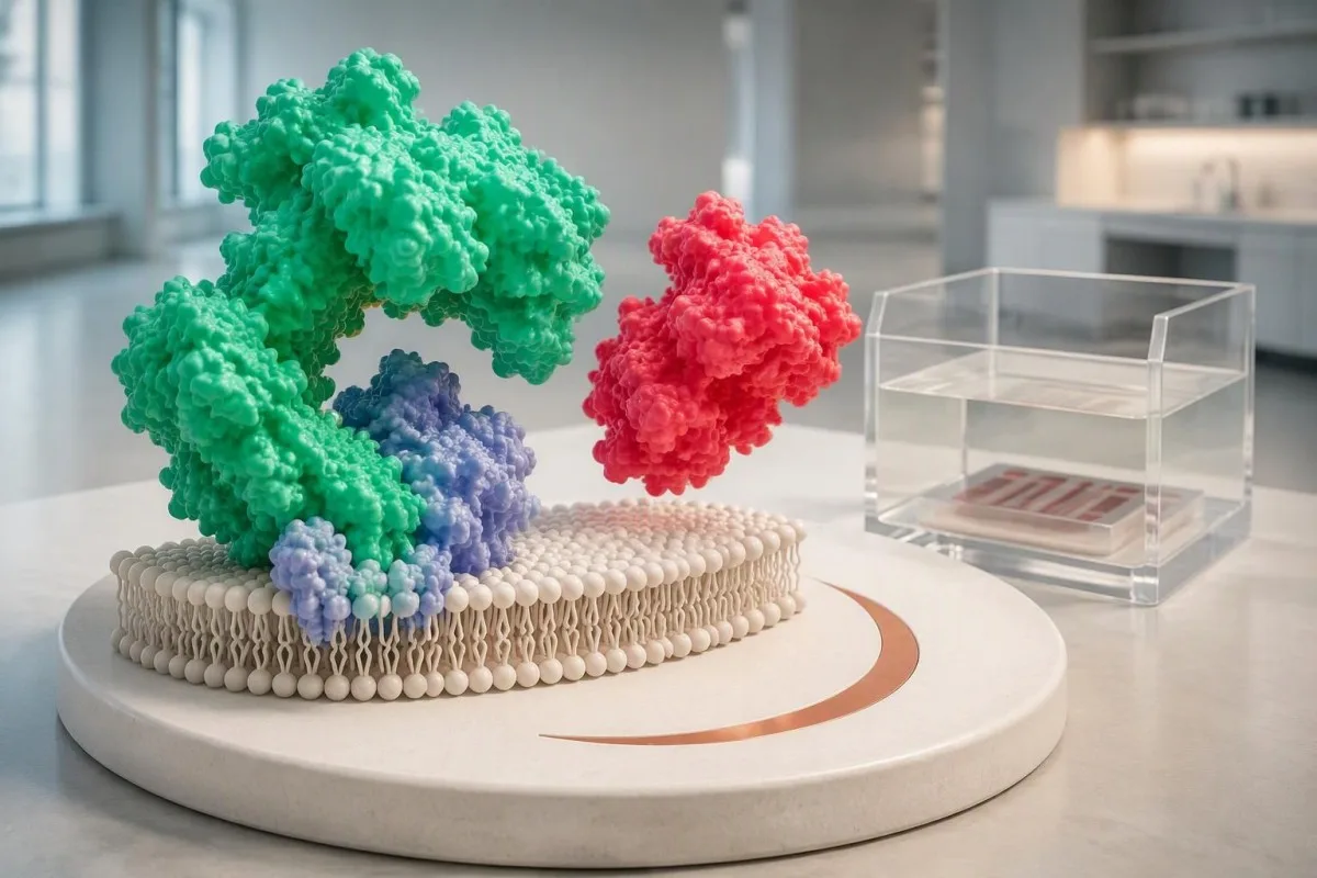
La proteina S aiuta la proteina C attivata a limitare la coagulazione favorendo l’inattivazione dei fattori Va e VIIIa. La sua concentrazione non corrisponde alla misurazione delle proteine totali in un profilo biochimico; un valore di proteine totali pari a 7.0 g/dL non fornisce alcuna stima utile della funzione della proteina S, perché i test misurano quantità biologiche del tutto diverse.
Un segnale di anomalia isolato è un punto di partenza, non una sentenza. Nel caso ipotetico di una persona con un’attività pari a 48% rispetto a un limite inferiore di 60% del laboratorio, la domanda successiva è se il campione sia stato raccolto durante la gravidanza, una terapia farmacologica o una malattia, non se il numero dimostri automaticamente un disturbo genetico; il nostro guida del risultato fuori intervallo spiega questa distinzione.
Kantesti è un analizzatore di esami del sangue basato sull’IA che aiuta a organizzare i risultati della proteina S insieme agli intervalli riportati e al contesto pertinente, anziché trattare un singolo valore segnalato come anomalo come una diagnosi. La nostra background aziendale spiega l’organizzazione che offre questo servizio educativo; la conferma di una carenza ereditaria richiede comunque una valutazione di laboratorio diretta dal medico e, occasionalmente, una valutazione genetica specialistica.
Come si interpretano la percentuale di attività e l’intervallo?
L’attività della proteina S viene solitamente riportata come percentuale dell’attività di riferimento del test, non come probabilità percentuale di trombosi. Gli intervalli di riferimento dipendono dal laboratorio, dal metodo, dall’età e dal contesto ormonale; un intervallo per adulti come 60–150% è un esempio illustrativo, non una soglia diagnostica universale.

Un’attività della proteina S pari a 45% indica un’attività funzionale ridotta in quel test, non che 45% della circolazione sia ostruito. Le percentuali si differenziano anche dalle unità di concentrazione, come mg/L, perciò un risultato di attività non può essere confrontato numericamente direttamente con una concentrazione dell’antigene; la distinzione è simile a quella di altri misurazioni quantitative di laboratorio che quantificano proprietà diverse.
I valori borderline richiedono una gestione particolarmente attenta. In un laboratorio ipotetico che usa 60% come limite inferiore, risultati di 58% e 61% si trovano ai lati opposti della soglia di anomalia pur distando solo 3 punti percentuali; la variabilità analitica, le fluttuazioni biologiche e i diversi reagenti del test possono spiegare tale differenza senza che vi sia un cambiamento significativo del rischio di coagulazione.
Non esiste un’unica percentuale di proteina S che determini autonomamente se sia necessaria una terapia anticoagulante. Un risultato di 25% merita più attenzione di un valore lievemente ridotto, ma può comunque verificarsi durante una riduzione acquisita; le decisioni dipendono dalla conferma, dall’anamnesi di trombosi, dai fattori scatenanti e dal rischio emorragico, anziché dal considerare una soglia numerica come un’indicazione a iniziare un farmaco.
| Livello | Intervallo | Significato |
|---|---|---|
| Nell’intervallo illustrativo per adulti | attività 60–150% | Solo a titolo di esempio. Usare l’intervallo adeguato all’età e al contesto indicato dal laboratorio che esegue l’analisi; un anticoagulante interferente può talvolta mascherare una carenza. |
| Al di sotto di tale intervallo illustrativo | attività <60% | Richiede una valutazione del contesto e, se opportuno, una conferma. Questo non dimostra una carenza ereditaria. |
| Risultato dell’antigene libero | Laboratory-specific % or concentration units | Measures the unbound protein pool. Do not apply an activity reference interval to an antigen result. |
| Pregnancy or childhood result | Population- and method-specific | A nonpregnant adult interval may be inappropriate. Diagnostic interpretation may need specialist advice. |
Cosa indicano insieme l’attività e l’antigene libero della proteina S?
Activity testing measures functional performance, while free protein S antigen measures the available unbound protein pool. Approximately 40% of circulating protein S is free and the remainder is bound mainly to C4b-binding protein; comparing these measurements helps identify a quantitative reduction, a possible functional defect or a misleading activity result.
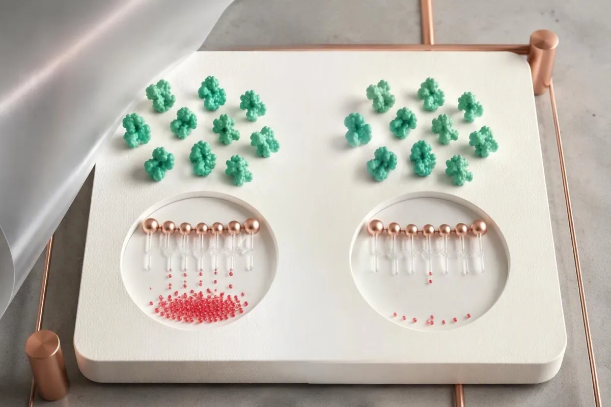
Type I protein S deficiency classically produces low total antigen, low free antigen and low activity; type III produces normal total antigen with low free antigen and activity. These 2 patterns can occur within the same family and are not necessarily separate biological diseases; total antigen alone can therefore miss a clinically relevant reduction in the free pool.
Type II protein S deficiency classically shows reduced activity despite normal free and total antigen. This uncommon pattern is not diagnosed from 1 discordant sample, because factor V Leiden and other assay problems can imitate functional deficiency; laboratories may repeat the activity using another method before considering a genuinely dysfunctional protein.
Paired results clarify the next step but do not identify the cause by themselves. For example, activity of 42% with free antigen of 38% could reflect either inherited quantitative deficiency or an acquired reduction, while activity of 42% with normal free antigen raises a different set of questions; our coagulation marker overview explains why complementary tests are interpreted together.
Perché la gravidanza riduce i risultati della proteina S?
Pregnancy physiologically lowers free protein S and often protein S activity, so a low pregnancy result cannot reliably establish inherited deficiency. Changes can begin early and become more pronounced later; the British Society for Haematology identifies pregnancy and the puerperium as confounders of protein S testing (Arachchillage et al., 2022).

A nonpregnant reference interval can create a misleading diagnosis during pregnancy. In a hypothetical second-trimester sample, activity of 43% and free antigen of 36% may both be flagged, but their agreement does not remove the pregnancy effect; both tests can reflect the same physiological reduction, just as l'interpretazione della ferritina in gravidanza requires pregnancy-specific context.
Reassessment for inherited protein S deficiency is commonly postponed until at least 6 weeks after delivery, and some services prefer 12 weeks or longer. Six weeks is not a guarantee of an unaffected baseline: recovery, continuing illness, a recent clot and newly started estrogen contraception can all influence whether a postpartum sample is suitable.
Protein S testing does not decide pregnancy anticoagulation by itself. When reviewing 2 pregnancy measurements, Kantesti AI can help distinguish activity from antigen on the report, but pregnancy prophylaxis requires obstetric and thrombosis assessment based on prior venous thromboembolism, family history and other risks; a low pregnancy value alone also does not establish the cause of a miscarriage.
I contraccettivi a base di estrogeni o la terapia ormonale sostitutiva possono causare un risultato basso?
Estrogen-containing contraception and oral estrogen therapy can lower free protein S and protein S activity without an inherited defect. The change is biological rather than merely a laboratory artifact; a result collected during estrogen use may therefore describe the treatment-associated state, not the person's untreated baseline.

The exact preparation matters more than the word contraception. A combined pill containing 30 micrograms of ethinylestradiol is not equivalent to a progestogen-only pill, and a contraceptive patch is not equivalent to a transdermal menopausal estrogen patch; hormone results during contraception require the active ingredients and route, not just the brand name.
Transdermal menopausal estrogen generally has less effect on hepatic coagulation pathways than oral estrogen, but this does not make every transdermal hormonal product risk-free. Evidence about clot risk and evidence about a specific protein S assay are different questions; an activity of 52% during treatment cannot be corrected mathematically by subtracting a standard estrogen effect.
There is no universally reliable 1-week estrogen washout for protein S diagnosis. If confirmation would change care, the prescribing clinician can discuss timing, alternative contraception and whether treatment should change at all; our pre-HRT testing considerations support that conversation, but never stop prescribed hormones independently just to remove a laboratory flag.
Perché il warfarin riduce l’attività della proteina S?
Warfarin lowers protein S because protein S is a vitamin K-dependent anticoagulant protein. This is an expected treatment effect rather than proof of inherited deficiency; free antigen can also be reduced, so replacing an activity test with antigen testing does not reliably reveal the untreated baseline during warfarin therapy.

A therapeutic INR does not make protein S testing diagnostically reliable during warfarin treatment. For a hypothetical patient taking 5 mg daily with an INR of 2.4 and activity of 31%, the low activity may be medication related; the meaning of an INR concerns anticoagulant monitoring, not confirmation of inherited protein S deficiency.
When an off-warfarin measurement is genuinely needed, laboratories commonly require at least 2 weeks after discontinuation, sometimes longer according to local policy and the clinical situation. That interval is not an instruction to stop treatment: a hematologist or anticoagulation clinician must decide whether interruption is safe, whether an alternative is needed and which assay remains interpretable.
Vitamin K deficiency can also reduce protein S, but supplements should not be used to self-correct an unexplained result. Taking even 1 new vitamin K-containing supplement can alter warfarin management; keep dietary intake reasonably consistent and discuss changes with the anticoagulation team, using our guida alimentare alla vitamina K for food context rather than as a deficiency treatment plan.
Quali malattie possono ridurre temporaneamente la proteina S?
Severe infection, disseminated intravascular coagulation, liver disease, nephrotic syndrome and recent thrombosis can reduce protein S measurements. Some reductions resolve with recovery, while others persist because the underlying condition persists; a low result during hospitalization therefore cannot be classified as inherited from 1 sample.
Disseminated intravascular coagulation can reduce natural anticoagulants while other coagulation markers change simultaneously. A hypothetical combination of protein S activity at 35%, platelets at 65 × 10^9/L and a prolonged prothrombin time calls for assessment of the acute illness, not immediate genetic labeling; our DIC laboratory pattern guide explains why multiple abnormal findings matter together.
The liver contributes to protein S production, but an isolated ALT of 75 IU/L does not prove that liver dysfunction caused a low protein S result. Clinicians look for impaired synthesis and the broader clinical picture, including albumin, bilirubin and coagulation results; modest enzyme elevation and advanced synthetic failure are different processes, with very different implications for interpretation.
Nephrotic-range protein loss can produce acquired protein S abnormalities, especially in the free fraction. An albumin of 2.2 g/dL with substantial urinary protein loss changes the interpretation of activity at 44%; persistent foamy urine assessment may help identify a reason to investigate renal protein loss, although bubbles alone neither diagnose nephrotic syndrome nor explain protein S.
In che modo le interferenze del test possono rendere fuorvianti i risultati della proteina S?
Clot-based protein S activity assays can report falsely low or falsely high results because medications and other coagulation abnormalities alter the endpoint. Direct oral anticoagulants often falsely increase activity in susceptible assays, whereas factor V Leiden can falsely decrease it; the direction and magnitude depend on the assay design.
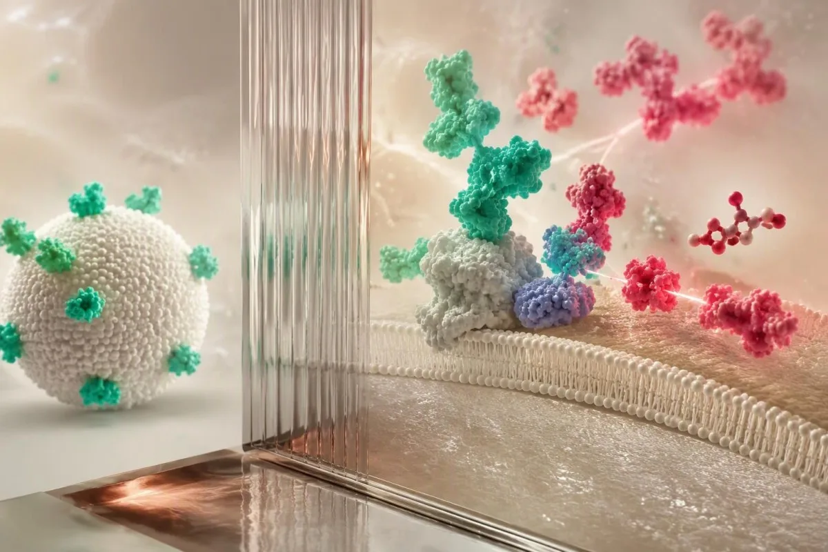
Factor V Leiden can interfere with some protein S activity assays by altering the activated protein C-dependent reaction. A hypothetical activity of 46% with normal free antigen could therefore mimic type II deficiency; the factor V Leiden result and the laboratory's assay characteristics should be reviewed before concluding that 2 inherited disorders coexist.
Direct oral anticoagulants can conceal deficiency rather than explain a low number. For example, an apparently normal activity of 110% during rivaroxaban treatment may be falsely increased in a susceptible clot-based method; lupus anticoagulant, high factor VIII and heparin can also complicate interpretation, so lupus anticoagulant testing and protein S testing need coordinated laboratory advice.
Kantesti is an AI lab test interpretation service that can place 2 discordant measurements—activity and free antigen—side by side for discussion, but cannot determine reagent interference from the numbers alone. Free antigen immunoassays generally avoid clot-endpoint interference, yet they still cannot remove warfarin's biological effect; the BSH guideline specifically lists medication- and assay-related confounders (Arachchillage et al., 2022).
Il prelievo o la manipolazione del campione possono causare un risultato basso?
Collection and handling problems can distort protein S testing, particularly functional assays that depend on correctly prepared citrated plasma. The laboratory should check tube filling, citrate balance, processing and sample quality before a discordant result becomes a diagnosis; repeating a technically unsuitable sample answers a different question from confirming inherited deficiency.
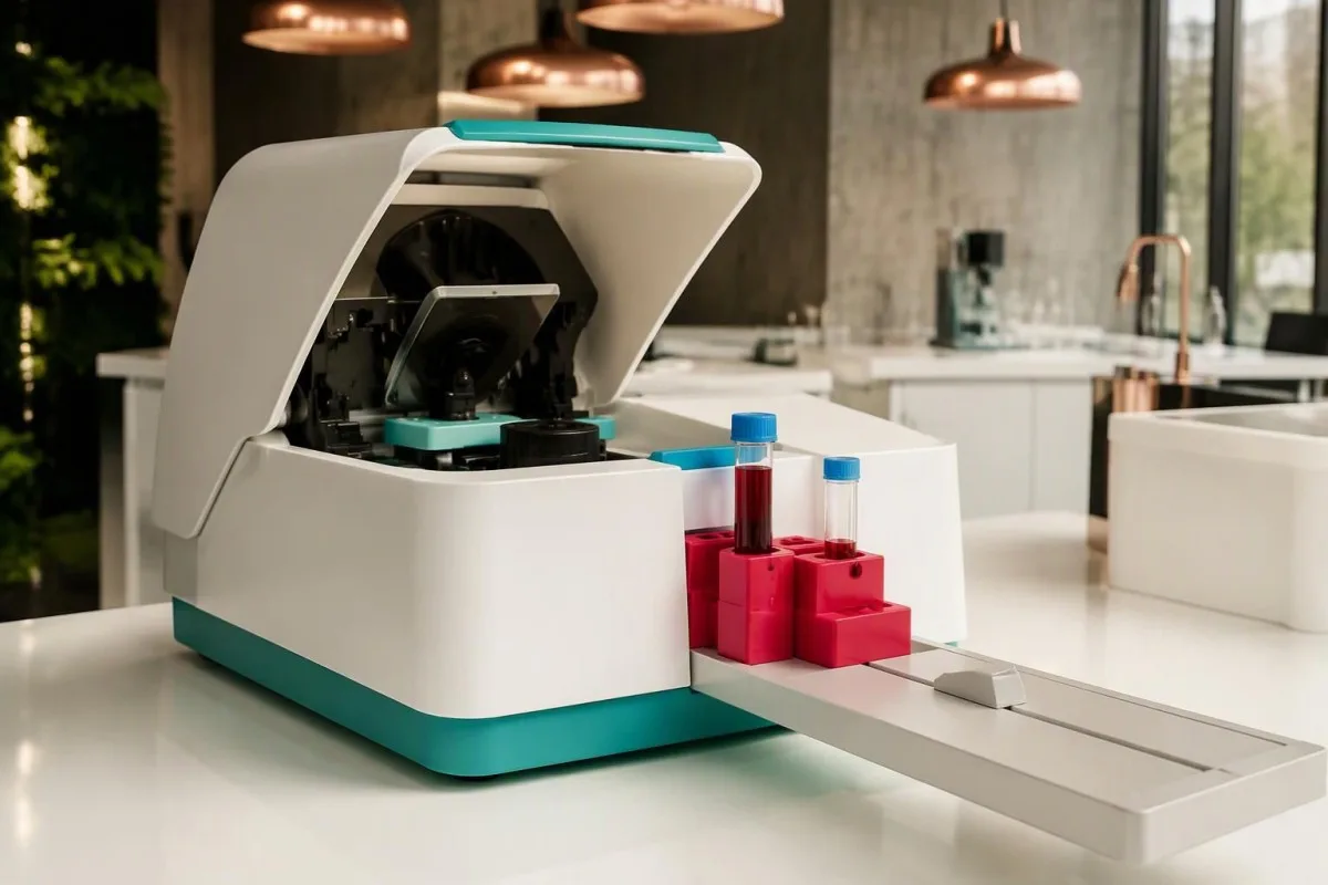
Standard coagulation collection uses approximately 9 parts sample to 1 part citrate anticoagulant. Underfilling changes that ratio and can distort clot-based measurements; tube-cap colors vary internationally, so the fill line and laboratory instructions matter more than recognizing a particular color, and a rejected specimen should not be interpreted as if its numerical result were trustworthy.
A hematocrit above 55% generally requires laboratory-directed adjustment of citrate volume for coagulation testing. A hypothetical patient with a hematocrit of 58% needs that issue addressed before a low functional result is taken at face value; otherwise, excess citrate relative to plasma can affect the assay even when the collection tube appears adequately filled.
Hemolysis, lipemia and delayed processing can affect methods differently, so there is no universal correction factor for a protein S result of 49%. Ask whether the laboratory accepted the specimen and whether it recommends recollection; our hemolysis interference guide explains why a sample-quality warning may affect some reported analytes but not others.
Quando è opportuno ripetere il test sotto la guida del medico?
Repeat protein S testing is appropriate when the first result was confounded, borderline or unexpected and confirmation could change care. After acute thrombosis, the British Society for Haematology recommends testing natural anticoagulant deficiencies only after 3 months of anticoagulation when indicated, with medication-related interference addressed separately (Arachchillage et al., 2022).

Three months of anticoagulation is a testing-timing recommendation, not permission to stop anticoagulants at 3 months. Treatment duration depends on the clot and the patient's risks, while assay suitability depends on the medication; clinicians may defer testing, choose an antigen assay or seek specialist laboratory methods without interrupting necessary protection.
Pregnancy, warfarin and acute illness each require a different reassessment plan. Common examples include at least 6 weeks postpartum, at least 2 weeks after clinician-approved warfarin discontinuation, or testing after recovery from significant illness; none is a universal washout schedule, and an unconfounded repeat may need to be postponed further if several influences overlap.
Kantesti's trend analysis can help display 2 measurements, but comparison is strongest when the method, laboratory and clinical conditions are comparable. Record pregnancy stage, illness dates, medicines and the exact timing of the last anticoagulant dose for the clinician; our guida alla tempistica di laboratorio explains why elapsed time alone does not establish that a change is biological.
Quali elementi rendono più probabile una carenza ereditaria di proteina S?
Inherited protein S deficiency becomes more likely when low results persist under suitable testing conditions and fit a personal or family history of venous thrombosis. A pathogenic PROS1 variant can support the diagnosis, but genetic testing is selective; 1 low activity result, especially during treatment or pregnancy, is insufficient.

Most inherited protein S deficiency associated with a single pathogenic PROS1 variant follows an autosomal dominant inheritance pattern. Each child has a 50% chance of inheriting that variant, not a 50% certainty of developing a clot; penetrance varies, and relatives with the same variant can have different protein measurements and clinical histories.
A first venous clot at age 28 with several affected close relatives raises more suspicion than an incidental borderline result at age 72 during pneumonia. Age is not a diagnostic cutoff, however: absence of family history does not exclude inheritance, and the BSH guideline recommends genetic testing for phenotypically identified deficiencies when the result would influence management (Arachchillage et al., 2022).
Family screening should be selective rather than a broad panel for every relative. Testing an unaffected first-degree relative may be useful if the result would change estrogen decisions or prophylaxis during high-risk periods; our guida ai test familiari basati sul rischio explains why testing plans should follow a confirmed family diagnosis instead of expanding from 1 uncertain laboratory flag.
Un risultato basso significa che hai un coagulo adesso?
Low protein S activity does not diagnose an existing deep vein thrombosis or pulmonary embolism. Confirmed inherited deficiency increases susceptibility to venous thrombosis, but current symptoms require the usual urgent assessment; the BSH guideline describes an approximately 5–7-fold increased first-event risk for heterozygous protein C or protein S deficiency (Arachchillage et al., 2022).
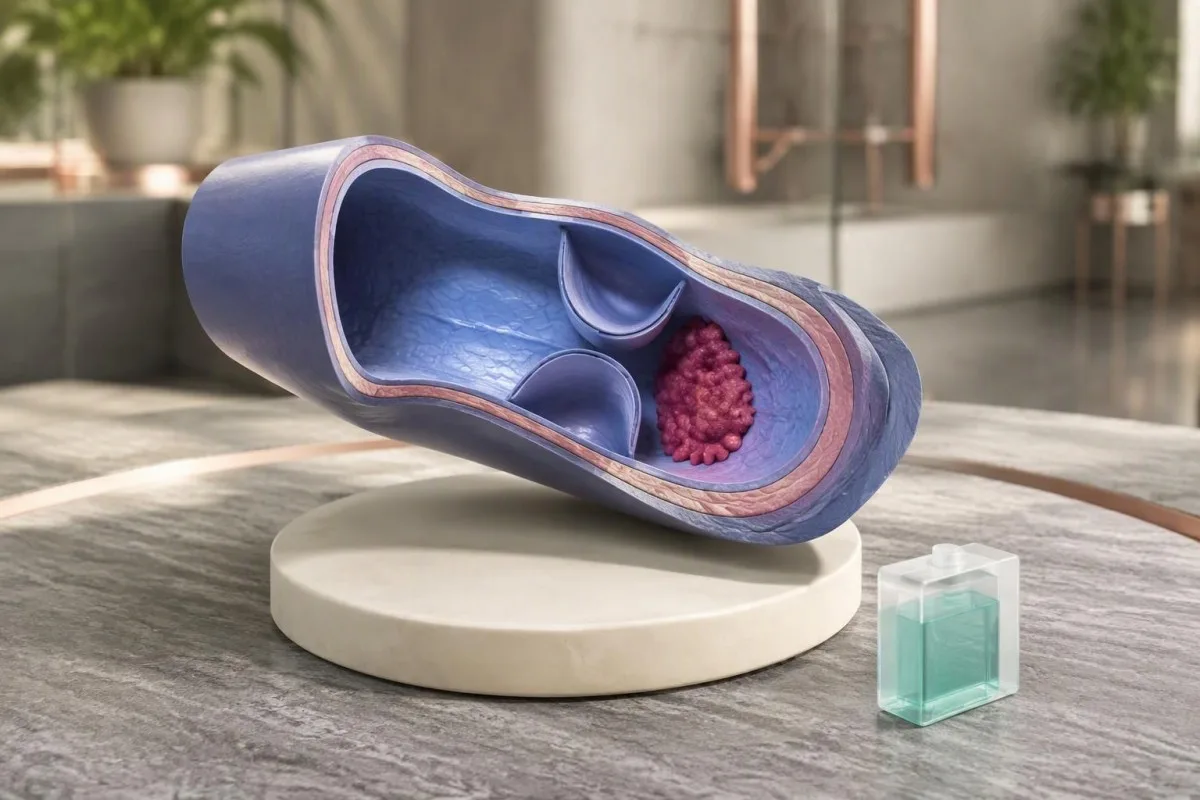
A 5–7-fold relative risk is not a 50–70% absolute probability of thrombosis. Absolute risk varies with age, family ascertainment, prior events and exposures such as surgery or estrogen; a person without symptoms should not interpret a low protein S value as evidence that a clot is silently present or start an anticoagulant independently.
Sudden unexplained breathlessness, chest pain, coughing blood or collapse requires emergency evaluation; new one-sided leg swelling or pain needs urgent assessment for deep vein thrombosis. A normal activity of 95% does not rule out either condition, and normal PT or aPTT also cannot exclude thrombophilia; our guida sui limiti del pannello della coagulazione explain that common misconception.
A confirmed protein S deficiency does not automatically require lifelong anticoagulation in someone who has never had thrombosis. The ASH 2023 guideline discourages testing unprovoked VTE solely to determine duration but conditionally supports testing in selected hormonal or nonsurgical provoked scenarios; those latter recommendations rely on very low-certainty evidence, so 1 label cannot replace individualized decisions (Middeldorp et al., 2023).
Cosa dovresti portare al prossimo appuntamento medico?
Bring the complete report, medication list, collection date and relevant clot or pregnancy history—not just the low protein S activity number. The most useful appointment establishes whether the result is interpretable, whether free antigen was measured and whether confirmation would change treatment; 3 focused questions often prevent unnecessary retesting.

Ask: Was the activity method vulnerable to my medication; was free antigen measured; and what would a confirmed deficiency change? For a hypothetical person with activity of 47% and free antigen of 41% while taking warfarin, both results may remain treatment-confounded; ordering a larger thrombophilia panel immediately does not solve the underlying baseline problem.
A different hypothetical pattern—activity of 47%, normal free antigen and known factor V Leiden—first raises an assay question rather than establishing type II deficiency. A third pattern, with activity of 39% and free antigen of 35% persisting on suitably timed samples without an acquired explanation, makes specialist evaluation more reasonable; these examples are illustrations, not diagnostic algorithms.
Kantesti can help organize these 3 patterns for discussion, but the clinician and testing laboratory must decide whether another assay or repeat specimen is needed. Our quadro di validazione clinica describes the role of evaluation and oversight; bring actual reference intervals and units, and do not rely on a cropped screenshot that omits the method or sample comments.
Evidenze scientifiche e limiti dell’interpretazione dell’IA
Clinical guidelines support context-sensitive protein S testing, but neither a laboratory flag nor an AI explanation confirms inherited deficiency. Kantesti is an AI blood test interpretation platform that helps explain activity and antigen results together; the diagnosis still depends on appropriate testing conditions, reproducibility and clinical assessment rather than 1 automated interpretation.
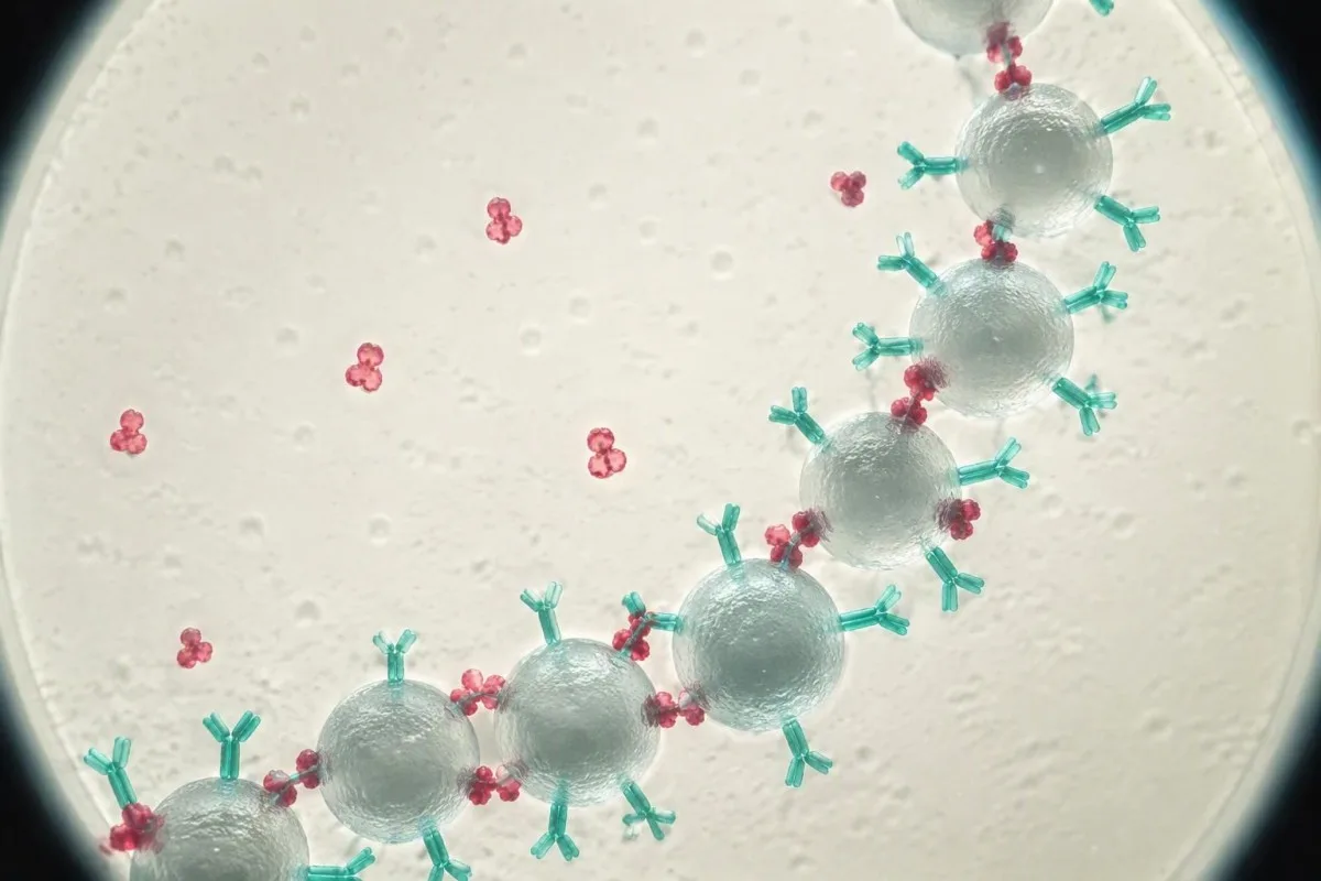
The 2022 BSH and 2023 ASH recommendations address different testing decisions, and neither recommends indiscriminate thrombophilia screening. BSH emphasizes validity and whether findings would change management, while ASH conditionally supports testing in some selected VTE scenarios using very low-certainty evidence; their differences reflect clinical questions and modeling assumptions, not proof that every low result needs genetic testing.
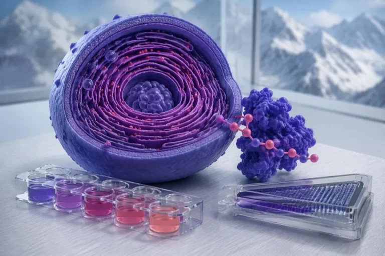
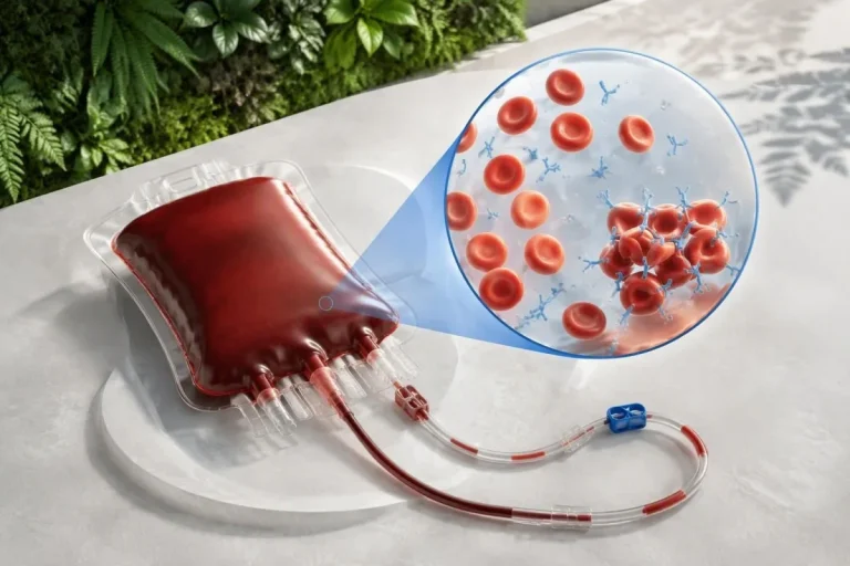
Protein S is not diagnosed by routine tissue microscopy, and the molecular illustrations here are educational models rather than images of an individual's deficiency. Our spiegazione della tecnologia AI describes the interpretation workflow; the hormonal health research overview supplies broader context but does not provide a validated protein S cutoff or a medication washout protocol.
The 2 Kantesti research publications listed below are Figshare-hosted materials covering hormonal health and multilingual clinical decision-support engineering; neither establishes diagnostic accuracy for inherited protein S deficiency. Repository hosting and DOI assignment do not establish peer review or protein S-specific clinical validation; our standard di contenuto medico explain why educational claims should remain separate from disease-specific evidence.
Domande frequenti
L’attività della proteina S può essere temporaneamente bassa?
Una bassa attività della proteina S può essere temporanea durante la gravidanza, in caso di esposizione agli estrogeni, trattamento con warfarin, malattia grave o convalescenza da una trombosi. Un risultato come 45% non consente, da solo, di distinguere una riduzione temporanea da una carenza ereditaria. L’attività e l’antigene della proteina S libera devono essere interpretati insieme, ripetendo il test quando il medico ritiene che le condizioni siano adeguate. Non interrompere autonomamente i farmaci prescritti per ottenere un risultato senza trattamento.
0 Qual è la differenza tra l’attività della proteina S e l’antigene della proteina S libera?
0 L'esame dell'attività della proteina S ne misura la funzionalità, mentre l'antigene libero della proteina S misura la quota di proteina non legata. Circa 40% della proteina S circolante è libera, mentre la parte restante è legata principalmente alla proteina legante C4b. Un'attività ridotta associata a una bassa concentrazione dell'antigene libero può verificarsi sia nelle riduzioni quantitative ereditarie sia in quelle acquisite. Un'attività ridotta con una concentrazione normale dell'antigene libero può riflettere un difetto funzionale o un'interferenza del test, pertanto il quadro deve essere confermato.
È normale che l’attività della proteina S sia bassa durante la gravidanza?
La gravidanza riduce fisiologicamente la proteina S libera e spesso l’attività della proteina S, quindi un risultato basso in gravidanza non consente di diagnosticare in modo affidabile una carenza ereditaria. Un valore di 40% deve essere valutato in relazione alla fase della gravidanza, al test utilizzato e al motivo clinico dell’esame, anziché basarsi soltanto su un intervallo di riferimento per adulti non in gravidanza. La rivalutazione diagnostica viene comunemente rimandata ad almeno 6 settimane dopo il parto, talvolta a 12 settimane o più. Le decisioni sull’anticoagulazione in gravidanza dipendono dalla storia di trombosi e da altri rischi, non dal solo valore della proteina S.
Quanto tempo dopo il warfarin è possibile eseguire il test della proteina S?
0 Molti laboratori richiedono di attendere almeno 2 settimane dalla sospensione del warfarin prima di valutare i livelli basali di proteina S non trattati, sebbene i requisiti locali e le circostanze cliniche varino. Il warfarin può ridurre sia l’attività sia l’antigene, poiché la proteina S è vitamina K-dipendente. Questo intervallo di attesa non autorizza a interrompere la terapia anticoagulante. Il medico prescrittore deve decidere se sia sicuro sospenderla, se sia necessario eseguire il test e se sia preferibile un altro approccio.
Apixaban o rivaroxaban possono influenzare un test di attività della proteina S?
Apixaban e rivaroxaban possono interferire con i test di attività della proteina S sensibili basati sulla coagulazione, causando spesso un’attività falsamente aumentata anziché una reale carenza. Un risultato apparentemente normale, come 100%, potrebbe quindi non escludere una ridotta attività sottostante della proteina S. I test immunologici per l’antigene libero in genere evitano l’effetto dell’endpoint della coagulazione, ma il laboratorio dovrebbe indicare quale metodo sia appropriato. Non saltare né interrompere le dosi di anticoagulante per prepararti al test, a meno che il medico prescrittore non indichi esplicitamente un piano sicuro.
Quando va ripetuta la misurazione dell’attività della proteina S bassa dopo un coagulo di sangue?
0. Quando è indicato eseguire gli esami dopo una trombosi acuta, la British Society for Haematology raccomanda di attendere il termine di 3 mesi di terapia anticoagulante prima di ricercare carenze degli anticoagulanti naturali. L’interferenza dei farmaci deve comunque essere valutata separatamente, quindi il raggiungimento dei 3 mesi non rende affidabile ogni test di attività. Ripetere gli esami è particolarmente utile quando il risultato potrebbe modificare la gestione clinica e il campione iniziale era condizionato da fattori confondenti o inatteso. Non si deve interrompere autonomamente il trattamento per ripetere il test.
La carenza di proteina S significa che dovrò assumere anticoagulanti per tutta la vita?
Confirmed protein S deficiency does not automatically mean lifelong anticoagulation, especially in someone who has never had venous thrombosis. The approximately 5–7-fold relative first-event risk described for heterozygous protein C or protein S deficiency is not an individual's absolute probability of a clot. Decisions depend on previous events, provoking factors, bleeding risk and whether prophylaxis is needed during high-risk periods. A single low activity result should not be used to start, stop or extend anticoagulant treatment independently.
Scopri come leggere le analisi del sangue
Carica il tuo referto di laboratorio e l'IA di Kantesti spiega ogni biomarcatore in un linguaggio semplice in pochi secondi. Utilizzata da oltre 2 milioni di persone in tutto il mondo.
Riferimenti
- Klein, T., Mitchell, S., & Weber, H. (2026). Women's Health Guide: Ovulation, Menopause & Hormonal Symptoms. Ricerca medica sull'IA di Kantesti. DOI
- Klein, T., Mitchell, S., & Weber, H. (2026). Multilingual AI Assisted Clinical Decision Support for Early Hantavirus Triage: Design, Engineering Validation, and Real-World Deployment Across 50,000 Interpreted Blood Test Reports. Ricerca medica sull'IA di Kantesti. DOI
- Arachchillage DJ et al. (2022). Thrombophilia testing: A British Society for Haematology guideline. British Journal of Haematology. DOI
- Middeldorp S et al. (2023). American Society of Hematology 2023 guidelines for management of venous thromboembolism: thrombophilia testing. Blood Advances. DOI
- 2 milioni+Esami analizzati
- 127+Paesi
- 100+Lingue
Informazioni sul team medico


Segnali di fiducia E-E-A-T
Esperienza
Revisione clinica guidata da un medico dei flussi di lavoro di interpretazione degli esami.
Competenza
Focus sulla medicina di laboratorio su come i biomarcatori si comportano nel contesto clinico.
autorevolezza
Revisione medica a cura del dott. Thomas Klein, con il contributo della dott.ssa Sarah Mitchell e del prof. Hans Weber.
Affidabilità
Interpretazione basata su evidenze, con percorsi di follow-up chiari per ridurre l’allarme.
Avvertenza medica
Questo articolo ha solo scopo educativo e non costituisce consulenza medica. Consulta sempre un operatore sanitario qualificato per decisioni su diagnosi e trattamento.
Pubblicato · Contatta il team editoriale
Pubblicato da Kantesti Ltd, registrata in Inghilterra e Galles, numero di società. 17090423, Londra, Regno Unito.
