
Una risposta neutrofila debole non indica sempre una malattia granulomatosa cronica. Il pattern di fluorescenza, il controllo sano e il percorso del campione determinano cosa si possa effettivamente stabilire dal risultato.



- Risultati anomali del test DHR indicano un'ossidazione ridotta o disomogenea della diidrorodamina 123 da parte dei neutrofili; richiedono un'interpretazione basata sul pattern, non una diagnosi automatica di CGD.
- Risposta quasi assente in quasi tutti i neutrofili, con un controllo sano soddisfacente, supporta fortemente un grave difetto della NADPH ossidasi.
- Due popolazioni di neutrofili possono indicare una portatrice di CGD legata all'X, ma una trasfusione di granulociti o un chimerismo correlato al trapianto possono produrre una miscela simile.
- Frazioni di portatrici inferiori a 10% di neutrofili funzionanti sono state associate a un rischio significativamente maggiore di infezioni di tipo CGD; 50% non è un risultato obbligatorio per una portatrice.
- Attività ossidativa residua può verificarsi nella CGD autentica, quindi una risposta ridotta non è necessariamente rassicurante.
- Tempistica del campione è importante: l’intervallo tra il prelievo e l’analisi è comunemente di circa 24 ore, mentre tempi più lunghi richiedono la convalida del laboratorio esecutore.
- Deficit di mieloperossidasi può ridurre la fluorescenza DHR nonostante l’attività conservata della NADPH ossidasi; un secondo metodo funzionale può distinguere le condizioni.
- Stimolazione PMA normale non esclude tutti i rari disturbi dei fagociti, in particolare i difetti dimostrabili meglio mediante stimolazione con particelle.
- Sintomi urgenti prevalgono sui piani di ripetizione del test: febbre con difficoltà respiratorie, malattia in rapida progressione o una nuova infezione profonda richiedono una valutazione clinica tempestiva.
Che cosa significano davvero risultati anomali del test DHR?
Risultati anomali del test DHR indicano che i neutrofili hanno prodotto meno fluorescenza, o una distribuzione diversa, dopo la stimolazione. Una risposta quasi assente è compatibile con la malattia granulomatosa cronica, 2 popolazioni distinte possono suggerire lo stato di portatrice X-linked e risposte deboli con controlli inadeguati possono riflettere problemi nella gestione del campione; nessuno di questi risultati dovrebbe essere interpretato basandosi solo sul numero segnalato.

Malattia granulomatosa cronica, o CGD, compromette il sistema della NADPH ossidasi dei fagociti, anziché limitarsi a ridurre la conta dei neutrofili. Un paziente può avere una conta assoluta dei neutrofili di 4.0 × 10^9/L e presentare comunque un grave difetto funzionale; contare le cellule e analizzarne i meccanismi antimicrobici risponde a domande diverse.
Si consideri un bambino di 7 anni con infezioni batteriche profonde ricorrenti e un tracciato DHR stimolato quasi piatto. Questa combinazione desta un livello di preoccupazione diverso rispetto a un’unica risposta borderline in un adulto per il resto in buona salute; il indagine immunologica iniziale dovrebbe accertare quali infezioni si sono verificate, dove e se gli organismi responsabili sono stati identificati.
Sono Thomas Klein, Chief Medical Officer di Kantesti, e la mia prima regola di lettura è distinguere 3 domande: il campione è valido, la risposta è anomala e quale meccanismo la spiega? È importante porsi le domande in quest’ordine, perché anche un grafico dall’aspetto convincente può derivare da cellule compromesse.
Kantesti è un analizzatore di esami del sangue basato sull’IA che può spiegare la terminologia riportata in un referto DHR insieme ai risultati di laboratorio correlati. La nostra background aziendale descrive il servizio; una spiegazione generata in circa 60 secondi non sostituisce la revisione specialistica dei dati di citometria a flusso né consente di diagnosticare la CGD.
Come funziona il test del burst ossidativo DHR?
IL Test del burst ossidativo DHR utilizza la citometria a flusso per misurare la conversione della diidrorodamina 123 in rodamina fluorescente dopo la stimolazione dei neutrofili. La NADPH ossidasi genera superossido, che contribuisce alla formazione di perossido di idrogeno; il segnale DHR dipende anche dalle reazioni ossidative a valle, compresa la mieloperossidasi, quindi la fluorescenza non misura direttamente il superossido.

PMA, o acetato di forbolo miristato, è un potente stimolo di laboratorio utilizzato in molti protocolli DHR. Attiva la segnalazione intracellulare senza richiedere il normale riconoscimento microbico sulla superficie cellulare; questo lo rende utile per molti difetti associati alla CGD, ma meno indicativo per alcune anomalie dipendenti dai recettori o dalle particelle.
Un’interpretazione standard confronta almeno 2 condizioni del paziente: cellule non stimolate e cellule stimolate. Il laboratorio esamina di norma anche un controllo sano; è importante sapere se tale controllo è stato trasportato insieme al campione del paziente, poiché un controllo locale appena prelevato non può riprodurre pienamente i danni subiti durante un lungo trasporto tramite corriere.
Il gate dei neutrofili seleziona la popolazione cellulare valutata e l’inclusione inappropriata di altri granulociti può distorcere il pattern apparente. Eosinofili sono particolarmente pertinenti nella diagnosi differenziale del deficit di mieloperossidasi; si tratta di un aspetto di laboratorio che non può essere chiarito da una percentuale del CBC o da una schermata isolata.
Una conta normale dei neutrofili non dimostra che la loro funzione sia normale. An adult ANC within a laboratory interval such as 1.5–7.5 × 10^9/L answers a counting question, not an oxidative-burst question; our neutrophil count guide explains why age, pregnancy, and reporting conventions affect that separate result.
Quali valori e pattern di fluorescenza DHR sono importanti?
DHR interpretation requires both the proportion of responding neutrophils and the strength of their response. A report showing 98% positive cells can still conceal reduced fluorescence intensity, while 2 separated populations can be clinically significant even when their combined average appears acceptable; laboratory-specific gates and controls determine the meaning.
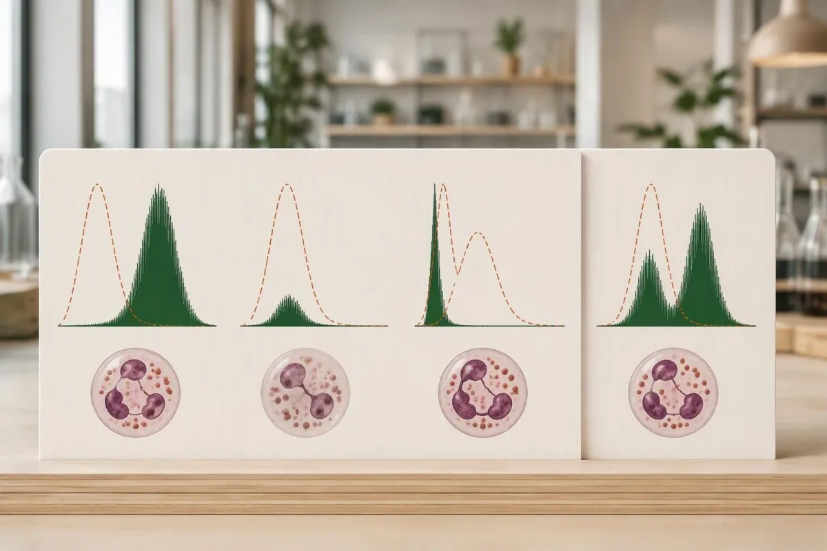
Mean fluorescence intensity, often abbreviated MFI, measures signal strength in a defined population. An illustrative MFI of 200 on one instrument cannot be compared directly with 200 on another; detector settings, reagents, stimulation time, and whether the laboratory averages all neutrophils or only responding cells affect the value.
Stimulation index is commonly calculated as stimulated fluorescence divided by baseline fluorescence, although laboratories may define the calculation differently. If stimulated MFI remains 100 but baseline rises from 1 to 5, the ratio falls from 100 to 20 without any change in the stimulated signal itself.
A histogram preserves information that a single summary number loses. The reason to request the full plot is similar to the reasoning behind flow-cytometry population analysis: the distribution can distinguish one uniformly dim population from 2 populations with very different behavior.
There is no universal DHR MFI or stimulation-index cutoff for CGD. A laboratory may also report a qualitative conclusion alongside quantitative measurements; our explanation of risultati qualitativi e quantitativi helps separate a categorical flag from the underlying data used to assign it.
Un burst ossidativo debole indica sempre una CGD grave?
A nearly absent oxidative burst in almost all neutrophils strongly supports severe NADPH oxidase dysfunction when controls are satisfactory, but CGD can also produce a partial response. A uniformly dim population is not equivalent to 2 separate carrier populations, and measurable activity does not automatically exclude clinically significant disease.

Residual oxidase activity has prognostic relevance in CGD. Kuhns et al. studied 287 patients and found that residual reactive-oxygen-intermediate production was associated with survival, beyond the affected gene alone; their 2010 New England Journal of Medicine paper supports measuring function rather than treating every abnormal response as equally severe.
An illustrative response at 5% of a concurrent control is different from an undetectable response, but that comparison is not a universal treatment threshold. A specialist must consider the assay's reproducibility, the genotype, and the patient's infection history before translating a small remaining signal into a prediction about future health.
Rare severe G6PD deficiency can affect neutrophil oxidative function, although ordinary G6PD deficiency is not synonymous with CGD. A low DHR response combined with episodes of oxidant-triggered hemolysis may justify a separate enzyme investigation; our G6PD interpretation guide explains why red-cell enzyme results need their own context.
Kantesti AI can help organize the distinction between an absent response, a partial response, and a mixed response when those descriptions appear in the report. The useful output is a set of follow-up questions—not a claim that 1 percentage or a particular shade of fluorescence establishes disease severity.
Perché le portatrici di CGD legata all'X mostrano due popolazioni?
X-linked CGD carriers often show 2 neutrophil populations because different cells keep different X chromosomes active. Cells expressing a functioning CYBB copy can respond strongly, while cells expressing the affected copy respond poorly; this is cellular mosaicism, not every neutrophil operating at approximately 50% capacity.
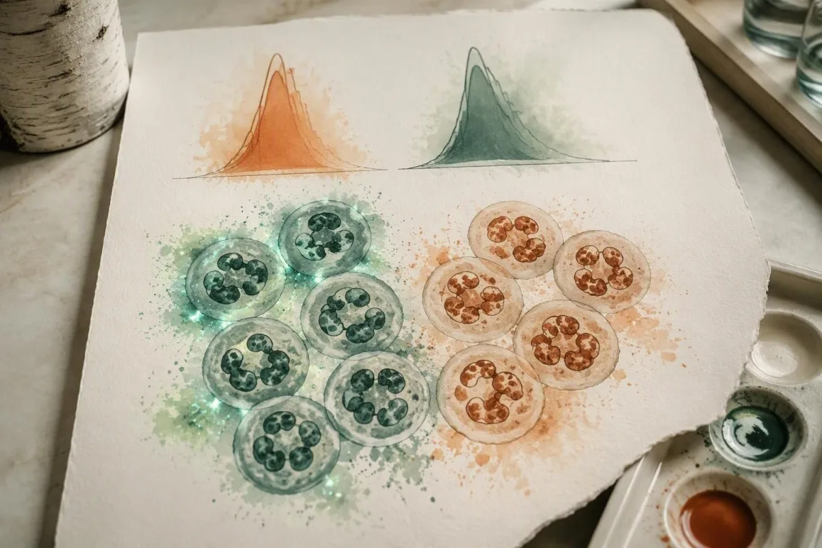
A 50:50 split is not required for X-linked carrier status. A carrier might have an illustrative 70:30 or 20:80 distribution, and the proportion can change over time; the brighter population's intensity and the dim population's behavior should be evaluated separately rather than compressed into one average.
The mechanistic distinction is useful when comparing a carrier with partial CGD. In a carrier example, 30% of cells may respond approximately like control neutrophils while 70% respond poorly; in a partial-deficiency example, almost all cells may respond weakly together, producing a shifted but largely single distribution.
DHR cannot identify every genetic carrier. Carriers of autosomal-recessive CGD commonly have a normal functional response because 1 functioning gene copy is sufficient; molecular testing is usually required to establish that status, and normal DHR should not be presented as a universal negative carrier screen.
Carrier terminology also differs across inherited conditions. Our genetic carrier screening explanation addresses why carrier labels require disease-specific interpretation; for CGD, the immediate questions are inheritance pattern, neutrophil function, and whether the person has symptoms—not simply whether a variant was found.
Quando una portatrice di CGD legata all'X può diventare sintomatica?
X-linked CGD carriers can develop infections or inflammatory complications, particularly when X-inactivation leaves very few functioning neutrophils. Fractions below 10% DHR-positive cells were strongly associated with CGD-type infections in a published carrier cohort, but no single percentage predicts every person's outcome or explains all autoimmune symptoms.

Marciano et al.'s 2018 Journal of Allergy and Clinical Immunology study connected low functioning-neutrophil fractions with CGD-type infection risk in X-linked carriers. Below 10% was a particularly concerning pattern; clinicians may also use a fraction below 20% to prompt closer assessment, but neither figure should become a stand-alone diagnostic rule.
Inflammatory and autoimmune manifestations do not track perfectly with the DHR-positive fraction. A carrier with 60% responding cells can still have clinically meaningful mouth sores, photosensitive rashes, or joint symptoms, so a reassuring percentage should not end the investigation when those complaints persist or recur.
A change from 35% to 18% functioning neutrophils deserves scrutiny, not an automatic conclusion that X-inactivation has worsened. First compare sample age, gating, stimulus, and the performing laboratory; repeat measurements under comparable conditions help distinguish a biological change from a technical difference.
Cellular mosaicism and chromosome mosaicism are different concepts. X-inactivation can create 2 functional neutrophil groups without 2 different chromosome counts; our chromosome mosaicism guide provides a useful contrast, although the diagnostic tests and inheritance counseling are not interchangeable.
Il deficit di mieloperossidasi può simulare un DHR anomalo?
Myeloperoxidase deficiency can produce an abnormal DHR response despite preserved NADPH oxidase activity. Because dihydrorhodamine 123 fluorescence depends on downstream oxidation, a weak signal does not uniquely identify CGD; an experienced laboratory may use direct superoxide testing, nitroblue tetrazolium testing, or cellular MPO assessment to investigate the discrepancy.
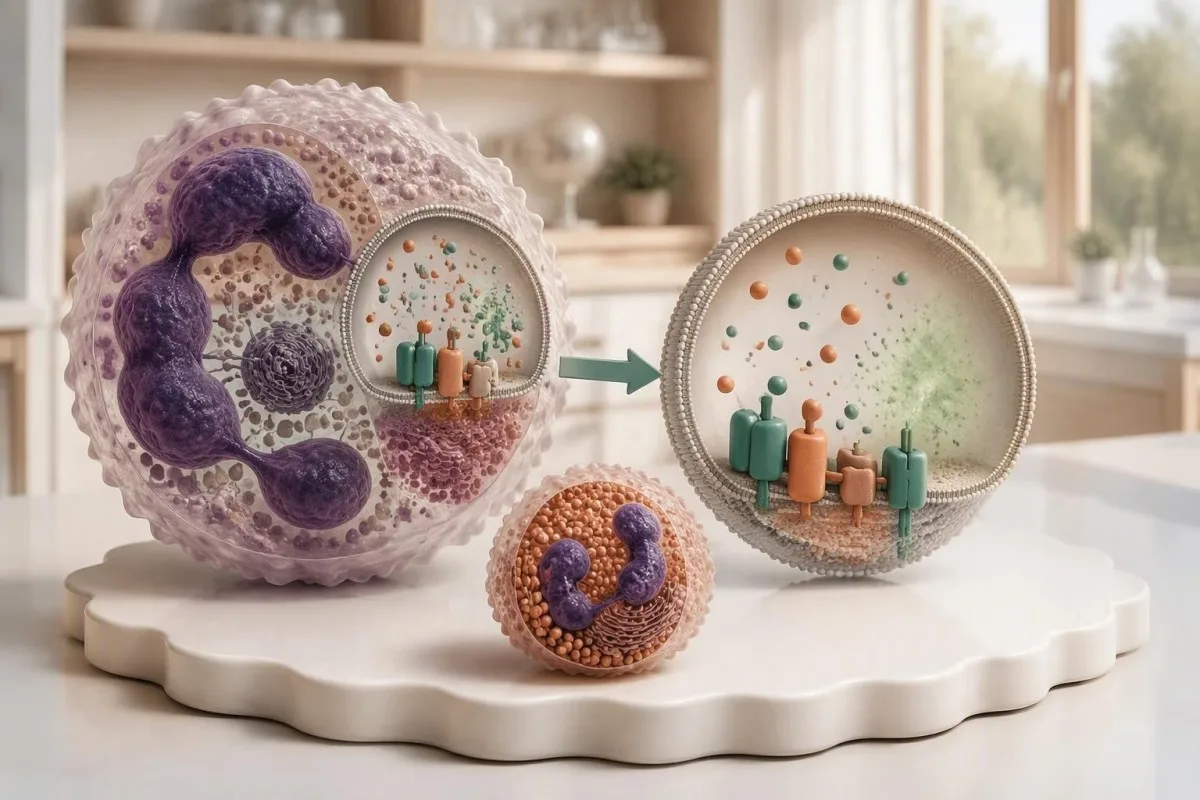
Preserved superoxide production argues against a classic NADPH oxidase defect. If a patient's DHR is low but a second method demonstrates satisfactory superoxide generation, the 2 results should be reconciled rather than forcing both into a CGD diagnosis; MPO deficiency is one recognized explanation discussed in Holland's 2010 clinical review.
The laboratory may inspect eosinophil behavior because eosinophil peroxidase can preserve a different response pattern in MPO deficiency. That distinction requires correctly identified cellular populations; asking whether at least 2 granulocyte groups were considered is more useful than trying to infer the answer from total eosinophil percentage alone.
A circulating MPO concentration is not a neutrophil MPO-deficiency assessment. A result reported in ng/mL for vascular-risk evaluation addresses a different question; our MPO test limitations explain why identical enzyme names on 2 reports do not make those tests interchangeable.
Kantesti is an AI blood test interpretation platform that can distinguish the purpose of these differently named or differently measured MPO tests in a written report. When 2 functional assays disagree, our explanation should preserve that uncertainty and direct attention to the performing laboratory rather than assigning a premature inherited-disease label.
In che modo la manipolazione del campione può causare un burst falsamente basso?
Delayed processing, unsuitable temperatures, an incorrect collection tube, or poor cell viability can weaken a DHR response. Many laboratories aim to analyze specimens within approximately 24 hours; acceptance at 48 hours or beyond depends on their validated protocol, and a weak transported control makes an immediate CGD diagnosis unreliable.

The correct tube is laboratory-specific, although sodium-heparin specimens are commonly requested for DHR testing. Before collection, confirm the anticoagulant, minimum volume, transport temperature, and arrival deadline; a tube suitable for 1 routine chemistry panel may be unsuitable for a living-cell functional assay.
A healthy control collected and shipped alongside the patient's specimen can reveal transport-related loss of function. If both samples show reduced fluorescence after a 36-hour journey, that pattern supports investigating the shipment; a satisfactory control is reassuring, but it cannot exclude every patient-specific viability or gating problem.
An elevated hemolysis index does not measure neutrophil viability. A chemistry sample and a functional-cell specimen can fail for different reasons, even when collected at the same appointment; our sample artifact explanation illustrates why a quality flag must be interpreted for the particular assay.
Ask for 4 pieces of documentation: collection time, analysis time, transport conditions, and the laboratory's control assessment. Do not refrigerate or freeze a DHR specimen merely because another test required cold transport; handling instructions are assay-specific, and the performing laboratory should decide whether a redraw is necessary.
Quando è opportuno ripetere un burst ossidativo anomalo?
Repeat DHR testing is appropriate when controls fail, handling is uncertain, results are borderline, or the pattern conflicts with the clinical picture. An almost absent response with a strong healthy control and characteristic infections warrants prompt immunology assessment; arranging a second specimen should not delay evaluation of an active infection.
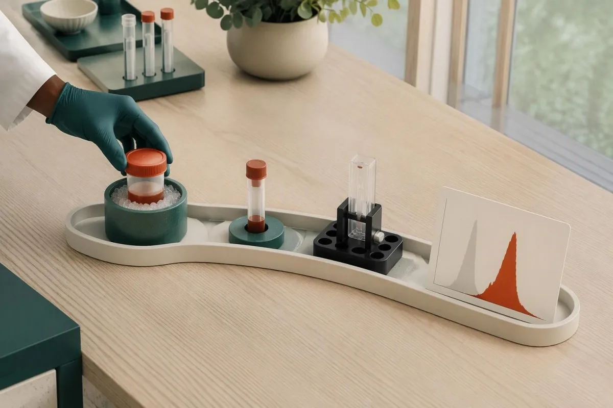
There is no universal waiting period for a repeat DHR. A transport problem can justify a fresh specimen at the next practical opportunity, while severe illness or treatment-related changes may require an individualized interval; repeating after 2 weeks without fixing the original handling issue adds little confidence.
A useful repeat controls the variables that were uncertain the first time. Arrange at least 2 appropriately identified specimens if the laboratory requests a patient sample and transported healthy control, confirm same-day dispatch, and ask whether the laboratory can reserve a processing slot before collection.
DHR generally does not require fasting by itself. Preparation should focus on assay instructions and recent treatment history rather than skipping breakfast; our guida alla preparazione della prima analisi helps distinguish general appointment preparation from requirements specific to a living-cell test.
Granulocyte transfusions can introduce functioning donor neutrophils and create 2 populations, while stem-cell transplantation can produce mixed donor-recipient responses. Routine leukoreduced red-cell transfusion is not usually the same confounder; tell the laboratory exactly which product was given and when, rather than reporting only that a transfusion occurred.
Quali reperti clinici rendono più preoccupante un DHR anomalo?
An abnormal DHR becomes more concerning when paired with recurrent deep infections, unusual organisms, granulomatous complications, or inflammatory bowel symptoms. An illustrative history of 2 liver abscess episodes carries more diagnostic weight than 2 uncomplicated colds; fever with breathing difficulty or rapidly worsening illness requires immediate assessment, not online interpretation.
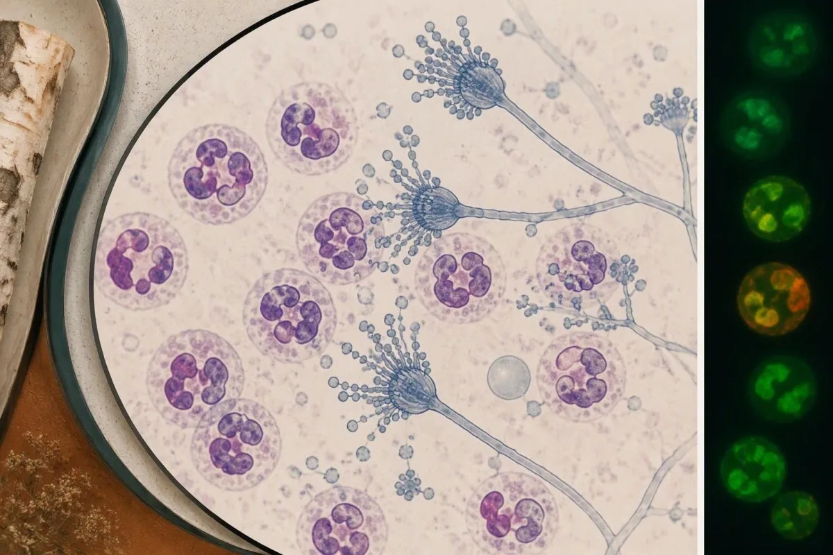
Aspergillus, Serratia, Burkholderia cepacia complex, Nocardia, and Staphylococcus aureus are recognized organisms in CGD, although none is exclusive to it. Holland's 2010 review describes the infection and inflammatory spectrum; the useful clinical question is whether 1 identified organism appeared in an unusual site or caused unexpectedly persistent disease.
An organism-specific test cannot measure oxidative burst function. Our fungal antigen interpretation illustrates the separate task of establishing fungal disease; a positive antigen and an abnormal DHR require different confirmation pathways, even when both are part of one patient's investigation.
CGD-related bowel inflammation can resemble inflammatory bowel disease. Persistent diarrhea, weight loss, or bloody stool warrants gastroenterology assessment, particularly alongside recurrent infections; a stool inflammatory marker measures intestinal inflammation, not neutrophil oxidase function, as our stool inflammation comparison .
A temperature of 38.0°C or higher in someone with suspected or confirmed CGD deserves a low threshold for contacting the treating team, especially with new respiratory or focal symptoms. Emergency assessment is appropriate for breathing difficulty, confusion, rapidly progressive illness, or inability to maintain hydration; prophylactic medication should not be assumed to exclude infection.
Che cosa conferma una diagnosi di malattia granulomatosa cronica?
Chronic granulomatous disease diagnosis combines a compatible functional defect with clinical assessment and usually molecular confirmation. Testing may examine at least 6 established CGD-associated genes—CYBB, CYBA, NCF1, NCF2, NCF4, and CYBC1—while broader panels can assess overlapping phagocyte disorders; a genetic variant of uncertain significance is not confirmation by itself.

CYBB-related CGD is X-linked; several other CGD forms are autosomal recessive. Gene identity influences inheritance counseling, but the DHR trace cannot reliably assign every molecular subtype; 2 patients with different genetic causes can produce similarly reduced responses, and variants within 1 gene can preserve different amounts of activity.
Genetic confirmation may require deletion-duplication analysis or specialized methods in addition to sequencing. NCF1 has closely related pseudogenes, which can complicate analysis; a negative generic panel should be reviewed for coverage and technical limitations before the clinician concludes that a convincing functional defect has no genetic explanation.
Normal PMA-stimulated DHR does not exclude every relevant phagocyte disorder. Some NCF4-related defects are better demonstrated with particulate stimulation, and other signaling disorders can affect receptor-mediated responses; when clinical suspicion remains high after 1 apparently normal test, the stimulus used becomes a critical detail.
Kantesti can explain why a functional test, gene panel, and clinical history sometimes disagree, but that disagreement belongs with an immunologist and the laboratory. The principle resembles borderline functional-test follow-up: neither 1 equivocal functional result nor 1 uncertain DNA variant should be stretched into a definitive diagnosis.
Chi altro in famiglia potrebbe aver bisogno di un test DHR o genetico?
Family testing should follow the confirmed inheritance pattern rather than an indiscriminate laboratory bundle. A person carrying a CYBB variant has a 50% chance of transmitting that X chromosome in each pregnancy; when both parents carry variants for the same autosomal-recessive condition, each pregnancy has a 25% chance of an affected child.

Those inheritance percentages apply independently to each pregnancy. For X-linked CYBB disease, the clinical consequence of inheriting the affected X differs according to the child's chromosome complement and X-inactivation; counseling should discuss affected sons, carrier daughters, and the possibility that a carrier can also develop significant symptoms.
A child with 1 affected CYBB variant may have inherited it or developed a new variant. Testing the mother can clarify family risk, but a normal maternal DHR does not exclude all forms of carrier mosaicism; targeted molecular testing is often needed once the child's causative variant is known.
Normal DHR usually cannot identify autosomal-recessive carriers. An unaffected sibling may still carry 1 familial variant, which matters for future reproductive counseling; targeted DNA testing answers that question more directly than repeating an oxidative-burst test that is expected to be normal in many such carriers.
A risk-based family testing plan should prioritize the relatives for whom the result changes care or reproductive decisions. For an illustrative family with 3 siblings, that might mean prompt evaluation of a symptomatic child, targeted testing of the others, and separate consent-based counseling for adults.
Come prepararsi alla valutazione specialistica del DHR?
Bring the complete DHR report, histogram, collection details, and infection history to specialist review. At least 4 elements should be clear: stimulus used, responding-cell fraction, fluorescence intensity or index, and healthy-control performance; a PDF containing only the word abnormal leaves too much unresolved for a reliable interpretation.

Kantesti is an AI-powered blood test analysis tool that can explain written DHR findings alongside CBC results and other uploaded laboratory information. Across a service used in 127+ countries, assay terminology may differ; our spiegazione tecnologica describes the interpretation workflow, not a substitute for direct inspection of raw cytometry files.
General interpretation performance does not establish DHR-specific diagnostic accuracy. A result delivered in approximately 60 seconds should still identify missing data and uncertainty; our validation and oversight information should be read for the actual evaluation scope rather than assumed to validate every rare functional assay.
The 3 questions I, Thomas Klein, would prioritize are practical: did the control pass, does the pattern suggest one deficient population or a mixture, and what confirmation will resolve the uncertainty? Our comitato consultivo medico provides organizational clinical context; a treating immunologist remains responsible for individualized diagnosis and management.
A family-history tool cannot replace Mendelian genetic counseling, even if it helps organize affected relatives. Before sharing 1 report, remove unnecessary identifiers and preserve the assay comments and reference information; our guida alla sicurezza dei report dell’IA explains why source verification matters more than the confidence of an automated explanation.
Pubblicazioni di ricerca e limiti di questa interpretazione
The evidence supporting DHR interpretation comes from CGD and phagocyte-function research, not unrelated digestive or hormonal publications. As of October 25, 2026, the 3 clinical references listed below support the disease mechanism, residual-function interpretation, and carrier discussion; this article does not claim that a new 2026 universal DHR cutoff exists.

Two additional publications are included as publisher-supplied background resources, not evidence for CGD diagnosis. The formal citations below reproduce the supplied titles and DOI identifiers; authorship and publication metadata have not been independently verified here, and ResearchGate and Academia.edu links are discovery searches rather than confirmed publication listings.
The citation beginning “Diarrhea After Fasting, Black Specks in Stool & GI Guide 2026” relates to broader digestive education, not oxidative-burst validation. Its associated digestive guide may help with symptom terminology, but it cannot establish whether 1 abnormal DHR result represents CGD-associated bowel disease.
The citation beginning “Women's Health Guide: Ovulation, Menopause & Hormonal Symptoms” is also separate from phagocyte-function evidence. Its associated hormonal health guide should not be used to explain carrier-related infection risk or to infer the significance of 2 neutrophil populations.
Kantesti educational content should distinguish verified clinical evidence from contextual publishing resources. Nostro standard di contenuto medico describe that boundary; the safest next step after an abnormal DHR is a documented laboratory interpretation and an appropriate clinical plan, not a diagnosis based on 1 flagged value.
Domande frequenti
Un test DHR anomalo significa che ho la malattia granulomatosa cronica?
Un test DHR anomalo non stabilisce automaticamente una malattia granulomatosa cronica. Una risposta quasi assente con un controllo sano soddisfacente è fortemente indicativa di un difetto dell’ossidasi, ma anche la carenza di mieloperossidasi, campioni compromessi e altre condizioni possono ridurre la fluorescenza. Due popolazioni neutrofile distinte possono indicare lo stato di portatrice legato al cromosoma X o una miscela di cellule del donatore e del ricevente, anziché una risposta uniformemente carente. La conferma di solito combina la valutazione specialistica, l’esame del risultato funzionale completo e i test molecolari.
Qual è l'intervallo normale dei risultati del test DHR?
I risultati del test DHR non hanno un intervallo di riferimento numerico universale applicabile a tutti i laboratori. La percentuale di cellule rispondenti, l’intensità di fluorescenza e l’indice di stimolazione dipendono dal test e dai relativi controlli. Ad esempio, la presenza di neutrofili rispondenti 98% non dimostra che la loro intensità di fluorescenza sia normale. Utilizzare i dati di riferimento del laboratorio che ha eseguito il test e interpretare la percentuale insieme all’istogramma e all’intensità della risposta.
Perché una portatrice di CGD avrebbe due picchi al test DHR?
0 Una portatrice di CGD legata al cromosoma X può presentare due picchi DHR perché l’inattivazione del cromosoma X crea 2 popolazioni di neutrofili che esprimono copie diverse di CYBB. Una popolazione può rispondere in modo marcato, mentre l’altra risponde scarsamente. Le proporzioni non devono necessariamente essere 50:50 e una frazione a bassa funzionalità può avere rilevanza clinica. I test genetici e l’anamnesi clinica aiutano a distinguere il mosaicismo da portatrice da altre cause di popolazioni miste.
Un campione ritardato può causare risultati anomali del test DHR?
Un campione ritardato o gestito in modo improprio può produrre una risposta DHR falsamente debole, perché il test dipende dalla vitalità dei neutrofili. Molti laboratori puntano a eseguire l’analisi entro circa 24 ore, mentre l’accettazione dopo 48 ore o più dipende da procedure locali validate. Un controllo sano trasportato insieme al campione del paziente può aiutare a identificare una perdita di funzionalità dovuta alla spedizione. Se la gestione o le prestazioni del controllo non sono soddisfacenti, il laboratorio può raccomandare un nuovo campione anziché formulare una diagnosi di malattia.
La carenza di mieloperossidasi può sembrare una CGD al test DHR?
La carenza di mieloperossidasi può ridurre la fluorescenza della diidrorodamina 123 anche quando la NADPH ossidasi produce ancora superossido. Un secondo metodo funzionale, come la misurazione diretta del superossido o un test al nitroblu tetrazolio eseguito correttamente, può aiutare a distinguerla dalla CGD classica. La valutazione cellulare della MPO e un’analisi dettagliata del pattern dei granulociti possono fornire ulteriori elementi. Una concentrazione circolante di MPO riportata in ng/mL non sostituisce questa indagine funzionale.
Un test DHR normale può escludere la CGD o lo stato di portatrice?
0 Un risultato DHR normale rende meno probabili molti difetti classici della CGD, ma non esclude tutte le patologie rilevanti dei fagociti né gli stati di portatore genetico. Alcuni difetti correlati a NCF4 possono mostrare una risposta soddisfacente al PMA, ma una risposta scarsa alla stimolazione con particolato. I portatori autosomici recessivi con 1 copia funzionante del gene hanno comunemente risposte DHR normali. Un sospetto clinico persistente o una variante familiare nota dovrebbero indurre a richiedere il parere di uno specialista su ulteriori test funzionali o molecolari.
Ottieni oggi l’analisi degli esami del sangue con IA (AI-Powered Blood Test Analysis)
Unisciti a oltre 2 milioni di utenti in tutto il mondo che si fidano di Kantesti per un’analisi istantanea e accurata degli esami di laboratorio. Carica i tuoi risultati analisi del sangue e ricevi un’interpretazione completa dei biomarcatori di 15,000+ in pochi secondi.
📚 Pubblicazioni di ricerca di riferimento
Klein, T., Mitchell, S., & Weber, H. (2026). Diarrea dopo il digiuno, macchie nere nelle feci e guida gastrointestinale 2026. Kantesti AI Medical Research.
Klein, T., Mitchell, S., & Weber, H. (2026). Guida alla salute femminile: ovulazione, menopausa e sintomi ormonali. Kantesti AI Medical Research.
📖 Riferimenti medici esterni
Holland SM. (2010). Malattia granulomatosa cronica. Clinical Reviews in Allergy & Immunology.
Kuhns DB et al. (2010). Residual NADPH oxidase and survival in chronic granulomatous disease. New England Journal of Medicine.
Marciano BE et al. (2018). X-linked carriers of chronic granulomatous disease: Illness, lyonization, and stability. Journal of Allergy and Clinical Immunology.





⚕️ Esclusione di responsabilità medica
Questo articolo ha solo scopo educativo e non costituisce consulenza medica. Consulta sempre un operatore sanitario qualificato per decisioni su diagnosi e trattamento.
Segnali di fiducia E-E-A-T
Esperienza
Revisione clinica guidata da un medico dei flussi di lavoro di interpretazione degli esami.
Competenza
Focus sulla medicina di laboratorio su come i biomarcatori si comportano nel contesto clinico.
autorevolezza
Scritto dal dott. Thomas Klein con revisione della dott.ssa Sarah Mitchell e del Prof. Dr. Hans Weber.
Affidabilità
Interpretazione basata su evidenze, con percorsi di follow-up chiari per ridurre l’allarme.
