
Respons neutrofil yang lemah tidak selalu berarti penyakit granulomatosa kronis. Pola fluoresensi, kontrol sehat, dan perjalanan sampel menentukan hal yang benar-benar dapat dipastikan dari hasil tersebut.



- Hasil tes DHR yang abnormal menunjukkan oksidasi dihidrorhodamin 123 oleh neutrofil yang berkurang atau tidak merata; hasil tersebut memerlukan interpretasi berdasarkan pola, bukan diagnosis CGD secara otomatis.
- Respons yang hampir tidak ada pada hampir semua neutrofil, dengan kontrol sehat yang memadai, sangat mendukung adanya defek oksidase NADPH yang berat.
- Dua populasi neutrofil dapat menunjukkan carrier CGD terkait-X, tetapi transfusi granulosit atau kimerisme terkait transplantasi dapat menghasilkan campuran serupa.
- Fraksi carrier di bawah 10% neutrofil fungsional telah dikaitkan dengan risiko infeksi tipe CGD yang jauh lebih tinggi; 50% bukan hasil carrier yang disyaratkan.
- Aktivitas oksidatif residual dapat terjadi pada CGD sejati, sehingga respons yang redup belum tentu meyakinkan.
- Waktu pengambilan sampel penting: target waktu dari pengambilan hingga analisis umumnya sekitar 24 jam, sedangkan rentang waktu yang lebih lama memerlukan validasi oleh laboratorium pelaksana.
- Defisiensi mieloperoksidase dapat mengurangi fluoresensi DHR meskipun aktivitas NADPH oksidase tetap terjaga; metode fungsional kedua dapat membedakan kondisi-kondisi tersebut.
- Stimulasi PMA normal tidak menyingkirkan semua kelainan fagosit yang langka, terutama defek yang lebih baik terlihat dengan stimulasi berbasis partikel.
- Gejala yang mendesak mengesampingkan rencana pengujian ulang: demam disertai kesulitan bernapas, penyakit yang berkembang cepat, atau infeksi baru yang dalam memerlukan penilaian klinis segera.
Apa sebenarnya arti hasil tes DHR yang abnormal?
Hasil tes DHR yang abnormal berarti neutrofil menghasilkan lebih sedikit fluoresensi—atau distribusi fluoresensi yang berbeda—setelah stimulasi. Respons yang hampir tidak ada mendukung penyakit granulomatosa kronis, 2 populasi yang berbeda dapat mengindikasikan status pembawa terkait-X, dan respons lemah dengan kontrol yang buruk dapat mencerminkan masalah penanganan; hasil tidak boleh ditafsirkan hanya berdasarkan angka yang ditandai.

Penyakit granulomatosa kronis, atau CGD, mengganggu sistem NADPH oksidase fagosit, bukan sekadar menurunkan jumlah neutrofil. Pasien dapat memiliki jumlah neutrofil absolut sebesar 4.0 × 10^9/L dan tetap mengalami defek fungsional yang berat; penghitungan sel dan pengujian mekanisme antimikrobanya menjawab pertanyaan yang berbeda.
Pertimbangkan anak berusia 7 tahun dengan infeksi bakteri dalam yang berulang dan kurva DHR terstimulasi yang hampir datar. Kombinasi itu menimbulkan tingkat kekhawatiran yang berbeda dibandingkan satu respons ambang pada orang dewasa yang secara umum sehat; pemeriksaan imun awal harus memastikan infeksi apa yang terjadi, di bagian tubuh mana, dan apakah organisme penyebabnya berhasil diidentifikasi.
Saya Thomas Klein, Chief Medical Officer Kantesti, dan aturan pertama saya dalam membaca hasil adalah memisahkan 3 pertanyaan: apakah spesimen valid, apakah responsnya abnormal, dan mekanisme apa yang menjelaskannya? Pertanyaan-pertanyaan itu harus dijawab secara berurutan, karena grafik yang tampak meyakinkan tetap dapat berasal dari sel yang kondisinya terganggu.
Kantesti adalah penganalisis tes darah berbasis AI yang dapat menjelaskan istilah tertulis dalam laporan DHR beserta temuan laboratorium terkait. latar belakang perusahaan menjelaskan layanan ini; penjelasan yang dihasilkan dalam waktu sekitar 60 detik tidak menggantikan tinjauan oleh spesialis atas data sitometri alir ataupun memastikan diagnosis CGD.
Bagaimana cara kerja tes oxidative burst DHR?
Itu Tes oxidative burst DHR menggunakan sitometri alir untuk mengukur perubahan dihidrorodamin 123 menjadi rodamin berfluoresensi setelah stimulasi neutrofil. NADPH oksidase menghasilkan superoksida, yang berkontribusi pada pembentukan hidrogen peroksida; hasil pembacaan DHR juga bergantung pada kimia oksidasi di tahap selanjutnya, termasuk mieloperoksidase, sehingga fluoresensi bukan pengukuran langsung superoksida.

PMA, atau phorbol myristate acetate, adalah stimulan laboratorium kuat yang digunakan dalam banyak protokol DHR. Zat ini mengaktifkan pensinyalan intraseluler tanpa memerlukan pengenalan mikroba normal pada permukaan sel, sehingga berguna untuk banyak defek CGD, tetapi kurang dapat mengungkap kelainan tertentu yang bergantung pada reseptor atau partikel.
Interpretasi standar membandingkan setidaknya 2 kondisi pasien: sel tanpa stimulasi dan sel terstimulasi. Laboratorium biasanya juga memeriksa kontrol sehat; apakah kontrol tersebut dikirim bersama spesimen pasien penting, karena kontrol lokal yang baru diambil tidak dapat sepenuhnya mereproduksi kerusakan akibat perjalanan kurir yang lama.
Gerbang neutrofil memilih populasi sel yang dievaluasi, dan masuknya granulosit lain secara tidak tepat dapat mendistorsi pola yang tampak. Eosinofil sangat relevan saat membedakan defisiensi mieloperoksidase; ini merupakan persoalan tingkat laboratorium yang tidak dapat diselesaikan berdasarkan persentase CBC atau tangkapan layar yang berdiri sendiri.
Jumlah neutrofil normal tidak membuktikan fungsi neutrofil yang normal. An adult ANC within a laboratory interval such as 1.5–7.5 × 10^9/L answers a counting question, not an oxidative-burst question; our neutrophil count guide explains why age, pregnancy, and reporting conventions affect that separate result.
Angka dan pola fluoresensi DHR mana yang penting?
DHR interpretation requires both the proportion of responding neutrophils and the strength of their response. A report showing 98% positive cells can still conceal reduced fluorescence intensity, while 2 separated populations can be clinically significant even when their combined average appears acceptable; laboratory-specific gates and controls determine the meaning.
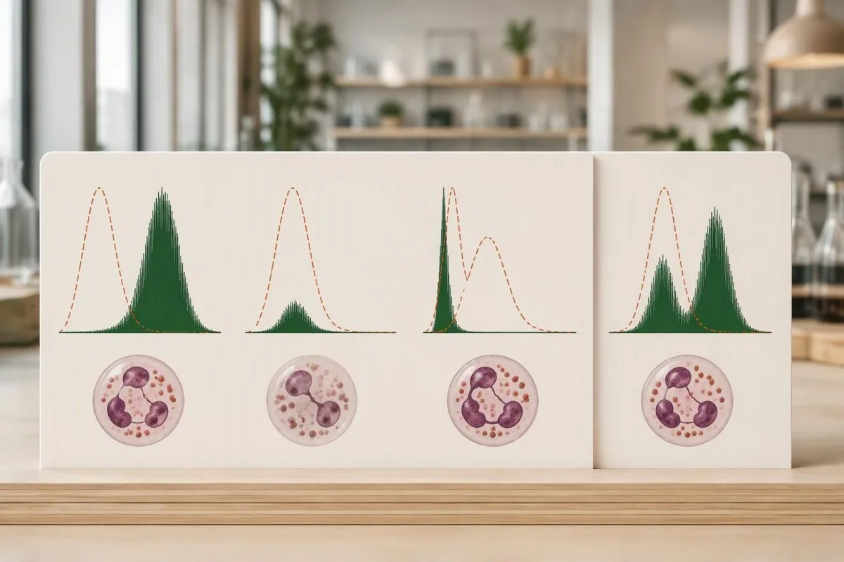
Mean fluorescence intensity, often abbreviated MFI, measures signal strength in a defined population. An illustrative MFI of 200 on one instrument cannot be compared directly with 200 on another; detector settings, reagents, stimulation time, and whether the laboratory averages all neutrophils or only responding cells affect the value.
Stimulation index is commonly calculated as stimulated fluorescence divided by baseline fluorescence, although laboratories may define the calculation differently. If stimulated MFI remains 100 but baseline rises from 1 to 5, the ratio falls from 100 to 20 without any change in the stimulated signal itself.
A histogram preserves information that a single summary number loses. The reason to request the full plot is similar to the reasoning behind flow-cytometry population analysis: the distribution can distinguish one uniformly dim population from 2 populations with very different behavior.
There is no universal DHR MFI or stimulation-index cutoff for CGD. A laboratory may also report a qualitative conclusion alongside quantitative measurements; our explanation of hasil kualitatif dan kuantitatif helps separate a categorical flag from the underlying data used to assign it.
Apakah oxidative burst yang lemah selalu menunjukkan CGD berat?
A nearly absent oxidative burst in almost all neutrophils strongly supports severe NADPH oxidase dysfunction when controls are satisfactory, but CGD can also produce a partial response. A uniformly dim population is not equivalent to 2 separate carrier populations, and measurable activity does not automatically exclude clinically significant disease.

Residual oxidase activity has prognostic relevance in CGD. Kuhns et al. studied 287 patients and found that residual reactive-oxygen-intermediate production was associated with survival, beyond the affected gene alone; their 2010 New England Journal of Medicine paper supports measuring function rather than treating every abnormal response as equally severe.
An illustrative response at 5% of a concurrent control is different from an undetectable response, but that comparison is not a universal treatment threshold. A specialist must consider the assay's reproducibility, the genotype, and the patient's infection history before translating a small remaining signal into a prediction about future health.
Rare severe G6PD deficiency can affect neutrophil oxidative function, although ordinary G6PD deficiency is not synonymous with CGD. A low DHR response combined with episodes of oxidant-triggered hemolysis may justify a separate enzyme investigation; our G6PD interpretation guide explains why red-cell enzyme results need their own context.
Kantesti AI can help organize the distinction between an absent response, a partial response, and a mixed response when those descriptions appear in the report. The useful output is a set of follow-up questions—not a claim that 1 percentage or a particular shade of fluorescence establishes disease severity.
Mengapa carrier CGD terkait-X menunjukkan dua populasi?
X-linked CGD carriers often show 2 neutrophil populations because different cells keep different X chromosomes active. Cells expressing a functioning CYBB copy can respond strongly, while cells expressing the affected copy respond poorly; this is cellular mosaicism, not every neutrophil operating at approximately 50% capacity.
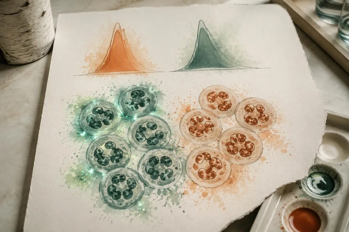
A 50:50 split is not required for X-linked carrier status. A carrier might have an illustrative 70:30 or 20:80 distribution, and the proportion can change over time; the brighter population's intensity and the dim population's behavior should be evaluated separately rather than compressed into one average.
The mechanistic distinction is useful when comparing a carrier with partial CGD. In a carrier example, 30% of cells may respond approximately like control neutrophils while 70% respond poorly; in a partial-deficiency example, almost all cells may respond weakly together, producing a shifted but largely single distribution.
DHR cannot identify every genetic carrier. Carriers of autosomal-recessive CGD commonly have a normal functional response because 1 functioning gene copy is sufficient; molecular testing is usually required to establish that status, and normal DHR should not be presented as a universal negative carrier screen.
Carrier terminology also differs across inherited conditions. Our genetic carrier screening explanation addresses why carrier labels require disease-specific interpretation; for CGD, the immediate questions are inheritance pattern, neutrophil function, and whether the person has symptoms—not simply whether a variant was found.
Kapan carrier CGD terkait-X dapat mengalami gejala?
X-linked CGD carriers can develop infections or inflammatory complications, particularly when X-inactivation leaves very few functioning neutrophils. Fractions below 10% DHR-positive cells were strongly associated with CGD-type infections in a published carrier cohort, but no single percentage predicts every person's outcome or explains all autoimmune symptoms.
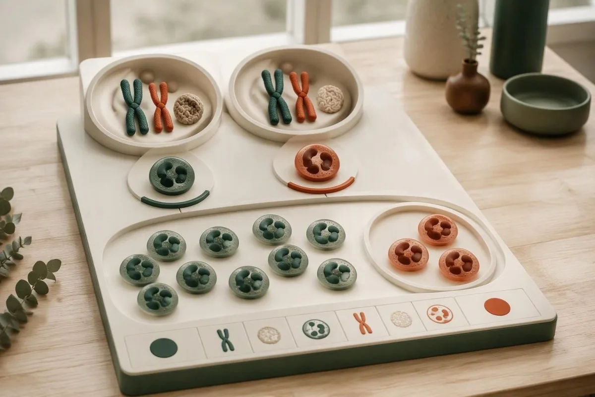
Marciano et al.'s 2018 Journal of Allergy and Clinical Immunology study connected low functioning-neutrophil fractions with CGD-type infection risk in X-linked carriers. Below 10% was a particularly concerning pattern; clinicians may also use a fraction below 20% to prompt closer assessment, but neither figure should become a stand-alone diagnostic rule.
Inflammatory and autoimmune manifestations do not track perfectly with the DHR-positive fraction. A carrier with 60% responding cells can still have clinically meaningful mouth sores, photosensitive rashes, or joint symptoms, so a reassuring percentage should not end the investigation when those complaints persist or recur.
A change from 35% to 18% functioning neutrophils deserves scrutiny, not an automatic conclusion that X-inactivation has worsened. First compare sample age, gating, stimulus, and the performing laboratory; repeat measurements under comparable conditions help distinguish a biological change from a technical difference.
Cellular mosaicism and chromosome mosaicism are different concepts. X-inactivation can create 2 functional neutrophil groups without 2 different chromosome counts; our chromosome mosaicism guide provides a useful contrast, although the diagnostic tests and inheritance counseling are not interchangeable.
Dapatkah defisiensi mieloperoksidase menyerupai hasil DHR yang abnormal?
Myeloperoxidase deficiency can produce an abnormal DHR response despite preserved NADPH oxidase activity. Because dihydrorhodamine 123 fluorescence depends on downstream oxidation, a weak signal does not uniquely identify CGD; an experienced laboratory may use direct superoxide testing, nitroblue tetrazolium testing, or cellular MPO assessment to investigate the discrepancy.
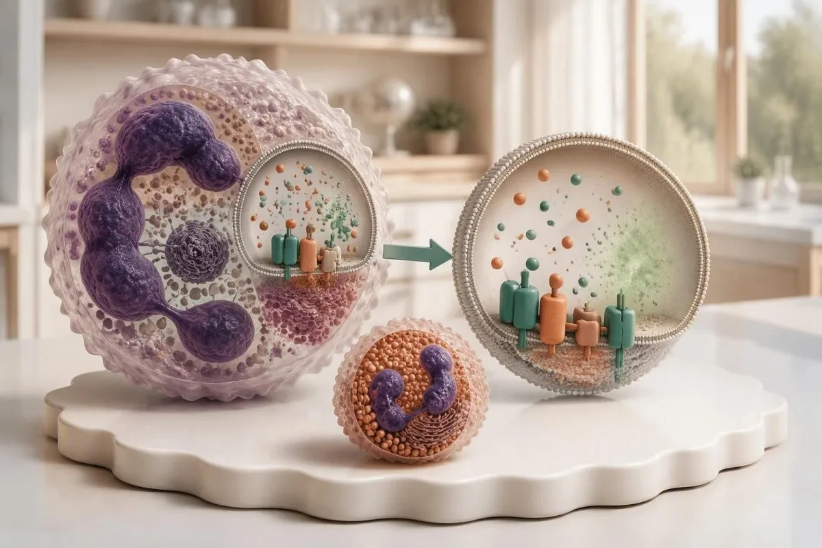
Preserved superoxide production argues against a classic NADPH oxidase defect. If a patient's DHR is low but a second method demonstrates satisfactory superoxide generation, the 2 results should be reconciled rather than forcing both into a CGD diagnosis; MPO deficiency is one recognized explanation discussed in Holland's 2010 clinical review.
The laboratory may inspect eosinophil behavior because eosinophil peroxidase can preserve a different response pattern in MPO deficiency. That distinction requires correctly identified cellular populations; asking whether at least 2 granulocyte groups were considered is more useful than trying to infer the answer from total eosinophil percentage alone.
A circulating MPO concentration is not a neutrophil MPO-deficiency assessment. A result reported in ng/mL for vascular-risk evaluation addresses a different question; our MPO test limitations explain why identical enzyme names on 2 reports do not make those tests interchangeable.
Kantesti is an AI blood test interpretation platform that can distinguish the purpose of these differently named or differently measured MPO tests in a written report. When 2 functional assays disagree, our explanation should preserve that uncertainty and direct attention to the performing laboratory rather than assigning a premature inherited-disease label.
Bagaimana penanganan sampel dapat menyebabkan oxidative burst yang tampak rendah secara keliru?
Delayed processing, unsuitable temperatures, an incorrect collection tube, or poor cell viability can weaken a DHR response. Many laboratories aim to analyze specimens within approximately 24 hours; acceptance at 48 hours or beyond depends on their validated protocol, and a weak transported control makes an immediate CGD diagnosis unreliable.

The correct tube is laboratory-specific, although sodium-heparin specimens are commonly requested for DHR testing. Before collection, confirm the anticoagulant, minimum volume, transport temperature, and arrival deadline; a tube suitable for 1 routine chemistry panel may be unsuitable for a living-cell functional assay.
A healthy control collected and shipped alongside the patient's specimen can reveal transport-related loss of function. If both samples show reduced fluorescence after a 36-hour journey, that pattern supports investigating the shipment; a satisfactory control is reassuring, but it cannot exclude every patient-specific viability or gating problem.
An elevated hemolysis index does not measure neutrophil viability. A chemistry sample and a functional-cell specimen can fail for different reasons, even when collected at the same appointment; our sample artifact explanation illustrates why a quality flag must be interpreted for the particular assay.
Ask for 4 pieces of documentation: collection time, analysis time, transport conditions, and the laboratory's control assessment. Do not refrigerate or freeze a DHR specimen merely because another test required cold transport; handling instructions are assay-specific, and the performing laboratory should decide whether a redraw is necessary.
Kapan oxidative burst yang abnormal perlu diperiksa ulang?
Repeat DHR testing is appropriate when controls fail, handling is uncertain, results are borderline, or the pattern conflicts with the clinical picture. An almost absent response with a strong healthy control and characteristic infections warrants prompt immunology assessment; arranging a second specimen should not delay evaluation of an active infection.
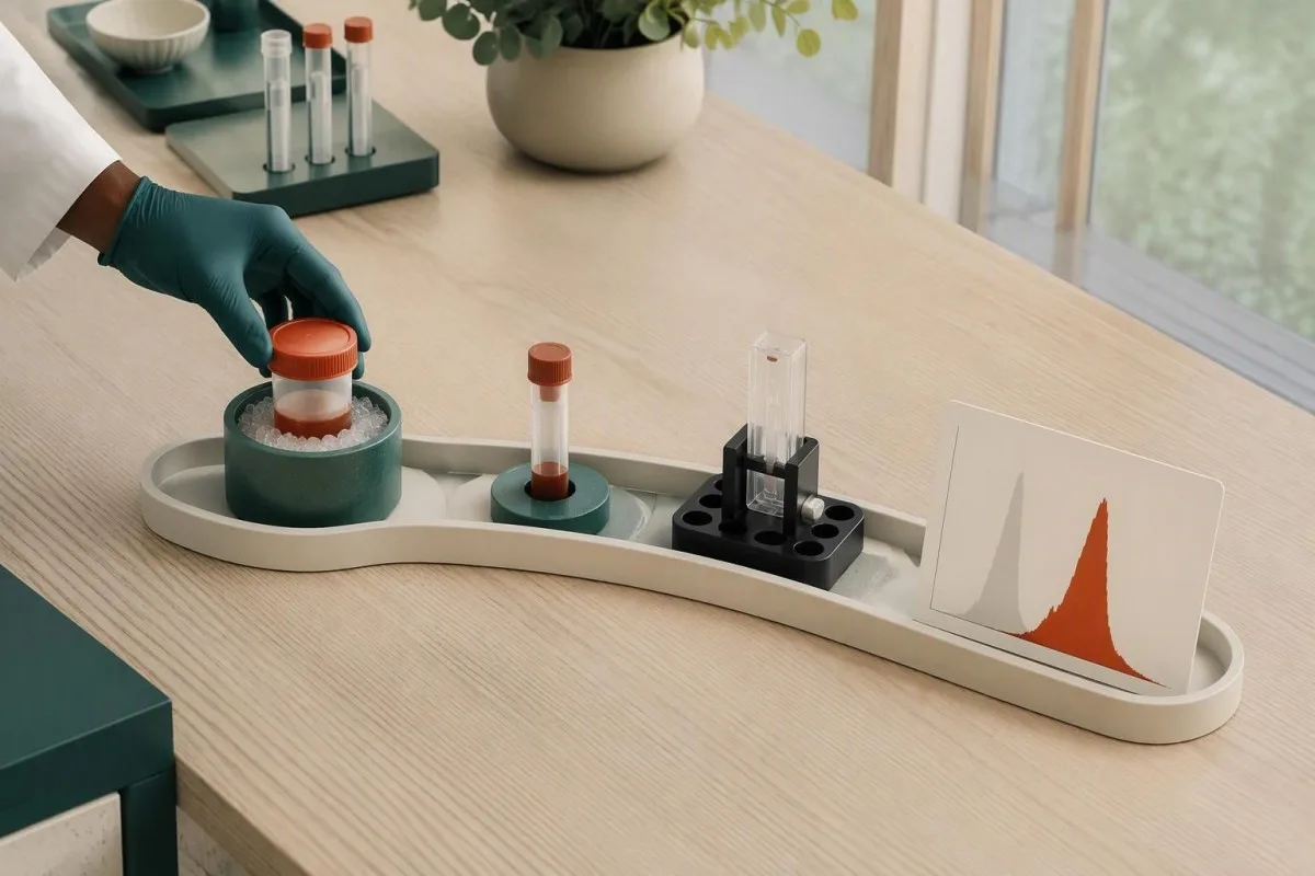
There is no universal waiting period for a repeat DHR. A transport problem can justify a fresh specimen at the next practical opportunity, while severe illness or treatment-related changes may require an individualized interval; repeating after 2 weeks without fixing the original handling issue adds little confidence.
A useful repeat controls the variables that were uncertain the first time. Arrange at least 2 appropriately identified specimens if the laboratory requests a patient sample and transported healthy control, confirm same-day dispatch, and ask whether the laboratory can reserve a processing slot before collection.
DHR generally does not require fasting by itself. Preparation should focus on assay instructions and recent treatment history rather than skipping breakfast; our menjelaskan cara mengurangi kebisingan yang dapat dihindari tersebut. helps distinguish general appointment preparation from requirements specific to a living-cell test.
Granulocyte transfusions can introduce functioning donor neutrophils and create 2 populations, while stem-cell transplantation can produce mixed donor-recipient responses. Routine leukoreduced red-cell transfusion is not usually the same confounder; tell the laboratory exactly which product was given and when, rather than reporting only that a transfusion occurred.
Temuan klinis apa yang membuat DHR abnormal lebih mengkhawatirkan?
An abnormal DHR becomes more concerning when paired with recurrent deep infections, unusual organisms, granulomatous complications, or inflammatory bowel symptoms. An illustrative history of 2 liver abscess episodes carries more diagnostic weight than 2 uncomplicated colds; fever with breathing difficulty or rapidly worsening illness requires immediate assessment, not online interpretation.
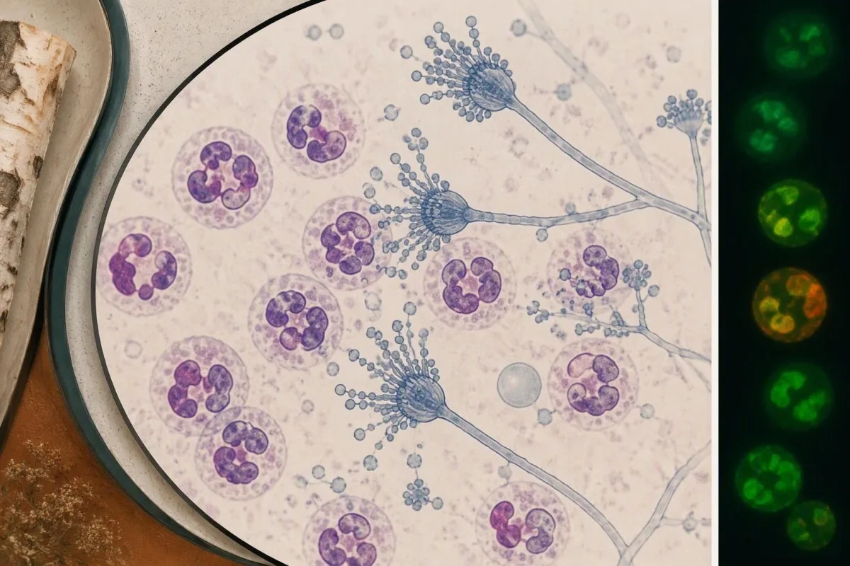
Aspergillus, Serratia, Burkholderia cepacia complex, Nocardia, and Staphylococcus aureus are recognized organisms in CGD, although none is exclusive to it. Holland's 2010 review describes the infection and inflammatory spectrum; the useful clinical question is whether 1 identified organism appeared in an unusual site or caused unexpectedly persistent disease.
An organism-specific test cannot measure oxidative burst function. Our fungal antigen interpretation illustrates the separate task of establishing fungal disease; a positive antigen and an abnormal DHR require different confirmation pathways, even when both are part of one patient's investigation.
CGD-related bowel inflammation can resemble inflammatory bowel disease. Persistent diarrhea, weight loss, or bloody stool warrants gastroenterology assessment, particularly alongside recurrent infections; a stool inflammatory marker measures intestinal inflammation, not neutrophil oxidase function, as our stool inflammation comparison .
A temperature of 38.0°C or higher in someone with suspected or confirmed CGD deserves a low threshold for contacting the treating team, especially with new respiratory or focal symptoms. Emergency assessment is appropriate for breathing difficulty, confusion, rapidly progressive illness, or inability to maintain hydration; prophylactic medication should not be assumed to exclude infection.
Apa yang mengonfirmasi diagnosis penyakit granulomatosa kronis?
Chronic granulomatous disease diagnosis combines a compatible functional defect with clinical assessment and usually molecular confirmation. Testing may examine at least 6 established CGD-associated genes—CYBB, CYBA, NCF1, NCF2, NCF4, and CYBC1—while broader panels can assess overlapping phagocyte disorders; a genetic variant of uncertain significance is not confirmation by itself.

CYBB-related CGD is X-linked; several other CGD forms are autosomal recessive. Gene identity influences inheritance counseling, but the DHR trace cannot reliably assign every molecular subtype; 2 patients with different genetic causes can produce similarly reduced responses, and variants within 1 gene can preserve different amounts of activity.
Genetic confirmation may require deletion-duplication analysis or specialized methods in addition to sequencing. NCF1 has closely related pseudogenes, which can complicate analysis; a negative generic panel should be reviewed for coverage and technical limitations before the clinician concludes that a convincing functional defect has no genetic explanation.
Normal PMA-stimulated DHR does not exclude every relevant phagocyte disorder. Some NCF4-related defects are better demonstrated with particulate stimulation, and other signaling disorders can affect receptor-mediated responses; when clinical suspicion remains high after 1 apparently normal test, the stimulus used becomes a critical detail.
Kantesti can explain why a functional test, gene panel, and clinical history sometimes disagree, but that disagreement belongs with an immunologist and the laboratory. The principle resembles borderline functional-test follow-up: neither 1 equivocal functional result nor 1 uncertain DNA variant should be stretched into a definitive diagnosis.
Siapa lagi dalam keluarga yang mungkin memerlukan pemeriksaan DHR atau genetik?
Family testing should follow the confirmed inheritance pattern rather than an indiscriminate laboratory bundle. A person carrying a CYBB variant has a 50% chance of transmitting that X chromosome in each pregnancy; when both parents carry variants for the same autosomal-recessive condition, each pregnancy has a 25% chance of an affected child.

Those inheritance percentages apply independently to each pregnancy. For X-linked CYBB disease, the clinical consequence of inheriting the affected X differs according to the child's chromosome complement and X-inactivation; counseling should discuss affected sons, carrier daughters, and the possibility that a carrier can also develop significant symptoms.
A child with 1 affected CYBB variant may have inherited it or developed a new variant. Testing the mother can clarify family risk, but a normal maternal DHR does not exclude all forms of carrier mosaicism; targeted molecular testing is often needed once the child's causative variant is known.
Normal DHR usually cannot identify autosomal-recessive carriers. An unaffected sibling may still carry 1 familial variant, which matters for future reproductive counseling; targeted DNA testing answers that question more directly than repeating an oxidative-burst test that is expected to be normal in many such carriers.
A risk-based family testing plan should prioritize the relatives for whom the result changes care or reproductive decisions. For an illustrative family with 3 siblings, that might mean prompt evaluation of a symptomatic child, targeted testing of the others, and separate consent-based counseling for adults.
Bagaimana sebaiknya Anda mempersiapkan diri untuk tinjauan spesialis DHR?
Bring the complete DHR report, histogram, collection details, and infection history to specialist review. At least 4 elements should be clear: stimulus used, responding-cell fraction, fluorescence intensity or index, and healthy-control performance; a PDF containing only the word abnormal leaves too much unresolved for a reliable interpretation.

Kantesti is an AI-powered blood test analysis tool that can explain written DHR findings alongside CBC results and other uploaded laboratory information. Across a service used in 127+ countries, assay terminology may differ; our penjelasan teknologi describes the interpretation workflow, not a substitute for direct inspection of raw cytometry files.
General interpretation performance does not establish DHR-specific diagnostic accuracy. A result delivered in approximately 60 seconds should still identify missing data and uncertainty; our validation and oversight information should be read for the actual evaluation scope rather than assumed to validate every rare functional assay.
The 3 questions I, Thomas Klein, would prioritize are practical: did the control pass, does the pattern suggest one deficient population or a mixture, and what confirmation will resolve the uncertainty? Our dewan penasihat medis provides organizational clinical context; a treating immunologist remains responsible for individualized diagnosis and management.
A family-history tool cannot replace Mendelian genetic counseling, even if it helps organize affected relatives. Before sharing 1 report, remove unnecessary identifiers and preserve the assay comments and reference information; our AI explains why source verification matters more than the confidence of an automated explanation.
Publikasi penelitian dan batasan interpretasi ini
The evidence supporting DHR interpretation comes from CGD and phagocyte-function research, not unrelated digestive or hormonal publications. As of October 25, 2026, the 3 clinical references listed below support the disease mechanism, residual-function interpretation, and carrier discussion; this article does not claim that a new 2026 universal DHR cutoff exists.

Two additional publications are included as publisher-supplied background resources, not evidence for CGD diagnosis. The formal citations below reproduce the supplied titles and DOI identifiers; authorship and publication metadata have not been independently verified here, and ResearchGate and Academia.edu links are discovery searches rather than confirmed publication listings.
The citation beginning “Diarrhea After Fasting, Black Specks in Stool & GI Guide 2026” relates to broader digestive education, not oxidative-burst validation. Its associated digestive guide may help with symptom terminology, but it cannot establish whether 1 abnormal DHR result represents CGD-associated bowel disease.
The citation beginning “Women's Health Guide: Ovulation, Menopause & Hormonal Symptoms” is also separate from phagocyte-function evidence. Its associated hormonal health guide should not be used to explain carrier-related infection risk or to infer the significance of 2 neutrophil populations.
Kantesti educational content should distinguish verified clinical evidence from contextual publishing resources. Kita standar konten medis describe that boundary; the safest next step after an abnormal DHR is a documented laboratory interpretation and an appropriate clinical plan, not a diagnosis based on 1 flagged value.
Pertanyaan yang Sering Diajukan
Apakah hasil tes DHR yang abnormal berarti saya menderita penyakit granulomatosa kronis?
Hasil tes DHR yang abnormal tidak secara otomatis menegakkan diagnosis penyakit granulomatosa kronis. Respons yang hampir tidak ada dengan kontrol sehat yang memadai sangat mendukung adanya defek oksidase, tetapi defisiensi mieloperoksidase, spesimen yang kualitasnya terganggu, dan kondisi lain juga dapat menurunkan fluoresensi. Dua populasi neutrofil yang terpisah dapat mengindikasikan status karier terkait-X atau campuran sel donor-penerima, bukan respons yang mengalami defisiensi secara merata. Konfirmasi biasanya menggabungkan penilaian spesialis, peninjauan hasil fungsional lengkap, dan pemeriksaan molekuler.
Berapa rentang normal untuk hasil tes DHR?
Hasil tes DHR tidak memiliki rentang referensi numerik universal yang berlaku di semua laboratorium. Persentase sel yang merespons, intensitas fluoresensi, dan indeks stimulasi bergantung pada pemeriksaan dan kontrolnya. Sebagai contoh, 98% neutrofil yang merespons tidak membuktikan bahwa intensitas fluoresensinya normal. Gunakan informasi referensi dari laboratorium yang melakukan pemeriksaan dan interpretasikan persentase tersebut bersama histogram dan kekuatan respons.
Mengapa seorang pembawa CGD memiliki dua puncak pada tes DHR?
0 An X-linked CGD carrier dapat memiliki dua puncak DHR karena inaktivasi kromosom X menghasilkan 2 populasi neutrofil yang mengekspresikan salinan CYBB yang berbeda. Satu populasi dapat merespons dengan kuat, sementara populasi lainnya merespons dengan buruk. Proporsinya tidak harus 50:50, dan fraksi dengan fungsi rendah dapat memiliki signifikansi klinis. Pemeriksaan genetik dan riwayat klinis membantu membedakan mosaikisme pembawa dari penyebab lain populasi campuran.
Dapatkah spesimen yang terlambat menyebabkan hasil tes DHR abnormal?
Spesimen yang terlambat atau ditangani secara tidak tepat dapat menghasilkan respons DHR yang tampak lemah secara keliru karena pemeriksaan ini bergantung pada neutrofil yang masih hidup. Banyak laboratorium menargetkan pemrosesan dalam waktu sekitar 24 jam, sementara penerimaan spesimen setelah 48 jam atau lebih bergantung pada prosedur lokal yang telah divalidasi. Kontrol sehat yang dikirim bersama sampel pasien dapat membantu mengidentifikasi hilangnya fungsi terkait pengiriman. Jika penanganan atau kinerja kontrol tidak memuaskan, laboratorium dapat merekomendasikan spesimen baru daripada diagnosis penyakit.
Dapatkah defisiensi mieloperoksidase tampak seperti CGD pada pemeriksaan DHR?
Defisiensi mieloperoksidase dapat mengurangi fluoresensi dihidrorodamin 123 meskipun NADPH oksidase masih menghasilkan superoksida. Metode fungsional kedua, seperti pengukuran langsung superoksida atau pemeriksaan nitroblue tetrazolium yang dilakukan dengan tepat, dapat membantu membedakannya dari CGD klasik. Penilaian MPO seluler dan analisis pola granulosit yang terperinci dapat memberikan bukti tambahan. Konsentrasi MPO yang bersirkulasi dan dilaporkan dalam ng/mL bukan pengganti pemeriksaan fungsional ini.
Apakah hasil tes DHR normal dapat menyingkirkan CGD atau status sebagai pembawa?
Hasil DHR normal membuat banyak kelainan CGD klasik menjadi kurang mungkin, tetapi tidak menyingkirkan setiap kelainan fagosit yang relevan atau status pembawa genetik. Beberapa kelainan terkait NCF4 dapat menunjukkan respons yang memadai terhadap PMA, tetapi merespons buruk terhadap stimulasi partikel. Individu pembawa sifat autosomal resesif dengan 1 salinan gen yang berfungsi umumnya memiliki respons DHR normal. Kecurigaan klinis yang menetap atau varian familial yang telah diketahui perlu ditindaklanjuti dengan saran dari spesialis mengenai pemeriksaan fungsional atau molekuler tambahan.
Dapatkan Analisis Tes Darah Berbasis AI Hari Ini
Bergabunglah dengan lebih dari 2 juta pengguna di seluruh dunia yang mempercayai Kantesti untuk analisis instan dan akurat terhadap tes lab. Unggah hasil tes darah Anda dan terima interpretasi komprehensif biomarker 15,000+ dalam hitungan detik.
📚 Publikasi Riset yang Dirujuk
Klein, T., Mitchell, S., & Weber, H. (2026). Diare Setelah Puasa, Bintik Hitam pada Tinja & Panduan GI 2026. Kantesti Penelitian Medis AI.
Klein, T., Mitchell, S., & Weber, H. (2026). Panduan Kesehatan Wanita: Ovulasi, Menopause & Gejala Hormonal. Kantesti Penelitian Medis AI.
📖 Referensi Medis Eksternal
Holland SM. (2010). Penyakit granulomatosa kronis. Clinical Reviews in Allergy & Immunology.
Kuhns DB et al. (2010). Residual NADPH oxidase and survival in chronic granulomatous disease. New England Journal of Medicine.
Marciano BE et al. (2018). X-linked carriers of chronic granulomatous disease: Illness, lyonization, and stability.





⚕️ Penafian Medis
Artikel ini hanya untuk tujuan edukasi dan tidak merupakan nasihat medis. Selalu konsultasikan dengan penyedia layanan kesehatan yang berkualifikasi untuk keputusan diagnosis dan perawatan.
Sinyal Kepercayaan E-E-A-T
Pengalaman
Tinjauan klinis yang dipimpin dokter terhadap alur kerja interpretasi hasil lab.
Keahlian
Fokus pada kedokteran laboratorium tentang bagaimana biomarker berperilaku dalam konteks klinis.
Kewenangan
Ditulis oleh Dr. Thomas Klein dengan peninjauan oleh Dr. Sarah Mitchell dan Prof. Dr. Hans Weber.
Kepercayaan
Interpretasi berbasis bukti dengan jalur tindak lanjut yang jelas untuk mengurangi kepanikan.
