Ниската активност на протеин S сама по себе си не доказва наследствен дефицит на протеин S. Бременността, естрогените, варфаринът, острото заболяване и влиянието на метода могат да обяснят резултата; изследването на активността и свободния антиген в двойка, повторено при условия, избрани от лекар, помага да се изясни дали понижението се запазва.

Основни изводи
- Един нисък резултат не могат да установят наследствен дефицит на протеин S, особено по време на бременност, остро заболяване или лечение с антикоагуланти.
- Процентите на активност описват функционалността спрямо калибратора на метода; активност от 45% не означава вероятност от 45% за развитие на тромб.
- Свободният антиген на протеин S измерва несвързаната фракция на протеина, която обикновено съставлява приблизително 40% от циркулиращия протеин S, а не неговата функционална активност.
- Тест за бременност може да покаже физиологично понижение; диагностичната преоценка обикновено се отлага до най-малко 6 седмици след раждането, а понякога и за по-дълго.
- Варфарин действително понижава нивата на протеин S, тъй като протеин S зависи от витамин K; дори изследването на свободния антиген не може надеждно да установи изходното ниво без лечение по време на терапията.
- Смущение в анализа може фалшиво да понижи или повиши активността. Директните перорални антикоагуланти често водят до фалшиво повишени резултати при чувствителни коагулационни методи, което може да прикрие дефицит.
- Повторното изследване след тромб обикновено се отлага до приключване на 3 месеца антикоагулантно лечение, когато изследването е показано, като лекарите определят приема на лекарства и условията на анализа.
- Безопасност на медикаментите идва първо: никога не спирайте самоволно антикоагуланти, лечение с естроген или други предписани лекарства, за да получите по-чист лабораторен резултат.
Означава ли ниската активност на протеин S наследствен дефицит?
Ниската активност на протеин S може да е наследствена, придобита или фалшиво ниска поради метода на изследване. Единичен отклонен резултат не може да разграничи тези възможности: преди поставяне на доживотна диагноза трябва да се вземат предвид бременност, излагане на естроген, варфарин, скорошна тромбоза и тежко заболяване, а активността обикновено трябва да се интерпретира заедно със свободния антиген.
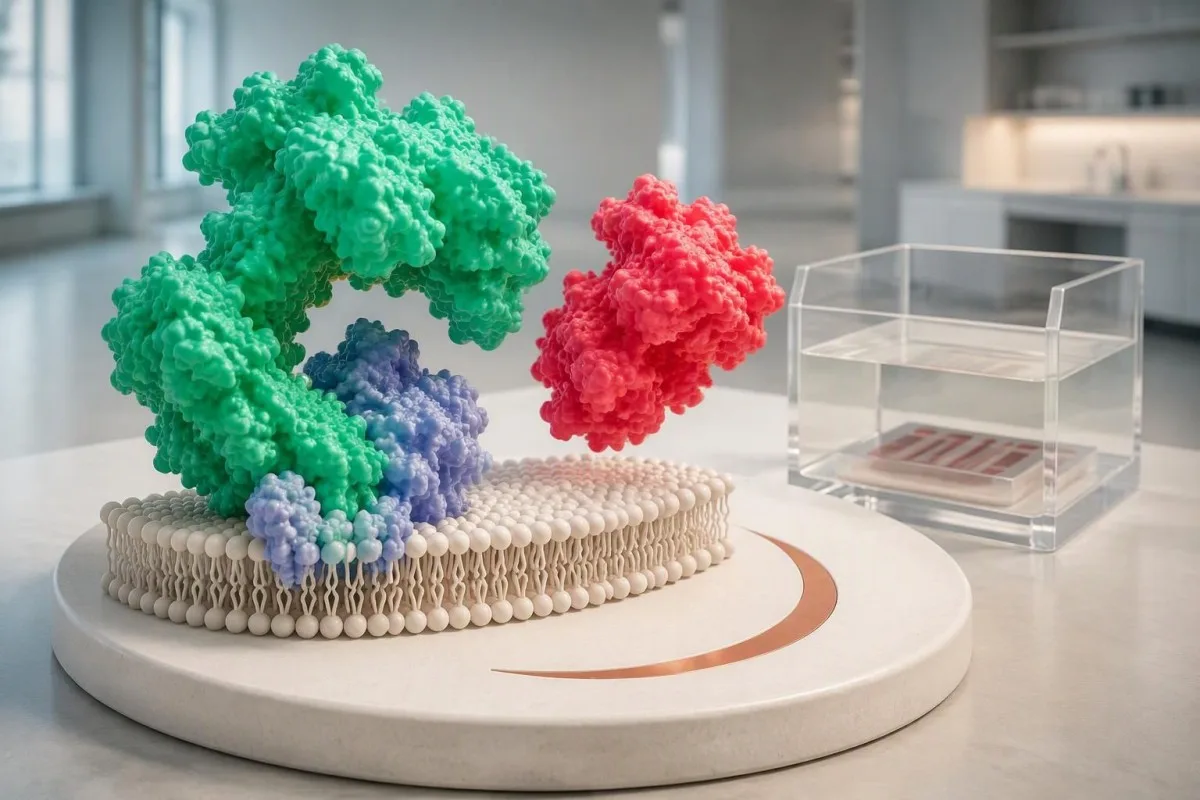
Протеин S помага на активирания протеин C да ограничава коагулацията, като подпомага инактивирането на фактори Va и VIIIa. Концентрацията му не е същото като общия протеин, измерен в биохимичен профил; общ протеин от 7.0 g/dL не дава полезна оценка за функцията на протеин S, тъй като тестовете измерват напълно различни биологични величини.
Единичният флаг е отправна точка, а не присъда. При хипотетичен човек, чиято активност е 48% при долна лабораторна граница 60%, следващият въпрос е дали пробата е взета по време на бременност, медикаментозно лечение или заболяване — а не дали числото автоматично доказва генетично заболяване; нашият извънреферентен резултат обяснява тази разлика.
Kantesti е анализатор на кръвни изследвания с ИИ, който помага за подреждането на резултатите за протеин S спрямо посочените референтни граници и релевантния контекст, вместо 1 маркиран резултат да се приема за диагноза. Нашата фирмен профил обяснява организацията, стояща зад тази образователна услуга; потвърждаването на наследствен дефицит все пак изисква лабораторна оценка по преценка на клиницист и понякога специализирано генетично изследване.
Как да разчитате процента на активност и референтния интервал?
Активността на протеин S обикновено се отчита като процент от референтната активност на теста, а не като вероятност за тромбоза в проценти. Референтните интервали зависят от лабораторията, метода, възрастта и хормоналния контекст; интервал за възрастни като 60–150% е илюстративен пример, а не универсален диагностичен праг.

Активност на протеин S от 45% означава намалена функционална активност в този тест, а не че 45% от кръвообращението ви е запушено. Процентите се различават и от концентрационни единици като mg/L, така че резултат за активност не може да се сравнява пряко по числена стойност с концентрация на антиген; разграничението наподобява други количествени лабораторни измервания които измерват различни характеристики.
Граничните стойности изискват особено внимателна преценка. В хипотетична лаборатория, използваща 60% като долна граница, резултати от 58% и 61% попадат от двете страни на флага, въпреки че разликата между тях е само 3 процентни пункта; аналитичната вариация, биологичните колебания и различните реактиви за теста може да обяснят тази разлика, без да има съществена промяна в риска от съсирване.
Няма единен процент на протеин S, който самостоятелно да определя дали е необходима антикоагулация. Резултат от 25% заслужава повече внимание от минимално понижената стойност, но все пак може да се наблюдава при придобито намаление; решенията зависят от потвърждението, анамнезата за съсиреци, провокиращите фактори и риска от кървене, а не от използването на 1 числен праг като сигнал за започване на медикамент.
| Ниво | Диапазон | Какво означава |
|---|---|---|
| В рамките на илюстративен интервал за възрастни | активност 60–150% | Само за пример. Използвайте интервала на лабораторията, която отчита резултата, съобразен с възрастта и контекста; понякога антикоагулант, който пречи на теста, може да прикрие дефицит. |
| Под този илюстративен интервал | активност <60% | Необходима е преценка на контекста и, когато е уместно, потвърждение. Това не установява наследствен дефицит. |
| Резултат за свободен антиген | Laboratory-specific % or concentration units | Measures the unbound protein pool. Do not apply an activity reference interval to an antigen result. |
| Pregnancy or childhood result | Population- and method-specific | A nonpregnant adult interval may be inappropriate. Diagnostic interpretation may need specialist advice. |
Какво показват заедно активността и свободният антиген на протеин S?
Activity testing measures functional performance, while free protein S antigen measures the available unbound protein pool. Approximately 40% of circulating protein S is free and the remainder is bound mainly to C4b-binding protein; comparing these measurements helps identify a quantitative reduction, a possible functional defect or a misleading activity result.
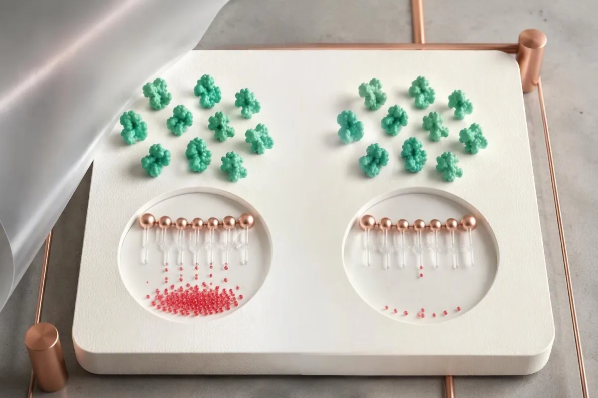
Type I protein S deficiency classically produces low total antigen, low free antigen and low activity; type III produces normal total antigen with low free antigen and activity. These 2 patterns can occur within the same family and are not necessarily separate biological diseases; total antigen alone can therefore miss a clinically relevant reduction in the free pool.
Type II protein S deficiency classically shows reduced activity despite normal free and total antigen. This uncommon pattern is not diagnosed from 1 discordant sample, because factor V Leiden and other assay problems can imitate functional deficiency; laboratories may repeat the activity using another method before considering a genuinely dysfunctional protein.
Paired results clarify the next step but do not identify the cause by themselves. For example, activity of 42% with free antigen of 38% could reflect either inherited quantitative deficiency or an acquired reduction, while activity of 42% with normal free antigen raises a different set of questions; our coagulation marker overview explains why complementary tests are interpreted together.
Защо бременността понижава резултатите за протеин S?
Pregnancy physiologically lowers free protein S and often protein S activity, so a low pregnancy result cannot reliably establish inherited deficiency. Changes can begin early and become more pronounced later; the British Society for Haematology identifies pregnancy and the puerperium as confounders of protein S testing (Arachchillage et al., 2022).

A nonpregnant reference interval can create a misleading diagnosis during pregnancy. In a hypothetical second-trimester sample, activity of 43% and free antigen of 36% may both be flagged, but their agreement does not remove the pregnancy effect; both tests can reflect the same physiological reduction, just as интерпретация на феритин по време на бременност requires pregnancy-specific context.
Reassessment for inherited protein S deficiency is commonly postponed until at least 6 weeks after delivery, and some services prefer 12 weeks or longer. Six weeks is not a guarantee of an unaffected baseline: recovery, continuing illness, a recent clot and newly started estrogen contraception can all influence whether a postpartum sample is suitable.
Protein S testing does not decide pregnancy anticoagulation by itself. When reviewing 2 pregnancy measurements, Kantesti AI can help distinguish activity from antigen on the report, but pregnancy prophylaxis requires obstetric and thrombosis assessment based on prior venous thromboembolism, family history and other risks; a low pregnancy value alone also does not establish the cause of a miscarriage.
Могат ли естрогенните контрацептиви или хормонозаместителната терапия да причинят нисък резултат?
Estrogen-containing contraception and oral estrogen therapy can lower free protein S and protein S activity without an inherited defect. The change is biological rather than merely a laboratory artifact; a result collected during estrogen use may therefore describe the treatment-associated state, not the person's untreated baseline.

The exact preparation matters more than the word contraception. A combined pill containing 30 micrograms of ethinylestradiol is not equivalent to a progestogen-only pill, and a contraceptive patch is not equivalent to a transdermal menopausal estrogen patch; hormone results during contraception require the active ingredients and route, not just the brand name.
Transdermal menopausal estrogen generally has less effect on hepatic coagulation pathways than oral estrogen, but this does not make every transdermal hormonal product risk-free. Evidence about clot risk and evidence about a specific protein S assay are different questions; an activity of 52% during treatment cannot be corrected mathematically by subtracting a standard estrogen effect.
There is no universally reliable 1-week estrogen washout for protein S diagnosis. If confirmation would change care, the prescribing clinician can discuss timing, alternative contraception and whether treatment should change at all; our pre-HRT testing considerations support that conversation, but never stop prescribed hormones independently just to remove a laboratory flag.
Защо варфаринът понижава активността на протеин S?
Warfarin lowers protein S because protein S is a vitamin K-dependent anticoagulant protein. This is an expected treatment effect rather than proof of inherited deficiency; free antigen can also be reduced, so replacing an activity test with antigen testing does not reliably reveal the untreated baseline during warfarin therapy.

A therapeutic INR does not make protein S testing diagnostically reliable during warfarin treatment. For a hypothetical patient taking 5 mg daily with an INR of 2.4 and activity of 31%, the low activity may be medication related; the meaning of an INR concerns anticoagulant monitoring, not confirmation of inherited protein S deficiency.
When an off-warfarin measurement is genuinely needed, laboratories commonly require at least 2 weeks after discontinuation, sometimes longer according to local policy and the clinical situation. That interval is not an instruction to stop treatment: a hematologist or anticoagulation clinician must decide whether interruption is safe, whether an alternative is needed and which assay remains interpretable.
Vitamin K deficiency can also reduce protein S, but supplements should not be used to self-correct an unexplained result. Taking even 1 new vitamin K-containing supplement can alter warfarin management; keep dietary intake reasonably consistent and discuss changes with the anticoagulation team, using our ръководство за храни, богати на витамин К for food context rather than as a deficiency treatment plan.
Кои заболявания могат временно да понижат протеин S?
Severe infection, disseminated intravascular coagulation, liver disease, nephrotic syndrome and recent thrombosis can reduce protein S measurements. Some reductions resolve with recovery, while others persist because the underlying condition persists; a low result during hospitalization therefore cannot be classified as inherited from 1 sample.
Disseminated intravascular coagulation can reduce natural anticoagulants while other coagulation markers change simultaneously. A hypothetical combination of protein S activity at 35%, platelets at 65 × 10^9/L and a prolonged prothrombin time calls for assessment of the acute illness, not immediate genetic labeling; our DIC laboratory pattern guide explains why multiple abnormal findings matter together.
The liver contributes to protein S production, but an isolated ALT of 75 IU/L does not prove that liver dysfunction caused a low protein S result. Clinicians look for impaired synthesis and the broader clinical picture, including albumin, bilirubin and coagulation results; modest enzyme elevation and advanced synthetic failure are different processes, with very different implications for interpretation.
Nephrotic-range protein loss can produce acquired protein S abnormalities, especially in the free fraction. An albumin of 2.2 g/dL with substantial urinary protein loss changes the interpretation of activity at 44%; persistent foamy urine assessment may help identify a reason to investigate renal protein loss, although bubbles alone neither diagnose nephrotic syndrome nor explain protein S.
Как интерференцията в лабораторния анализ може да доведе до подвеждащ резултат за протеин S?
Clot-based protein S activity assays can report falsely low or falsely high results because medications and other coagulation abnormalities alter the endpoint. Direct oral anticoagulants often falsely increase activity in susceptible assays, whereas factor V Leiden can falsely decrease it; the direction and magnitude depend on the assay design.
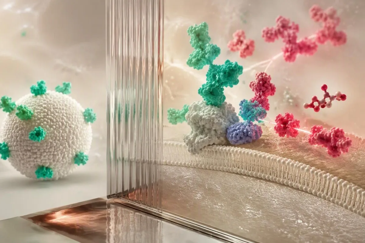
Factor V Leiden can interfere with some protein S activity assays by altering the activated protein C-dependent reaction. A hypothetical activity of 46% with normal free antigen could therefore mimic type II deficiency; the factor V Leiden result and the laboratory's assay characteristics should be reviewed before concluding that 2 inherited disorders coexist.
Direct oral anticoagulants can conceal deficiency rather than explain a low number. For example, an apparently normal activity of 110% during rivaroxaban treatment may be falsely increased in a susceptible clot-based method; lupus anticoagulant, high factor VIII and heparin can also complicate interpretation, so lupus anticoagulant testing and protein S testing need coordinated laboratory advice.
Kantesti is an AI lab test interpretation service that can place 2 discordant measurements—activity and free antigen—side by side for discussion, but cannot determine reagent interference from the numbers alone. Free antigen immunoassays generally avoid clot-endpoint interference, yet they still cannot remove warfarin's biological effect; the BSH guideline specifically lists medication- and assay-related confounders (Arachchillage et al., 2022).
Могат ли вземането или обработката на пробата да доведат до нисък резултат?
Collection and handling problems can distort protein S testing, particularly functional assays that depend on correctly prepared citrated plasma. The laboratory should check tube filling, citrate balance, processing and sample quality before a discordant result becomes a diagnosis; repeating a technically unsuitable sample answers a different question from confirming inherited deficiency.
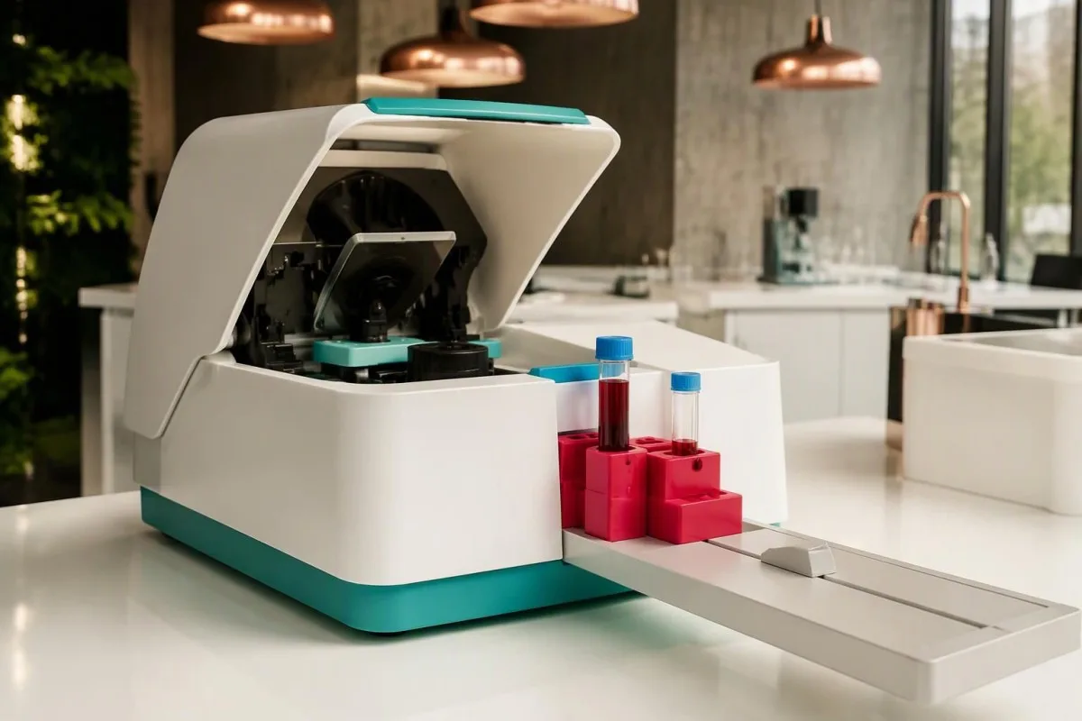
Standard coagulation collection uses approximately 9 parts sample to 1 part citrate anticoagulant. Underfilling changes that ratio and can distort clot-based measurements; tube-cap colors vary internationally, so the fill line and laboratory instructions matter more than recognizing a particular color, and a rejected specimen should not be interpreted as if its numerical result were trustworthy.
A hematocrit above 55% generally requires laboratory-directed adjustment of citrate volume for coagulation testing. A hypothetical patient with a hematocrit of 58% needs that issue addressed before a low functional result is taken at face value; otherwise, excess citrate relative to plasma can affect the assay even when the collection tube appears adequately filled.
Hemolysis, lipemia and delayed processing can affect methods differently, so there is no universal correction factor for a protein S result of 49%. Ask whether the laboratory accepted the specimen and whether it recommends recollection; our hemolysis interference guide explains why a sample-quality warning may affect some reported analytes but not others.
Кога е уместно повторно изследване под лекарско наблюдение?
Repeat protein S testing is appropriate when the first result was confounded, borderline or unexpected and confirmation could change care. After acute thrombosis, the British Society for Haematology recommends testing natural anticoagulant deficiencies only after 3 months of anticoagulation when indicated, with medication-related interference addressed separately (Arachchillage et al., 2022).

Three months of anticoagulation is a testing-timing recommendation, not permission to stop anticoagulants at 3 months. Treatment duration depends on the clot and the patient's risks, while assay suitability depends on the medication; clinicians may defer testing, choose an antigen assay or seek specialist laboratory methods without interrupting necessary protection.
Pregnancy, warfarin and acute illness each require a different reassessment plan. Common examples include at least 6 weeks postpartum, at least 2 weeks after clinician-approved warfarin discontinuation, or testing after recovery from significant illness; none is a universal washout schedule, and an unconfounded repeat may need to be postponed further if several influences overlap.
Kantesti's trend analysis can help display 2 measurements, but comparison is strongest when the method, laboratory and clinical conditions are comparable. Record pregnancy stage, illness dates, medicines and the exact timing of the last anticoagulant dose for the clinician; our ръководство за лабораторни срокове explains why elapsed time alone does not establish that a change is biological.
Какво прави наследствения дефицит на протеин S по-вероятен?
Inherited protein S deficiency becomes more likely when low results persist under suitable testing conditions and fit a personal or family history of venous thrombosis. A pathogenic PROS1 variant can support the diagnosis, but genetic testing is selective; 1 low activity result, especially during treatment or pregnancy, is insufficient.

Most inherited protein S deficiency associated with a single pathogenic PROS1 variant follows an autosomal dominant inheritance pattern. Each child has a 50% chance of inheriting that variant, not a 50% certainty of developing a clot; penetrance varies, and relatives with the same variant can have different protein measurements and clinical histories.
A first venous clot at age 28 with several affected close relatives raises more suspicion than an incidental borderline result at age 72 during pneumonia. Age is not a diagnostic cutoff, however: absence of family history does not exclude inheritance, and the BSH guideline recommends genetic testing for phenotypically identified deficiencies when the result would influence management (Arachchillage et al., 2022).
Family screening should be selective rather than a broad panel for every relative. Testing an unaffected first-degree relative may be useful if the result would change estrogen decisions or prophylaxis during high-risk periods; our ръководство за семейно тестване въз основа на риск explains why testing plans should follow a confirmed family diagnosis instead of expanding from 1 uncertain laboratory flag.
Означава ли ниският резултат, че в момента имате тромб?
Low protein S activity does not diagnose an existing deep vein thrombosis or pulmonary embolism. Confirmed inherited deficiency increases susceptibility to venous thrombosis, but current symptoms require the usual urgent assessment; the BSH guideline describes an approximately 5–7-fold increased first-event risk for heterozygous protein C or protein S deficiency (Arachchillage et al., 2022).
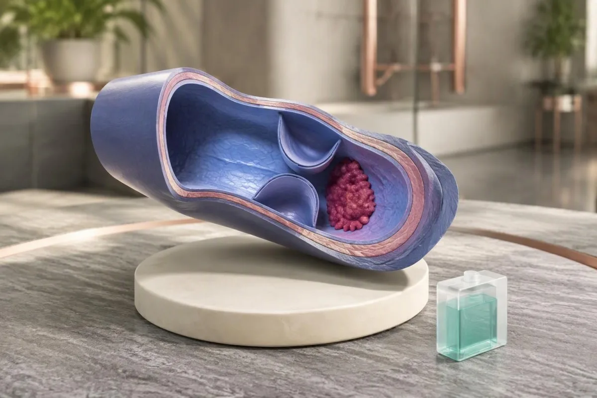
A 5–7-fold relative risk is not a 50–70% absolute probability of thrombosis. Absolute risk varies with age, family ascertainment, prior events and exposures such as surgery or estrogen; a person without symptoms should not interpret a low protein S value as evidence that a clot is silently present or start an anticoagulant independently.
Sudden unexplained breathlessness, chest pain, coughing blood or collapse requires emergency evaluation; new one-sided leg swelling or pain needs urgent assessment for deep vein thrombosis. A normal activity of 95% does not rule out either condition, and normal PT or aPTT also cannot exclude thrombophilia; our ограничения на коагулационния панел explain that common misconception.
A confirmed protein S deficiency does not automatically require lifelong anticoagulation in someone who has never had thrombosis. The ASH 2023 guideline discourages testing unprovoked VTE solely to determine duration but conditionally supports testing in selected hormonal or nonsurgical provoked scenarios; those latter recommendations rely on very low-certainty evidence, so 1 label cannot replace individualized decisions (Middeldorp et al., 2023).
Какво трябва да донесете на следващия клиничен преглед?
Bring the complete report, medication list, collection date and relevant clot or pregnancy history—not just the low protein S activity number. The most useful appointment establishes whether the result is interpretable, whether free antigen was measured and whether confirmation would change treatment; 3 focused questions often prevent unnecessary retesting.

Ask: Was the activity method vulnerable to my medication; was free antigen measured; and what would a confirmed deficiency change? For a hypothetical person with activity of 47% and free antigen of 41% while taking warfarin, both results may remain treatment-confounded; ordering a larger thrombophilia panel immediately does not solve the underlying baseline problem.
A different hypothetical pattern—activity of 47%, normal free antigen and known factor V Leiden—first raises an assay question rather than establishing type II deficiency. A third pattern, with activity of 39% and free antigen of 35% persisting on suitably timed samples without an acquired explanation, makes specialist evaluation more reasonable; these examples are illustrations, not diagnostic algorithms.
Kantesti can help organize these 3 patterns for discussion, but the clinician and testing laboratory must decide whether another assay or repeat specimen is needed. Our клинична рамка за валидиране describes the role of evaluation and oversight; bring actual reference intervals and units, and do not rely on a cropped screenshot that omits the method or sample comments.
Научни данни и ограничения на интерпретацията с ИИ
Clinical guidelines support context-sensitive protein S testing, but neither a laboratory flag nor an AI explanation confirms inherited deficiency. Kantesti is an AI blood test interpretation platform that helps explain activity and antigen results together; the diagnosis still depends on appropriate testing conditions, reproducibility and clinical assessment rather than 1 automated interpretation.
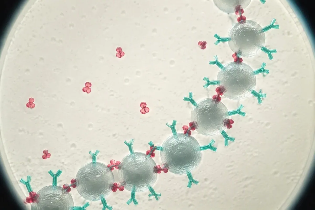
The 2022 BSH and 2023 ASH recommendations address different testing decisions, and neither recommends indiscriminate thrombophilia screening. BSH emphasizes validity and whether findings would change management, while ASH conditionally supports testing in some selected VTE scenarios using very low-certainty evidence; their differences reflect clinical questions and modeling assumptions, not proof that every low result needs genetic testing.
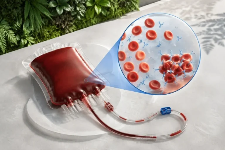
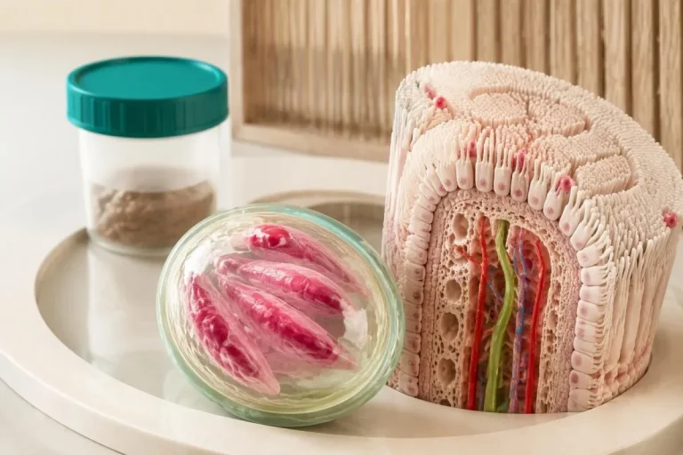
Protein S is not diagnosed by routine tissue microscopy, and the molecular illustrations here are educational models rather than images of an individual's deficiency. Our обяснение на AI технологията describes the interpretation workflow; the hormonal health research overview supplies broader context but does not provide a validated protein S cutoff or a medication washout protocol.
The 2 Kantesti research publications listed below are Figshare-hosted materials covering hormonal health and multilingual clinical decision-support engineering; neither establishes diagnostic accuracy for inherited protein S deficiency. Repository hosting and DOI assignment do not establish peer review or protein S-specific clinical validation; our стандарти за медицинско съдържание explain why educational claims should remain separate from disease-specific evidence.
Често задавани въпроси
Може ли ниската активност на протеин S да бъде временна?
Ниската активност на протеин S може да бъде временна по време на бременност, при излагане на естрогени, лечение с варфарин, тежко заболяване или възстановяване след тромбоза. Резултат като 45% сам по себе си не може да разграничи временното понижение от наследствения дефицит. Активността и свободният антиген на протеин S трябва да се интерпретират заедно, като изследването се повтори, когато клиницистът прецени, че условията са подходящи. Не спирайте самостоятелно предписани лекарства, за да получите резултат без лечение.
Каква е разликата между активността на протеин S и свободния антиген на протеин S?
Тестът за активност на протеин S измерва функционалните му характеристики, докато тестът за свободен антиген на протеин S измерва несвързания протеинов пул. Приблизително 40% от циркулиращия протеин S е свободен, а останалата част е свързана главно с C4b-свързващия протеин. Ниска активност при нисък свободен антиген може да се наблюдава както при наследствено, така и при придобито количествено намаление. Ниска активност при нормален свободен антиген може да отразява функционален дефект или интерференция с теста, затова този модел трябва да бъде потвърден.
Нормална ли е ниската активност на протеин S по време на бременност?
Бременността физиологично понижава свободния протеин S и често активността на протеин S, затова нисък резултат по време на бременност не позволява надеждно да се диагностицира наследствен дефицит. Стойност от 40% трябва да се оценява спрямо етапа на бременността, метода на изследване и клиничната причина за изследването, а не само спрямо референтния интервал за небременни възрастни. Диагностичната преоценка обикновено се отлага най-малко до 6 седмици след раждането, понякога до 12 или повече седмици. Решенията за антикоагулация по време на бременност зависят от анамнезата за тромбоза и други рискови фактори, а не само от стойността на протеин S.
Колко време след приема на варфарин може да се изследва протеин S?
Много лаборатории изискват изчакване от поне 2 седмици след прекратяване на варфарина, преди да оценят изходното ниво на протеин S без лечение, въпреки че местните изисквания и клиничните обстоятелства се различават. Варфаринът може да понижи както активността, така и антигена, тъй като протеин S зависи от витамин K. Този период на изчакване не е разрешение за прекъсване на антикоагулацията. Лекуващият лекар трябва да реши дали спирането е безопасно, дали са необходими изследвания и дали е за предпочитане друг подход.
Могат ли апиксабан или ривароксабан да повлияят на тест за активност на протеин S?
0 Апиксабан и ривароксабан могат да повлияят на чувствителните функционални тестове за протеин S, базирани на съсирването, като често водят до фалшиво повишена активност, а не до истински дефицит. Привидно нормален резултат като 100% следователно може да не изключва ниска действителна активност на протеин S. Имуноанализите за свободен антиген обикновено избягват ефекта върху крайната точка на съсирването, но лабораторията трябва да посочи кой метод е подходящ. Не пропускайте и не спирайте дози от антикоагуланта с цел подготовка за изследването, освен ако предписалият го клиницист изрично не е указал безопасен план.
Кога трябва да се повтори изследването на ниската активност на протеин S след кръвен съсирек?
Когато е показано изследване след остра тромбоза, Британското дружество по хематология препоръчва да се изчакат 3 месеца антикоагулантно лечение, преди да се изследват дефицити на естествени антикоагуланти. Влиянието на лекарствата все още трябва да се отчита отделно, така че изтичането на 3 месеца не прави всеки тест за активност надежден. Повторното изследване е най-полезно, когато резултатът може да промени лечението и първоначалната проба е била повлияна или неочаквана. Лечението не бива да се спира самоволно за повторното изследване.
Означава ли дефицитът на протеин S, че трябва да приемам антикоагуланти до края на живота си?
Confirmed protein S deficiency does not automatically mean lifelong anticoagulation, especially in someone who has never had venous thrombosis. The approximately 5–7-fold relative first-event risk described for heterozygous protein C or protein S deficiency is not an individual's absolute probability of a clot. Decisions depend on previous events, provoking factors, bleeding risk and whether prophylaxis is needed during high-risk periods. A single low activity result should not be used to start, stop or extend anticoagulant treatment independently.
Получете обяснение на резултатите от кръвните си изследвания
Качете лабораторния си доклад и AI на Kantesti ще обясни всеки биомаркер на разбираем език за секунди. Използва се от над 2 милиона души по света.
Референции
- Klein, T., Mitchell, S., & Weber, H. (2026). Women's Health Guide: Ovulation, Menopause & Hormonal Symptoms. Медицински изследвания с Kantesti AI. ДОИ
- Klein, T., Mitchell, S., & Weber, H. (2026). Multilingual AI Assisted Clinical Decision Support for Early Hantavirus Triage: Design, Engineering Validation, and Real-World Deployment Across 50,000 Interpreted Blood Test Reports. Медицински изследвания с Kantesti AI. ДОИ
- Arachchillage DJ et al. (2022). Thrombophilia testing: A British Society for Haematology guideline. British Journal of Haematology. ДОИ
- Middeldorp S et al. (2023). American Society of Hematology 2023 guidelines for management of venous thromboembolism: thrombophilia testing. Blood Advances. ДОИ
- 2 милиона+Анализирани тестове
- 127+Държави
- 100+Езици
За медицинския екип


Сигнали за доверие E-E-A-T
Опит
Медицински преглед, воден от лекар, на работните процеси за интерпретация на лабораторни резултати.
Експертиза
Фокус в лабораторната медицина върху това как се държат биомаркерите в клиничен контекст.
Авторитетност
Медицинска рецензия от д-р Thomas Klein, с принос от д-р Sarah Mitchell и проф. д-р Hans Weber.
Надеждност
Интерпретация, основана на доказателства, с ясни последващи стъпки за намаляване на тревогата.
Медицински отказ от отговорност
Тази статия е само с образователна цел и не представлява медицински съвет. Винаги се консултирайте с квалифициран медицински специалист за решения относно диагностика и лечение.
Публикувано · Свържете се с редакционния екип
Издава се от Kantesti Ltd, регистрирана в Англия и Уелс, дружество №. 17090423, Лондон, Обединено кралство.
