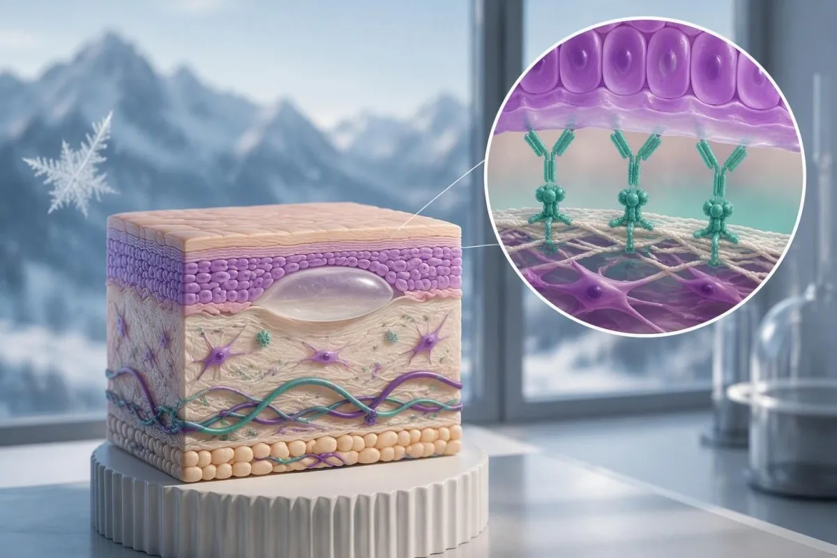
ผล BP180 เป็นบวกสนับสนุนว่ามีความผิดปกติของผิวหนังจากภูมิคุ้มกันทำลายตนเอง แต่ตัวเลขนี้ไม่สามารถใช้วินิจฉัยได้เพียงลำพัง อาการ การตรวจที่ใช้ และตัวอย่างผิวหนังที่เก็บอย่างถูกต้องเป็นตัวกำหนดขั้นตอนต่อไป.

ประเด็นสำคัญ
- แอนติบอดี BP180 เป็นบวก หมายความว่าการตรวจพบแอนติบอดีต่อเป้าหมาย BP180 ของการตรวจ แต่ไม่ได้ยืนยันโรคเพมฟิกอยด์ชนิดตุ่มพองโดยลำพัง.
- ค่าตัดของห้องปฏิบัติการ แตกต่างกัน: การตรวจ BP180 บางชนิดใช้ค่าประมาณ 9 U/mL ขณะที่บางชนิดใช้ค่าประมาณ 20 U/mL โปรดดูช่วงค่าอ้างอิงของห้องปฏิบัติการที่ตรวจ.
- ผลบวกระดับต่ำ หากไม่มีอาการคัน ผื่น หรือตุ่มพอง อาจเป็นการตรวจพบแอนติบอดีโดยบังเอิญ หรือเป็นผลตรวจที่ให้ผลบวกลวง ไม่ใช่โรคผิวหนังที่ได้รับการยืนยัน.
- เพมฟิกอยด์ชนิดไม่มีตุ่มพอง อาจทำให้คันต่อเนื่องโดยไม่มีตุ่มพอง โดยในผู้ป่วยเพมฟิกอยด์ 343 รายจากการศึกษาวินิจฉัยเฉพาะทาง มี 74 รายที่เป็นชนิดไม่มีตุ่มพอง.
- แอนติบอดี BP230 เป็นหลักฐานเสริม แต่ผลแอนติบอดี BP230 ที่เป็นลบไม่ได้ตัดโรคเพมฟิกอยด์ชนิดตุ่มพองออกไป.
- การตรวจอิมมูโนฟลูออเรสเซนซ์โดยตรง ตรวจหาการสะสมของแอนติบอดีในผิวหนัง; การพบ IgG และ/หรือ C3 เรียงตัวตามแนวเยื่อฐานสนับสนุนว่าเป็นโรคในกลุ่มเพมฟิกอยด์.
- ตัวอย่างผิวหนังสองชิ้น มักเก็บจากบริเวณที่ได้รับผลกระทบหนึ่งชิ้นเพื่อตรวจด้วยกล้องจุลทรรศน์ตามปกติ และอีกชิ้นจากบริเวณใกล้เคียงที่เลือกอย่างเหมาะสมเพื่อตรวจด้วย direct immunofluorescence.
- อาการที่ควรรีบด่วน ได้แก่ ปวดตา การมองเห็นเปลี่ยนแปลง กลืนลำบาก มีไข้ร่วมกับความผิดปกติของผิวหนังเป็นบริเวณกว้าง หรือมีตุ่มพองลุกลามอย่างรวดเร็ว; อย่ารอผลตรวจแอนติบอดีอีกครั้ง.
ผล BP180 เป็นบวกยืนยันโรคเพมฟิกอยด์ชนิดตุ่มพองหรือไม่?
ไม่ใช่—ผลตรวจแอนติบอดี BP180 เป็นบวกเพียงอย่างเดียวไม่ยืนยันว่าเป็นโรคตุ่มพองเพมฟิกอยด์. ผลดังกล่าวสนับสนุนการวินิจฉัยเมื่ออาการและความผิดปกติของผิวหนังสอดคล้องกัน แต่ผลบวกระดับต่ำโดยไม่มีตุ่มพองอาจเป็นผลที่พบโดยบังเอิญ; แพทย์ผิวหนังมักต้องตรวจชิ้นเนื้อผิวหนังด้วย direct immunofluorescence และบางครั้งต้องตรวจแอนติบอดีเพิ่มเติมเพื่อระบุสาเหตุ.
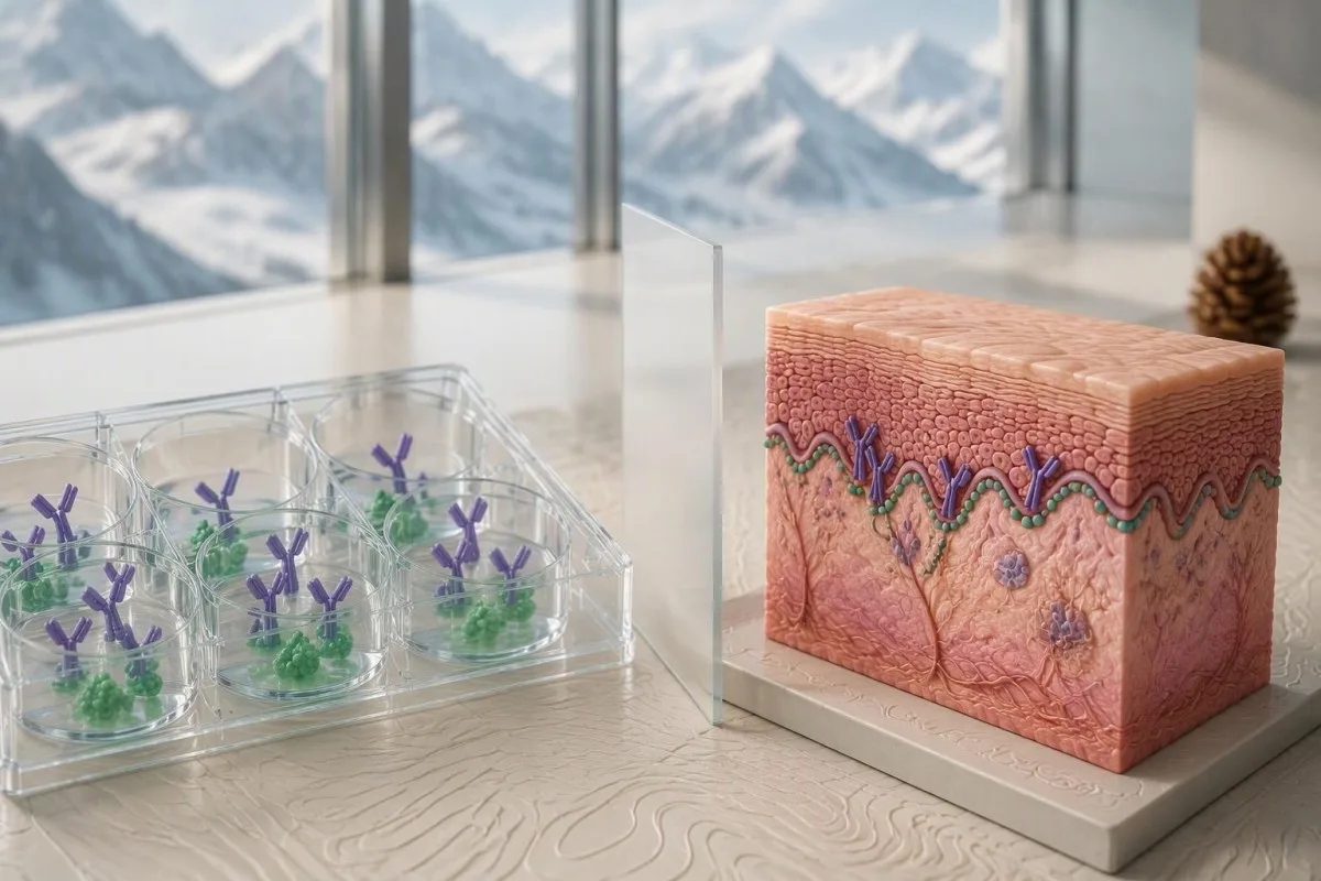
การวินิจฉัยโรคตุ่มพองเพมฟิกอยด์ต้องอาศัยความสอดคล้องระหว่างข้อมูลทางคลินิกและอิมมูโนพยาธิวิทยา, ไม่ใช่เพียงค่าผลตรวจทางห้องปฏิบัติการที่ถูกระบุว่าผิดปกติค่าเดียว แนวทาง EADV ปี 2022 ระบุว่าการวินิจฉัยพิจารณาจากภาพทางคลินิกและการตรวจเนื้อเยื่อกับแอนติบอดีในกระแสเลือดที่เหมาะสม (Borradori et al., 2022); ประเด็นในทางปฏิบัติคือแอนติบอดีที่ตรวจพบอธิบายภาวะผิวหนังของบุคคลนั้นได้หรือไม่.
ผลตรวจแอนติบอดีเป็นบวกและการมีโรคภูมิต้านตนเองที่กำลังดำเนินอยู่เป็นข้อค้นพบที่แตกต่างกัน ตัวอย่างเช่น ผลตรวจสมมติที่ 12 U/mL อาจเป็นบวกในห้องปฏิบัติการแห่งหนึ่ง แต่บุคคลที่ไม่มีอาการและได้ผลเช่นนั้นมีความน่าจะเป็นในการวินิจฉัยแตกต่างจากผู้ที่มีตุ่มพองตึงคันเป็นบริเวณกว้างและมีค่าความเข้มข้นเท่ากัน.
Kantesti เป็นเครื่องวิเคราะห์ผลตรวจเลือดด้วย AI ที่ช่วยอธิบายผล BP180 โดยพิจารณาค่าตัดของห้องปฏิบัติการและข้อมูลที่คุณให้ไว้ประกอบ. คำอธิบายของเราไม่สามารถตรวจผิวหนังของคุณหรือทำ direct immunofluorescence ได้; ส่วน ข้อมูลพื้นฐานของบริษัท ระบุองค์กรที่อยู่เบื้องหลังการสนับสนุนด้านความรู้นี้.
ให้มองผลตรวจนี้เป็นเหตุให้ตั้งคำถามที่เฉพาะเจาะจงยิ่งขึ้น แทนที่จะมองว่าเป็นป้ายระบุโรคถาวร คู่มือของเราเกี่ยวกับ ผลการตรวจแอนติบอดีเป็นบวก อธิบายว่าเหตุใดการตรวจพบแอนติบอดี กิจกรรมของโรค และการยืนยันการวินิจฉัยจึงเป็นแนวคิดสามประการที่แยกจากกัน.
การตรวจแอนติบอดี BP180 วัดอะไร?
การตรวจแอนติบอดี BP180 โดยทั่วไปวัด IgG ในซีรัมที่มุ่งต่อส่วนหนึ่งของ BP180 ซึ่งเรียกอีกชื่อว่าคอลลาเจนชนิด XVII. BP180 เป็นโปรตีนที่มีขนาดประมาณ 180-kDa ซึ่งมีส่วนในการยึดหนังกำพร้าเข้ากับเนื้อเยื่อด้านล่าง; การตรวจแบบมาตรฐานหลายชนิดตรวจบริเวณ NC16A ของโปรตีน แทนที่จะตรวจโปรตีนทั้งโมเลกุล.
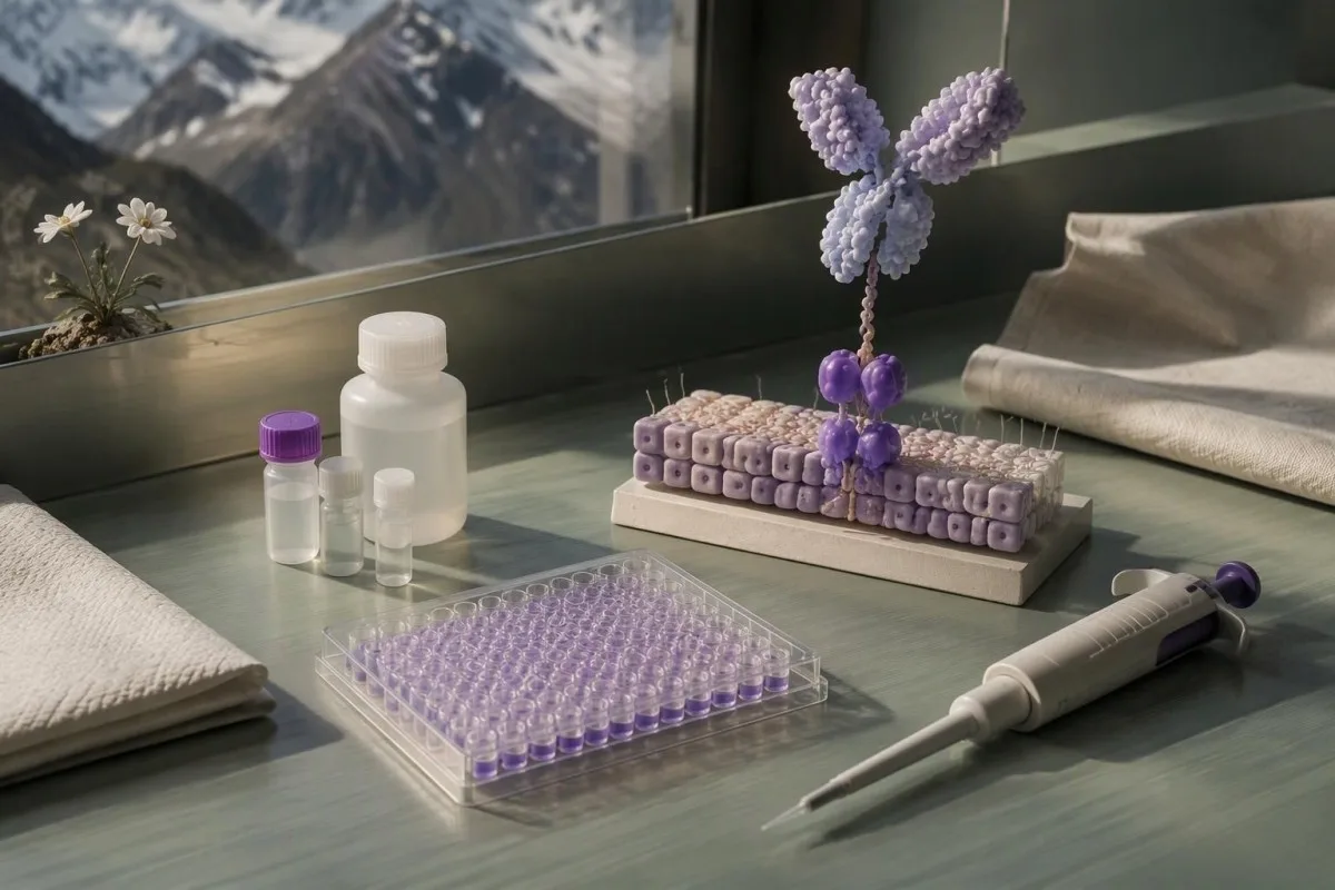
BP180 เป็นโปรตีนเชิงโครงสร้างของผิวหนัง ไม่ใช่สารที่มีการวัดความเข้มข้นในเลือด. รายงานผลระบุแอนติบอดีต่อโปรตีนนั้น ซึ่งมักตรวจด้วยวิธีอิมมูโนแอสเซย์แบบเอนไซม์ลิงก์ หรือ ELISA; ค่า 30 U/mL จึงไม่ได้หมายความว่ามีโปรตีนผิวหนัง 30 หน่วยไหลเวียนอยู่ในกระแสเลือด.
การ โดเมน NC16A อยู่ใกล้กับเยื่อหุ้มเซลล์บริเวณส่วนด้านนอกของ BP180 และเป็นเป้าหมายของแอนติบอดีที่พบบ่อยในโรคตุ่มน้ำพองเพมฟิกอยด์ชนิดคลาสสิก การตรวจบริเวณนี้ทำให้สามารถใช้การตรวจที่สะดวกได้ แต่ก็ทำให้เกิดจุดบอดขึ้น กล่าวคือ แอนติบอดีที่มุ่งเป้าไปยังบริเวณอื่นเป็นหลักอาจตรวจไม่พบ.
ค่าความเข้มข้นของแอนติบอดีที่เป็นตัวเลขใช้แทนค่าไตเตอร์จากการตรวจอิมมูโนฟลูออเรสเซนซ์ไม่ได้ ผลที่รายงานเป็น 1:40 แสดงปฏิกิริยาที่การเจือจางระดับหนึ่ง ขณะที่ 40 U/mL แสดงค่าความเข้มข้นที่ปรับเทียบตามการตรวจวิเคราะห์ คำอธิบายของเราเกี่ยวกับ การตรวจเชิงปริมาณเทียบกับเชิงคุณภาพ ช่วยให้แยกแยะรูปแบบการรายงานเหล่านี้ได้.
ตรวจสอบว่ารายงานระบุว่าเป็น BP180 NC16A IgG, การตรวจ BP180 แบบอื่น หรือแผงตรวจเพมฟิกอยด์แบบครอบคลุม การตรวจเหล่านี้ไม่ได้เทียบเท่ากันโดยอัตโนมัติ และผล ELISA IgG ที่เป็นลบไม่ได้ตอบทุกข้อสงสัยเกี่ยวกับแอนติบอดีชนิดอื่นหรือเป้าหมายอื่นในเยื่อฐาน.
ผลตรวจแอนติบอดี BP180 ระดับบวกต่ำหมายถึงเท่าใด?
ผล BP180 ที่เป็นบวกระดับต่ำคือค่าที่สูงกว่าจุดตัดของการตรวจเฉพาะที่ใช้เพียงเล็กน้อย. ไม่มีช่วงผลบวกระดับต่ำที่เป็นสากล ห้องปฏิบัติการบางแห่งใช้เกณฑ์ใกล้เคียง 9 U/mL, ขณะที่บางแห่งใช้เกณฑ์ใกล้เคียง 20 U/mL, โดยมีการปรับเทียบและหมวดหมู่การรายงานที่แตกต่างกัน.
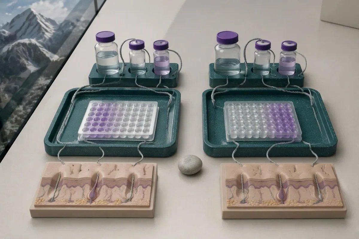
หน่วยของการตรวจ BP180 ไม่มีการกำหนดมาตรฐานที่ใช้ร่วมกันในห้องปฏิบัติการทุกแห่ง. ผลของ 15 U/mL อาจถือว่าเป็นผลบวกตามเกณฑ์ 9 U/mL และเป็นผลลบตามเกณฑ์ 20 U/mL แต่ไม่ได้ยืนยันว่าการตรวจชนิดที่สองตรวจไม่พบโรค เพราะวิธีการและสารมาตรฐานที่ใช้ปรับเทียบอาจแตกต่างกัน.
จุดตัดเป็นการสร้างสมดุลระหว่างความไวกับความจำเพาะในประชากรที่ใช้ประเมินการตรวจ การลดเกณฑ์ลงอาจตรวจพบผู้ที่เป็นโรคได้มากขึ้น ขณะเดียวกันก็อาจระบุผู้ที่ไม่ได้เป็นโรคได้มากขึ้นด้วย จุดตัดเป็นเส้นแบ่งการตัดสินใจของห้องปฏิบัติการ ไม่ใช่จุดทางชีววิทยาที่ผิวหนังเริ่มเกิดตุ่มพองขึ้นอย่างฉับพลัน.
Kantesti AI ช่วยแสดงค่าที่รายงานของ BP180 ควบคู่กับช่วงค่าอ้างอิงที่ให้มาได้, แต่ภาพที่ครอบตัดซึ่งแสดงเพียง 18 U/mL ทำให้ขาดข้อมูลสำคัญ โปรดระบุชื่อการตรวจ หน่วย หมวดหมู่การแปลผล และความเห็นจากห้องปฏิบัติการ คำอธิบายทั่วไป เครื่องหมายเกินช่วง ไม่อาจใช้แทนรายละเอียดเหล่านั้นได้.
ตัวอย่างด้านล่างแสดงนโยบายการรายงานที่เป็นไปได้สองแบบ ไม่ใช่มาตรวัดความรุนแรงแบบสากล. ความเข้มข้นของแอนติบอดี BP180 เพียงอย่างเดียวไม่สามารถระบุภาวะฉุกเฉินทางการแพทย์ได้; ความรุนแรงของการเกี่ยวข้องกับผิวหนัง ภาวะขาดน้ำ อาการเจ็บป่วยทั่วร่างกาย และอาการทางตาหรือทางเดินหายใจเป็นตัวกำหนดความเร่งด่วน.
| ระดับ | ช่วง | ความหมาย |
|---|---|---|
| การทดสอบตัวอย่าง A: ต่ำกว่าค่าตัด | <9 U/mL | เป็นผลลบภายใต้นโยบายห้องปฏิบัติการเพื่อการอธิบายนี้; ไม่ได้ตัดโรคออกหากยังมีความสงสัยทางคลินิกสูง. |
| การทดสอบตัวอย่าง A: เท่ากับหรือสูงกว่าค่าตัด | ≥9 U/mL | เป็นผลบวกภายใต้นโยบายนี้; ความใกล้เคียงกับค่าตัดและผลตรวจทางคลินิกมีผลต่อการแปลผล. |
| การทดสอบตัวอย่าง B: ต่ำกว่าค่าตัด | <20 U/mL | เป็นผลลบภายใต้นโยบายตัวอย่างอีกแบบหนึ่ง; ห้ามแปลงค่าโดยตรงระหว่างระบบการทดสอบ. |
| การทดสอบตัวอย่าง B: เท่ากับหรือสูงกว่าค่าตัด | ≥20 U/mL | เป็นผลบวกภายใต้นโยบายนี้; ตัวเลขนี้ไม่สามารถยืนยันการวินิจฉัยหรือความรุนแรงได้ด้วยตัวเอง. |
เหตุใดผล BP180 ที่เป็นบวกจึงอาจทำให้เข้าใจผิด?
ผล BP180 ที่เป็นบวกมีน้ำหนักน้อยลงเมื่อบุคคลนั้นมีโอกาสทางคลินิกที่จะเป็นเพมฟิกอยด์ต่ำก่อนตรวจ. แม้การทดสอบที่มีความแม่นยำพอสมควรก็ยังให้ผลบวกลวงได้ ดังนั้นความหมายของผลตรวจจึงเปลี่ยนไปตามว่าเป็นประชากรที่ไม่มีอาการซึ่งเข้ารับการคัดกรอง หรือเป็นผู้ป่วยในคลินิกเฉพาะทางที่กำลังตรวจประเมินตุ่มพองซึ่งมีลักษณะจำเพาะ.
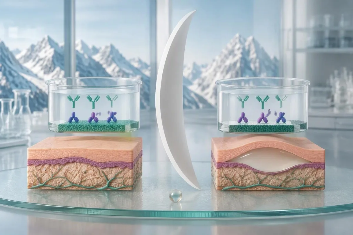
ความไวและความจำเพาะไม่เท่ากับความน่าจะเป็นที่คุณจะเป็นโรคหลังได้ผลบวก. ความน่าจะเป็นดังกล่าวยังขึ้นอยู่กับความชุกของภาวะนี้ในกลุ่มที่ได้รับการตรวจด้วย; ความแตกต่างนี้สำคัญที่สุดเมื่อมีการสั่งตรวจแผงแอนติบอดีในวงกว้าง แทนที่จะสั่งตรวจเพื่อหาสาเหตุของรูปแบบอาการทางผิวหนังที่เฉพาะเจาะจง.
พิจารณาการทดสอบสมมติที่ชัดเจนว่าเป็นเพียงตัวอย่าง โดยมี ความไว 90% และความจำเพาะ 95%, ซึ่งใช้ตรวจคน 1,000 คนที่มีความน่าจะเป็นของโรค 1% โดยจะตรวจพบประมาณ ผู้ที่เป็นโรคจริง 9 คน และผลบวกลวง 50 คน, ทำให้ค่าพยากรณ์ผลบวกอยู่ที่ประมาณ 15%; ตัวเลขเหล่านี้เป็นสมมติฐานเพื่อการเรียนรู้ ไม่ใช่คำกล่าวอ้างถึงประสิทธิภาพของการทดสอบ BP180 ของคุณ.
ใช้การทดสอบสมมติเดียวกันในกรณีที่ความน่าจะเป็นของโรคคือ 20%, and approximately 180 true positives and 40 false positives would occur per 1,000 tests. The positive predictive value becomes about 82%—a large change caused by clinical context, not by a different laboratory method.
These examples explain why a dermatologist may request tissue testing despite a positive result, or take characteristic symptoms seriously despite a negative one. Our discussion of การแปลผลแอนติบอดี MOG illustrates the same probability principle in another autoimmune condition, although its diagnostic criteria cannot be transferred to pemphigoid.
จะเป็นอย่างไรหากผล BP180 เป็นบวกแต่ไม่มีตุ่มพอง?
A low-positive BP180 result without blisters does not establish bullous pemphigoid or reliably predict future blistering. The next distinction is whether you have no skin symptoms at all, or persistent itching and an unexplained rash that could represent a nonbullous pemphigoid presentation.

Incidental BP180 antibody detection can occur without clinically established pemphigoid. Possible explanations include low-level autoreactivity and assay-related false positivity; there is no validated conversion rule that turns an isolated result of 11 U/mL into a percentage risk of developing blisters later.
A hypothetical person with 11 U/mL, no itching, no rash, and no mucosal symptoms usually needs a contextual review rather than automatic immunosuppressive treatment. The clinician should check why the assay was ordered, whether the result was unexpected, and whether a skin examination reveals findings the person had not recognized.
Persistent dryness and itching still deserve assessment because common explanations remain common. Eczema, medication effects, thyroid disease, and other causes may be more likely than pemphigoid; our dry skin testing guide explains why additional blood tests should follow symptoms rather than a blanket panel.
ไม่มี no universal repeat-testing interval for an asymptomatic low-positive BP180 result. A clinician may choose observation, a repeat using the same method, or specialist review depending on the history; new persistent itching, urticarial plaques, or blisters should change that plan rather than waiting for a scheduled repeat.
โรคเพมฟิกอยด์ชนิดตุ่มพองเริ่มต้นด้วยอาการคันเพียงอย่างเดียวได้หรือไม่?
Pemphigoid can present with persistent itching, eczema-like patches, urticarial plaques, or scratch-related changes without visible blisters. In a specialist diagnostic study, 74 of 343 pemphigoid cases—21.6%—were nonbullous, showing that the absence of blisters cannot independently exclude the disorder (Meijer et al., 2019).

Nonbullous pemphigoid is not simply every itchy rash with a positive BP180 result. Diagnosis still needs compatible symptoms and appropriate immunopathological evidence; the 21.6% study figure came from a referred population and should not be interpreted as the probability that any person with itching has pemphigoid.
The symptom pattern often matters more than whether one small blister has appeared. Persistent, widespread itching in an older adult, especially with recurrent raised plaques or poor response to ordinary eczema care, warrants a dermatological assessment; our itchy skin laboratory guide separates skin-directed testing from tests for systemic causes.
Eosinophilia can accompany bullous pemphigoid, but it is neither required nor diagnostic. An absolute eosinophil count above approximately 0.5 × 10^9/L is often flagged in adults, yet allergy, medications, and parasites can produce the same finding; our guide to จำนวน eosinophil สูง explains those alternatives.
A dermatologist does not always need to wait for a blister before collecting a useful sample for direct immunofluorescence. In nonbullous disease, sample selection is tailored to the available rash and laboratory protocol; document the distribution and duration of itching, because a disappearing rash can make later assessment harder.
แอนติบอดี BP230 ช่วยเสริมการตรวจ BP180 อย่างไร?
BP230 antibodies provide complementary evidence for pemphigoid but cannot confirm or exclude bullous pemphigoid alone. BP230 is an approximately 230-kDa intracellular anchoring protein, whereas BP180 crosses the cell membrane; their different locations and antibody targets help explain why the two results can disagree.
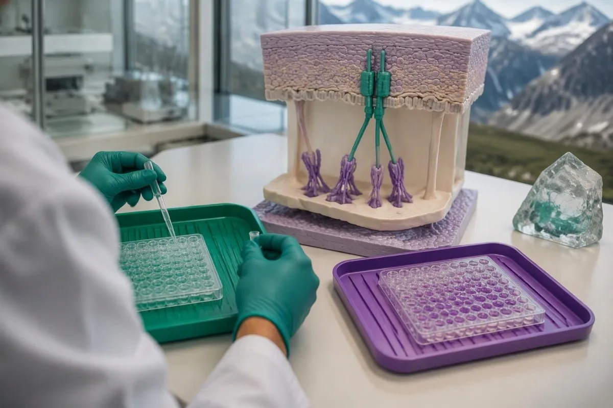
A negative BP230 antibody result does not cancel a positive BP180 result. Some people with confirmed pemphigoid have antibodies detectable against only one tested antigen, and each assay has its own sensitivity; interpreting the pair as a two-test pass-or-fail rule creates avoidable diagnostic errors.
บุคคลสมมติ BP180 result of 45 U/mL with negative BP230 antibodies still warrants assessment if tense blisters and severe itching are present. Conversely, isolated low-positive BP230 antibodies without compatible findings are not enough to diagnose pemphigoid; a second positive marker strengthens a pattern only when the overall evidence fits.
Kantesti is an AI biomarker interpretation platform that can explain BP180 and BP230 antibodies as complementary findings rather than interchangeable scores. This is similar to the reasoning used for antibody panels in connective disease, but skin-specific tissue findings remain central to pemphigoid assessment.
Do not add BP180 and BP230 concentrations together, even if both are expressed in U/mL. They represent different antigen-specific measurements with different calibration systems; BP180 levels generally have a better-established relationship with disease activity than BP230 levels, but neither replaces examination.
เหตุใดคุณจึงอาจต้องตัดชิ้นเนื้อผิวหนังเพื่อตรวจอิมมูโนฟลูออเรสเซนซ์โดยตรง?
Skin biopsy with direct immunofluorescence can show whether antibodies or complement are deposited at the skin's basement membrane. Dermatologists often collect two separate specimens because routine microscopy and direct immunofluorescence answer different questions and require different sample handling.
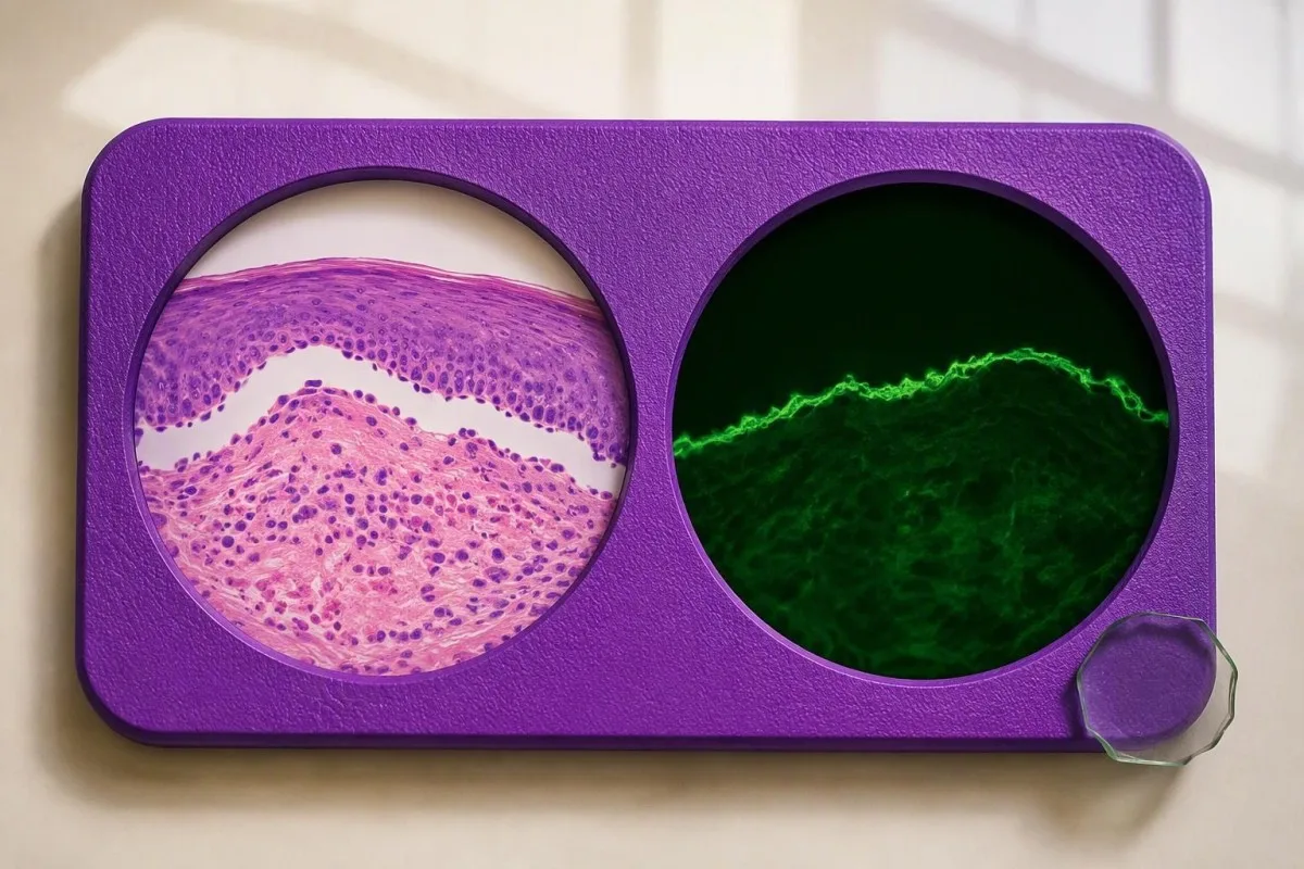
Routine histology examines tissue structure and inflammatory cells. A sample from a fresh blister or suitable affected area may show separation beneath the epidermis and eosinophils, but these features are not unique to bullous pemphigoid; the second sample looks for the immune-deposit pattern that helps classify the disorder.
For classic blistering presentations, the direct-immunofluorescence sample is commonly taken from appropriately selected skin near a fresh blister rather than its eroded center. A small 3–4-mm punch is often used, although technique and location depend on the case; sampling heavily damaged tissue can remove the structures the laboratory needs to examine.
The specimen for direct immunofluorescence must not be routinely placed in formalin. Laboratories generally require a dedicated transport medium or another explicitly agreed handling method, while the routine histology specimen commonly goes into formalin; ask whether the two containers have been prepared for their separate purposes.
In Meijer et al.'s 2019 diagnostic cohort, direct immunofluorescence had a sensitivity of 88.3%, so even this valuable test was not infallible. Kantesti's ข้อมูลการยืนยันทางคลินิก concerns our interpretation methodology—not a claim that AI can reproduce skin immunofluorescence or establish its findings.
ผลตรวจอิมมูโนฟลูออเรสเซนซ์และการทดสอบแยกชั้นด้วยเกลือที่เป็นบวกหมายถึงอะไร?
Linear IgG and/or C3 along the basement membrane on direct immunofluorescence supports a pemphigoid-spectrum disorder, but the pattern is not unique to bullous pemphigoid. Indirect immunofluorescence on salt-split skin can add evidence by showing where circulating antibodies bind relative to an experimentally created tissue separation.
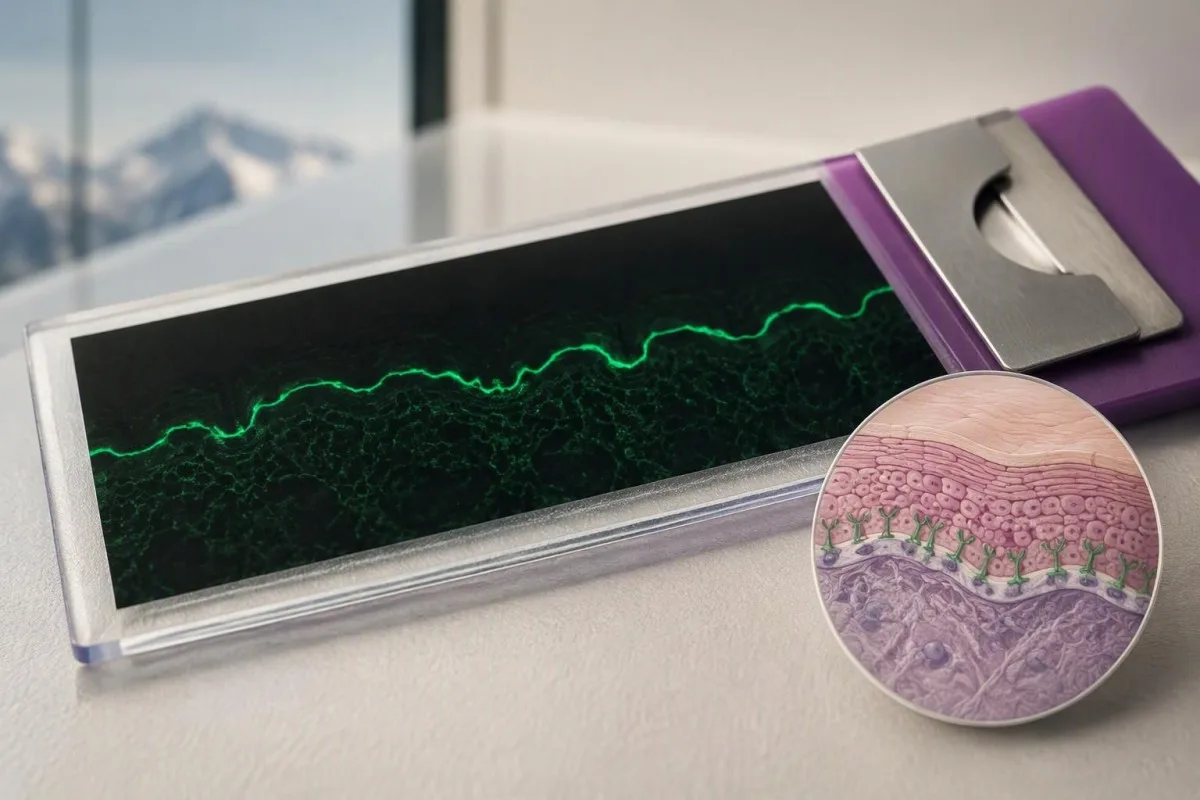
Direct and indirect immunofluorescence use different materials. Direct testing examines deposits already present in the person's skin, while indirect testing applies the person's serum to a laboratory substrate; a positive result in one test and a negative result in the other is therefore possible without a reporting error.
On salt-split skin, bullous pemphigoid antibodies commonly bind the epidermal side, often called the roof. Antibodies binding the dermal side, or floor, raise consideration of other disorders such as epidermolysis bullosa acquisita or anti-p200 pemphigoid, but binding location alone does not fully classify every case.
Meijer et al. reported 99.9% specificity for indirect immunofluorescence on salt-split skin in their 2019 diagnostic cohort. That high figure applies to that study and testing pathway; a negative result still cannot exclude pemphigoid because specificity describes false positives, not how many cases the test misses.
C3 detected in skin is not the same measurement as a serum C3 concentration. A normal circulating complement result does not negate local deposits at the basement membrane; our complement testing explanation helps prevent this common cross-test misunderstanding.
จะเป็นอย่างไรหากผล BP180 การตัดชิ้นเนื้อผิวหนัง และอาการไม่สอดคล้องกัน?
Discordant BP180 and skin-test results require a review of the clinical picture, assay target, sampling site, and specimen handling. A positive ELISA with negative direct immunofluorescence can reflect incidental antibodies or an uninformative skin sample; a negative NC16A ELISA can miss disease directed against other targets.
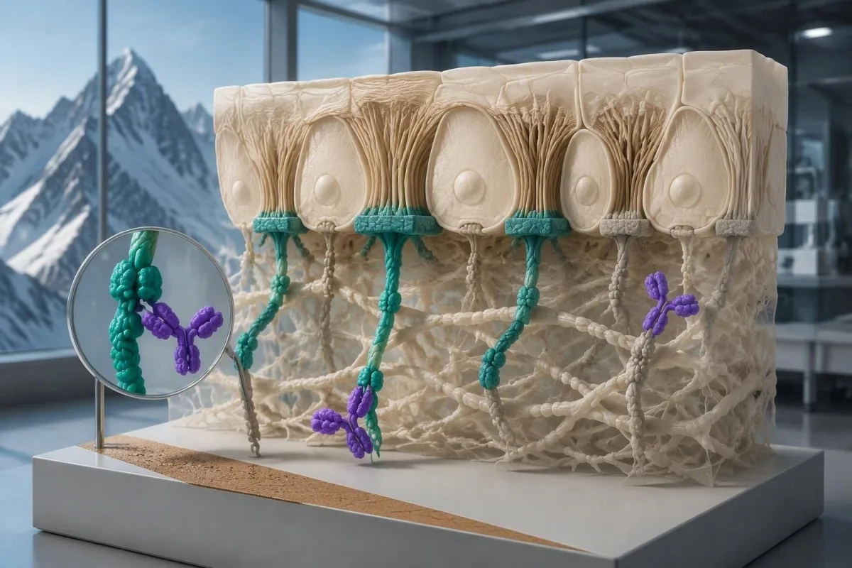
A negative direct-immunofluorescence result is less reassuring if the sample was poorly selected or incorrectly transported. Treatment before sampling may also influence findings, so clinicians need to know which creams or systemic medicines were used and where; never stop prescribed treatment solely to make a test more likely to turn positive.
A hypothetical person with characteristic new blisters, BP180 of 80 U/mL, and a negative direct-immunofluorescence specimen from an old eroded site may need repeat sampling. The same antibody result with no compatible skin findings demands a different investigation; the discrepancy does not have one automatic explanation.
A negative BP180 NC16A result does not exclude every form of pemphigoid. Further testing may include BP230 antibodies, indirect immunofluorescence, or specialized assays for other antigens; our guide to negative myasthenia antibody tests illustrates why a limited antigen panel cannot exclude an entire autoimmune disease category.
Repeat testing should address a specific uncertainty rather than create an endless sequence of panels. Ask whether the next investigation is checking the same antigen by another method, examining an additional antigen, or correcting a tissue-sampling problem; each strategy answers a different diagnostic question.
ภาวะอื่นใดบ้างที่อาจมีลักษณะคล้ายเพมฟิกอยด์ที่ผล BP180 เป็นบวก?
BP180 antibodies can occur in other pemphigoid-spectrum conditions, including mucous membrane pemphigoid and pemphigoid gestationis. Eczema, scabies, and drug-related eruptions can resemble early bullous pemphigoid clinically, while pemphigus involves different adhesion proteins and usually a different level of skin separation.
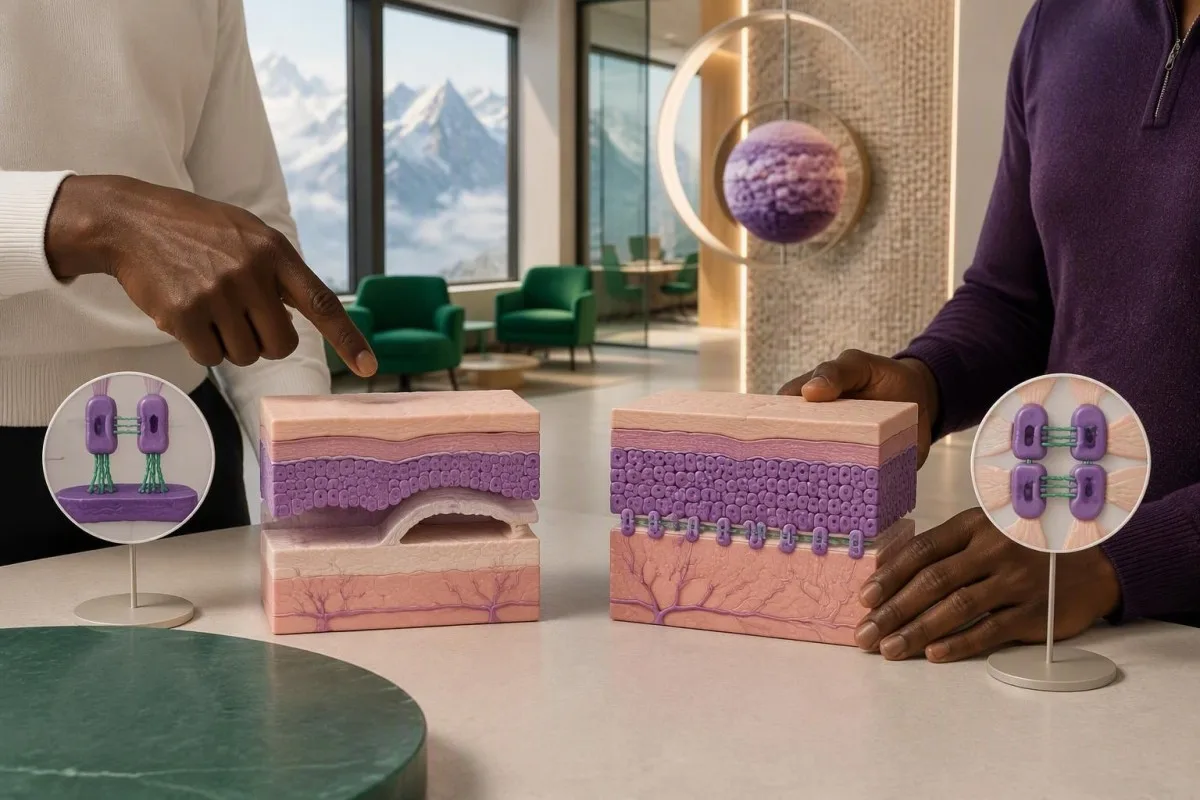
Bullous pemphigoid typically causes tense blisters because separation occurs beneath the epidermis. Pemphigus often produces more fragile blisters or erosions through loss of adhesion within the epidermis; our desmoglein antibody guide explains why BP180 testing cannot substitute for a pemphigus evaluation.
Predominantly mouth, eye, genital, or other mucosal involvement raises concern for mucous membrane pemphigoid, especially when scarring occurs. Eye pain, light sensitivity, or vision changes require urgent assessment because waiting for a repeat antibody concentration can delay treatment of a sight-threatening process.
Pemphigoid gestationis is associated with pregnancy and can produce BP180 antibodies. Pregnancy or recent childbirth therefore changes the interpretation of the same laboratory result; the distinction requires obstetric and dermatological coordination rather than assuming that every BP180-positive blistering disorder is classic older-adult bullous pemphigoid.
Scabies can cause intense itching and may coexist with an unrelated positive antibody result. Household itching, characteristic distribution, and burrows alter the differential; our scabies symptom guide is useful for that comparison, but neither household exposure nor BP180 positivity independently settles the diagnosis.
ยามีผลต่อการแปลผล BP180 หรือการตัดสินใจรักษาหรือไม่?
Medication history can change the likelihood and presentation of bullous pemphigoid, but a positive BP180 result alone is not a reason to stop a medicine. DPP-4 inhibitors used for diabetes and immune checkpoint inhibitors are recognized associations; treatment decisions depend on confirmed disease, severity, and the person's other health risks.

DPP-4 inhibitor-associated pemphigoid can have a less typical inflammatory presentation and may show limited NC16A reactivity. This is not universal, and exposure does not prove causation; bring a complete medicine list, including start dates, to a review using our senior medication effects guide as a preparation aid.
การ 2022 EADV guideline recommends potent topical corticosteroids as a mainstay when feasible and discusses oral prednisone at approximately 0.5 mg/kg/day as an alternative in suitable circumstances (Borradori et al., 2022). These are clinician-directed treatment options for disease, not instructions to treat an isolated antibody result or begin steroids without assessment.
Systemic corticosteroids can create additional risks that are unrelated to the BP180 concentration. People with relevant residence or travel exposure may need assessment for Strongyloides before significant immunosuppression; our Strongyloides antibody explanation describes why an apparently unrelated parasite history can become a treatment-safety issue.
Kidney function, glucose, infection risk, and the ability to apply topical treatment may influence the choice of therapy more than whether BP180 is 40 or 80 U/mL. ของเรา คู่มือการตีความการทำงานของไต covers supportive laboratory assessment, but renal markers do not confirm pemphigoid.
สามารถใช้ระดับ BP180 ติดตามกิจกรรมของโรคและการฟื้นตัวได้หรือไม่?
BP180 antibody levels can support monitoring in confirmed bullous pemphigoid, but they do not replace examination or determine treatment alone. Concentrations often track disease activity at a group level; individual patients can remain antibody-positive while their skin is controlled or experience symptoms without a proportionate rise.
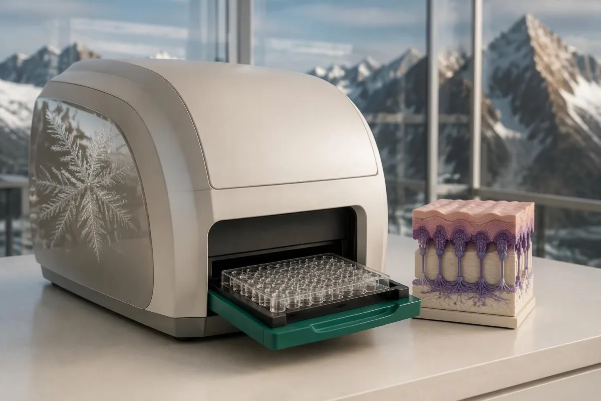
A falling BP180 concentration is most useful when it agrees with fewer new blisters and less itching. A hypothetical decline from 120 to 35 U/mL on the same assay supports an improving pattern, but it does not independently establish remission or justify stopping treatment.
An increase from 12 to 14 U/mL near a cutoff may reflect analytical or biological variation rather than a meaningful change in disease. There is no universal percentage increase that proves relapse across all BP180 assays; the method, timing, symptoms, and previous response need to be considered together.
Kantesti is an AI blood test interpretation platform that can organize serial BP180 results without treating a laboratory trend as a diagnosis. ของเรา คำอธิบายเทคโนโลยี AI describes the interpretation workflow; preserving each report's assay name and reference interval matters when comparing two or more measurements.
Changing laboratories can create an apparent rise or fall that is partly methodological. Our คู่มือแนวโน้มความปลอดภัยของยา explains why dates, doses, and laboratory context belong beside the numbers; photograph new skin findings and record new-blister counts when your dermatologist requests them.
คุณควรนำอะไรไปในการติดตามผล และหลักฐานใดเกี่ยวข้อง?
Bring the full BP180 report, any BP230 result, symptom photographs, and your complete medication list to follow-up. These four information groups help a clinician decide whether you need observation, additional serum testing, properly handled skin samples, or urgent assessment rather than another isolated antibody measurement.
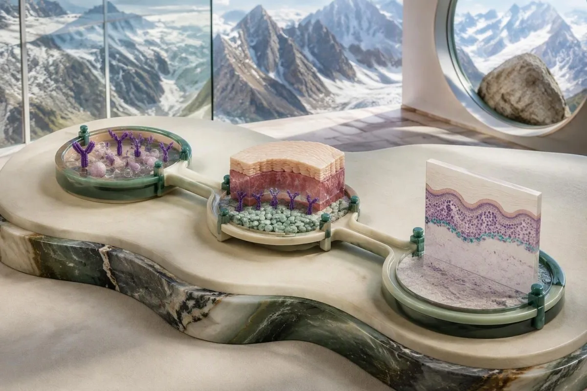
A useful report includes the exact assay name, numerical value, units, cutoff, and collection date. If a hypothetical BP180 result reads 16 U/mL, the missing cutoff may reverse whether it is considered positive; also request the actual histology and direct-immunofluorescence reports rather than relying on the phrase 'skin biopsy normal.'
New eye symptoms, swallowing difficulty, extensive rapidly spreading blisters, fever, or inability to maintain fluids need prompt clinical attention. Do not wait for a scheduled second test when those symptoms appear; supportive urinalysis can answer separate medical questions, as explained in our คู่มือการแปลผลการตรวจปัสสาวะ, but it cannot establish a pemphigoid diagnosis.
Kantesti helps explain the distinctions between a flagged antibody result, a probable diagnosis, and confirmed disease. ของเรา หลักการ AI ที่มีความรับผิดชอบ describe the boundaries of educational interpretation; only a clinician with access to your examination and appropriate investigations can decide whether a แอนติบอดี BP180 เป็นบวก result represents active pemphigoid.
The directly relevant evidence here comes from the 2022 EADV guideline and the 2019 JAMA Dermatology diagnostic study. The two Zenodo publications listed below concern kidney-function and urinalysis interpretation and are included as background educational publications, not evidence that BP180 confirms bullous pemphigoid or that our AI has validated diagnostic accuracy for this condition.
คำถามที่พบบ่อย
ผลตรวจแอนติบอดี BP180 เป็นบวก หมายความว่าฉันเป็นโรคตุ่มพองเพมฟิกอยด์แน่นอนหรือไม่?
ผลตรวจแอนติบอดี BP180 เป็นบวกไม่ได้หมายความแน่นอนว่าคุณเป็นโรค bullous pemphigoid ห้องปฏิบัติการบางแห่งใช้ค่าตัดใกล้ 9 U/mL ขณะที่บางแห่งใช้ใกล้ 20 U/mL ดังนั้นวิธีตรวจและช่วงอ้างอิงที่ใช้จึงมีความสำคัญ อาการคันหรือตุ่มพองที่สอดคล้องกันทำให้ผลตรวจมีความหมายมากขึ้น ขณะที่ผลบวกอ่อน ๆ เพียงอย่างเดียวอาจเป็นผลที่พบโดยบังเอิญหรือเป็นผลบวกลวง อาจจำเป็นต้องตรวจชิ้นเนื้อผิวหนังด้วย direct immunofluorescence และตรวจแอนติบอดีเพิ่มเติมเพื่อยืนยันการวินิจฉัย.
ผลตรวจ BP180 ที่เป็นบวกระดับต่ำหมายความว่าอย่างไร หากฉันไม่มีตุ่มพอง?
ผล BP180 ที่เป็นบวกเล็กน้อยโดยไม่มีตุ่มพอง อาจสะท้อนการตรวจพบแอนติบอดีโดยบังเอิญ ผลบวกลวงที่เกี่ยวข้องกับการตรวจวิเคราะห์ หรือโรคเพมฟิกอยด์ที่แสดงอาการโดยไม่มีตุ่มพอง ตัวอย่างเช่น 12 U/mL ถือว่าเป็นผลบวกเมื่อใช้เกณฑ์ตัดที่ 9 U/mL แต่ไม่สามารถทำนายได้ว่าผู้ที่ไม่มีอาการจะเกิดโรคหรือไม่ อาการคันที่เป็นต่อเนื่องหรือผื่นที่ไม่ทราบสาเหตุควรได้รับการประเมินจากแพทย์ผิวหนัง แม้ไม่มีตุ่มพองก็ตาม ไม่มีช่วงเวลาการตรวจซ้ำหรือคำแนะนำในการรักษาที่ใช้ได้กับทุกกรณี โดยพิจารณาจากผลบวกเล็กน้อยที่พบเพียงครั้งเดียวเท่านั้น.
ฉันเป็นเพมฟิกอยด์ชนิดตุ่มพองโดยมีผลตรวจแอนติบอดี BP180 เป็นลบได้หรือไม่?
โรคตุ่มพองเพมฟิกอยด์อาจเกิดขึ้นได้แม้ผลตรวจแอนติบอดีต่อ BP180 ด้วยการทดสอบ NC16A ตามปกติจะเป็นลบ การทดสอบนี้ตรวจบริเวณเฉพาะส่วนหนึ่งของโปรตีนขนาดประมาณ 180 kDa และอาจตรวจไม่พบแอนติบอดีที่มุ่งเป้าไปยังบริเวณอื่นเป็นหลัก การตรวจ BP230, การตรวจอิมมูโนฟลูออเรสเซนซ์โดยตรง และการตรวจอิมมูโนฟลูออเรสเซนซ์โดยอ้อม อาจให้หลักฐานเพิ่มเติม ผลตรวจเลือดที่เป็นลบไม่ควรทำให้ยุติการประเมิน หากลักษณะทางผิวหนังบ่งชี้อย่างชัดเจนว่าเป็นโรคเพมฟิกอยด์.
ผลแอนติบอดีต่อ BP230 ที่เป็นลบสามารถตัดโรคเพมฟิกอยด์ชนิดตุ่มพองออกได้หรือไม่?
แอนติบอดีต่อ BP230 ที่ให้ผลลบไม่ได้ตัดโรคตุ่มพองเพมฟิกอยด์ออกไป BP230 เป็นโปรตีนยึดเกาะภายในเซลล์ที่มีขนาดประมาณ 230 kDa และผู้ป่วยบางรายที่ยืนยันว่าเป็นโรคนี้ไม่มีแอนติบอดีที่ตรวจพบได้ต่อโปรตีนดังกล่าว ผล BP180 เป็นบวกร่วมกับแอนติบอดีต่อ BP230 เป็นลบยังคงสอดคล้องกับการวินิจฉัยได้ เมื่อผลตรวจทางคลินิกและผลตรวจเนื้อเยื่อสอดคล้องกัน ไม่ควรนำความเข้มข้นของแอนติบอดีทั้งสองชนิดมาบวกกัน หรือถือว่าเป็นเกณฑ์ยืนยันผลจากการตรวจสองรายการ.
เหตุใดบางครั้งจึงต้องเก็บชิ้นเนื้อผิวหนังสองชิ้นเพื่อตรวจหาโรคเพมฟิกอยด์?
มักจำเป็นต้องตัดชิ้นเนื้อผิวหนัง 2 ชิ้น เนื่องจากการตรวจทางจุลพยาธิวิทยาตามปกติและการตรวจอิมมูโนฟลูออเรสเซนซ์โดยตรงตอบคำถามด้านการวินิจฉัยที่แตกต่างกัน โดยทั่วไปใช้เครื่องมือตัดชิ้นเนื้อแบบ punch ขนาดเล็ก 3–4-mm แม้แพทย์ผิวหนังจะเป็นผู้เลือกวิธีและตำแหน่งให้เหมาะกับอาการของแต่ละราย ตัวอย่างหนึ่งใช้ตรวจโครงสร้างเนื้อเยื่อ ส่วนอีกตัวอย่างใช้ตรวจหาสารภูมิคุ้มกันที่สะสมตามแนวเยื่อฐาน ตัวอย่างสำหรับการตรวจอิมมูโนฟลูออเรสเซนซ์โดยตรงต้องใช้วิธีขนส่งที่ตกลงกันไว้โดยเฉพาะ และโดยปกติไม่ควรแช่ในฟอร์มาลิน.
ระดับ BP180 ที่สูงหมายความว่าโรคเพมฟิกอยด์ของฉันรุนแรงหรือไม่?
A high BP180 level does not independently determine how severe pemphigoid is or whether it is an emergency. A result of 100 U/mL must be interpreted using that assay's cutoff and alongside the extent of skin involvement, itching, new blisters, and general health. Antibody levels often relate to disease activity, but individual results and symptoms can disagree. Eye pain, vision changes, difficulty swallowing, or rapidly spreading extensive blistering require prompt assessment regardless of the antibody concentration.
แอนติบอดีต่อ BP180 ควรมีผลเป็นลบก่อนหยุดการรักษาหรือไม่?
แอนติบอดีต่อ BP180 ไม่จำเป็นต้องลดลงจนเป็นลบเสมอไปก่อนที่แพทย์จะพิจารณาลดหรือหยุดการรักษาโรคเพมฟิกอยด์ การลดลงโดยสมมติจาก 120 เป็น 35 U/mL อาจบ่งชี้ว่าอาการดีขึ้น แต่การตัดสินใจจะพิจารณาจากอาการ การเกิดตุ่มพองใหม่ การตรวจร่างกาย และประวัติการกลับเป็นซ้ำ อาจยังตรวจพบแอนติบอดีได้แม้ผิวหนังจะควบคุมโรคได้ และไม่มีค่าตัดสำหรับการหยุดยาที่ใช้ได้กับทุกวิธีตรวจ อย่าปรับเปลี่ยนการรักษาที่แพทย์สั่งโดยอาศัยแนวโน้มของแอนติบอดีเพียงอย่างเดียว.
อ่านผลตรวจเลือดอย่างไร
อัปโหลดรายงานผลตรวจของคุณ แล้ว AI ของ Kantesti จะอธิบายตัวบ่งชี้ทางชีวภาพแต่ละรายการด้วยภาษาที่เข้าใจง่ายภายในไม่กี่วินาที มีผู้ใช้ทั่วโลกมากกว่า 2 ล้านคน.
เอกสารอ้างอิง
- ไคลน์, ที., มิตเชลล์, เอส., & เวเบอร์, เอช. (2026). อธิบายอัตราส่วน BUN/Creatinine: คู่มือการทดสอบการทำงานของไต. Kantesti AI การวิจัยทางการแพทย์. โดอิ
- ไคลน์, ที., มิตเชลล์, เอส., & เวเบอร์, เอช. (2026). การทดสอบยูโรบิลิโนเจนในปัสสาวะ: คู่มือการวิเคราะห์ปัสสาวะครบถ้วน 2026. Kantesti AI การวิจัยทางการแพทย์. โดอิ
- Borradori L et al. (2022). Updated S2 K guidelines for the management of bullous pemphigoid initiated by the European Academy of Dermatology and Venereology (EADV). Journal of the European Academy of Dermatology and Venereology. โดอิ
- Meijer JM et al. (2019). Assessment of Diagnostic Strategy for Early Recognition of Bullous and Nonbullous Variants of Pemphigoid. JAMA Dermatology. โดอิ
- 2 ล้าน+การตรวจที่วิเคราะห์
- 127+ประเทศ
- 100+ภาษา
เกี่ยวกับทีมแพทย์


สัญญาณความน่าเชื่อถือ E-E-A-T
ประสบการณ์
การทบทวนเชิงคลินิกโดยแพทย์ที่นำโดยกระบวนการตีความผลตรวจในห้องแล็บ.
ความเชี่ยวชาญ
โฟกัสด้านเวชศาสตร์ห้องปฏิบัติการเกี่ยวกับพฤติกรรมของไบโอมาร์กเกอร์ในบริบททางคลินิก.
อำนาจ
ทบทวนทางการแพทย์โดย นพ. Thomas Klein พร้อมผลงานจาก นพ. Sarah Mitchell และศาสตราจารย์ ดร. Hans Weber.
ความน่าเชื่อถือ
การตีความที่อิงหลักฐาน พร้อมเส้นทางการติดตามที่ชัดเจนเพื่อลดความตื่นตระหนก.


ข้อจำกัดความรับผิดชอบทางการแพทย์
บทความนี้จัดทำเพื่อวัตถุประสงค์ด้านการศึกษาเท่านั้น และไม่ถือเป็นคำแนะนำทางการแพทย์ โปรดปรึกษาผู้ให้บริการด้านสุขภาพที่มีคุณสมบัติเหมาะสมเสมอสำหรับการตัดสินใจด้านการวินิจฉัยและการรักษา.
เผยแพร่แล้ว · ติดต่อทีมบรรณาธิการ
เผยแพร่โดย Kantesti Ltd จดทะเบียนในอังกฤษและเวลส์ เลขทะเบียนบริษัท. 17090423, ลอนดอน สหราชอาณาจักร.
