
ผลตรวจแอนติบอดี anti-Hu เป็นบวกทำให้เกิดความกังวลเกี่ยวกับการตอบสนองทางภูมิคุ้มกันทางระบบประสาทที่สัมพันธ์กับมะเร็ง โดยเฉพาะมะเร็งปอดชนิดเซลล์เล็ก แต่ไม่ได้วินิจฉัยว่าเป็นมะเร็ง แพทย์จะประเมินอาการ ยืนยันผลตรวจทางห้องปฏิบัติการ และจัดให้มีการตรวจหามะเร็ง อาจต้องตรวจคัดกรองซ้ำแม้ผลสแกนครั้งแรกจะเป็นลบ.



- Anti-Hu / ANNA-1 เป็นแอนติบอดีพารานีโอพลาสติกความเสี่ยงสูง ซึ่งเป็นกลุ่มที่สัมพันธ์กับมะเร็งในกลุ่มตัวอย่างทางคลินิกที่มีการยืนยันแล้วมากกว่า 70%—ไม่ใช่ผลตรวจคัดกรองที่ยังไม่ได้รับการยืนยันทั้งหมด.
- การวินิจฉัยมะเร็ง ต้องมีหลักฐานของมะเร็งที่เป็นต้นเหตุ โดยปกติรวมถึงการตรวจเนื้อเยื่อ ผลแอนติบอดีเป็นบวกเพียงอย่างเดียวไม่ใช่หลักฐานดังกล่าว.
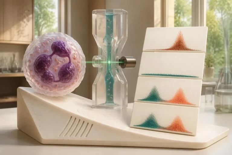
- การยืนยันผลตรวจ มักใช้วิธีเสริมกัน 2 วิธี ได้แก่ รูปแบบการติดสีของเนื้อเยื่อที่มีลักษณะจำเพาะ และการตรวจที่จำเพาะต่อแอนติเจน.
- อาการทางระบบประสาท เช่น การสูญเสียความรู้สึกที่ค่อย ๆ รุนแรงขึ้น การทรงตัวผิดปกติ อาการชัก หรือการทำงานผิดปกติของระบบประสาทอัตโนมัติ ส่งผลต่อความสำคัญและความเร่งด่วนของผลตรวจ.
- ผลตรวจภาพวินิจฉัยเป็นลบ หมายความว่าการตรวจครั้งนั้นไม่พบมะเร็ง ณ เวลานั้น แต่ไม่ได้ตัดความเป็นไปได้ที่จะมีมะเร็งขนาดเล็กที่ยังตรวจไม่พบในระยะแรกออกไป.
- การตรวจคัดกรองซ้ำ แนะนำให้ตรวจทุก 4–6 เดือนเป็นเวลา 2 ปี เมื่อมีฟีโนไทป์ทางระบบประสาทที่มีความเสี่ยงสูงร่วมกับแอนติบอดีที่มีความเสี่ยงสูง ตามเกณฑ์ PNS ปี 2021.
- การเฝ้าระวังที่นานขึ้น อาจเหมาะสมสำหรับผู้ป่วยบางราย; กลุ่มผู้ป่วย anti-Hu ในปี 2023 สนับสนุนให้พิจารณาการตรวจคัดกรองนานกว่า 2 ปี ซึ่งบางครั้งอาจนานถึง 5 ปี.
- อาการฉุกเฉิน รวมถึงอาการหายใจลำบากที่เกิดขึ้นใหม่ กลืนอย่างปลอดภัยไม่ได้ อ่อนแรงลงอย่างรวดเร็ว ชักครั้งแรก หรือมีความบกพร่องทางระบบประสาทเกิดขึ้นฉับพลัน.
ผลตรวจแอนติบอดี anti-Hu เป็นบวกหมายความว่าอย่างไร?
หนึ่ง ผลตรวจแอนติบอดี anti-Hu เป็นบวก หมายความว่าห้องปฏิบัติการตรวจพบปฏิกิริยาต่อโปรตีน Hu ของเซลล์ประสาท; ผลนี้ไม่ได้ยืนยันการเป็นมะเร็งหรือกลุ่มอาการทางระบบประสาทด้วยตัวมันเอง การแปลผลมี 3 ส่วน ได้แก่ ผลตรวจเป็นผลบวกจริงหรือไม่ อาการทางระบบประสาทสอดคล้องกันหรือไม่ และตรวจพบมะเร็งที่เกี่ยวข้องได้หรือไม่.
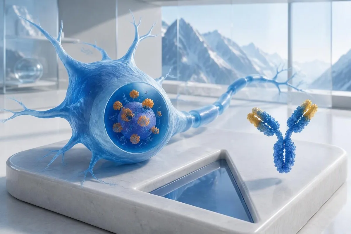
Anti-Hu จัดเป็นแอนติบอดีที่เกี่ยวข้องกับมะเร็งซึ่งมีความเสี่ยงสูง, โดยกำหนดประเภทนี้จากการมีความสัมพันธ์กับมะเร็งมากกว่า 70% ในประชากรทางคลินิกที่เกี่ยวข้อง (Graus และคณะ, 2021) ไม่สามารถนำเปอร์เซ็นต์ดังกล่าวไปใช้กับบุคคลที่มีสุขภาพดีโดยทั่วไป ซึ่งมีผลตรวจจากแผงตรวจที่ยังไม่ยืนยันและให้ผลอ่อน ๆ ได้โดยตรง: ผู้ป่วยที่ศึกษา วิธีตรวจ และอาการทางระบบประสาทล้วนส่งผลต่อความน่าจะเป็น.
ผู้ที่สมมติว่าอายุ 62 ปี มีอาการทรงตัวแย่ลงเป็นเวลา 8 สัปดาห์และสูญเสียความรู้สึกแบบไม่สมมาตร ควรได้รับการประเมินต่างจากผู้ที่ตรวจพบแอนติบอดีโดยบังเอิญระหว่างการตรวจแผงอาการอ่อนเพลียแบบครอบคลุม ไม่ควรบอกคนใดคนหนึ่งว่า “คุณเป็นมะเร็ง” โดยดูจากผลแล็บเพียงอย่างเดียว คำอธิบายของเราเกี่ยวกับ ผลตรวจแอนติบอดีเป็นบวก ช่วยแยกตัวบ่งชี้ทางภูมิคุ้มกันออกจากการวินิจฉัยที่ครบถ้วนแล้ว.
คันเตสตีเป็น เครื่องวิเคราะห์ผลเลือด AI ซึ่งอาจช่วยอธิบายข้อความในรายงาน anti-Hu แต่การยืนยันผลจากห้องปฏิบัติการและการประเมินมะเร็งควรดำเนินการโดยแพทย์ผู้รักษาของคุณ ในฐานะ Thomas Klein, MD, Chief Medical Officer ของ Kantesti LTD คำถามแรกที่ผมจะถามคืออะไรเป็นเหตุให้ตรวจรายการนี้โดยเฉพาะ ไม่ใช่สัญญาณเตือนดูน่ากังวลเพียงใด; ของเรา องค์กรและภารกิจทางคลินิกของเรา อธิบายความแตกต่างนั้น.
ANNA-1 เป็นแอนติบอดีชนิดเดียวกับ anti-Hu หรือไม่?
โดยทั่วไป แอนติบอดี ANNA-1 และแอนติบอดี anti-Hu หมายถึงแอนติบอดีกลุ่มเดียวกัน. ANNA-1 ย่อมาจากแอนติบอดีต่อนิวเคลียสของเซลล์ประสาทชนิดที่ 1; ชื่อ Hu หมายถึงโปรตีนภายในเซลล์ประสาทที่ภูมิคุ้มกันตอบสนอง ไม่ใช่โรคหรือระยะของมะเร็งที่แยกต่างหาก.
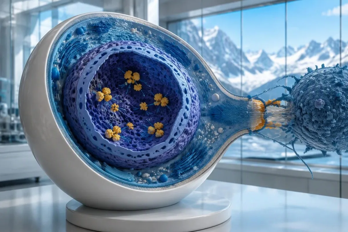
โปรตีน Hu เป็นโปรตีนจับ RNA ภายในเซลล์, และควรเข้าใจ anti-Hu ว่าเป็นตัวบ่งชี้ของการตอบสนองทางภูมิคุ้มกันที่มักเกี่ยวข้องกับเซลล์ T ที่เป็นพิษต่อเซลล์ ซึ่งแตกต่างจากโรคที่แอนติบอดีจับกับโปรตีนที่เข้าถึงได้บนผิวเซลล์ประสาท ความแตกต่างนี้อธิบายได้ว่าเหตุใดการลดระดับแอนติบอดีเพียงอย่างเดียวจึงไม่สามารถฟื้นฟูความเสียหายทางระบบประสาทจาก anti-Hu ที่เกิดขึ้นแล้วได้อย่างน่าเชื่อถือ แม้รายงาน 2 ฉบับจะแสดงระดับไตเตอร์ที่แตกต่างกันมากก็ตาม.
รายงานที่ระบุว่า “ตรวจพบ ANNA-1” ไม่เหมือนกับผล ANA เป็นบวกจากการตรวจทั่วไป แม้ตัวอักษรจะคล้ายกัน. อนา โดยทั่วไปหมายถึงแอนติบอดีต่อนิวเคลียสที่ใช้ในการประเมินโรคภูมิคุ้มกันทำลายตนเองทั่วร่างกาย ขณะที่ ANNA-1 ระบุรูปแบบแอนติบอดีต่อเซลล์ประสาท ควรแยกตัวย่อ 2 คำนี้ออกจากกันเมื่อพูดคุยเกี่ยวกับผลตรวจของคุณ; ของเรา คู่มือคำศัพท์ ANA อธิบายการตรวจที่คุ้นเคยมากกว่า.
อาจตรวจพบ Anti-Hu ในผู้ป่วยมะเร็งปอดชนิดเซลล์เล็กบางรายที่ไม่มีอาการทางระบบประสาทที่ระบุได้ ในทางกลับกัน อาการทางระบบประสาทอาจนำไปสู่การตรวจพบแอนติบอดีก่อนที่มะเร็งจะมองเห็นได้ ทั้งสองกรณีเป็นแนวทางทางคลินิกที่แตกต่างกัน: แอนติบอดีเพียงอย่างเดียวไม่ได้บอกแพทย์ว่าเส้นประสาทกำลังได้รับความเสียหายอยู่หรือไม่ หรือมะเร็งที่ซ่อนอยู่จะอยู่ตรงไหน.
ควรอ่านรายงานผลตรวจแอนติบอดี anti-Hu อย่างไร?
An anti-Hu antibody test has no universal numerical cutoff that can be applied across laboratories. Read the specimen type, assay method, result category, laboratory reference interval and confirmation comments together; a dilution titer, band intensity and numerical index are 3 different ways of reporting reactivity.
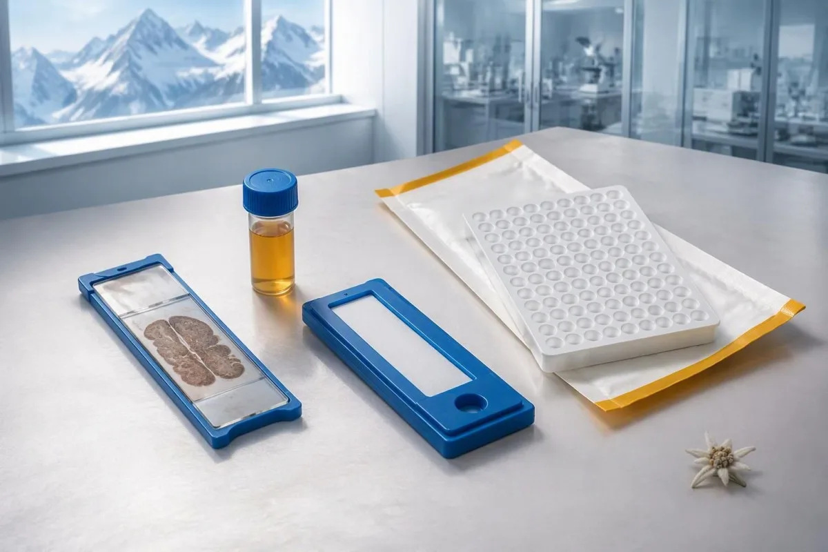
A hypothetical titer of 1:240 describes detectable reactivity after a particular dilution; it is not a cancer probability of 240, nor does it establish cancer stage. A numerical index from another method may use entirely different calibration. Our guide to ผลเชิงคุณภาพเทียบกับผลเชิงปริมาณ explains why “positive” and “how much” answer different laboratory questions.
Weak or borderline reactivity deserves method-specific interpretation, particularly when a broad panel was ordered without a convincing neurological indication. Ask whether the laboratory observed the characteristic neuronal staining pattern and whether an antigen-specific confirmation was performed. Two tests using the same underlying strip technology may reproduce the same analytical problem; agreement is more informative when the methods are genuinely complementary.
Bring the complete report, not just a screenshot of the word positive. The 5 most useful details are the specimen, collection date, assay name, reported strength or titer and laboratory interpretation. Also list recent intravenous immunoglobulin and other immune treatments, because passive antibodies or treatment effects can complicate some serological results; your laboratory can advise whether that issue plausibly affects this particular finding.
เมื่อใดที่ผลตรวจ anti-Hu เป็นบวกจำเป็นต้องได้รับการยืนยัน?
Unexpected, weak or clinically discordant anti-Hu results should be reviewed for confirmation, preferably with an experienced neuroimmunology laboratory. The 2021 PNS criteria emphasize interpreting neuronal antibody testing alongside the phenotype and using appropriate confirmatory methods rather than accepting an isolated commercial-panel result as a diagnosis.

Tissue-based immunohistochemistry or immunofluorescence and an antigen-specific assay provide different information. The first identifies a characteristic distribution of neuronal staining; the second tests reactivity against the relevant antigen. Graus et al. (2021) describe these complementary approaches, with specialist reassessment particularly useful when 1 method gives a result that does not fit the clinical picture.
Repeating a sample can address collection errors or a transient analytical concern, but repeating the same kit is not always enough. In a hypothetical 45-year-old with no objective neurological findings and a faint isolated band, I would want the laboratory’s explanation before treating the report as established anti-Hu autoimmunity. Our GAD65 interpretation guide illustrates another antibody for which context and assay characteristics change meaning.
Confirmation and urgent assessment can happen in parallel. A person who develops seizures, substantial walking difficulty or progressive swallowing impairment should not wait several weeks for a reference-laboratory answer before receiving neurological care. The practical question is not “repeat or investigate?” but which of those 2 tasks can safely proceed together, given the symptoms and the initial laboratory evidence.
อาการทางระบบประสาทใดที่ทำให้ผล anti-Hu น่ากังวลมากขึ้น?
Progressive sensory neuronopathy, encephalomyelitis, limbic encephalitis and marked autonomic dysfunction are recognized anti-Hu presentations. Symptoms can involve more than 1 part of the nervous system, so the pattern and speed of change are usually more informative than a single symptom such as tingling or fatigue.

Sensory neuronopathy can cause asymmetric numbness, impaired position sense and sensory ataxia, sometimes involving the hands or trunk early rather than following a simple feet-first pattern. A patient may lose balance in the dark because vision had been compensating for reduced position feedback. That is different from the focal finger distribution discussed in our carpal tunnel symptom guide, although neither pattern can be diagnosed from description alone.
Anti-Hu autonomic involvement can affect gut movement, blood-pressure regulation and bladder function. Severe constipation, unexplained intestinal pseudo-obstruction or dizziness on standing becomes more relevant when accompanied by objective neurological abnormalities. Recording symptoms over 7 days can reveal combinations that otherwise get presented as separate stomach, balance and urinary complaints; sudden severe abdominal symptoms still require assessment for non-neurological emergencies.
New memory impairment, behavioral change or seizures may indicate central nervous system involvement, while rapid weakness has a broader emergency differential. The สัญญาณเตือนอาการอ่อนแรงที่ต้องไปพบแพทย์ด่วน in our neurological safety guide apply regardless of the antibody label. Call emergency services for new breathing difficulty, unsafe swallowing, a first seizure or sudden stroke-like symptoms rather than waiting for a scheduled follow-up.
อาจมีการตรวจทางระบบประสาทใดบ้างหลังได้ผลตรวจเป็นบวก?
Neurological follow-up may include examination, nerve conduction studies, MRI and cerebrospinal fluid testing, selected according to the symptoms. There is no mandatory battery for every anti-Hu-positive person; the purpose is to document which structures are affected and exclude other causes, not merely to collect additional positive tests.
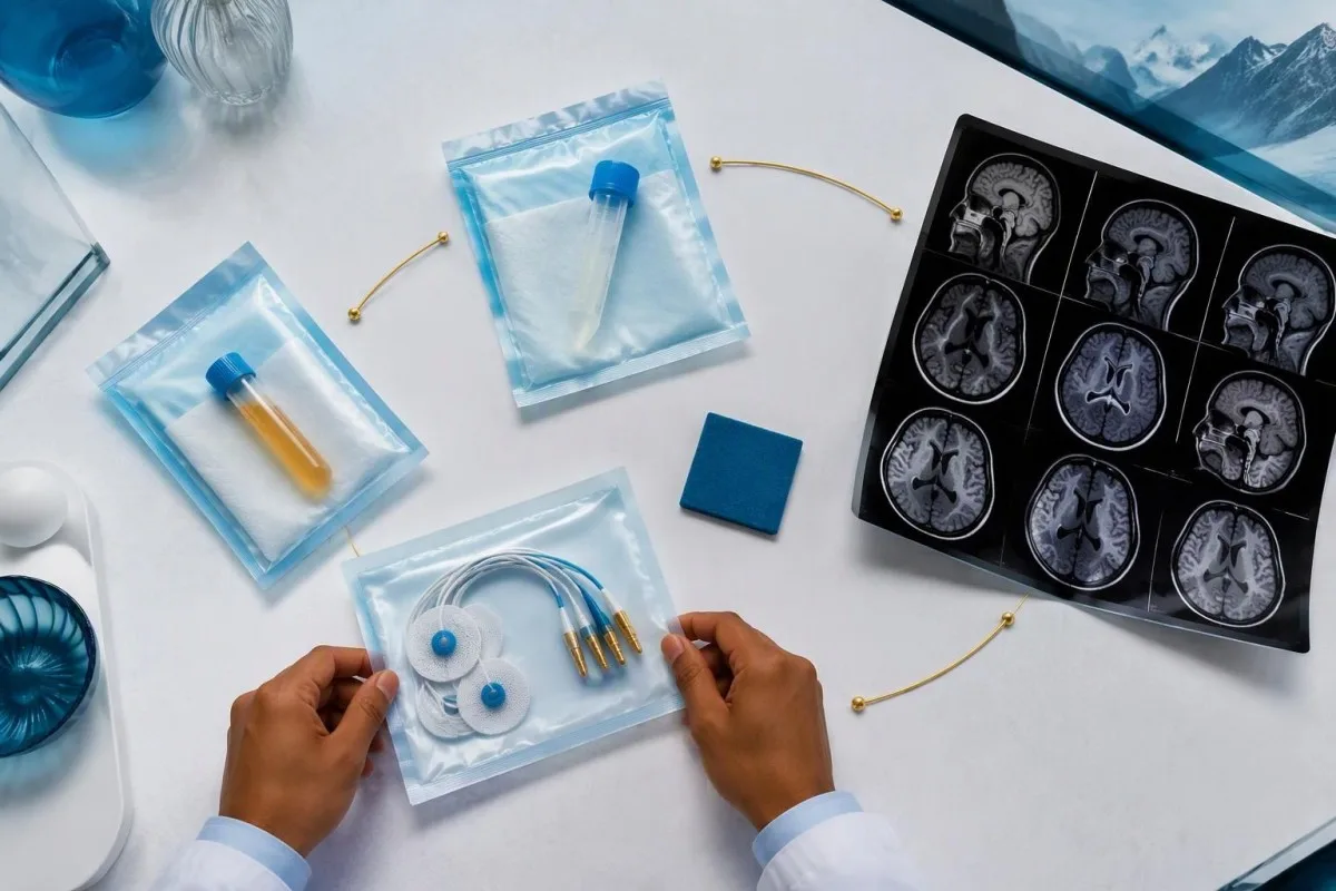
Nerve conduction studies can support a sensory neuronopathy pattern by showing reduced sensory responses across distributions that do not fit a single compressed nerve. Brain or spinal MRI addresses different questions, including structural disease and some inflammatory changes. A normal MRI does not exclude peripheral sensory ganglion involvement; these are 2 distinct anatomical compartments with different diagnostic tools.
Cerebrospinal fluid may show increased cells, protein or evidence of intrathecal immune activity, but none of those findings is specific to anti-Hu. A lumbar puncture is most useful when the neurological syndrome warrants it, rather than as an automatic response to 1 weak serum band. Our discussion of การแปลผลแถบโอลิโกโคลนัลในน้ำไขสันหลัง explains why immune activity in spinal fluid does not identify a single disease.
Basic investigations still matter: glucose control, vitamin status, renal function and serum protein assessment can uncover treatable contributors to neuropathy. A clinician may request 2 or more of these alongside specialist testing, depending on the examination. The คู่มือการประเมินโปรตีนในซีรัม provides background, but normal routine blood work neither confirms nor excludes an anti-Hu-associated neurological process.
การมี anti-Hu เป็นบวกยืนยันกลุ่มอาการพารานีโอพลาสติกหรือไม่?
A positive antibody alone does not confirm anti-Hu paraneoplastic syndrome. The PNS-Care approach combines the neurological phenotype, antibody risk category, cancer findings and follow-up; clinicians also check for direct cancer effects, treatment toxicity and unrelated neurological conditions before attributing symptoms to a paraneoplastic immune response.
Definite, probable and possible PNS are diagnostic categories, not cancer stages. The 2021 criteria use a structured scoring system rather than allowing 1 antibody to determine the whole diagnosis (Graus et al., 2021). A confirmed anti-Hu result with a strongly compatible syndrome remains clinically significant even before a cancer is identified, but clinicians should state clearly which parts of the assessment remain unresolved.
An older patient can have anti-Hu reactivity and a separate cause of numbness, such as diabetes, nutritional deficiency or a monoclonal protein disorder. Coexistence is possible; either-or reasoning can miss treatable disease. If serum protein studies reveal a band, our immunofixation result guide explains why that finding requires its own interpretation rather than being automatically folded into the anti-Hu diagnosis.
คันเตสตีเป็น แพลตฟอร์มการแปลผลตรวจเลือดของ AI that can help organize these distinctions in a laboratory explanation, but it cannot assign a reliable PNS-Care diagnosis from a report alone. Our กรอบการตรวจยืนยันทางคลินิก separates technical evaluation from clinical conclusions. In practice, I would document 3 separate judgments: confidence in the antibody, confidence in the neurological syndrome and the current evidence for cancer.
แพทย์ตรวจหามะเร็งที่เกี่ยวข้องอย่างไร?
Cancer evaluation usually prioritizes the chest because small-cell lung cancer is the strongest adult anti-Hu association. Doctors commonly begin with appropriate chest CT and may add FDG-PET/CT or targeted investigations if suspicion remains; the exact plan depends on age, symptoms, examination, smoking history and previous imaging.
The cancer association is substantial in established anti-Hu clinical cohorts. Villagrán-García et al. (2023) reported identified cancer in 349 of 466 patients, approximately 75%, in a specialist anti-Hu cohort. This is valuable evidence for taking confirmed, clinically relevant anti-Hu seriously; it is not a personalized prediction for an asymptomatic person with an unconfirmed positive screening strip.
A chest X-ray is not equivalent to a chest CT, and routine low-dose screening is not necessarily the correct examination for a suspected paraneoplastic syndrome. The clinician chooses the protocol and body regions based on the diagnostic question. Likewise, neurological symptoms do not automatically mean cancer has spread to the brain; our brain imaging warning-sign guide distinguishes structural investigation from immune-mediated disease.
A suspicious scan often leads to tissue sampling through a method chosen by respiratory or oncology specialists, such as bronchoscopic assessment of an accessible area. Imaging suspicion and cancer confirmation are 2 separate steps. Never-smoking status lowers the baseline probability of small-cell lung cancer but does not cancel a well-confirmed anti-Hu finding with a convincing neurological presentation; other cancers and non-cancer explanations still need consideration.
เหตุใดผลสแกนมะเร็งครั้งแรกจึงอาจเป็นลบ?
Initial imaging can be negative because an associated cancer is still too small to detect, lies outside the examined area or is not readily visible with that technique. Neurological autoimmunity may precede cancer detection, so a negative scan reduces concern without necessarily ending follow-up in a high-risk clinical presentation.
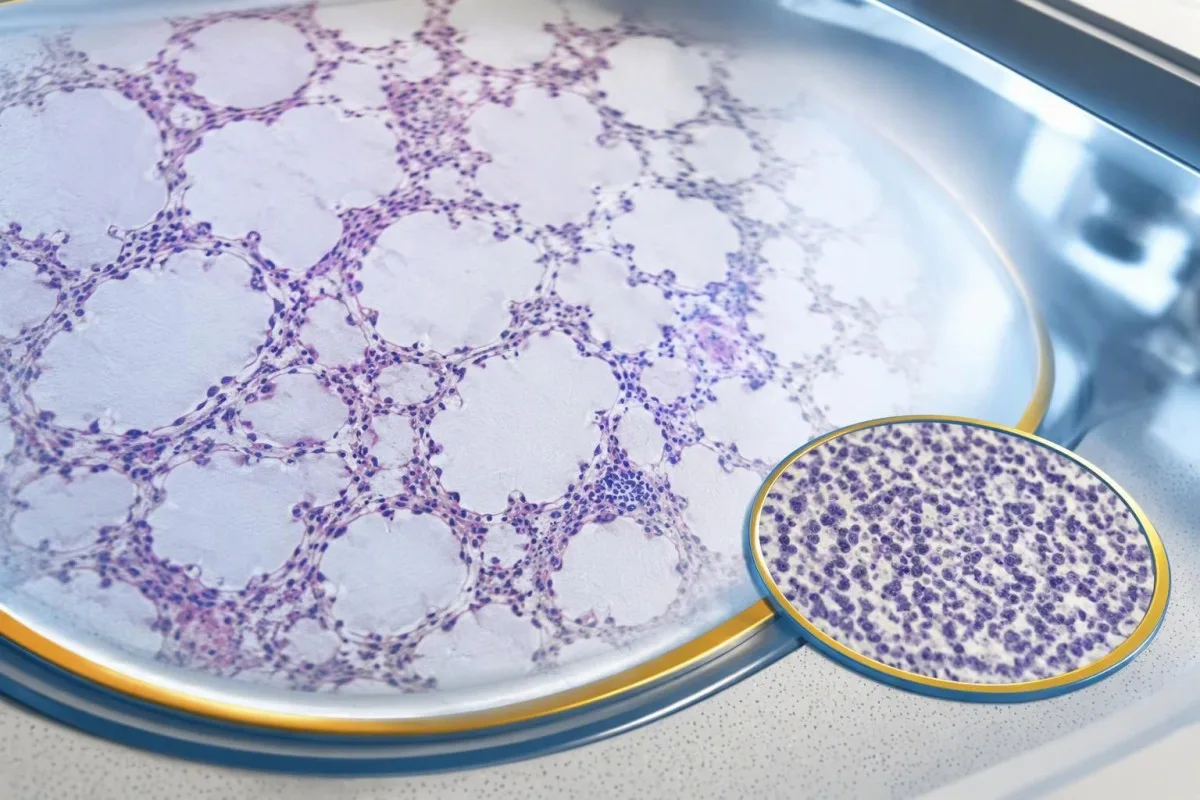
A negative scan is time-specific and method-specific. A normal chest CT performed on 1 date answers whether that examination identified a suspicious chest finding then; it does not guarantee that every possible associated cancer is absent indefinitely. Ask what was actually imaged, whether the protocol was suitable and whether the specialist recommends PET/CT or targeted investigation despite the initial result.
Villagrán-García et al. (2023) described cancers detected beyond the first 2 years in a minority of their anti-Hu cohort, supporting individualized longer surveillance. Their findings also support considering PET-based investigation when initial CT is unrevealing in an appropriate clinical context. This is an escalation decision for the treating team, not a reason to independently purchase repeated whole-body scans without weighing radiation, incidental findings and cost.
Neurological deterioration after negative imaging still needs reassessment. Cancer surveillance and neurological monitoring are 2 parallel tracks; one reassuring scan does not explain new loss of function. A marker such as neurofilament light chain may indicate neuronal injury in selected settings, but our NfL interpretation guide explains why it cannot establish anti-Hu disease, locate cancer or replace examination.
ควรตรวจคัดกรองมะเร็งต่อเนื่องนานเท่าใด?
The 2021 PNS criteria recommend repeat cancer screening every 4–6 months for 2 years when a high-risk neurological phenotype accompanies a high-risk antibody. Longer surveillance may be appropriate for selected anti-Hu patients; the interval and duration should reflect confirmation, clinical evolution, initial investigations and individual cancer risk.

The 2-year schedule is not an instruction to scan every positive antibody result repeatedly. Graus et al. (2021) link that recommendation to the combination of high-risk phenotype and high-risk antibody. An incidental, weak result without a matching syndrome needs confirmation and individualized assessment first; repeating imaging mechanically can create incidental findings and unnecessary procedures without resolving the original uncertainty.
Older European guidance recommended longer surveillance. The EFNS task force advised repeating screening after 3–6 months and then every 6 months for up to 4 years in relevant antibody-positive paraneoplastic presentations (Titulaer et al., 2011). The 2023 anti-Hu cohort supported considering screening up to 5 years in selected patients, so a specialist may reasonably extend follow-up beyond the newer general 2-year framework.
Keep a dated plan stating the next examination, the clinician responsible and what symptoms should trigger earlier contact. Kantesti explanations can help you understand terminology, but the platform should not be treated as a scan-reminder service or substitute for that plan. Our guide to risk-based cancer screening addresses a different setting: routine prevention is not the same as investigating a possible paraneoplastic syndrome.
การรักษาใดช่วยรักษาโรคทางระบบประสาทจาก anti-Hu?
Treatment focuses on an identified cancer, selected immune therapy and symptom-directed rehabilitation. Anti-Hu-associated neurological disease often responds less predictably to immunotherapy than neuronal surface-antibody disorders because intracellular targets and T-cell-mediated injury are involved; treatment may aim to stabilize function rather than promise complete recovery.

Treating an associated cancer is a central part of care, but established neurological injury may persist even when cancer treatment succeeds. Neurology and oncology teams may consider corticosteroids, intravenous immunoglobulin, plasma exchange or other immune treatments in selected situations. There is no single evidence-based dose or 1 universal regimen appropriate for everyone with anti-Hu positivity, especially without a confirmed clinical syndrome.
An antibody titer is not a dependable treatment target on its own. A lower number does not necessarily mean restored nerve function, and a persistent positive result does not by itself prove cancer progression. This differs from some surface-antibody disorders; our MOG antibody diagnosis guide explains another immune neurological condition, but its diagnostic rules and treatment expectations should not be transferred to anti-Hu.
Immune checkpoint cancer therapies deserve an explicit discussion when anti-Hu neurological disease is present, because they can provoke or worsen paraneoplastic neurological autoimmunity. This is a specialist risk-benefit decision, not an automatic prohibition based on 1 laboratory result. Tell the oncologist about previous neurological symptoms and confirmed neuronal antibodies before treatment decisions are finalized; coordinating the teams early can prevent avoidable misunderstanding.
คุณสามารถทำอะไรได้อย่างปลอดภัยระหว่างที่การตรวจประเมินยังดำเนินอยู่?
While investigations continue, protect mobility, swallowing and medication safety rather than trying to lower anti-Hu antibodies with supplements. Use the neurological symptoms—not the appearance of the laboratory flag—to decide whether ordinary follow-up is sufficient or urgent care is needed; a new functional loss warrants contact before the next planned scan.

Position-sense loss increases fall risk, particularly in poor lighting or on uneven ground. A physiotherapist may recommend a suitable walking aid and individualized balance strategies; occupational therapy can address hand coordination and home hazards. A short 7-day record of falls, near-falls and walking changes is often more useful at review than the vague statement “my balance is worse,” but stop activities that feel unsafe.
Excess vitamin B6 can itself cause sensory neuropathy, making unsupervised high-dose nerve supplements particularly unhelpful during this assessment. Review every supplement and its daily dose in mg, including overlapping multivitamins and combination products. Our คู่มือความปลอดภัย B-complex explains that problem; correcting a documented deficiency is different from taking progressively larger doses in the hope of treating anti-Hu autoimmunity.
Vitamin B12 deficiency can contribute to sensory and gait problems and may need investigation even when anti-Hu is confirmed. There is no universally interchangeable B12 cutoff: results in ห้องแล็บส่วนใหญ่รายงาน B12 เป็น และ pmol/L require their own laboratory context, sometimes with functional testing. Our คู่มือการติดตามผล B12 ก้ำกึ่งของเรา explains those next steps; new choking, breathing difficulty or rapid weakness should be assessed urgently instead of managed with vitamins.
AI ช่วยอธิบายรายงานผล anti-Hu อย่างปลอดภัยได้อย่างไร?
AI can help explain report terminology and organize questions, but it cannot confirm anti-Hu specificity, diagnose paraneoplastic syndrome or rule out cancer. Safe interpretation preserves the original assay details and separates 3 issues: what the laboratory reported, what the result may mean and what a clinician has actually established.

คันเตสตีเป็น เครื่องมือวิเคราะห์ผลตรวจเลือดที่ขับเคลื่อนด้วย AI that can provide a plain-language explanation of uploaded laboratory information, including the distinction between an antibody screen and a confirmed clinical diagnosis. A rapid explanation—often around 60 seconds for report processing—is not a reference-laboratory assay. Our คู่มือเทคโนโลยี AI describes the interpretation workflow without making anti-Hu-specific diagnostic guarantees.
The complete source report should remain available for verification. An explanation should not silently replace “weakly reactive” with “confirmed,” drop the specimen type or invent a universal reference interval. Our guide to การตรวจสอบคำอธิบายทางการแพทย์จาก AI describes practical source checks; for this antibody, compare at least the assay wording, confirmation comment and collection date against the original document.
Kantesti cannot examine sensation, review tissue staining directly through a report explanation or establish that negative imaging has excluded a tiny cancer. Those are 3 different clinical capabilities, not features that follow from understanding laboratory language. Share the explanation with the treating team as a discussion aid, and do not delay urgent assessment while uploading documents or waiting for a second automated interpretation.
การนัดหมายครั้งถัดไปและความโปร่งใสด้านการวิจัย
Bring the complete antibody report, a dated symptom history, previous imaging reports and a current medication list to your next appointment. Ask the clinician to clarify antibody confirmation, the neurological diagnosis and cancer surveillance separately; those 3 answers may have different levels of certainty and different follow-up dates.

A useful 1-page symptom history records onset, progression, sensory distribution, walking changes, memory problems and autonomic symptoms. Include prior cancer treatment and any recent immunoglobulin therapy, then ask who will coordinate neurology and oncology if both are involved. As Thomas Klein, MD, I would rather see a clear timeline than 20 disconnected screenshots; it makes discrepancies between the laboratory result and clinical course easier to spot.
Medical authorship is not a substitute for an individual neurological examination. ของเรา คณะกรรมการที่ปรึกษาทางการแพทย์ของ Kantesti describes the clinicians associated with our work, while your own specialist determines the appropriate next investigation. As of 25 ตุลาคม 2026, the recommendations discussed here draw on the 2021 international PNS criteria, the 2011 EFNS screening guidance and the 2023 anti-Hu cohort—not an invented 2026 guideline.
The 2 Figshare publications below are platform-related research references, not evidence validating anti-Hu diagnosis or cancer exclusion. Their supplied titles describe hantavirus triage deployment and a synthetic-case technical benchmark; neither title establishes anti-Hu-specific clinical performance. Because publication authors and dates were not supplied for verification, the citations use title-first APA formatting with “n.d.”; the ResearchGate and Academia.edu links are searches, not claims of verified publication copies.
คำถามที่พบบ่อย
ผลตรวจแอนติบอดี anti-Hu เป็นบวกหมายความว่าฉันเป็นมะเร็งหรือไม่?
0 ผลตรวจแอนติบอดี anti-Hu เป็นบวกไม่ได้วินิจฉัยว่าเป็นมะเร็งได้ด้วยตัวมันเอง Anti-Hu เป็นแอนติบอดีที่สัมพันธ์กับความเสี่ยงสูงต่อมะเร็ง และพบมะเร็งในผู้ป่วย 349 จาก 466 รายในกลุ่มผู้ป่วย anti-Hu ที่ดูแลโดยผู้เชี่ยวชาญในปี 2023 แต่ตัวเลขดังกล่าวไม่ได้บ่งชี้ความเสี่ยงจากผลคัดกรองที่พบโดยบังเอิญและยังไม่ได้รับการยืนยัน แพทย์จะประเมินอาการทางระบบประสาท ยืนยันผลตรวจทางห้องปฏิบัติการที่ผิดคาด และตรวจหามะเร็งที่อาจเป็นสาเหตุ โดยทั่วไปจะให้ความสำคัญกับทรวงอกในผู้ใหญ่เป็นลำดับแรก การวินิจฉัยมะเร็งโดยทั่วไปต้องอาศัยหลักฐานทางคลินิก ภาพถ่ายทางการแพทย์ และชิ้นเนื้อแยกต่างหาก.
anti-Hu กับ ANNA-1 เป็นสิ่งเดียวกันหรือไม่?
Anti-Hu และ ANNA-1 โดยทั่วไปหมายถึงแอนติบอดีต่อเซลล์ประสาทในตระกูลเดียวกัน ANNA-1 หมายถึงแอนติบอดีต่อนิวเคลียสของเซลล์ประสาทชนิดที่ 1 ส่วน Hu หมายถึงโปรตีนภายในเซลล์ประสาทที่แอนติบอดีจดจำ ANNA-1 ไม่ใช่ ANA ที่ตรวจเป็นประจำในการตรวจภูมิต้านตนเองทั่วร่างกาย ตรวจสอบรายละเอียดการตรวจและข้อความยืนยันผลให้แน่ชัด เนื่องจากห้องปฏิบัติการอาจรายงานแอนติบอดีในตระกูลเดียวกันด้วยรูปแบบที่แตกต่างกัน.
ผลตรวจแอนติบอดี anti-Hu อาจให้ผลบวกลวงได้หรือไม่?
การตรวจแอนติบอดี anti-Hu อาจให้ปฏิกิริยาที่ทำให้เข้าใจผิดหรือไม่จำเพาะ โดยเฉพาะเมื่อผลคัดกรองที่ให้ปฏิกิริยาอ่อน ๆ ไม่สอดคล้องกับอาการทางระบบประสาท โดยทั่วไปการยืนยันผลใช้แนวทางเสริมกัน 2 วิธี ได้แก่ รูปแบบการติดสีเนื้อเยื่อที่เป็นลักษณะเฉพาะ และการตรวจที่จำเพาะต่อแอนติเจนซึ่งแปลผลโดยห้องปฏิบัติการที่มีประสบการณ์ การตรวจซ้ำด้วยวิธีเชิงพาณิชย์แบบเดิมอาจทำให้เกิดปัญหาด้านการวิเคราะห์แบบเดิมซ้ำ แทนที่จะแก้ปัญหานั้น อาการทางระบบประสาทที่ดำเนินเร็วขึ้นยังคงต้องได้รับการประเมินอย่างทันท่วงทีระหว่างรอการยืนยันผล.
ควรตรวจสแกนซ้ำบ่อยเพียงใดหากผล anti-Hu เป็นบวก?
เกณฑ์ PNS ปี 2021 แนะนำให้ตรวจคัดกรองมะเร็งซ้ำทุก 4–6 เดือนเป็นเวลา 2 ปี เมื่อมีฟีโนไทป์ทางระบบประสาทที่มีความเสี่ยงสูงร่วมกับแอนติบอดีที่มีความเสี่ยงสูง ตารางการตรวจนี้ไม่ได้ใช้โดยอัตโนมัติกับผล anti-Hu เป็นบวกที่ตรวจพบโดยบังเอิญทุกราย แนวทาง EFNS ฉบับเก่าแนะนำให้เฝ้าระวังนานถึง 4 ปี และการศึกษา anti-Hu cohort ในปี 2023 สนับสนุนให้พิจารณาตรวจนานถึง 5 ปีในผู้ป่วยบางราย แพทย์ผู้เชี่ยวชาญของคุณควรระบุวิธีการตรวจภาพ ช่วงเวลาระหว่างการตรวจ ระยะเวลา และอาการที่ควรเข้ารับการตรวจเร็วกว่ากำหนด.
ผลตรวจ anti-Hu เป็นบวกได้หรือไม่ เมื่อผล CT และ PET ปกติ?
ผลตรวจ Anti-Hu อาจยังคงเป็นบวกได้เมื่อการตรวจภาพด้วย CT หรือ PET ยังไม่พบมะเร็ง ภูมิคุ้มกันทำลายระบบประสาทอาจเกิดขึ้นก่อนตรวจพบมะเร็ง และผลสแกนที่เป็นลบสะท้อนบริเวณที่ตรวจ เทคนิคที่ใช้ และวันที่ตรวจ ไม่ใช่การรับประกันถาวรว่าจะไม่มีมะเร็ง สำหรับอาการที่เข้าได้กับภาวะเสี่ยงสูง เกณฑ์ปี 2021 แนะนำให้ตรวจคัดกรองซ้ำทุก 4–6 เดือนเป็นเวลา 2 ปี โดยติดตามต่อให้นานขึ้นเป็นรายบุคคลในผู้ป่วย Anti-Hu ที่คัดเลือกไว้ นอกจากนี้ ควรพิจารณาผลปฏิกิริยาของแอนติบอดีที่ยังไม่ยืนยันและสาเหตุอื่นที่ไม่ใช่มะเร็งอีกครั้ง แทนที่จะสันนิษฐานว่ามีมะเร็งที่มองไม่เห็นอยู่แน่นอน.
การลดระดับแอนติบอดีต่อต้าน Hu หมายความว่าการรักษาได้ผลหรือไม่?
ระดับแอนติบอดี anti-Hu ที่ลดลงไม่ได้ยืนยันการฟื้นตัวทางระบบประสาทหรือความสำเร็จของการรักษามะเร็งได้อย่างน่าเชื่อถือ anti-Hu จดจำโปรตีนภายในเซลล์ และการบาดเจ็บทางระบบประสาทที่เกี่ยวข้องมักเกี่ยวข้องกับกลไกของทีเซลล์ มากกว่าผลของแอนติบอดีเพียงอย่างเดียว ค่าไตเตอร์สองค่าจากห้องปฏิบัติการต่างแห่งอาจเปรียบเทียบกันไม่ได้เช่นกัน เนื่องจากใช้วิธีการที่แตกต่างกัน แพทย์ประเมินการทำงาน ผลการตรวจร่างกาย การตรวจที่เกี่ยวข้อง และการตอบสนองของมะเร็ง แทนการใช้ตัวเลขแอนติบอดีเป็นเป้าหมายการรักษาเพียงอย่างเดียว.
รับการวิเคราะห์ผลตรวจเลือดด้วย AI วันนี้
เข้าร่วมผู้ใช้งานมากกว่า 2 ล้านคนทั่วโลกที่ไว้วางใจ Kantesti สำหรับการวิเคราะห์ผลตรวจทางห้องแล็บแบบทันทีและแม่นยำ อัปโหลดผลตรวจเลือดของคุณ แล้วรับการอ่านผลตรวจเลือดอย่างครอบคลุมของไบโอมาร์กเกอร์ 15,000+ ภายในไม่กี่วินาที.
📚 งานวิจัยที่อ้างอิง
Klein, T., Mitchell, S., & Weber, H. (2026). AI ช่วยแปลภาษาสำหรับการสนับสนุนการตัดสินใจทางคลินิกเพื่อคัดกรองโรคฮันทาไวรัสระยะเริ่มต้น: การออกแบบ การตรวจสอบความถูกต้องทางวิศวกรรม และการนำไปใช้จริงในรายงานผลเลือดที่แปลแล้วกว่า 50,000 ฉบับ.
Klein, T., Mitchell, S., & Weber, H. (2026). การประเมินเชิงเทคนิคอัตโนมัติแบบลงทะเบียนล่วงหน้าและยึดตามรูบริกของเครื่องมือการตีความผลตรวจเลือด Kantesti บนเคสทดสอบสังเคราะห์ 100,000 รายการ.
📖 อ้างอิงทางการแพทย์ภายนอก
Graus F et al. (2021). Updated Diagnostic Criteria for Paraneoplastic Neurologic Syndromes. Neurology: Neuroimmunology & Neuroinflammation.
Titulaer MJ et al. (2011). Screening for tumours in paraneoplastic syndromes: report of an EFNS Task Force. European Journal of Neurology.
Villagrán-García D et al. (2023). Revisiting anti-Hu paraneoplastic autoimmunity: phenotypic characterization and cancer diagnosis. Brain Communications.
⚕️ ข้อสงวนสิทธิ์ทางการแพทย์
บทความนี้จัดทำเพื่อวัตถุประสงค์ด้านการศึกษาเท่านั้น และไม่ถือเป็นคำแนะนำทางการแพทย์ โปรดปรึกษาผู้ให้บริการด้านสุขภาพที่มีคุณสมบัติเหมาะสมเสมอสำหรับการตัดสินใจด้านการวินิจฉัยและการรักษา.
สัญญาณความน่าเชื่อถือ E-E-A-T
ประสบการณ์
การทบทวนเชิงคลินิกโดยแพทย์ที่นำโดยกระบวนการตีความผลตรวจในห้องแล็บ.
ความเชี่ยวชาญ
โฟกัสด้านเวชศาสตร์ห้องปฏิบัติการเกี่ยวกับพฤติกรรมของไบโอมาร์กเกอร์ในบริบททางคลินิก.
อำนาจ
เขียนโดย ดร. โธมัส ไคลน์ (Dr. Thomas Klein) พร้อมทบทวนโดย ดร. ซาราห์ มิตเชลล์ (Dr. Sarah Mitchell) และ ศ.ดร. ฮันส์ เวเบอร์ (Prof. Dr. Hans Weber).
ความน่าเชื่อถือ
การตีความที่อิงหลักฐาน พร้อมเส้นทางการติดตามที่ชัดเจนเพื่อลดความตื่นตระหนก.
