
An anti-Hu antibody positive result raises concern for a cancer-associated neurological immune response, especially involving small-cell lung cancer, but it does not diagnose cancer. Doctors assess symptoms, confirm the laboratory finding and arrange cancer evaluation; repeat screening may be needed even when the first scans are negative.



- Anti-Hu / ANNA-1 is a high-risk paraneoplastic antibody, a category associated with cancer in more than 70% of established clinical cohorts—not every unconfirmed screening result.
- Cancer diagnosis requires evidence of an underlying malignancy, usually including tissue examination; one positive antibody result is not that evidence.
- Assay confirmation often combines 2 complementary approaches: a characteristic tissue-staining pattern and an antigen-specific test.
- Neurological symptoms such as progressive sensory loss, imbalance, seizures or autonomic dysfunction change the significance and urgency of the result.
- Negative imaging means no cancer was identified by that examination at that time; it does not eliminate the possibility of a small, initially undetectable cancer.
- Repeat screening is recommended every 4–6 months for 2 years when a high-risk neurological phenotype accompanies a high-risk antibody, according to the 2021 PNS criteria.
- Longer surveillance may be appropriate for selected patients; a 2023 anti-Hu cohort supported considering screening beyond 2 years, sometimes up to 5 years.
- የአስቸኳይ ምልክቶች include new breathing difficulty, inability to swallow safely, rapidly worsening weakness, a first seizure or sudden neurological deficits.
What does an anti-Hu antibody positive result mean?
አንድ anti-Hu antibody positive result means the laboratory detected reactivity against Hu neuronal proteins; it does not establish cancer or a neurological syndrome by itself. Interpretation has 3 parts: whether the result is genuine, whether the neurological presentation fits and whether an associated cancer can be found.
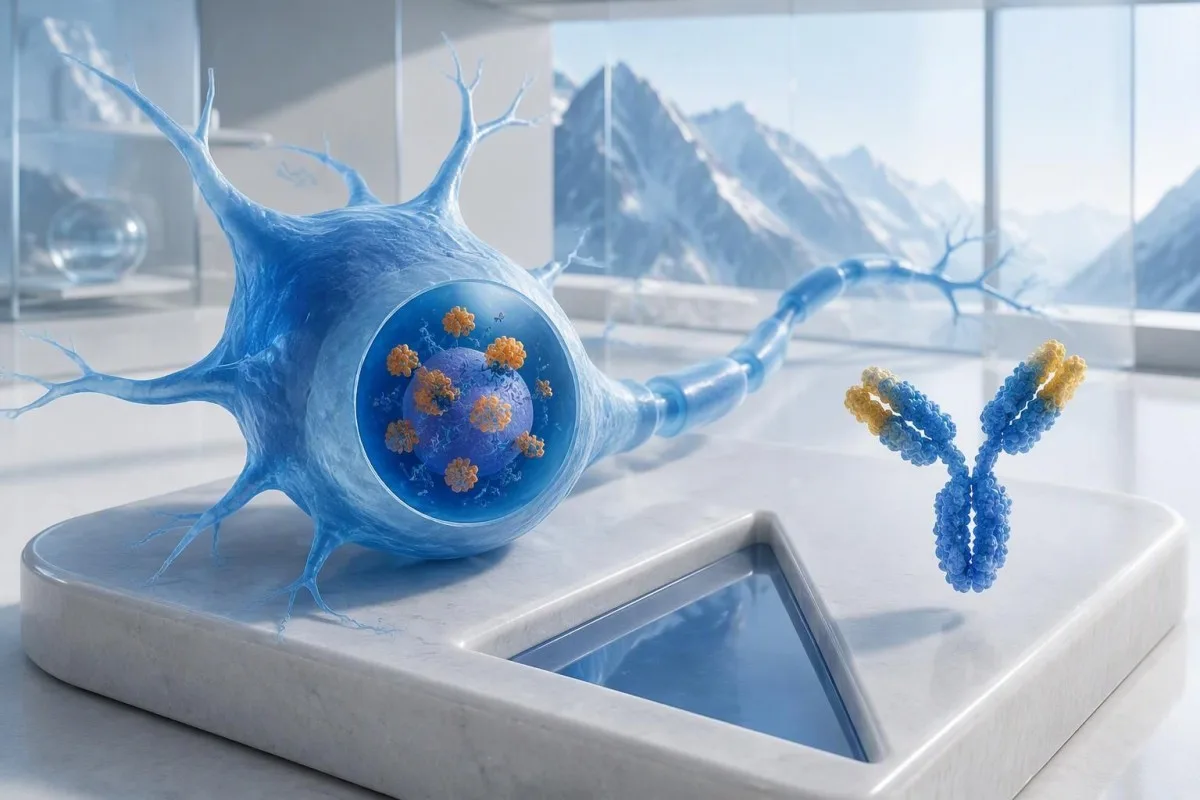
Anti-Hu is classified as a high-risk cancer-associated antibody, with the category defined by a cancer association above 70% in relevant clinical populations (Graus et al., 2021). That percentage cannot simply be assigned to an otherwise well person with a weak, unconfirmed panel result: the patients studied, the testing method and the neurological presentation all affect the probability.
A hypothetical 62-year-old with 8 weeks of worsening imbalance and asymmetric sensory loss deserves a different assessment from someone whose antibody appeared incidentally during a broad fatigue panel. Neither person should be told, “You have cancer,” from the laboratory page alone. Our explanation of አዎንታዊ የፀረ እንግዳ አካላት ግኝቶች helps separate an immune marker from a completed diagnosis.
ካንቴስቲ እ.ኤ.አ. AI የደም ምርመራ ተንታኝ that can help explain the wording of an anti-Hu report, but laboratory confirmation and cancer assessment belong with your treating clinicians. As Thomas Klein, MD, Chief Medical Officer at Kantesti LTD, my first question would be what prompted this particular test—not how alarming the red flag looks; our ድርጅት እና ክሊኒካዊ ተልዕኮ ያንን ልዩነት ያብራራሉ።.
Is ANNA-1 the same antibody as anti-Hu?
ANNA-1 antibody and anti-Hu antibody generally refer to the same antibody family. ANNA-1 stands for antineuronal nuclear antibody type 1; the Hu designation refers to the intracellular neuronal proteins recognized by the immune response, rather than to a separate disease or a cancer stage.
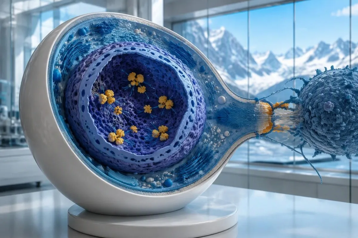
Hu proteins are intracellular RNA-binding proteins, and anti-Hu is best understood as a marker of an immune response that often involves cytotoxic T cells. This differs from disorders in which antibodies bind accessible proteins on the neuronal surface. The distinction explains why simply lowering antibody levels does not reliably reverse established anti-Hu neurological injury, even when 2 reports show markedly different titers.
A report reading “ANNA-1 detected” is not the same as a routine positive ANA, despite the similar letters. ኤኤንኤ usually means antinuclear antibodies used in systemic autoimmune assessment, whereas ANNA-1 identifies a neuronal antibody pattern. Keep those 2 abbreviations separate when discussing your results; our ANA terminology guide explains the more familiar test.
Anti-Hu can be detected in some people with small-cell lung cancer who do not have a recognizable neurological syndrome. Conversely, neurological symptoms can lead to antibody detection before the cancer becomes visible. Those are 2 different clinical pathways: the antibody does not tell a doctor, on its own, whether nerves are currently being injured or where an underlying cancer might be located.
How should the anti-Hu antibody test report be read?
An anti-Hu antibody test has no universal numerical cutoff that can be applied across laboratories. Read the specimen type, assay method, result category, laboratory reference interval and confirmation comments together; a dilution titer, band intensity and numerical index are 3 different ways of reporting reactivity.
A hypothetical titer of 1:240 describes detectable reactivity after a particular dilution; it is not a cancer probability of 240, nor does it establish cancer stage. A numerical index from another method may use entirely different calibration. Our guide to የጥራት ንፅፅር ከቁጥር ጋር explains why “positive” and “how much” answer different laboratory questions.
Weak or borderline reactivity deserves method-specific interpretation, particularly when a broad panel was ordered without a convincing neurological indication. Ask whether the laboratory observed the characteristic neuronal staining pattern and whether an antigen-specific confirmation was performed. Two tests using the same underlying strip technology may reproduce the same analytical problem; agreement is more informative when the methods are genuinely complementary.
Bring the complete report, not just a screenshot of the word positive. The 5 most useful details are the specimen, collection date, assay name, reported strength or titer and laboratory interpretation. Also list recent intravenous immunoglobulin and other immune treatments, because passive antibodies or treatment effects can complicate some serological results; your laboratory can advise whether that issue plausibly affects this particular finding.
When does a positive anti-Hu result need confirmation?
Unexpected, weak or clinically discordant anti-Hu results should be reviewed for confirmation, preferably with an experienced neuroimmunology laboratory. The 2021 PNS criteria emphasize interpreting neuronal antibody testing alongside the phenotype and using appropriate confirmatory methods rather than accepting an isolated commercial-panel result as a diagnosis.
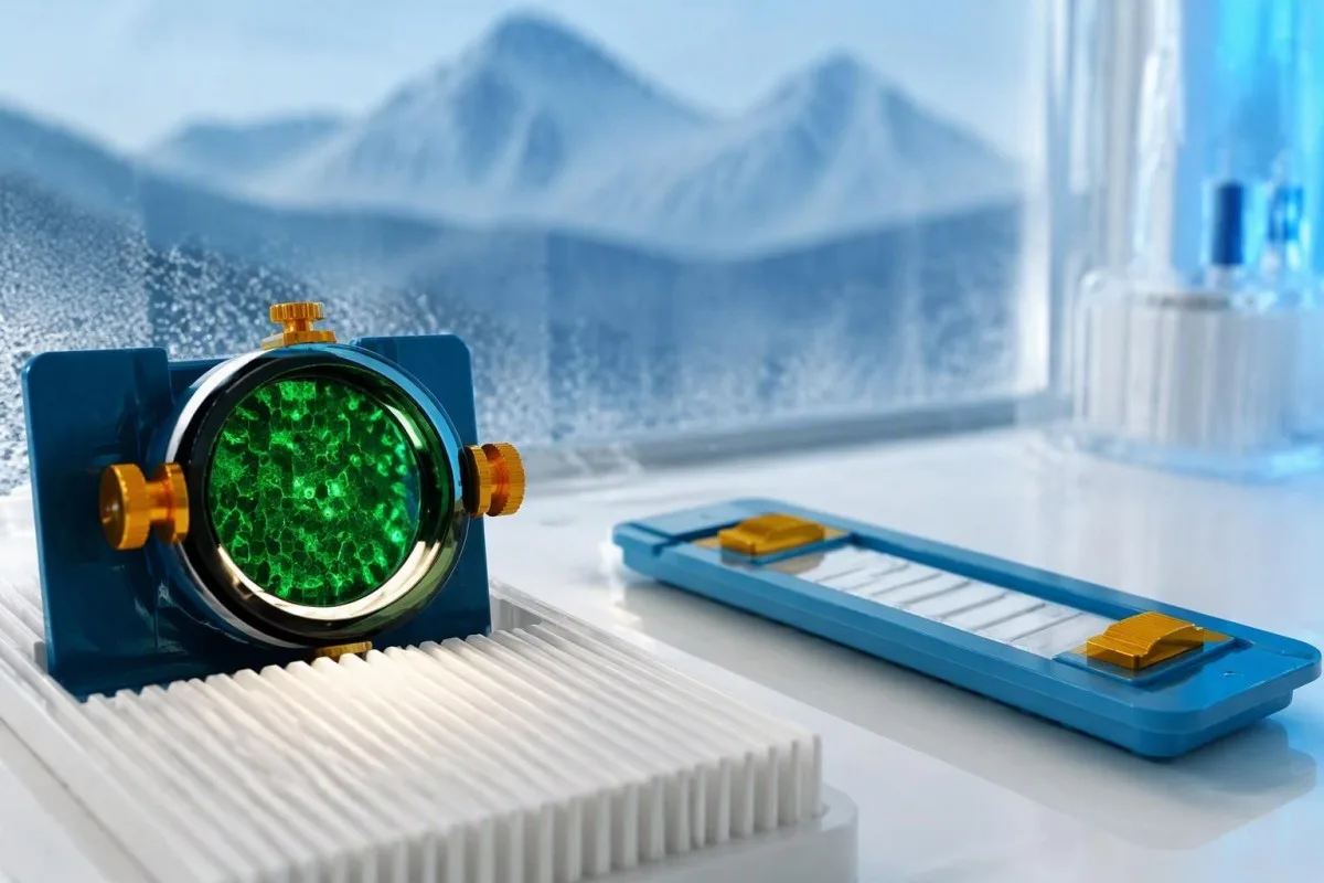
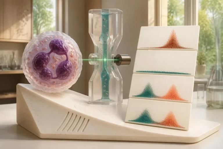
Tissue-based immunohistochemistry or immunofluorescence and an antigen-specific assay provide different information. The first identifies a characteristic distribution of neuronal staining; the second tests reactivity against the relevant antigen. Graus et al. (2021) describe these complementary approaches, with specialist reassessment particularly useful when 1 method gives a result that does not fit the clinical picture.
Repeating a sample can address collection errors or a transient analytical concern, but repeating the same kit is not always enough. In a hypothetical 45-year-old with no objective neurological findings and a faint isolated band, I would want the laboratory’s explanation before treating the report as established anti-Hu autoimmunity. Our GAD65 interpretation guide illustrates another antibody for which context and assay characteristics change meaning.
Confirmation and urgent assessment can happen in parallel. A person who develops seizures, substantial walking difficulty or progressive swallowing impairment should not wait several weeks for a reference-laboratory answer before receiving neurological care. The practical question is not “repeat or investigate?” but which of those 2 tasks can safely proceed together, given the symptoms and the initial laboratory evidence.
Which neurological symptoms make anti-Hu more concerning?
Progressive sensory neuronopathy, encephalomyelitis, limbic encephalitis and marked autonomic dysfunction are recognized anti-Hu presentations. Symptoms can involve more than 1 part of the nervous system, so the pattern and speed of change are usually more informative than a single symptom such as tingling or fatigue.

Sensory neuronopathy can cause asymmetric numbness, impaired position sense and sensory ataxia, sometimes involving the hands or trunk early rather than following a simple feet-first pattern. A patient may lose balance in the dark because vision had been compensating for reduced position feedback. That is different from the focal finger distribution discussed in our carpal tunnel symptom guide, although neither pattern can be diagnosed from description alone.
Anti-Hu autonomic involvement can affect gut movement, blood-pressure regulation and bladder function. Severe constipation, unexplained intestinal pseudo-obstruction or dizziness on standing becomes more relevant when accompanied by objective neurological abnormalities. Recording symptoms over 7 days can reveal combinations that otherwise get presented as separate stomach, balance and urinary complaints; sudden severe abdominal symptoms still require assessment for non-neurological emergencies.
New memory impairment, behavioral change or seizures may indicate central nervous system involvement, while rapid weakness has a broader emergency differential. The አስቸኳይ የድክመት ማስጠንቀቂያ ምልክቶች in our neurological safety guide apply regardless of the antibody label. Call emergency services for new breathing difficulty, unsafe swallowing, a first seizure or sudden stroke-like symptoms rather than waiting for a scheduled follow-up.
What neurological tests may follow a positive result?
Neurological follow-up may include examination, nerve conduction studies, MRI and cerebrospinal fluid testing, selected according to the symptoms. There is no mandatory battery for every anti-Hu-positive person; the purpose is to document which structures are affected and exclude other causes, not merely to collect additional positive tests.
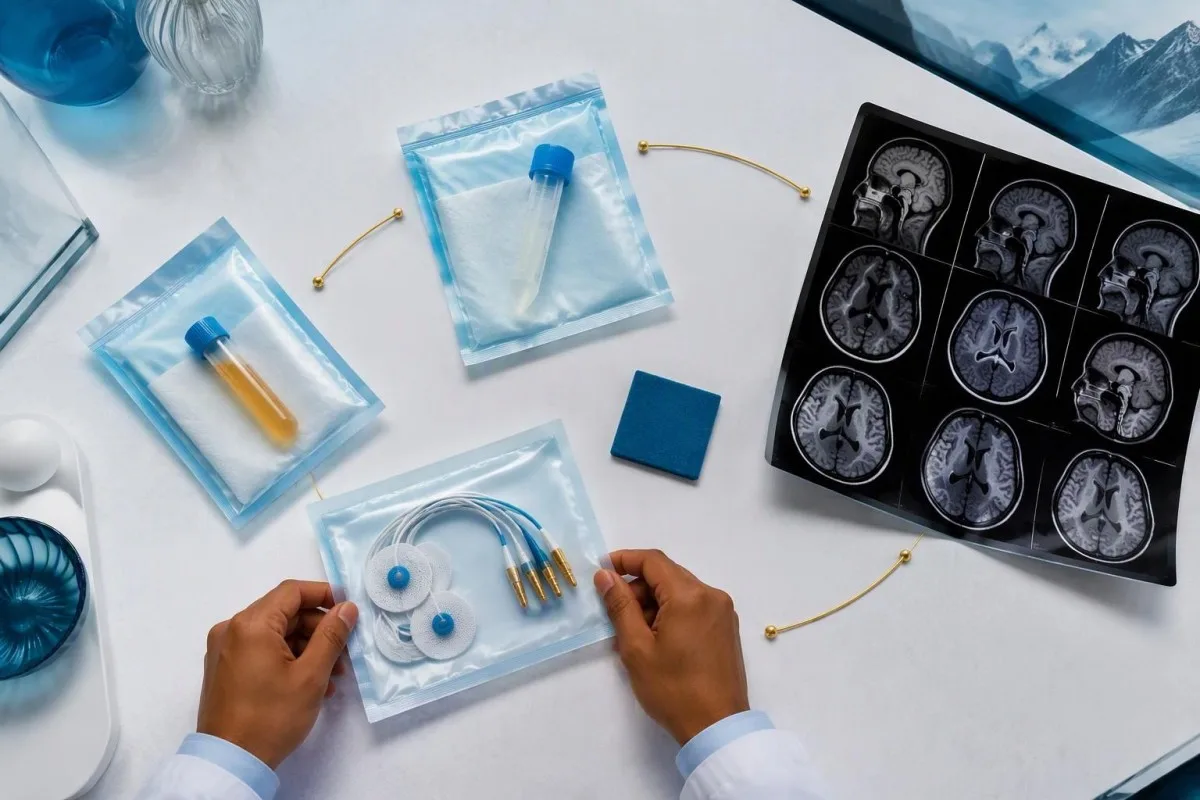
Nerve conduction studies can support a sensory neuronopathy pattern by showing reduced sensory responses across distributions that do not fit a single compressed nerve. Brain or spinal MRI addresses different questions, including structural disease and some inflammatory changes. A normal MRI does not exclude peripheral sensory ganglion involvement; these are 2 distinct anatomical compartments with different diagnostic tools.
Cerebrospinal fluid may show increased cells, protein or evidence of intrathecal immune activity, but none of those findings is specific to anti-Hu. A lumbar puncture is most useful when the neurological syndrome warrants it, rather than as an automatic response to 1 weak serum band. Our discussion of የCSF ኦሊጎክሎናል ባንድ ትርጓሜ explains why immune activity in spinal fluid does not identify a single disease.
Basic investigations still matter: glucose control, vitamin status, renal function and serum protein assessment can uncover treatable contributors to neuropathy. A clinician may request 2 or more of these alongside specialist testing, depending on the examination. The የሴረም ፕሮቲን ግምገማ መመሪያ provides background, but normal routine blood work neither confirms nor excludes an anti-Hu-associated neurological process.
Does anti-Hu positivity confirm a paraneoplastic syndrome?
A positive antibody alone does not confirm anti-Hu paraneoplastic syndrome. The PNS-Care approach combines the neurological phenotype, antibody risk category, cancer findings and follow-up; clinicians also check for direct cancer effects, treatment toxicity and unrelated neurological conditions before attributing symptoms to a paraneoplastic immune response.
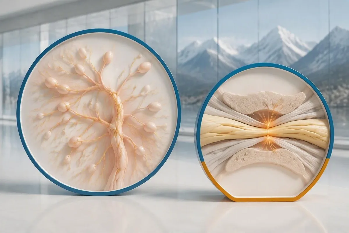
Definite, probable and possible PNS are diagnostic categories, not cancer stages. The 2021 criteria use a structured scoring system rather than allowing 1 antibody to determine the whole diagnosis (Graus et al., 2021). A confirmed anti-Hu result with a strongly compatible syndrome remains clinically significant even before a cancer is identified, but clinicians should state clearly which parts of the assessment remain unresolved.
An older patient can have anti-Hu reactivity and a separate cause of numbness, such as diabetes, nutritional deficiency or a monoclonal protein disorder. Coexistence is possible; either-or reasoning can miss treatable disease. If serum protein studies reveal a band, our immunofixation result guide explains why that finding requires its own interpretation rather than being automatically folded into the anti-Hu diagnosis.
ካንቴስቲ እ.ኤ.አ. AI የደም ምርመራ ውጤት ትርጓሜ መድረክ that can help organize these distinctions in a laboratory explanation, but it cannot assign a reliable PNS-Care diagnosis from a report alone. Our ክሊኒካዊ ማረጋገጫ ክፈፍ (clinical validation framework) separates technical evaluation from clinical conclusions. In practice, I would document 3 separate judgments: confidence in the antibody, confidence in the neurological syndrome and the current evidence for cancer.
How do doctors look for an associated cancer?
Cancer evaluation usually prioritizes the chest because small-cell lung cancer is the strongest adult anti-Hu association. Doctors commonly begin with appropriate chest CT and may add FDG-PET/CT or targeted investigations if suspicion remains; the exact plan depends on age, symptoms, examination, smoking history and previous imaging.
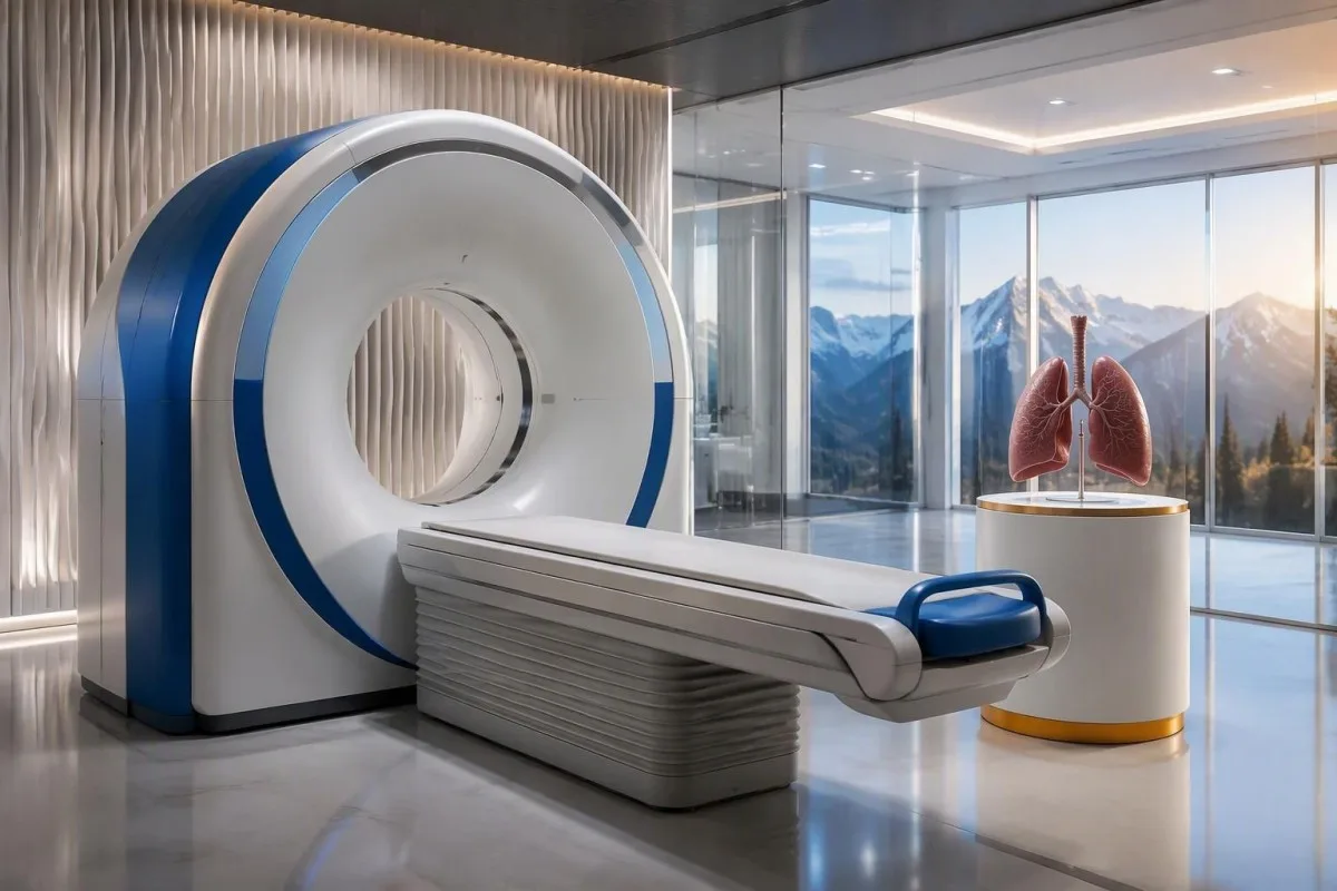
The cancer association is substantial in established anti-Hu clinical cohorts. Villagrán-García et al. (2023) reported identified cancer in 349 of 466 patients, approximately 75%, in a specialist anti-Hu cohort. This is valuable evidence for taking confirmed, clinically relevant anti-Hu seriously; it is not a personalized prediction for an asymptomatic person with an unconfirmed positive screening strip.
A chest X-ray is not equivalent to a chest CT, and routine low-dose screening is not necessarily the correct examination for a suspected paraneoplastic syndrome. The clinician chooses the protocol and body regions based on the diagnostic question. Likewise, neurological symptoms do not automatically mean cancer has spread to the brain; our brain imaging warning-sign guide distinguishes structural investigation from immune-mediated disease.
A suspicious scan often leads to tissue sampling through a method chosen by respiratory or oncology specialists, such as bronchoscopic assessment of an accessible area. Imaging suspicion and cancer confirmation are 2 separate steps. Never-smoking status lowers the baseline probability of small-cell lung cancer but does not cancel a well-confirmed anti-Hu finding with a convincing neurological presentation; other cancers and non-cancer explanations still need consideration.
Why can the first cancer scans be negative?
Initial imaging can be negative because an associated cancer is still too small to detect, lies outside the examined area or is not readily visible with that technique. Neurological autoimmunity may precede cancer detection, so a negative scan reduces concern without necessarily ending follow-up in a high-risk clinical presentation.
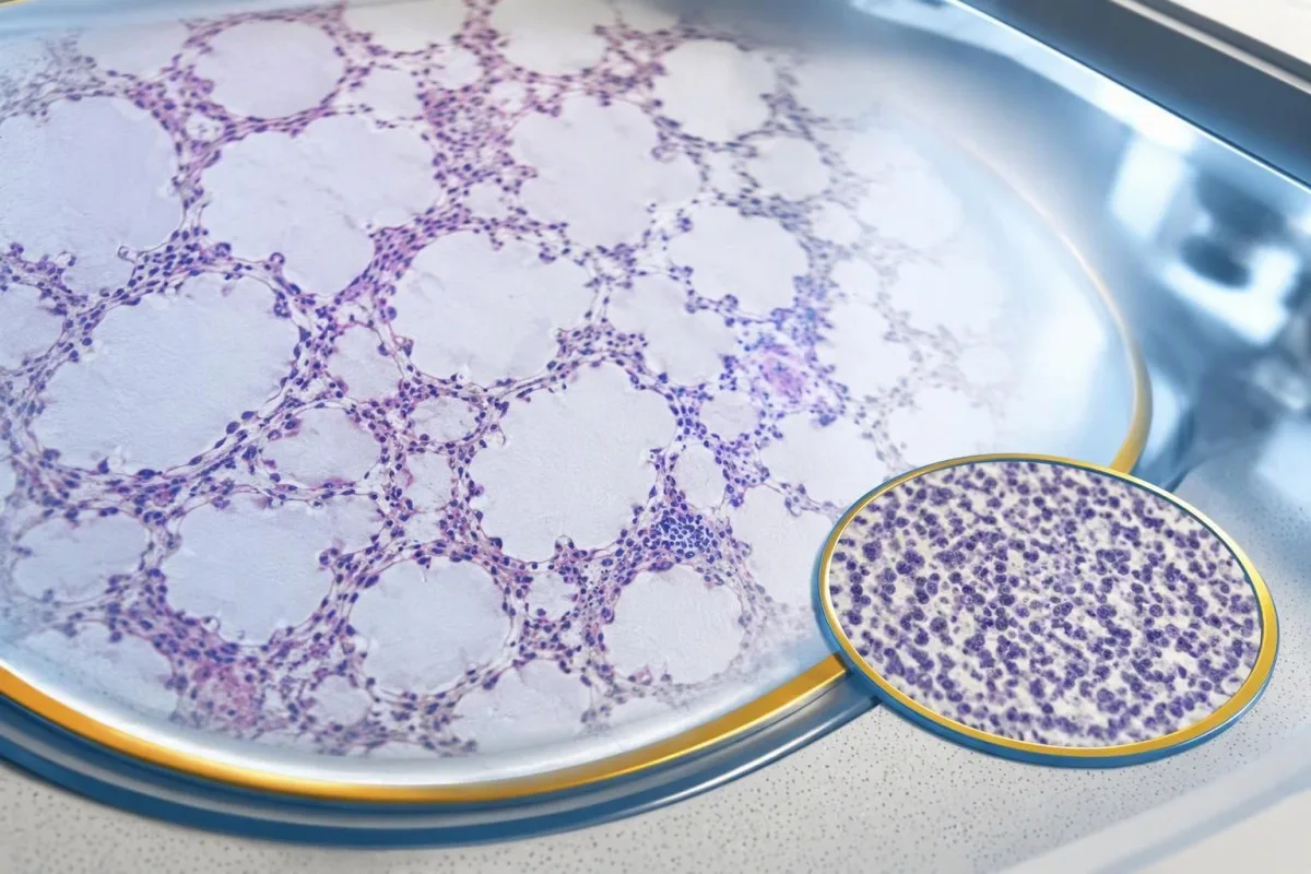
A negative scan is time-specific and method-specific. A normal chest CT performed on 1 date answers whether that examination identified a suspicious chest finding then; it does not guarantee that every possible associated cancer is absent indefinitely. Ask what was actually imaged, whether the protocol was suitable and whether the specialist recommends PET/CT or targeted investigation despite the initial result.
Villagrán-García et al. (2023) described cancers detected beyond the first 2 years in a minority of their anti-Hu cohort, supporting individualized longer surveillance. Their findings also support considering PET-based investigation when initial CT is unrevealing in an appropriate clinical context. This is an escalation decision for the treating team, not a reason to independently purchase repeated whole-body scans without weighing radiation, incidental findings and cost.
Neurological deterioration after negative imaging still needs reassessment. Cancer surveillance and neurological monitoring are 2 parallel tracks; one reassuring scan does not explain new loss of function. A marker such as neurofilament light chain may indicate neuronal injury in selected settings, but our NfL interpretation guide explains why it cannot establish anti-Hu disease, locate cancer or replace examination.
How long should cancer screening continue?
The 2021 PNS criteria recommend repeat cancer screening every 4–6 months for 2 years when a high-risk neurological phenotype accompanies a high-risk antibody. Longer surveillance may be appropriate for selected anti-Hu patients; the interval and duration should reflect confirmation, clinical evolution, initial investigations and individual cancer risk.

The 2-year schedule is not an instruction to scan every positive antibody result repeatedly. Graus et al. (2021) link that recommendation to the combination of high-risk phenotype and high-risk antibody. An incidental, weak result without a matching syndrome needs confirmation and individualized assessment first; repeating imaging mechanically can create incidental findings and unnecessary procedures without resolving the original uncertainty.
Older European guidance recommended longer surveillance. The EFNS task force advised repeating screening after 3–6 months and then every 6 months for up to 4 years in relevant antibody-positive paraneoplastic presentations (Titulaer et al., 2011). The 2023 anti-Hu cohort supported considering screening up to 5 years in selected patients, so a specialist may reasonably extend follow-up beyond the newer general 2-year framework.
Keep a dated plan stating the next examination, the clinician responsible and what symptoms should trigger earlier contact. Kantesti explanations can help you understand terminology, but the platform should not be treated as a scan-reminder service or substitute for that plan. Our guide to risk-based cancer screening addresses a different setting: routine prevention is not the same as investigating a possible paraneoplastic syndrome.
What treatment helps anti-Hu neurological disease?
Treatment focuses on an identified cancer, selected immune therapy and symptom-directed rehabilitation. Anti-Hu-associated neurological disease often responds less predictably to immunotherapy than neuronal surface-antibody disorders because intracellular targets and T-cell-mediated injury are involved; treatment may aim to stabilize function rather than promise complete recovery.

Treating an associated cancer is a central part of care, but established neurological injury may persist even when cancer treatment succeeds. Neurology and oncology teams may consider corticosteroids, intravenous immunoglobulin, plasma exchange or other immune treatments in selected situations. There is no single evidence-based dose or 1 universal regimen appropriate for everyone with anti-Hu positivity, especially without a confirmed clinical syndrome.
An antibody titer is not a dependable treatment target on its own. A lower number does not necessarily mean restored nerve function, and a persistent positive result does not by itself prove cancer progression. This differs from some surface-antibody disorders; our MOG antibody diagnosis guide explains another immune neurological condition, but its diagnostic rules and treatment expectations should not be transferred to anti-Hu.
Immune checkpoint cancer therapies deserve an explicit discussion when anti-Hu neurological disease is present, because they can provoke or worsen paraneoplastic neurological autoimmunity. This is a specialist risk-benefit decision, not an automatic prohibition based on 1 laboratory result. Tell the oncologist about previous neurological symptoms and confirmed neuronal antibodies before treatment decisions are finalized; coordinating the teams early can prevent avoidable misunderstanding.
What can you do safely while investigations continue?
While investigations continue, protect mobility, swallowing and medication safety rather than trying to lower anti-Hu antibodies with supplements. Use the neurological symptoms—not the appearance of the laboratory flag—to decide whether ordinary follow-up is sufficient or urgent care is needed; a new functional loss warrants contact before the next planned scan.

Position-sense loss increases fall risk, particularly in poor lighting or on uneven ground. A physiotherapist may recommend a suitable walking aid and individualized balance strategies; occupational therapy can address hand coordination and home hazards. A short 7-day record of falls, near-falls and walking changes is often more useful at review than the vague statement “my balance is worse,” but stop activities that feel unsafe.
Excess vitamin B6 can itself cause sensory neuropathy, making unsupervised high-dose nerve supplements particularly unhelpful during this assessment. Review every supplement and its daily dose in mg, including overlapping multivitamins and combination products. Our B-complex ደህንነት መመሪያ explains that problem; correcting a documented deficiency is different from taking progressively larger doses in the hope of treating anti-Hu autoimmunity.
Vitamin B12 deficiency can contribute to sensory and gait problems and may need investigation even when anti-Hu is confirmed. There is no universally interchangeable B12 cutoff: results in ፒጂ/ሚሊ እና pmol/L require their own laboratory context, sometimes with functional testing. Our ድንበር B12 የክትትል መመሪያ explains those next steps; new choking, breathing difficulty or rapid weakness should be assessed urgently instead of managed with vitamins.
How can AI help explain an anti-Hu report safely?
AI can help explain report terminology and organize questions, but it cannot confirm anti-Hu specificity, diagnose paraneoplastic syndrome or rule out cancer. Safe interpretation preserves the original assay details and separates 3 issues: what the laboratory reported, what the result may mean and what a clinician has actually established.

ካንቴስቲ እ.ኤ.አ. በ AI የተጎላበተ የደም ምርመራ ትንተና መሳሪያ that can provide a plain-language explanation of uploaded laboratory information, including the distinction between an antibody screen and a confirmed clinical diagnosis. A rapid explanation—often around 60 seconds for report processing—is not a reference-laboratory assay. Our የAI ቴክኖሎጂ መመሪያ describes the interpretation workflow without making anti-Hu-specific diagnostic guarantees.
The complete source report should remain available for verification. An explanation should not silently replace “weakly reactive” with “confirmed,” drop the specimen type or invent a universal reference interval. Our guide to የAI የሕክምና ማብራሪያዎችን መፈተሽ describes practical source checks; for this antibody, compare at least the assay wording, confirmation comment and collection date against the original document.
Kantesti cannot examine sensation, review tissue staining directly through a report explanation or establish that negative imaging has excluded a tiny cancer. Those are 3 different clinical capabilities, not features that follow from understanding laboratory language. Share the explanation with the treating team as a discussion aid, and do not delay urgent assessment while uploading documents or waiting for a second automated interpretation.
Your next appointment and research transparency
Bring the complete antibody report, a dated symptom history, previous imaging reports and a current medication list to your next appointment. Ask the clinician to clarify antibody confirmation, the neurological diagnosis and cancer surveillance separately; those 3 answers may have different levels of certainty and different follow-up dates.

A useful 1-page symptom history records onset, progression, sensory distribution, walking changes, memory problems and autonomic symptoms. Include prior cancer treatment and any recent immunoglobulin therapy, then ask who will coordinate neurology and oncology if both are involved. As Thomas Klein, MD, I would rather see a clear timeline than 20 disconnected screenshots; it makes discrepancies between the laboratory result and clinical course easier to spot.
Medical authorship is not a substitute for an individual neurological examination. የእኛ የህክምና አማካሪ ቦርድ መረጃ describes the clinicians associated with our work, while your own specialist determines the appropriate next investigation. As of ጥቅምት 25፣ 2026, the recommendations discussed here draw on the 2021 international PNS criteria, the 2011 EFNS screening guidance and the 2023 anti-Hu cohort—not an invented 2026 guideline.
The 2 Figshare publications below are platform-related research references, not evidence validating anti-Hu diagnosis or cancer exclusion. Their supplied titles describe hantavirus triage deployment and a synthetic-case technical benchmark; neither title establishes anti-Hu-specific clinical performance. Because publication authors and dates were not supplied for verification, the citations use title-first APA formatting with “n.d.”; the ResearchGate and Academia.edu links are searches, not claims of verified publication copies.
በተደጋጋሚ የሚጠየቁ ጥያቄዎች
የፀረ-Hu ፀረ እንግዳ አካል ውጤት አዎንታዊ መሆኑ ካንሰር አለብኝ ማለት ነው?
0 ፀረ-Hu አንቲቦዲ አዎንታዊ ውጤት በራሱ ካንሰርን አያረጋግጥም። ፀረ-Hu ከካንሰር ጋር የተያያዘ ከፍተኛ አደጋ ያለው አንቲቦዲ ሲሆን፣ በ2023 በተደረገ የልዩ ባለሙያዎች ፀረ-Hu ቡድን ጥናት ከ466 ታካሚዎች ውስጥ በ349 ካንሰር ተለይቷል፤ ይሁን እንጂ ይህ አሃዝ በድንገት የተገኘና ያልተረጋገጠ የማጣሪያ ውጤት ስጋትን አይወስንም። ሐኪሞች የነርቭ ምልክቶችን ይገመግማሉ፣ ያልተጠበቁ የላቦራቶሪ ግኝቶችን ያረጋግጣሉ፣ እንዲሁም ዋና የሆነ ካንሰር መኖሩን ይመረምራሉ፤ በአዋቂዎች ላይ ብዙውን ጊዜ በደረት ምርመራ ላይ ቅድሚያ ይሰጣሉ። የካንሰር ምርመራ በአጠቃላይ የተለየ ክሊኒካዊ፣ የምስል እና የቲሹ ማስረጃ ይፈልጋል።.
0. ፀረ-Hu እና ANNA-1 አንድ ነገር ናቸው?
Anti-Hu እና ANNA-1 በአጠቃላይ አንድና ያው የነርቭ ሴል ፀረ እንግዳ አካል ቤተሰብን ይለያሉ። ANNA-1 ማለት የነርቭ ሴሎች አንቲኒውክሌር ፀረ እንግዳ አካል ዓይነት 1 ማለት ሲሆን፣ Hu ደግሞ ፀረ እንግዳ አካሉ የሚያውቃቸውን በሴል ውስጥ የሚገኙ የነርቭ ፕሮቲኖችን ያመለክታል። ANNA-1 በስርዓታዊ ራስ-ሰር በሽታ ምርመራ ውስጥ ከሚጠቀምበት መደበኛ ANA ጋር አንድ አይደለም። ላቦራቶሪዎች አንድን የፀረ እንግዳ አካል ቤተሰብ በተለያዩ ቅርጸቶች ሊያቀርቡ ስለሚችሉ፣ ትክክለኛውን የምርመራ ዘዴ እና የማረጋገጫ ቃላት ያረጋግጡ።.
የፀረ-Hu ፀረ እንግዳ አካል ምርመራ በስህተት አዎንታዊ ሊሆን ይችላል?
የፀረ-Hu ፀረ-እንግዳ አካል ምርመራ አሳሳች ወይም ልዩ ያልሆነ ምላሽ ሊያሳይ ይችላል፤ በተለይም ደካማ የመጀመሪያ ምርመራ ውጤት ከኒውሮሎጂያዊ ምልክቶቹ ጋር በማይጣጣምበት ጊዜ። ማረጋገጫ ብዙውን ጊዜ 2 ተጓዳኝ ዘዴዎችን ይጠቀማል፦ የተለየ የቲሹ ቀለም አያያዝ ንድፍ እና ልምድ ባለው ላቦራቶሪ የሚተረጎም አንቲጂን-ተኮር ምርመራ። ተመሳሳዩን የንግድ ዘዴ መድገም ችግሩን ከመፍታት ይልቅ ተመሳሳይ የትንታኔ ችግርን እንደገና ሊያመጣ ይችላል። በፍጥነት እየተባባሱ የሚሄዱ የኒውሮሎጂ ምልክቶች ማረጋገጫው እየተጠበቀ እያለም በወቅቱ ግምገማ ያስፈልጋቸዋል።.
ፀረ-Hu አዎንታዊ ከሆነ ስካኖች ምን ያህል ጊዜ ሊደገሙ ይገባል?
የ2021 PNS መስፈርቶች ከከፍተኛ አደጋ አንቲቦዲ ጋር ከፍተኛ አደጋ ያለው የኒውሮሎጂ ፊኖታይፕ ሲኖር በየ4–6 ወሩ ለ2 ዓመታት የካንሰር ምርመራን እንዲደግሙ ይመክራሉ። ይህ የጊዜ ሰሌዳ በአጋጣሚ anti-Hu አዎንታዊ ውጤት ለታየባቸው ሁሉ በራስ-ሰር አይተገበርም። ከዚህ ቀደም የወጣው የEFNS መመሪያ እስከ4 ዓመት ድረስ ክትትል እንዲደረግ ይመክር ነበር፣ እና የ2023 የanti-Hu ቡድን ጥናት ለተመረጡ ታካሚዎች እስከ5 ዓመት ድረስ ማሰብን ደግፏል። ስፔሻሊስትዎ የምስል ምርመራ ዘዴውን፣ የምርመራ ልዩነቱን፣ የሚቆይበትን ጊዜ እና ቀደም ብሎ እንዲታዩ የሚያስፈልጉ ምልክቶችን መግለጽ አለበት።.
ሲቲ እና ፒኢቲ ስካኖች መደበኛ ሲሆኑ ፀረ-Hu ፖዘቲቭ ሊሆን ይችላል?
Anti-Hu can remain positive when CT or PET imaging has not identified cancer. Neurological autoimmunity may precede detection, and a negative scan reflects the examined regions, technique and date rather than a permanent guarantee that cancer is absent. For a compatible high-risk presentation, the 2021 criteria recommend repeat screening every 4–6 months for 2 years, with individualized longer follow-up in selected anti-Hu patients. Unconfirmed antibody reactivity and non-cancer explanations should also be reconsidered rather than assuming an invisible cancer is definitely present.
Does lowering the anti-Hu antibody level mean treatment is working?
A falling anti-Hu antibody level does not reliably prove neurological recovery or successful cancer treatment. Anti-Hu recognizes intracellular proteins, and associated neurological injury often involves T-cell mechanisms rather than antibody effects alone. Two titers from different laboratories may also be non-comparable because the methods differ. Clinicians assess function, examination findings, relevant investigations and cancer response instead of using the antibody number as a stand-alone treatment target.
ዛሬ የAI-የኃይል የደም ምርመራ ትንተና ያግኙ
በፍጥነት እና ትክክለኛ የላቦራቶሪ ምርመራ ትንተና ለማግኘት Kantestiን የሚያምኑ ከ2 ሚሊዮን በላይ ተጠቃሚዎችን ይቀላቀሉ። የደም ምርመራ ውጤትዎን ይስቀሉ እና በ15,000+ ባዮማርከሮች ላይ የተሟላ ትርጓሜን በሰከንዶች ውስጥ ይቀበሉ።.
📚 የተጠቀሱ የምርምር ህትመቶች
📖 ውጫዊ የሕክምና ማጣቀሻዎች
Graus F et al. (2021). Updated Diagnostic Criteria for Paraneoplastic Neurologic Syndromes. Neurology: Neuroimmunology & Neuroinflammation.
Titulaer MJ et al. (2011). Screening for tumours in paraneoplastic syndromes: report of an EFNS Task Force.። የአውሮፓ የነርቭ ሕክምና ጆርናል።.
Villagrán-García D et al. (2023). Revisiting anti-Hu paraneoplastic autoimmunity: phenotypic characterization and cancer diagnosis.። Brain Communications።.
⚕️ የሕክምና ማስተባበያ
ይህ ጽሑፍ ለትምህርታዊ ዓላማ ብቻ ነው እና የሕክምና ምክር አይደለም። ለምርመራ እና ለሕክምና ውሳኔዎች ሁልጊዜ ብቁ የጤና ባለሙያን ያማክሩ።.
የE-E-A-T እምነት ምልክቶች
ልምድ
በሐኪም መሪነት የላቦራቶሪ ትርጓሜ የስራ ፍሰቶች ክሊኒካዊ ግምገማ።.
ባለሙያነት
በክሊኒካዊ አውድ ውስጥ ባዮማርከሮች እንዴት እንደሚሰሩ ላይ የላቦራቶሪ ሕክምና ትኩረት።.
ስልጣን ያለው
በዶክተር ቶማስ ክላይን የተፃፈ ከዶክተር ሳራ ሚቸል እና ፕሮፌሰር ዶክተር ሃንስ ዌበር ግምገማ ጋር።.
አስተማማኝነት
ለማስጠንቀቂያ ምላሽ መቀነስ ግልጽ የቀጣይ መንገዶች ያለው በማስረጃ የተደገፈ ትርጓሜ።.
