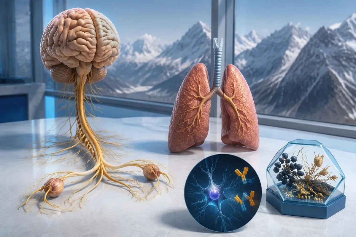
Позитивниот резултат на антитела против Hu предизвикува сомнеж за невролошки имунолошки одговор поврзан со рак, особено со ситноклеточен рак на белите дробови, но не поставува дијагноза на рак. Лекарите ги проценуваат симптомите, го потврдуваат лабораторискиот наод и организираат проценка за рак; може да биде потребно повторно скринирање дури и кога првите снимки се негативни.



- Анти-Hu / ANNA-1 е паранеопластично антитело со висок ризик, категорија поврзана со рак кај повеќе од 70% од утврдените клинички кохорти — но не и со секој непотврден резултат од скрининг.
- Дијагноза на рак бара докази за основен малигнитет, обично вклучително и испитување на ткиво; еден позитивен резултат на антитела не е таков доказ.
- Потврда со анализа често комбинира 2 комплементарни пристапи: карактеристичен образец на боење на ткивото и тест специфичен за антигенот.
- Невролошките симптоми како прогресивно губење на сетилноста, нарушена рамнотежа, напади или автономна дисфункција ја менуваат важноста и итноста на резултатот.
- Негативни снимки значат дека при тој преглед во тој момент не бил откриен рак; тоа не ја исклучува можноста за мал, првично неоткрилив рак.
- Повторниот скрининг се препорачува на секои 4–6 месеци во текот на 2 години кога високо ризичен невролошки фенотип е придружен со високо ризично антитело, според критериумите на PNS од 2021 година.
- Подолготрајното следење може да биде соодветно за одредени пациенти; кохортна студија за anti-Hu од 2023 година поддржа разгледување на скрининг и по 2 години, понекогаш до 5 години.
- Итни симптоми вклучуваат нови тешкотии со дишењето, неможност за безбедно голтање, брзо влошување на слабоста, прв епилептичен напад или ненадејни невролошки дефицити.
Што значи позитивен резултат на антитела против Hu?
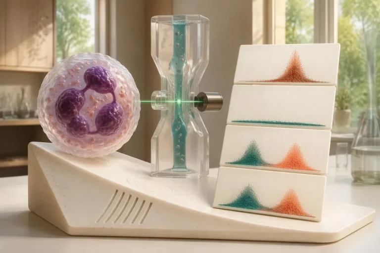
Ан позитивен резултат за anti-Hu антитела значи дека лабораторијата открила реактивност кон Hu невронските протеини; тоа само по себе не потврдува рак или невролошки синдром. Толкувањето има 3 дела: дали резултатот е вистински, дали невролошката клиничка слика одговара и дали може да се открие поврзан рак.
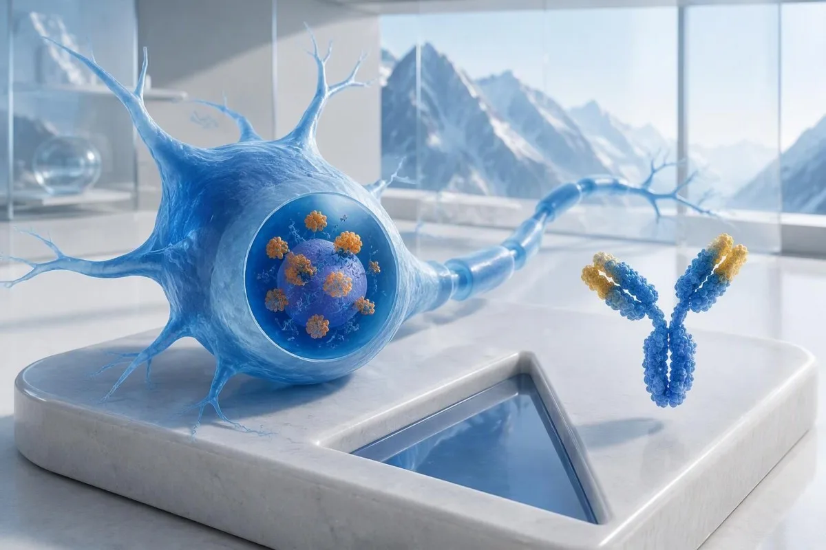
Anti-Hu е класифицирано како антитело со висок ризик поврзано со рак, при што категоријата е дефинирана со поврзаност со рак над 70% кај релевантни клинички популации (Graus et al., 2021). Тој процент не може едноставно да му се припише на инаку здраво лице со слаб, непотврден резултат од панелот: испитуваните пациенти, методот на тестирање и невролошката клиничка слика влијаат врз веројатноста.
На хипотетички 62-годишник со 8 недели влошување на рамнотежата и асиметрично губење на сетилноста треба да му се направи поинаква процена отколку на лице кај кое антителото било случајно откриено во широк панел за замор. На ниту едно од овие лица не треба да му се каже: “Имате рак”, само врз основа на лабораторискиот извештај. Нашето објаснување за позитивни наоди за антитела помага да се разграничи имунолошкиот маркер од поставена дијагноза.
Кантести е анализатор на крвна слика со вештачка интелигенција што може да помогне да се објасни формулацијата во извештајот за anti-Hu, но лабораториската потврда и процената за рак спаѓаат во надлежност на лекарите што ве лекуваат. Како Thomas Klein, MD, главен медицински директор во Kantesti LTD, првото прашање што би го поставил е што го поттикнало ова конкретно тестирање — а не колку загрижувачки изгледа црвеното предупредување; нашето организација и клиничка мисија ја објаснуваат таа разлика.
Дали ANNA-1 е истото антитело како анти-Hu?
ANNA-1 антитело и anti-Hu антитело обично се однесуваат на истото семејство антитела. ANNA-1 е кратенка од антиневронско нуклеарно антитело тип 1; ознаката Hu се однесува на внатреклеточните невронски протеини што ги препознава имунолошкиот одговор, а не на посебна болест или стадиум на рак.
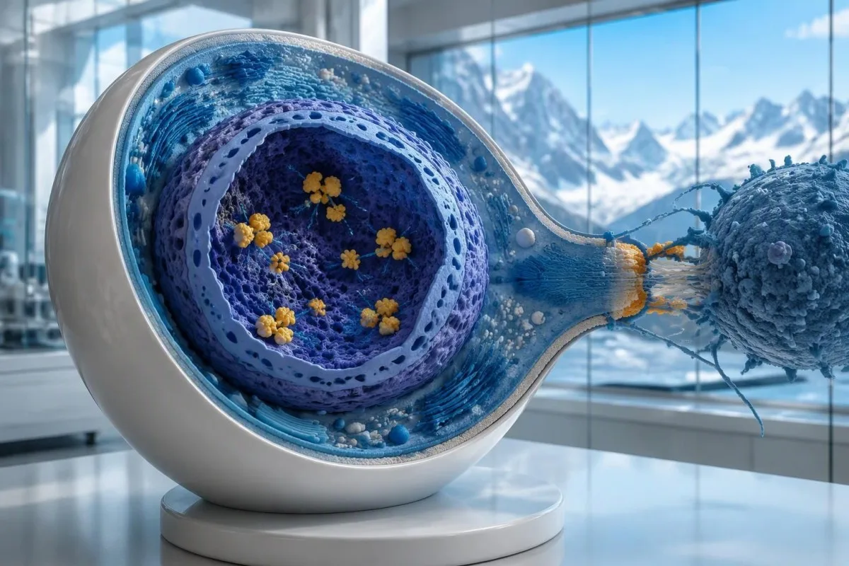
Hu протеините се внатреклеточни РНК-врзувачки протеини, а anti-Hu најдобро се разбира како маркер на имунолошки одговор во кој често учествуваат цитотоксичните Т-клетки. Ова се разликува од нарушувањата кај кои антителата се врзуваат за достапни протеини на површината на невроните. Оваа разлика објаснува зошто самото намалување на нивото на антителата не ја повлекува сигурно веќе настанатата невролошка повреда поврзана со anti-Hu, дури и кога 2 извештаи покажуваат значително различни титри.
Извештај во кој пишува “откриено е ANNA-1” не е исто што и рутински позитивен ANA, и покрај сличните букви. АНА обично се однесува на антинуклеарни антитела што се користат при процена на системски автоимуни заболувања, додека ANNA-1 означува невронски образец на антитела. Разликувајте ги овие 2 кратенки кога разговарате за резултатите; нашиот водич за терминологијата ANA го објаснува попознатиот тест.
Anti-Hu може да се открие кај некои лица со ситноклеточен рак на белите дробови кои немаат препознатлив невролошки синдром. Обратно, невролошките симптоми може да доведат до откривање антитела пред ракот да стане видлив. Тоа се 2 различни клинички текови: само по себе, антителото не му кажува на лекарот дали нервите моментално се оштетуваат или каде би можел да се наоѓа основниот рак.
Како треба да се толкува извештајот од тестот за антитела против Hu?
An anti-Hu antibody test has no universal numerical cutoff that can be applied across laboratories. Read the specimen type, assay method, result category, laboratory reference interval and confirmation comments together; a dilution titer, band intensity and numerical index are 3 different ways of reporting reactivity.
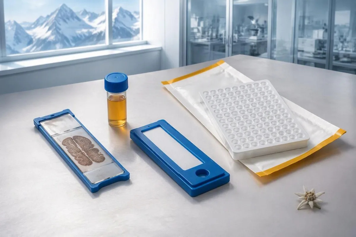
A hypothetical titer of 1:240 describes detectable reactivity after a particular dilution; it is not a cancer probability of 240, nor does it establish cancer stage. A numerical index from another method may use entirely different calibration. Our guide to квалитативни наспроти квантитативни резултати explains why “positive” and “how much” answer different laboratory questions.
Weak or borderline reactivity deserves method-specific interpretation, particularly when a broad panel was ordered without a convincing neurological indication. Ask whether the laboratory observed the characteristic neuronal staining pattern and whether an antigen-specific confirmation was performed. Two tests using the same underlying strip technology may reproduce the same analytical problem; agreement is more informative when the methods are genuinely complementary.
Bring the complete report, not just a screenshot of the word positive. The 5 most useful details are the specimen, collection date, assay name, reported strength or titer and laboratory interpretation. Also list recent intravenous immunoglobulin and other immune treatments, because passive antibodies or treatment effects can complicate some serological results; your laboratory can advise whether that issue plausibly affects this particular finding.
Кога е потребно потврдување на позитивен резултат за анти-Hu?
Unexpected, weak or clinically discordant anti-Hu results should be reviewed for confirmation, preferably with an experienced neuroimmunology laboratory. The 2021 PNS criteria emphasize interpreting neuronal antibody testing alongside the phenotype and using appropriate confirmatory methods rather than accepting an isolated commercial-panel result as a diagnosis.
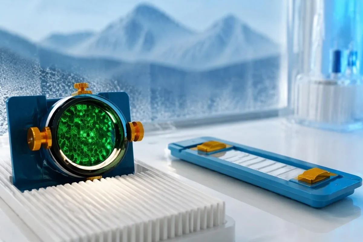
Tissue-based immunohistochemistry or immunofluorescence and an antigen-specific assay provide different information. The first identifies a characteristic distribution of neuronal staining; the second tests reactivity against the relevant antigen. Graus et al. (2021) describe these complementary approaches, with specialist reassessment particularly useful when 1 method gives a result that does not fit the clinical picture.
Repeating a sample can address collection errors or a transient analytical concern, but repeating the same kit is not always enough. In a hypothetical 45-year-old with no objective neurological findings and a faint isolated band, I would want the laboratory’s explanation before treating the report as established anti-Hu autoimmunity. Our GAD65 interpretation guide illustrates another antibody for which context and assay characteristics change meaning.
Confirmation and urgent assessment can happen in parallel. A person who develops seizures, substantial walking difficulty or progressive swallowing impairment should not wait several weeks for a reference-laboratory answer before receiving neurological care. The practical question is not “repeat or investigate?” but which of those 2 tasks can safely proceed together, given the symptoms and the initial laboratory evidence.
Кои невролошки симптоми предизвикуваат поголема загриженост во врска со анти-Hu?
Progressive sensory neuronopathy, encephalomyelitis, limbic encephalitis and marked autonomic dysfunction are recognized anti-Hu presentations. Symptoms can involve more than 1 part of the nervous system, so the pattern and speed of change are usually more informative than a single symptom such as tingling or fatigue.
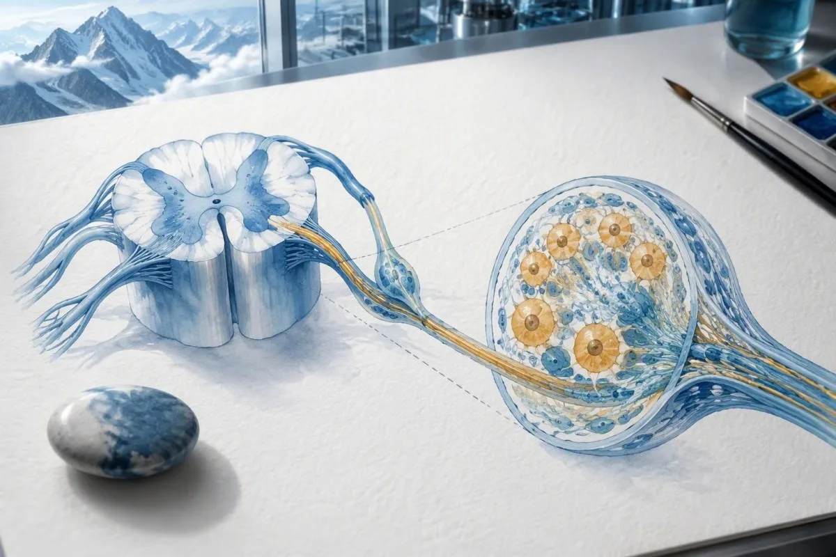
Sensory neuronopathy can cause asymmetric numbness, impaired position sense and sensory ataxia, sometimes involving the hands or trunk early rather than following a simple feet-first pattern. A patient may lose balance in the dark because vision had been compensating for reduced position feedback. That is different from the focal finger distribution discussed in our carpal tunnel symptom guide, although neither pattern can be diagnosed from description alone.
Anti-Hu autonomic involvement can affect gut movement, blood-pressure regulation and bladder function. Severe constipation, unexplained intestinal pseudo-obstruction or dizziness on standing becomes more relevant when accompanied by objective neurological abnormalities. Recording symptoms over 7 days can reveal combinations that otherwise get presented as separate stomach, balance and urinary complaints; sudden severe abdominal symptoms still require assessment for non-neurological emergencies.
New memory impairment, behavioral change or seizures may indicate central nervous system involvement, while rapid weakness has a broader emergency differential. The предупредувачки знаци на итна слабост in our neurological safety guide apply regardless of the antibody label. Call emergency services for new breathing difficulty, unsafe swallowing, a first seizure or sudden stroke-like symptoms rather than waiting for a scheduled follow-up.
Кои невролошки тестови може да следат по позитивен резултат?
Neurological follow-up may include examination, nerve conduction studies, MRI and cerebrospinal fluid testing, selected according to the symptoms. There is no mandatory battery for every anti-Hu-positive person; the purpose is to document which structures are affected and exclude other causes, not merely to collect additional positive tests.
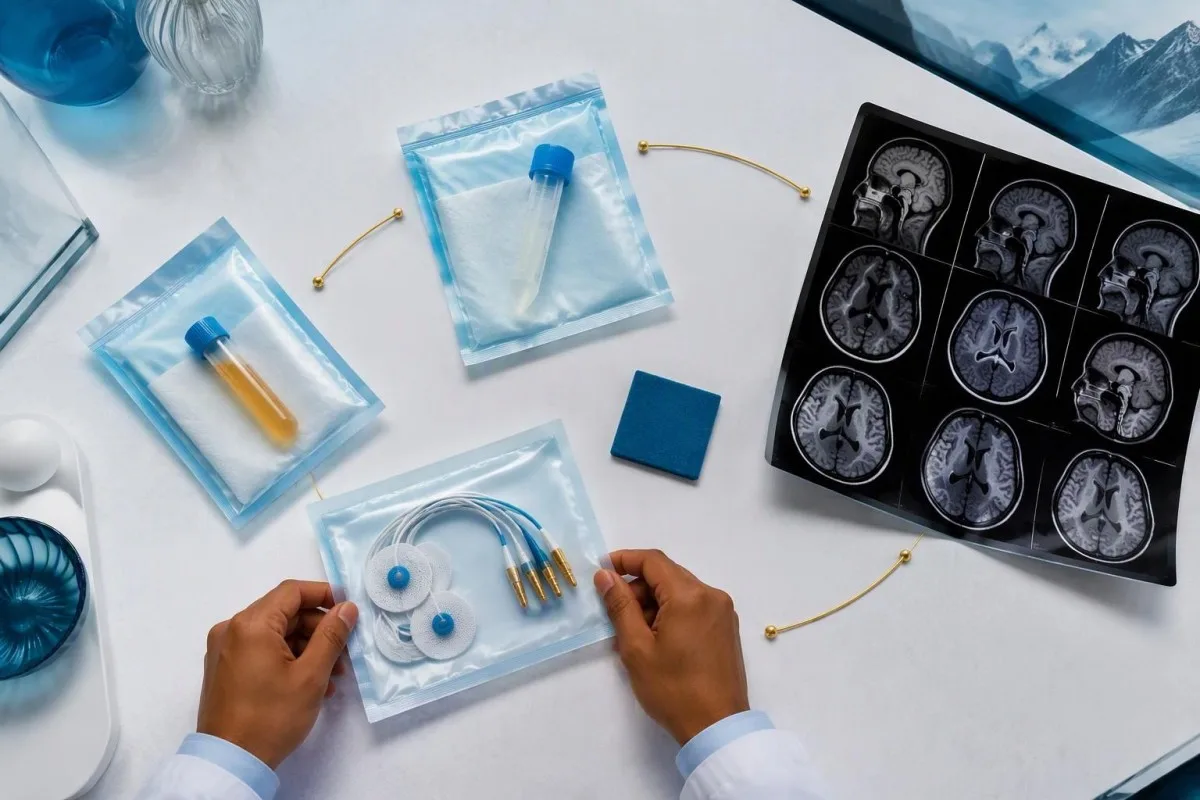
Nerve conduction studies can support a sensory neuronopathy pattern by showing reduced sensory responses across distributions that do not fit a single compressed nerve. Brain or spinal MRI addresses different questions, including structural disease and some inflammatory changes. A normal MRI does not exclude peripheral sensory ganglion involvement; these are 2 distinct anatomical compartments with different diagnostic tools.
Cerebrospinal fluid may show increased cells, protein or evidence of intrathecal immune activity, but none of those findings is specific to anti-Hu. A lumbar puncture is most useful when the neurological syndrome warrants it, rather than as an automatic response to 1 weak serum band. Our discussion of толкување на олигоклонални ленти во ЦСТ explains why immune activity in spinal fluid does not identify a single disease.
Basic investigations still matter: glucose control, vitamin status, renal function and serum protein assessment can uncover treatable contributors to neuropathy. A clinician may request 2 or more of these alongside specialist testing, depending on the examination. The водич за испитување на серумските протеини provides background, but normal routine blood work neither confirms nor excludes an anti-Hu-associated neurological process.
Дали позитивноста на анти-Hu потврдува паранеопластичен синдром?
A positive antibody alone does not confirm anti-Hu paraneoplastic syndrome. The PNS-Care approach combines the neurological phenotype, antibody risk category, cancer findings and follow-up; clinicians also check for direct cancer effects, treatment toxicity and unrelated neurological conditions before attributing symptoms to a paraneoplastic immune response.
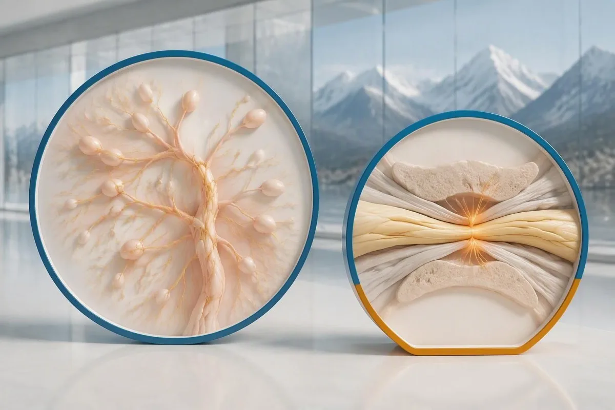
Definite, probable and possible PNS are diagnostic categories, not cancer stages. The 2021 criteria use a structured scoring system rather than allowing 1 antibody to determine the whole diagnosis (Graus et al., 2021). A confirmed anti-Hu result with a strongly compatible syndrome remains clinically significant even before a cancer is identified, but clinicians should state clearly which parts of the assessment remain unresolved.
An older patient can have anti-Hu reactivity and a separate cause of numbness, such as diabetes, nutritional deficiency or a monoclonal protein disorder. Coexistence is possible; either-or reasoning can miss treatable disease. If serum protein studies reveal a band, our immunofixation result guide explains why that finding requires its own interpretation rather than being automatically folded into the anti-Hu diagnosis.
Кантести е AI платформа за толкување крвна слика that can help organize these distinctions in a laboratory explanation, but it cannot assign a reliable PNS-Care diagnosis from a report alone. Our клиничка рамка за валидација separates technical evaluation from clinical conclusions. In practice, I would document 3 separate judgments: confidence in the antibody, confidence in the neurological syndrome and the current evidence for cancer.
Како лекарите бараат поврзан рак?
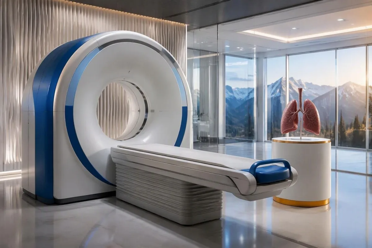
Cancer evaluation usually prioritizes the chest because small-cell lung cancer is the strongest adult anti-Hu association. Doctors commonly begin with appropriate chest CT and may add FDG-PET/CT or targeted investigations if suspicion remains; the exact plan depends on age, symptoms, examination, smoking history and previous imaging.
The cancer association is substantial in established anti-Hu clinical cohorts. Villagrán-García et al. (2023) reported identified cancer in 349 of 466 patients, approximately 75%, in a specialist anti-Hu cohort. This is valuable evidence for taking confirmed, clinically relevant anti-Hu seriously; it is not a personalized prediction for an asymptomatic person with an unconfirmed positive screening strip.
A chest X-ray is not equivalent to a chest CT, and routine low-dose screening is not necessarily the correct examination for a suspected paraneoplastic syndrome. The clinician chooses the protocol and body regions based on the diagnostic question. Likewise, neurological symptoms do not automatically mean cancer has spread to the brain; our brain imaging warning-sign guide distinguishes structural investigation from immune-mediated disease.
A suspicious scan often leads to tissue sampling through a method chosen by respiratory or oncology specialists, such as bronchoscopic assessment of an accessible area. Imaging suspicion and cancer confirmation are 2 separate steps. Never-smoking status lowers the baseline probability of small-cell lung cancer but does not cancel a well-confirmed anti-Hu finding with a convincing neurological presentation; other cancers and non-cancer explanations still need consideration.
Зошто првите снимки за откривање рак може да бидат негативни?
Initial imaging can be negative because an associated cancer is still too small to detect, lies outside the examined area or is not readily visible with that technique. Neurological autoimmunity may precede cancer detection, so a negative scan reduces concern without necessarily ending follow-up in a high-risk clinical presentation.
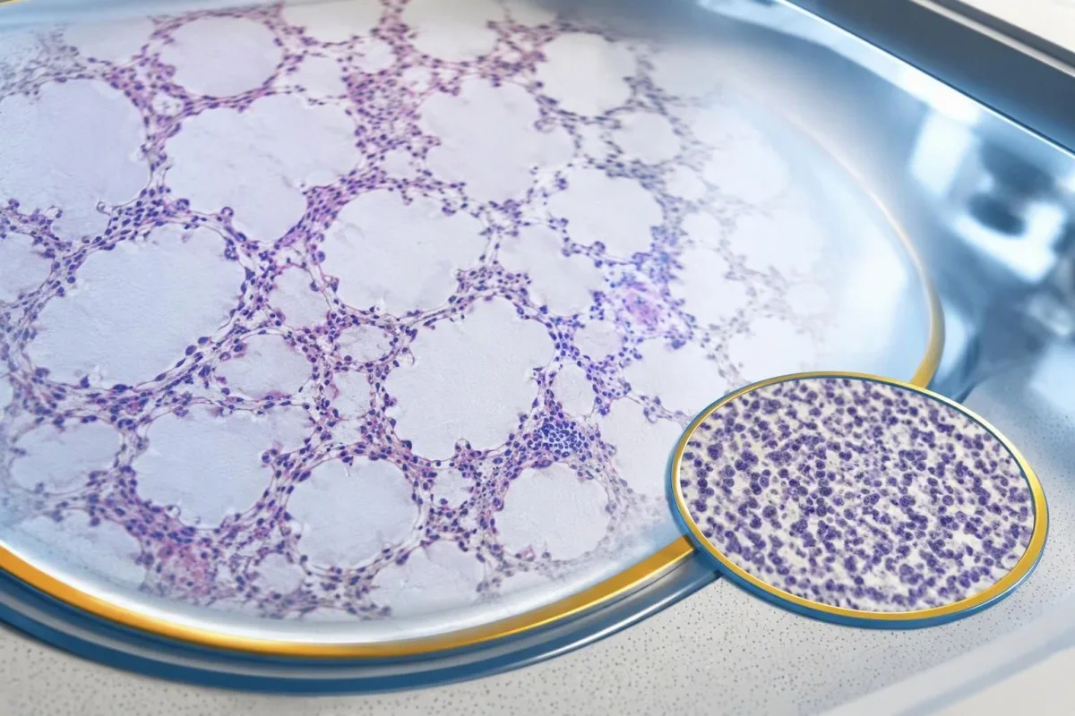
A negative scan is time-specific and method-specific. A normal chest CT performed on 1 date answers whether that examination identified a suspicious chest finding then; it does not guarantee that every possible associated cancer is absent indefinitely. Ask what was actually imaged, whether the protocol was suitable and whether the specialist recommends PET/CT or targeted investigation despite the initial result.
Villagrán-García et al. (2023) described cancers detected beyond the first 2 years in a minority of their anti-Hu cohort, supporting individualized longer surveillance. Their findings also support considering PET-based investigation when initial CT is unrevealing in an appropriate clinical context. This is an escalation decision for the treating team, not a reason to independently purchase repeated whole-body scans without weighing radiation, incidental findings and cost.
Neurological deterioration after negative imaging still needs reassessment. Cancer surveillance and neurological monitoring are 2 parallel tracks; one reassuring scan does not explain new loss of function. A marker such as neurofilament light chain may indicate neuronal injury in selected settings, but our NfL interpretation guide explains why it cannot establish anti-Hu disease, locate cancer or replace examination.
Колку долго треба да продолжи скринирањето за рак?
The 2021 PNS criteria recommend repeat cancer screening every 4–6 months for 2 years when a high-risk neurological phenotype accompanies a high-risk antibody. Longer surveillance may be appropriate for selected anti-Hu patients; the interval and duration should reflect confirmation, clinical evolution, initial investigations and individual cancer risk.

The 2-year schedule is not an instruction to scan every positive antibody result repeatedly. Graus et al. (2021) link that recommendation to the combination of high-risk phenotype and high-risk antibody. An incidental, weak result without a matching syndrome needs confirmation and individualized assessment first; repeating imaging mechanically can create incidental findings and unnecessary procedures without resolving the original uncertainty.
Older European guidance recommended longer surveillance. The EFNS task force advised repeating screening after 3–6 months and then every 6 months for up to 4 years in relevant antibody-positive paraneoplastic presentations (Titulaer et al., 2011). The 2023 anti-Hu cohort supported considering screening up to 5 years in selected patients, so a specialist may reasonably extend follow-up beyond the newer general 2-year framework.
Keep a dated plan stating the next examination, the clinician responsible and what symptoms should trigger earlier contact. Kantesti explanations can help you understand terminology, but the platform should not be treated as a scan-reminder service or substitute for that plan. Our guide to risk-based cancer screening addresses a different setting: routine prevention is not the same as investigating a possible paraneoplastic syndrome.
Кој третман помага кај невролошко заболување поврзано со анти-Hu?
Treatment focuses on an identified cancer, selected immune therapy and symptom-directed rehabilitation. Anti-Hu-associated neurological disease often responds less predictably to immunotherapy than neuronal surface-antibody disorders because intracellular targets and T-cell-mediated injury are involved; treatment may aim to stabilize function rather than promise complete recovery.

Treating an associated cancer is a central part of care, but established neurological injury may persist even when cancer treatment succeeds. Neurology and oncology teams may consider corticosteroids, intravenous immunoglobulin, plasma exchange or other immune treatments in selected situations. There is no single evidence-based dose or 1 universal regimen appropriate for everyone with anti-Hu positivity, especially without a confirmed clinical syndrome.
An antibody titer is not a dependable treatment target on its own. A lower number does not necessarily mean restored nerve function, and a persistent positive result does not by itself prove cancer progression. This differs from some surface-antibody disorders; our MOG antibody diagnosis guide explains another immune neurological condition, but its diagnostic rules and treatment expectations should not be transferred to anti-Hu.
Immune checkpoint cancer therapies deserve an explicit discussion when anti-Hu neurological disease is present, because they can provoke or worsen paraneoplastic neurological autoimmunity. This is a specialist risk-benefit decision, not an automatic prohibition based on 1 laboratory result. Tell the oncologist about previous neurological symptoms and confirmed neuronal antibodies before treatment decisions are finalized; coordinating the teams early can prevent avoidable misunderstanding.
Што можете безбедно да направите додека траат испитувањата?
While investigations continue, protect mobility, swallowing and medication safety rather than trying to lower anti-Hu antibodies with supplements. Use the neurological symptoms—not the appearance of the laboratory flag—to decide whether ordinary follow-up is sufficient or urgent care is needed; a new functional loss warrants contact before the next planned scan.
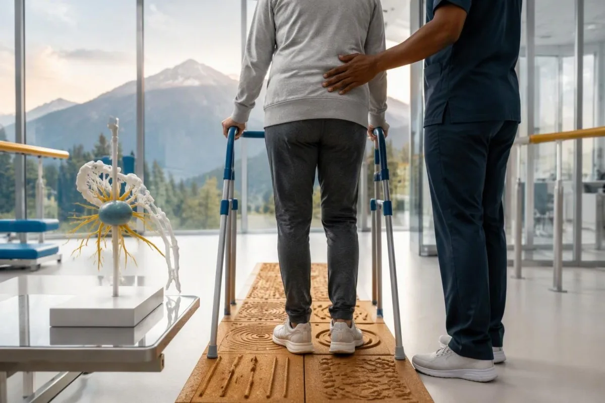
Position-sense loss increases fall risk, particularly in poor lighting or on uneven ground. A physiotherapist may recommend a suitable walking aid and individualized balance strategies; occupational therapy can address hand coordination and home hazards. A short 7-day record of falls, near-falls and walking changes is often more useful at review than the vague statement “my balance is worse,” but stop activities that feel unsafe.
Excess vitamin B6 can itself cause sensory neuropathy, making unsupervised high-dose nerve supplements particularly unhelpful during this assessment. Review every supplement and its daily dose in mg, including overlapping multivitamins and combination products. Our Водич за безбедност на Б-комплекс explains that problem; correcting a documented deficiency is different from taking progressively larger doses in the hope of treating anti-Hu autoimmunity.
Vitamin B12 deficiency can contribute to sensory and gait problems and may need investigation even when anti-Hu is confirmed. There is no universally interchangeable B12 cutoff: results in pg/mL и pmol/L require their own laboratory context, sometimes with functional testing. Our водич за следење на граничниот Б12 explains those next steps; new choking, breathing difficulty or rapid weakness should be assessed urgently instead of managed with vitamins.
Како AI може безбедно да помогне во објаснувањето на извештај за анти-Hu?
AI can help explain report terminology and organize questions, but it cannot confirm anti-Hu specificity, diagnose paraneoplastic syndrome or rule out cancer. Safe interpretation preserves the original assay details and separates 3 issues: what the laboratory reported, what the result may mean and what a clinician has actually established.

Кантести е Алатка за анализа на крвни тестови со вештачка интелигенција (AI) that can provide a plain-language explanation of uploaded laboratory information, including the distinction between an antibody screen and a confirmed clinical diagnosis. A rapid explanation—often around 60 seconds for report processing—is not a reference-laboratory assay. Our Водич за AI технологија describes the interpretation workflow without making anti-Hu-specific diagnostic guarantees.
The complete source report should remain available for verification. An explanation should not silently replace “weakly reactive” with “confirmed,” drop the specimen type or invent a universal reference interval. Our guide to проверка на објаснувањата за медицински теми од AI describes practical source checks; for this antibody, compare at least the assay wording, confirmation comment and collection date against the original document.
Kantesti cannot examine sensation, review tissue staining directly through a report explanation or establish that negative imaging has excluded a tiny cancer. Those are 3 different clinical capabilities, not features that follow from understanding laboratory language. Share the explanation with the treating team as a discussion aid, and do not delay urgent assessment while uploading documents or waiting for a second automated interpretation.
Вашиот следен преглед и транспарентноста на истражувањето
Bring the complete antibody report, a dated symptom history, previous imaging reports and a current medication list to your next appointment. Ask the clinician to clarify antibody confirmation, the neurological diagnosis and cancer surveillance separately; those 3 answers may have different levels of certainty and different follow-up dates.
A useful 1-page symptom history records onset, progression, sensory distribution, walking changes, memory problems and autonomic symptoms. Include prior cancer treatment and any recent immunoglobulin therapy, then ask who will coordinate neurology and oncology if both are involved. As Thomas Klein, MD, I would rather see a clear timeline than 20 disconnected screenshots; it makes discrepancies between the laboratory result and clinical course easier to spot.
Medical authorship is not a substitute for an individual neurological examination. Нашето информации за медицинскиот советодавен одбор describes the clinicians associated with our work, while your own specialist determines the appropriate next investigation. As of 25 октомври 2026 година, the recommendations discussed here draw on the 2021 international PNS criteria, the 2011 EFNS screening guidance and the 2023 anti-Hu cohort—not an invented 2026 guideline.
The 2 Figshare publications below are platform-related research references, not evidence validating anti-Hu diagnosis or cancer exclusion. Their supplied titles describe hantavirus triage deployment and a synthetic-case technical benchmark; neither title establishes anti-Hu-specific clinical performance. Because publication authors and dates were not supplied for verification, the citations use title-first APA formatting with “n.d.”; the ResearchGate and Academia.edu links are searches, not claims of verified publication copies.
Често поставувани прашања
Дали позитивен резултат за анти-Hу антитела значи дека имам рак?
0 Позитивниот резултат за антитела anti-Hu сам по себе не дијагностицира рак. Anti-Hu е антитело поврзано со висок ризик од рак, а рак бил утврден кај 349 од 466 пациенти во специјализирана група со anti-Hu од 2023 година, но таа бројка не го утврдува ризикот од случаен, непотврден скрининг-резултат. Лекарите ја проценуваат невролошката клиничка слика, ги потврдуваат неочекуваните лабораториски наоди и испитуваат дали постои скриен рак, обично давајќи им приоритет на градниот кош кај возрасните. За дијагноза на рак обично се потребни посебни клинички, снимачки и ткивни докази.
Дали anti-Hu и ANNA-1 се исто?
Anti-Hu и ANNA-1 генерално го означуваат истото семејство невронски антитела. ANNA-1 значи антиневронско нуклеарно антитело тип 1, додека Hu се однесува на интрацелуларните невронски протеини што ги препознава антителото. ANNA-1 не е исто што и рутинскиот ANA што се користи при тестирање за системски автоимуни заболувања. Проверете го точниот тест и формулацијата за потврда, бидејќи лабораториите може да го пријават истото семејство антитела во различни формати.
Може ли тестот за анти-Hу антитела да биде лажно позитивен?
Тестот за анти-Hу антитела може да даде погрешна или неспецифична реактивност, особено кога слаб резултат од скринингот не се совпаѓа со невролошката клиничка слика. Потврдата најчесто се врши со 2 комплементарни пристапа: карактеристичен образец на боење на ткивото и анализа специфична за антигенот, што ја толкува искусна лабораторија. Повторувањето на истиот комерцијален метод може да го повтори истиот аналитички проблем наместо да го реши. Брзо прогресивните невролошки симптоми и понатаму бараат навремена проценка додека се чека потврдата.
Колку често треба да се повторуваат снимањата ако анти-Hу е позитивен?
Критериумите на PNS од 2021 година препорачуваат повторување на скринингот за рак на секои 4–6 месеци во текот на 2 години кога високоризичен невролошки фенотип е придружен со високоризично антитело. Овој распоред не важи автоматски за секој случајно откриен позитивен резултат за анти-Hu. Постарите упатства на EFNS препорачуваа следење до 4 години, а една кохорта од 2023 година на пациенти со анти-Hu поддржа разгледување на следење до 5 години кај избрани пациенти. Вашиот специјалист треба да ги наведе методот на снимање, интервалот, времетраењето и симптомите поради кои е потребен порано направен преглед.
0 Може ли anti-Hu да биде позитивен кога КТ и ПЕТ-снимките се нормални?
Анти-Hu може да остане позитивен и кога со КТ или ПЕТ-снимање не е откриен карцином. Невролошкиот автоимунитет може да ѝ претходи на детекцијата, а негативниот наод од снимањето се однесува на испитаните региони, техниката и датумот, а не претставува трајна гаранција дека карциномот е отсутен. Кај компатибилна клиничка слика со висок ризик, критериумите од 2021 година препорачуваат повторен скрининг на секои 4–6 месеци во текот на 2 години, со индивидуализирано подолго следење кај избрани пациенти со анти-Hu. Треба повторно да се разгледаат и непотврдената реактивност на антителата и објаснувањата што не се поврзани со карцином, наместо да се претпоставува дека дефинитивно е присутен карцином што не може да се види.
Дали намалувањето на нивото на анти-Hу антитела значи дека третманот делува?
Намалувањето на нивото на anti-Hu антителата не е сигурен доказ за невролошко закрепнување или успешно лекување на ракот. Anti-Hu препознава интрацелуларни протеини, а поврзаното невролошко оштетување често вклучува механизми посредувани од Т-клетките, а не само дејства на антителата. Исто така, титрите од две различни лаборатории можеби не се споредливи поради разликите во методите. Клиничарите ја проценуваат функционалноста, наодите од прегледот, релевантните испитувања и одговорот на ракот наместо бројот на антителата да го користат како самостојна цел на лекувањето.
Добијте AI анализа на крв со моќ на вештачка интелигенција денес
Придружете се на над 2 милиони корисници ширум светот кои му веруваат на Kantesti за инстантна, точна анализа на лабораториски тестови. Поставете ги вашите резултати од крвна слика и добијте сеопфатно толкување на 15,000+ биомаркери за секунди.
📚 Реферирани научни публикации
Клајн, Т., Мичел, С., и Вебер, Х. (2026). Мултијазична поддршка за клиничко одлучување асистирана од вештачка интелигенција за рано тријажирање на Хан-тавирус: дизајн, инженерска валидација и примена во реалниот свет на 50.000 толкувани извештаи од крвни тестови. AI Medical Research.
Клајн, Т., Мичел, С., и Вебер, Х. (2026). Пререгистрирана, рубрика-базирана автоматизирана техничка бенчмарка на Kantesti Blood-Test Interpretation Engine на 100.000 синтетички тест-случаи. AI Medical Research.
📖 Надворешни медицински референци
Graus F et al. (2021). Updated Diagnostic Criteria for Paraneoplastic Neurologic Syndromes. Neurology: Neuroimmunology & Neuroinflammation.
Titulaer MJ et al. (2011). Screening for tumours in paraneoplastic syndromes: report of an EFNS Task Force. Европски журнал за неврологија.
Villagrán-García D et al. (2023). Revisiting anti-Hu paraneoplastic autoimmunity: phenotypic characterization and cancer diagnosis. Brain Communications.
⚕️ Медицинско одрекување од одговорност
Овој напис е само за едукативни цели и не претставува медицински совет. Секогаш консултирајте се со квалификуван здравствен работник за одлуки за дијагноза и третман.
Сигнали за доверба E-E-A-T
Искуство
Клиничка ревизија водена од лекар за работните текови на толкување на лабораториски резултати.
Експертиза
Фокус на лабораториска медицина за тоа како биомаркерите се однесуваат во клинички контекст.
Авторитивност
Напишано од д-р Thomas Klein, со ревизија од д-р Sarah Mitchell и проф. д-р Hans Weber.
Доверливост
Толкување засновано на докази со јасни патеки за следење за да се намали алармирањето.
