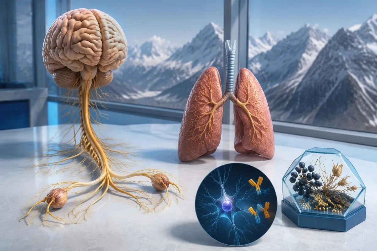
抗Hu抗体陽性という結果は、がんに関連する神経免疫反応、特に小細胞肺がんに関与する反応への懸念を高めますが、がんの診断にはなりません。医師は症状を評価し、検査所見を確認したうえで、がんの評価を手配します。初回の画像検査で異常がなくても、再検査が必要になることがあります。.



- 抗Hu/ANNA-1 は高リスクの傍腫瘍抗体であり、確立された臨床コホートでは70%を超える症例ががんに関連するカテゴリーに属します。ただし、未確認のスクリーニング結果すべてに当てはまるわけではありません。.
- がんの診断 には基礎にある悪性腫瘍の証拠が必要で、通常は組織検査も含まれます。抗体陽性という結果だけでは、その証拠にはなりません。.
- 検査法による確認 では、多くの場合、相補的な2つの方法を組み合わせます。特徴的な組織染色パターンの確認と、抗原特異的検査です。.
- 神経症状 進行性の感覚低下、平衡障害、けいれん、自律神経機能障害などがあると、結果の意味合いと緊急性が変わります。.
- 画像検査で異常がない とは、その時点でその検査によってがんが確認されなかったという意味です。小さく、初期には検出できないがんの可能性がなくなるわけではありません。.
- 再スクリーニング は、2021年PNS基準によると、高リスクの神経学的表現型を高リスク抗体が伴う場合、2年間は4~6か月ごとに推奨されます。.
- より長期の経過観察 は、選択された患者に適切な場合があります。2023年の抗Huコホートでは、2年を超えるスクリーニングを検討することが支持され、場合によっては最長5年まで行われました。.
- 緊急症状 新たな呼吸困難、安全に嚥下できない状態、急速に悪化する筋力低下、初回の発作、または突然の神経学的障害が含まれます。.
抗Hu抗体陽性という結果は何を意味しますか?
アン 抗Hu抗体陽性 という結果は、検査室がHu神経タンパク質に対する反応性を検出したことを意味します。それだけでがんや神経症候群が確定するわけではありません。解釈には3つの要素があります。結果が真の陽性か、神経学的な症状が合致するか、関連するがんが見つかるかです。.
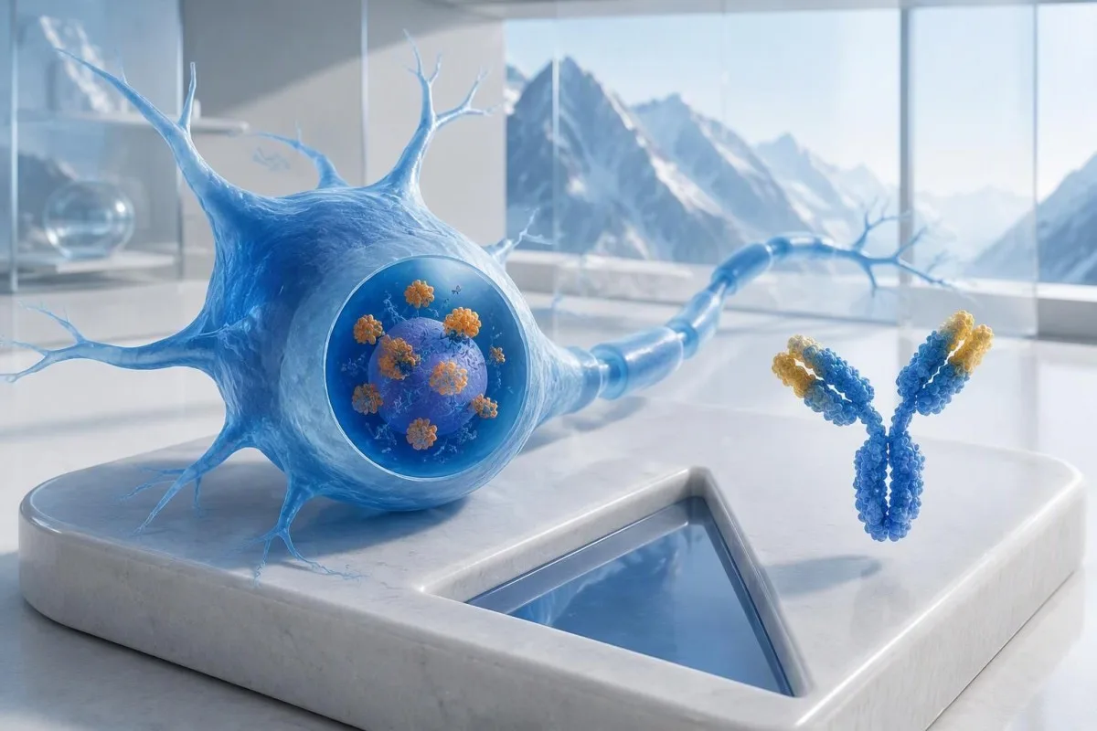
抗Hu抗体は、がんとの関連性が高い抗体に分類されます, 。このカテゴリーは、該当する臨床集団におけるがんとの関連率が70%を超えることで定義されています(Grausら、2021年)。この割合を、健康状態に問題がなく、パネル検査で弱陽性かつ未確認の結果が出た人にそのまま当てはめることはできません。対象患者、検査方法、神経学的症状のいずれもが確率に影響します。.
たとえば、平衡感覚の悪化と左右非対称の感覚低下が8週間続く62歳の人は、広範な疲労パネル検査で抗体が偶然検出された人とは異なる評価が必要です。どちらの人にも、検査結果だけをもとに「あなたはがんです」と告げるべきではありません。 抗体陽性所見に関するガイド は、免疫マーカーと確定診断を区別するのに役立ちます。.
カンテスティは AI血液検査分析装置 は抗Hu検査報告書の文言を理解するのに役立ちますが、検査室での確認とがんの評価は担当医が行うものです。Kantesti LTDの最高医療責任者であるThomas Klein, MDとして、私がまず尋ねるのは、この特定の検査を受けることになった経緯であり、赤旗所見がどれほど深刻に見えるかではありません。当社の 組織と臨床的使命 はその区別を説明します。.
ANNA-1は抗Hu抗体と同じ抗体ですか?
ANNA-1抗体と抗Hu抗体は、一般に同じ抗体ファミリーを指します。. ANNA-1は抗神経核抗体1型を意味します。Huという名称は、別の疾患やがんの病期ではなく、免疫応答が認識する細胞内神経タンパク質を指します。.
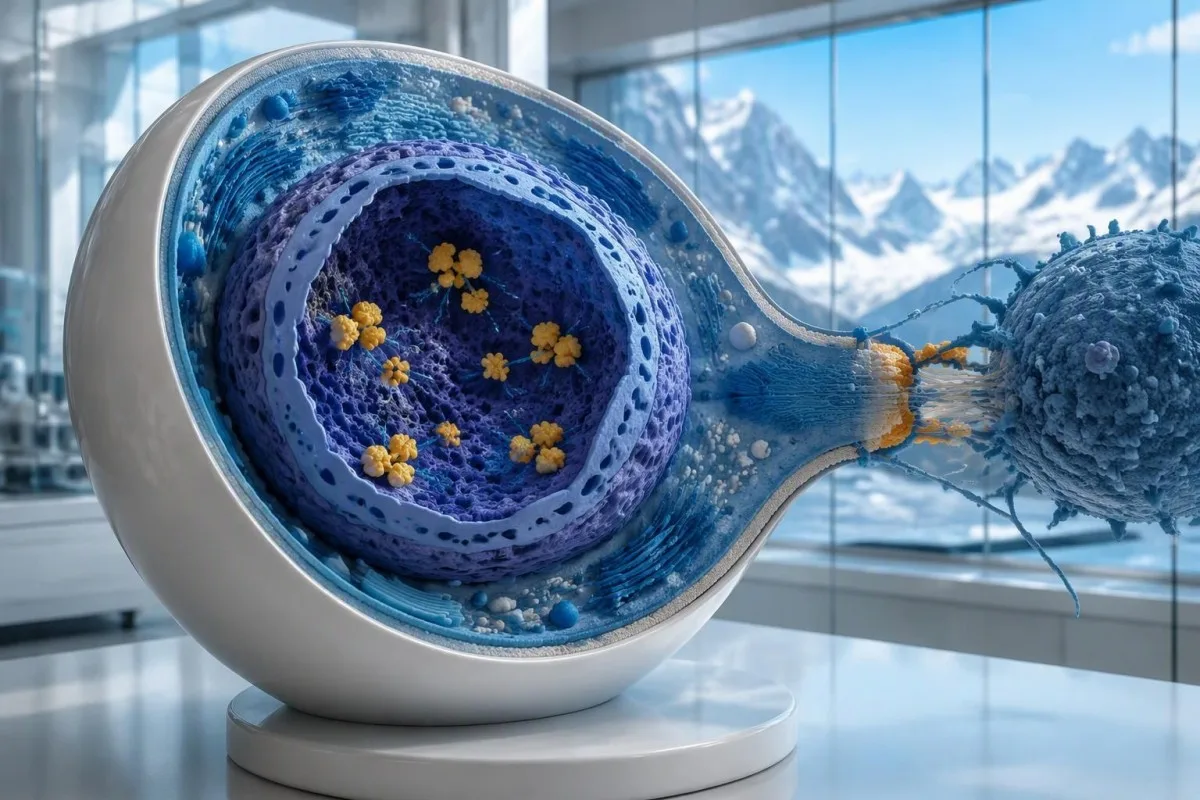
Huタンパク質は細胞内RNA結合タンパク質です, 。抗Hu抗体は、細胞傷害性T細胞が関与することの多い免疫応答のマーカーとして理解するのが最適です。これは、抗体が神経細胞表面の接近可能なタンパク質に結合する疾患とは異なります。この違いにより、2件の報告で抗体価に大きな差があっても、抗体価を下げるだけでは、すでに成立した抗Hu関連の神経障害が確実に回復するわけではない理由が説明できます。.
「ANNA-1検出」と記載された報告は、文字が似ていても、一般的なANA陽性と同じではありません。. 全日空 は通常、全身性自己免疫疾患の評価に用いられる抗核抗体を意味しますが、 ANNA-1 は神経抗体のパターンを示します。結果について話す際は、この2つの略語を区別してください。当社の ANA用語ガイド では、より一般的な検査について説明しています。.
抗Hu抗体は、神経症候群が明らかでない小細胞肺がん患者でも検出されることがあります。反対に、神経症状をきっかけに抗体が検出され、その後にがんが可視化されることもあります。これらは異なる2つの臨床経過です。抗体だけでは、神経が現在損傷を受けているか、潜在するがんがどこにあるかを医師が判断することはできません。.
抗Hu抗体検査の報告書はどのように読み取ればよいですか?
An anti-Hu antibody test has no universal numerical cutoff that can be applied across laboratories. Read the specimen type, assay method, result category, laboratory reference interval and confirmation comments together; a dilution titer, band intensity and numerical index are 3 different ways of reporting reactivity.
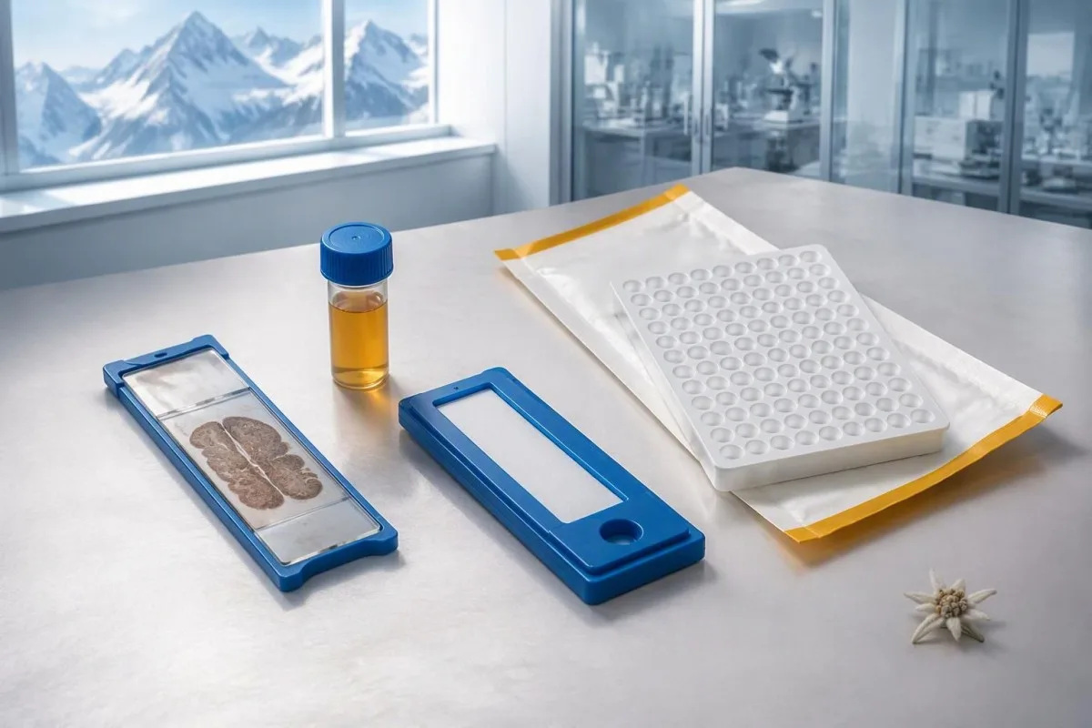
A hypothetical titer of 1:240 describes detectable reactivity after a particular dilution; it is not a cancer probability of 240, nor does it establish cancer stage. A numerical index from another method may use entirely different calibration. Our guide to 定性検査結果と定量検査結果 explains why “positive” and “how much” answer different laboratory questions.
Weak or borderline reactivity deserves method-specific interpretation, particularly when a broad panel was ordered without a convincing neurological indication. Ask whether the laboratory observed the characteristic neuronal staining pattern and whether an antigen-specific confirmation was performed. Two tests using the same underlying strip technology may reproduce the same analytical problem; agreement is more informative when the methods are genuinely complementary.
Bring the complete report, not just a screenshot of the word positive. The 5 most useful details are the specimen, collection date, assay name, reported strength or titer and laboratory interpretation. Also list recent intravenous immunoglobulin and other immune treatments, because passive antibodies or treatment effects can complicate some serological results; your laboratory can advise whether that issue plausibly affects this particular finding.
抗Hu陽性の結果はどのような場合に確認が必要ですか?
Unexpected, weak or clinically discordant anti-Hu results should be reviewed for confirmation, preferably with an experienced neuroimmunology laboratory. The 2021 PNS criteria emphasize interpreting neuronal antibody testing alongside the phenotype and using appropriate confirmatory methods rather than accepting an isolated commercial-panel result as a diagnosis.
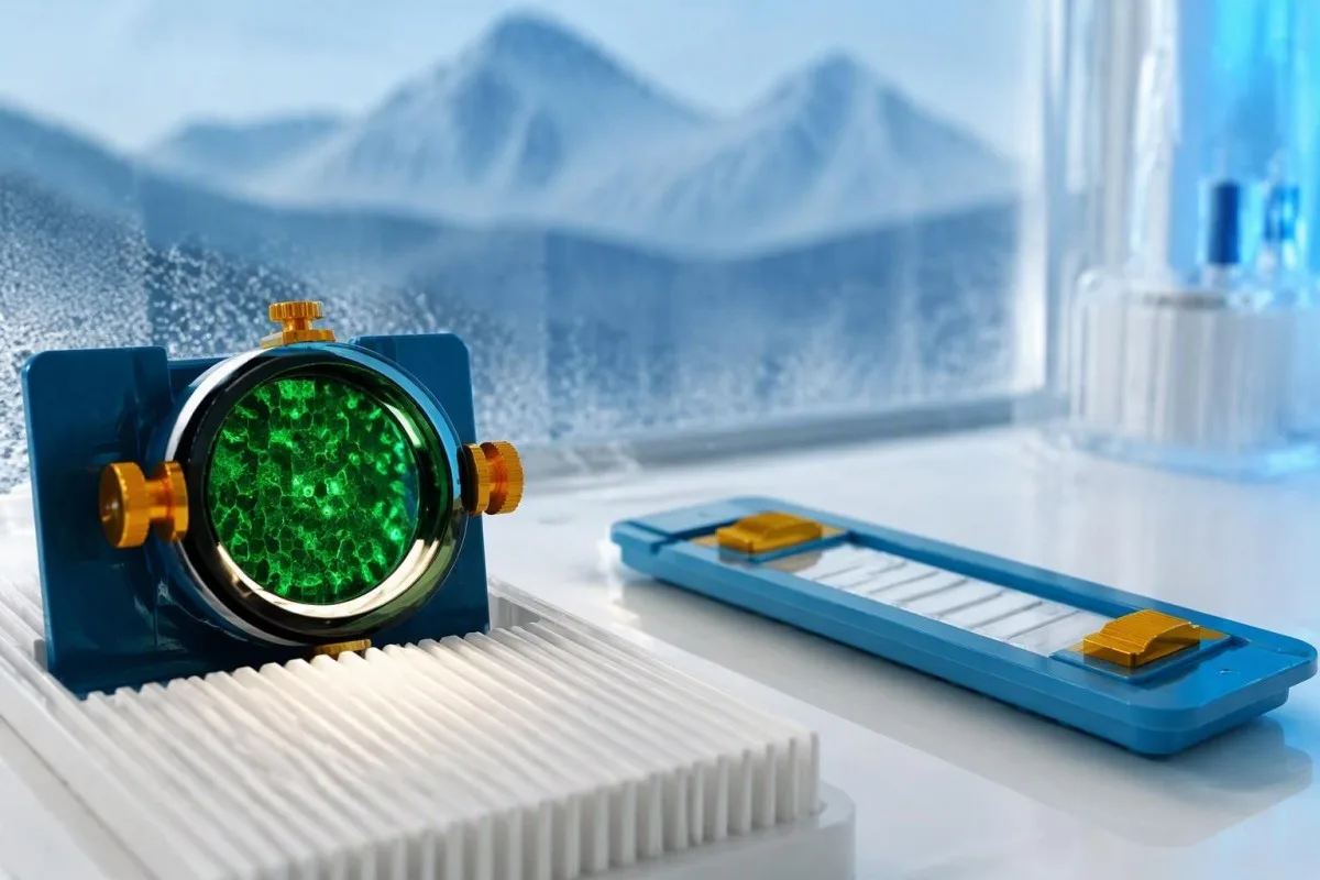
Tissue-based immunohistochemistry or immunofluorescence and an antigen-specific assay provide different information. The first identifies a characteristic distribution of neuronal staining; the second tests reactivity against the relevant antigen. Graus et al. (2021) describe these complementary approaches, with specialist reassessment particularly useful when 1 method gives a result that does not fit the clinical picture.
Repeating a sample can address collection errors or a transient analytical concern, but repeating the same kit is not always enough. In a hypothetical 45-year-old with no objective neurological findings and a faint isolated band, I would want the laboratory’s explanation before treating the report as established anti-Hu autoimmunity. Our GAD65 interpretation guide illustrates another antibody for which context and assay characteristics change meaning.
Confirmation and urgent assessment can happen in parallel. A person who develops seizures, substantial walking difficulty or progressive swallowing impairment should not wait several weeks for a reference-laboratory answer before receiving neurological care. The practical question is not “repeat or investigate?” but which of those 2 tasks can safely proceed together, given the symptoms and the initial laboratory evidence.
どのような神経症状があると、抗Huへの懸念が高まりますか?
Progressive sensory neuronopathy, encephalomyelitis, limbic encephalitis and marked autonomic dysfunction are recognized anti-Hu presentations. Symptoms can involve more than 1 part of the nervous system, so the pattern and speed of change are usually more informative than a single symptom such as tingling or fatigue.
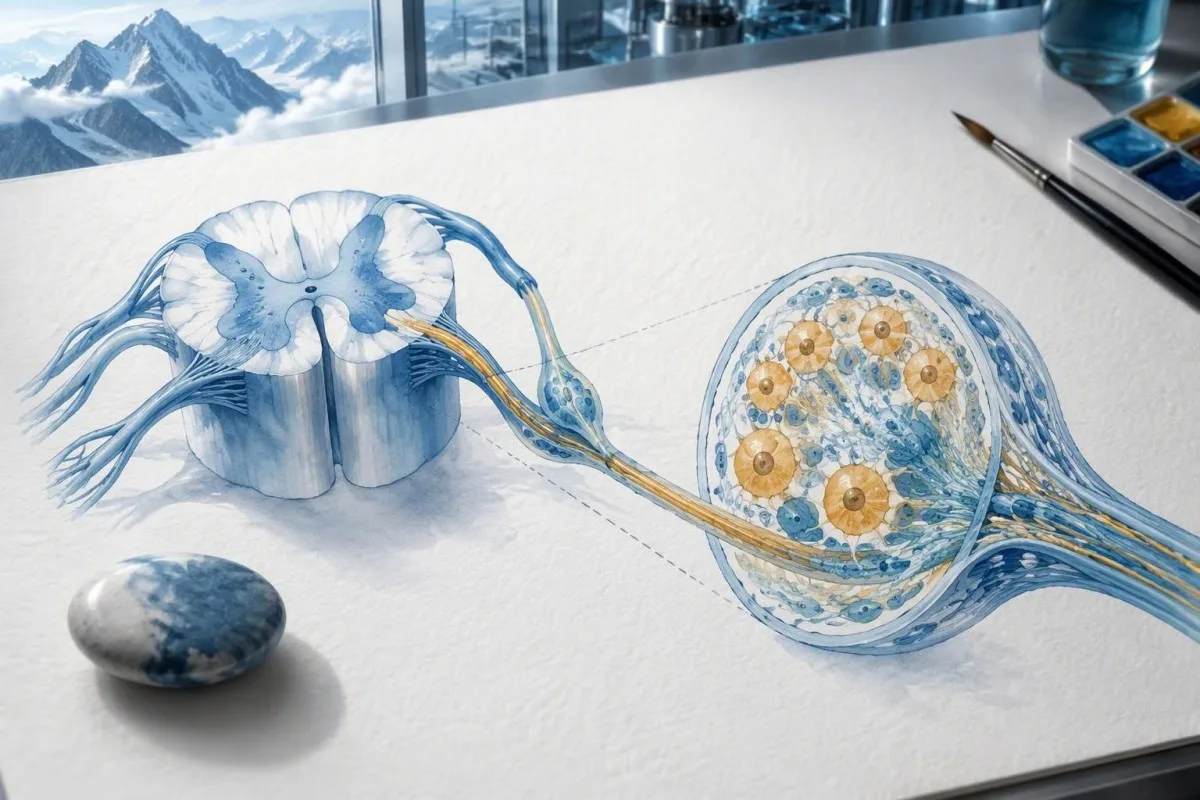
Sensory neuronopathy can cause asymmetric numbness, impaired position sense and sensory ataxia, sometimes involving the hands or trunk early rather than following a simple feet-first pattern. A patient may lose balance in the dark because vision had been compensating for reduced position feedback. That is different from the focal finger distribution discussed in our carpal tunnel symptom guide, although neither pattern can be diagnosed from description alone.
Anti-Hu autonomic involvement can affect gut movement, blood-pressure regulation and bladder function. Severe constipation, unexplained intestinal pseudo-obstruction or dizziness on standing becomes more relevant when accompanied by objective neurological abnormalities. Recording symptoms over 7 days can reveal combinations that otherwise get presented as separate stomach, balance and urinary complaints; sudden severe abdominal symptoms still require assessment for non-neurological emergencies.
New memory impairment, behavioral change or seizures may indicate central nervous system involvement, while rapid weakness has a broader emergency differential. The 緊急性のある筋力低下の警告サイン in our neurological safety guide apply regardless of the antibody label. Call emergency services for new breathing difficulty, unsafe swallowing, a first seizure or sudden stroke-like symptoms rather than waiting for a scheduled follow-up.
陽性結果の後に、どのような神経学的検査が行われることがありますか?
Neurological follow-up may include examination, nerve conduction studies, MRI and cerebrospinal fluid testing, selected according to the symptoms. There is no mandatory battery for every anti-Hu-positive person; the purpose is to document which structures are affected and exclude other causes, not merely to collect additional positive tests.
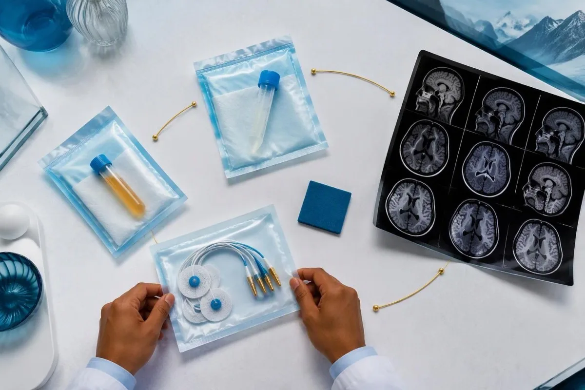
Nerve conduction studies can support a sensory neuronopathy pattern by showing reduced sensory responses across distributions that do not fit a single compressed nerve. Brain or spinal MRI addresses different questions, including structural disease and some inflammatory changes. A normal MRI does not exclude peripheral sensory ganglion involvement; these are 2 distinct anatomical compartments with different diagnostic tools.
Cerebrospinal fluid may show increased cells, protein or evidence of intrathecal immune activity, but none of those findings is specific to anti-Hu. A lumbar puncture is most useful when the neurological syndrome warrants it, rather than as an automatic response to 1 weak serum band. Our discussion of 脳脊髄液オリゴクローナルバンドの読み方 explains why immune activity in spinal fluid does not identify a single disease.
Basic investigations still matter: glucose control, vitamin status, renal function and serum protein assessment can uncover treatable contributors to neuropathy. A clinician may request 2 or more of these alongside specialist testing, depending on the examination. The 血清タンパク評価ガイド provides background, but normal routine blood work neither confirms nor excludes an anti-Hu-associated neurological process.
抗Hu陽性は傍腫瘍症候群の確定診断になりますか?
A positive antibody alone does not confirm anti-Hu paraneoplastic syndrome. The PNS-Care approach combines the neurological phenotype, antibody risk category, cancer findings and follow-up; clinicians also check for direct cancer effects, treatment toxicity and unrelated neurological conditions before attributing symptoms to a paraneoplastic immune response.
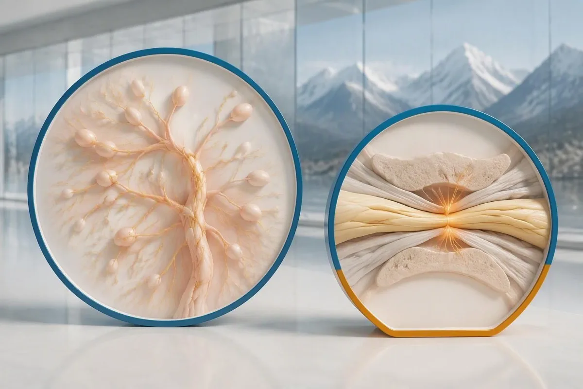
Definite, probable and possible PNS are diagnostic categories, not cancer stages. The 2021 criteria use a structured scoring system rather than allowing 1 antibody to determine the whole diagnosis (Graus et al., 2021). A confirmed anti-Hu result with a strongly compatible syndrome remains clinically significant even before a cancer is identified, but clinicians should state clearly which parts of the assessment remain unresolved.
An older patient can have anti-Hu reactivity and a separate cause of numbness, such as diabetes, nutritional deficiency or a monoclonal protein disorder. Coexistence is possible; either-or reasoning can miss treatable disease. If serum protein studies reveal a band, our immunofixation result guide explains why that finding requires its own interpretation rather than being automatically folded into the anti-Hu diagnosis.
カンテスティは AI血液検査の読み方プラットフォーム that can help organize these distinctions in a laboratory explanation, but it cannot assign a reliable PNS-Care diagnosis from a report alone. Our 臨床検証フレームワーク separates technical evaluation from clinical conclusions. In practice, I would document 3 separate judgments: confidence in the antibody, confidence in the neurological syndrome and the current evidence for cancer.
医師は関連するがんをどのように調べますか?
Cancer evaluation usually prioritizes the chest because small-cell lung cancer is the strongest adult anti-Hu association. Doctors commonly begin with appropriate chest CT and may add FDG-PET/CT or targeted investigations if suspicion remains; the exact plan depends on age, symptoms, examination, smoking history and previous imaging.
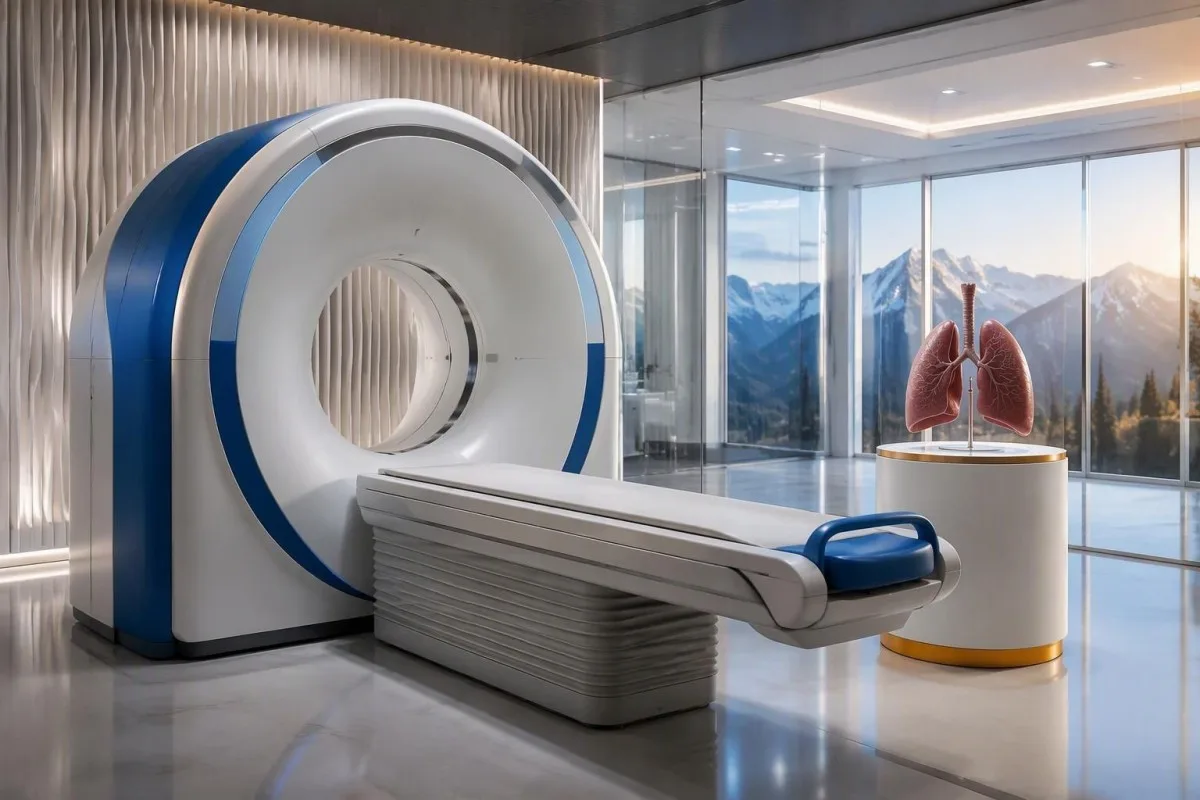
The cancer association is substantial in established anti-Hu clinical cohorts. Villagrán-García et al. (2023) reported identified cancer in 349 of 466 patients, approximately 75%, in a specialist anti-Hu cohort. This is valuable evidence for taking confirmed, clinically relevant anti-Hu seriously; it is not a personalized prediction for an asymptomatic person with an unconfirmed positive screening strip.
A chest X-ray is not equivalent to a chest CT, and routine low-dose screening is not necessarily the correct examination for a suspected paraneoplastic syndrome. The clinician chooses the protocol and body regions based on the diagnostic question. Likewise, neurological symptoms do not automatically mean cancer has spread to the brain; our brain imaging warning-sign guide distinguishes structural investigation from immune-mediated disease.
A suspicious scan often leads to tissue sampling through a method chosen by respiratory or oncology specialists, such as bronchoscopic assessment of an accessible area. Imaging suspicion and cancer confirmation are 2 separate steps. Never-smoking status lowers the baseline probability of small-cell lung cancer but does not cancel a well-confirmed anti-Hu finding with a convincing neurological presentation; other cancers and non-cancer explanations still need consideration.
初回のがん画像検査で異常が見つからないことがあるのはなぜですか?
Initial imaging can be negative because an associated cancer is still too small to detect, lies outside the examined area or is not readily visible with that technique. Neurological autoimmunity may precede cancer detection, so a negative scan reduces concern without necessarily ending follow-up in a high-risk clinical presentation.
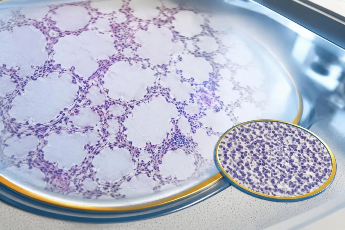
A negative scan is time-specific and method-specific. A normal chest CT performed on 1 date answers whether that examination identified a suspicious chest finding then; it does not guarantee that every possible associated cancer is absent indefinitely. Ask what was actually imaged, whether the protocol was suitable and whether the specialist recommends PET/CT or targeted investigation despite the initial result.
Villagrán-García et al. (2023) described cancers detected beyond the first 2 years in a minority of their anti-Hu cohort, supporting individualized longer surveillance. Their findings also support considering PET-based investigation when initial CT is unrevealing in an appropriate clinical context. This is an escalation decision for the treating team, not a reason to independently purchase repeated whole-body scans without weighing radiation, incidental findings and cost.
Neurological deterioration after negative imaging still needs reassessment. Cancer surveillance and neurological monitoring are 2 parallel tracks; one reassuring scan does not explain new loss of function. A marker such as neurofilament light chain may indicate neuronal injury in selected settings, but our NfL interpretation guide explains why it cannot establish anti-Hu disease, locate cancer or replace examination.
がんのスクリーニングはどのくらい続けるべきですか?
The 2021 PNS criteria recommend repeat cancer screening every 4–6 months for 2 years when a high-risk neurological phenotype accompanies a high-risk antibody. Longer surveillance may be appropriate for selected anti-Hu patients; the interval and duration should reflect confirmation, clinical evolution, initial investigations and individual cancer risk.

The 2-year schedule is not an instruction to scan every positive antibody result repeatedly. Graus et al. (2021) link that recommendation to the combination of high-risk phenotype and high-risk antibody. An incidental, weak result without a matching syndrome needs confirmation and individualized assessment first; repeating imaging mechanically can create incidental findings and unnecessary procedures without resolving the original uncertainty.
Older European guidance recommended longer surveillance. The EFNS task force advised repeating screening after 3–6 months and then every 6 months for up to 4 years in relevant antibody-positive paraneoplastic presentations (Titulaer et al., 2011). The 2023 anti-Hu cohort supported considering screening up to 5 years in selected patients, so a specialist may reasonably extend follow-up beyond the newer general 2-year framework.
Keep a dated plan stating the next examination, the clinician responsible and what symptoms should trigger earlier contact. Kantesti explanations can help you understand terminology, but the platform should not be treated as a scan-reminder service or substitute for that plan. Our guide to risk-based cancer screening addresses a different setting: routine prevention is not the same as investigating a possible paraneoplastic syndrome.
抗Hu関連の神経疾患にはどのような治療が有効ですか?
Treatment focuses on an identified cancer, selected immune therapy and symptom-directed rehabilitation. Anti-Hu-associated neurological disease often responds less predictably to immunotherapy than neuronal surface-antibody disorders because intracellular targets and T-cell-mediated injury are involved; treatment may aim to stabilize function rather than promise complete recovery.

Treating an associated cancer is a central part of care, but established neurological injury may persist even when cancer treatment succeeds. Neurology and oncology teams may consider corticosteroids, intravenous immunoglobulin, plasma exchange or other immune treatments in selected situations. There is no single evidence-based dose or 1 universal regimen appropriate for everyone with anti-Hu positivity, especially without a confirmed clinical syndrome.
An antibody titer is not a dependable treatment target on its own. A lower number does not necessarily mean restored nerve function, and a persistent positive result does not by itself prove cancer progression. This differs from some surface-antibody disorders; our MOG antibody diagnosis guide explains another immune neurological condition, but its diagnostic rules and treatment expectations should not be transferred to anti-Hu.
Immune checkpoint cancer therapies deserve an explicit discussion when anti-Hu neurological disease is present, because they can provoke or worsen paraneoplastic neurological autoimmunity. This is a specialist risk-benefit decision, not an automatic prohibition based on 1 laboratory result. Tell the oncologist about previous neurological symptoms and confirmed neuronal antibodies before treatment decisions are finalized; coordinating the teams early can prevent avoidable misunderstanding.
検査が続いている間、安全にできることは何ですか?
While investigations continue, protect mobility, swallowing and medication safety rather than trying to lower anti-Hu antibodies with supplements. Use the neurological symptoms—not the appearance of the laboratory flag—to decide whether ordinary follow-up is sufficient or urgent care is needed; a new functional loss warrants contact before the next planned scan.
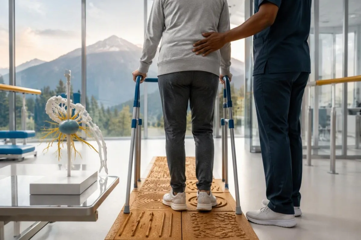
Position-sense loss increases fall risk, particularly in poor lighting or on uneven ground. A physiotherapist may recommend a suitable walking aid and individualized balance strategies; occupational therapy can address hand coordination and home hazards. A short 7-day record of falls, near-falls and walking changes is often more useful at review than the vague statement “my balance is worse,” but stop activities that feel unsafe.
Excess vitamin B6 can itself cause sensory neuropathy, making unsupervised high-dose nerve supplements particularly unhelpful during this assessment. Review every supplement and its daily dose in mg, including overlapping multivitamins and combination products. Our B複合体安全ガイド explains that problem; correcting a documented deficiency is different from taking progressively larger doses in the hope of treating anti-Hu autoimmunity.
Vitamin B12 deficiency can contribute to sensory and gait problems and may need investigation even when anti-Hu is confirmed. There is no universally interchangeable B12 cutoff: results in pg/mL そして pmol/L require their own laboratory context, sometimes with functional testing. Our 境界型B12フォローアップガイド explains those next steps; new choking, breathing difficulty or rapid weakness should be assessed urgently instead of managed with vitamins.
AIは抗Huの検査報告書を安全に理解するうえで、どのように役立ちますか?
AI can help explain report terminology and organize questions, but it cannot confirm anti-Hu specificity, diagnose paraneoplastic syndrome or rule out cancer. Safe interpretation preserves the original assay details and separates 3 issues: what the laboratory reported, what the result may mean and what a clinician has actually established.

カンテスティは AI搭載の血液検査解析ツール that can provide a plain-language explanation of uploaded laboratory information, including the distinction between an antibody screen and a confirmed clinical diagnosis. A rapid explanation—often around 60 seconds for report processing—is not a reference-laboratory assay. Our AIテクノロジーガイド describes the interpretation workflow without making anti-Hu-specific diagnostic guarantees.
The complete source report should remain available for verification. An explanation should not silently replace “weakly reactive” with “confirmed,” drop the specimen type or invent a universal reference interval. Our guide to AIによる医療情報の説明の確認 describes practical source checks; for this antibody, compare at least the assay wording, confirmation comment and collection date against the original document.
Kantesti cannot examine sensation, review tissue staining directly through a report explanation or establish that negative imaging has excluded a tiny cancer. Those are 3 different clinical capabilities, not features that follow from understanding laboratory language. Share the explanation with the treating team as a discussion aid, and do not delay urgent assessment while uploading documents or waiting for a second automated interpretation.
次回の診察と研究の透明性
Bring the complete antibody report, a dated symptom history, previous imaging reports and a current medication list to your next appointment. Ask the clinician to clarify antibody confirmation, the neurological diagnosis and cancer surveillance separately; those 3 answers may have different levels of certainty and different follow-up dates.
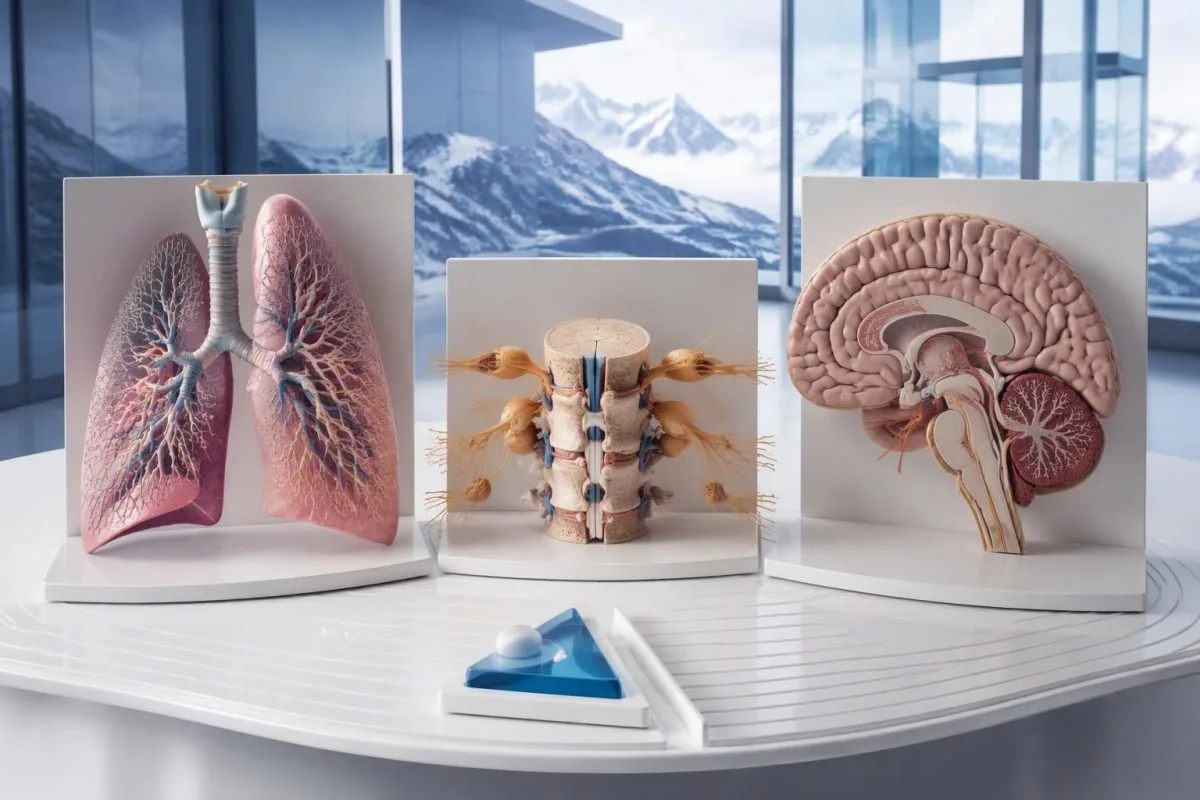
A useful 1-page symptom history records onset, progression, sensory distribution, walking changes, memory problems and autonomic symptoms. Include prior cancer treatment and any recent immunoglobulin therapy, then ask who will coordinate neurology and oncology if both are involved. As Thomas Klein, MD, I would rather see a clear timeline than 20 disconnected screenshots; it makes discrepancies between the laboratory result and clinical course easier to spot.
Medical authorship is not a substitute for an individual neurological examination. 私たちの 医療顧問委員会の情報 describes the clinicians associated with our work, while your own specialist determines the appropriate next investigation. As of 2026年10月25日, the recommendations discussed here draw on the 2021 international PNS criteria, the 2011 EFNS screening guidance and the 2023 anti-Hu cohort—not an invented 2026 guideline.
The 2 Figshare publications below are platform-related research references, not evidence validating anti-Hu diagnosis or cancer exclusion. Their supplied titles describe hantavirus triage deployment and a synthetic-case technical benchmark; neither title establishes anti-Hu-specific clinical performance. Because publication authors and dates were not supplied for verification, the citations use title-first APA formatting with “n.d.”; the ResearchGate and Academia.edu links are searches, not claims of verified publication copies.
よくある質問
抗Hu抗体陽性という結果は、がんがあることを意味しますか?
0 抗Hu抗体陽性という結果だけで、がんと診断されるわけではありません。抗Hu抗体はがんとの関連性が高い抗体であり、2023年の抗Hu抗体専門コホートでは466人中349人にがんが確認されましたが、この数値から、偶発的に得られた未確認のスクリーニング結果に伴うリスクを判断することはできません。医師は神経症状を評価し、予想外の検査所見を確認したうえで、通常は成人では胸部を優先して、潜在するがんを調べます。一般に、がんの診断には、臨床所見、画像所見、組織所見という別個の根拠が必要です。.
抗Hu抗体とANNA-1は同じものですか?
Anti-HuとANNA-1は一般に、同じ神経抗体ファミリーを指します。ANNA-1は抗神経核抗体1型を意味し、Huは抗体が認識する細胞内神経タンパク質を指します。ANNA-1は、全身性自己免疫疾患の検査で用いられる通常のANAとは異なります。検査室によって同じ抗体ファミリーが異なる形式で報告されることがあるため、検査法と確認検査の記載を正確に確認してください。.
抗Hu抗体検査が偽陽性になることはありますか?
抗Hu抗体検査では、特にスクリーニング検査の弱陽性結果が神経学的所見と一致しない場合、誤解を招く反応や非特異的反応が生じることがあります。確認には通常、特徴的な組織染色パターンと、経験豊富な検査室が判定する抗原特異的検査という、相補的な2つの方法を用います。同じ市販法を繰り返しても、分析上の問題が解決するどころか、同じ問題が再現されることがあります。確認結果を待つ間も、急速に進行する神経症状については速やかな評価が必要です。.
抗Hu抗体が陽性の場合、スキャンはどのくらいの頻度で繰り返すべきですか?
2021年のPNS診断基準では、高リスクの神経学的表現型に高リスク抗体が伴う場合、2年間にわたり4~6か月ごとにがん検診を繰り返すことが推奨されています。このスケジュールが、偶発的に抗Hu抗体陽性となったすべての結果に自動的に適用されるわけではありません。旧EFNSガイダンスでは最長4年間のサーベイランスが推奨されており、2023年の抗Hu抗体コホート研究では、選択された患者について最長5年間の検討が支持されています。専門医が、画像検査の方法、間隔、期間、およびより早い受診が必要となる症状を明確にすべきです。.
CT検査とPET検査が正常でも、抗Hu抗体が陽性になることはありますか?
CTまたはPET画像検査でがんが確認されていない場合でも、抗Hu抗体は陽性のままであることがあります。神経学的自己免疫はがんの検出に先行することがあり、陰性の画像検査が示すのは、検査した部位、検査手法および検査日についてであり、がんが存在しないことを恒久的に保証するものではありません。臨床像が合致する高リスク例では、2021年の基準は2年間、4~6か月ごとのスクリーニング再検を推奨しており、抗Hu抗体陽性患者の一部では個別に、さらに長期の経過観察が行われます。確認されていない抗体反応や、がん以外の原因についても再検討すべきであり、見えないがんが確実に存在すると決めつけるべきではありません。.
抗Hu抗体値の低下は、治療が効いていることを意味しますか?
抗Hu抗体価の低下は、神経学的回復やがん治療の成功を確実に証明するものではありません。抗Hu抗体は細胞内タンパク質を認識し、関連する神経障害には、抗体の作用だけでなくT細胞が関与する機序が関わることが少なくありません。また、検査法が異なるため、異なる検査機関で測定された2つの抗体価は比較できない場合があります。臨床医は、抗体価を単独の治療目標として用いるのではなく、機能、診察所見、関連する検査結果、がんの治療反応を評価します。.
今日、AIによる血液検査分析を
いますぐ利用しませんか。即時で正確な検査分析を提供するKantestiを信頼する、世界中の200万人以上のユーザーに参加してください。血液検査結果をアップロードすると、15,000+のバイオマーカーについて数秒で包括的な解釈が得られます。.
📚 参考文献
📖 外部の医学的参考文献
Graus F et al. (2021). Updated Diagnostic Criteria for Paraneoplastic Neurologic Syndromes. Neurology: Neuroimmunology & Neuroinflammation.
Titulaer MJ et al. (2011). Screening for tumours in paraneoplastic syndromes: report of an EFNS Task Force.。European Journal of Neurology。.
Villagrán-García D et al. (2023). Revisiting anti-Hu paraneoplastic autoimmunity: phenotypic characterization and cancer diagnosis. Brain Communications.
⚕️ 医療免責事項
この記事は教育目的のみを対象としており、医療助言を構成するものではありません。診断や治療の判断を行う際は、必ず有資格の医療提供者にご相談ください。.
E-E-A-T 信頼性シグナル
経験
医師主導による、検査結果解釈ワークフローの臨床レビュー。.
専門知識
臨床的な文脈においてバイオマーカーがどのように振る舞うかに焦点を当てた検査医学。.
権威
トーマス・クライン博士が執筆し、サラ・ミッチェル博士およびハンス・ヴェーバー教授によるレビュー。.
信頼性
アラームを減らすための明確なフォローアップ経路を備えた、エビデンスに基づく解釈。.
