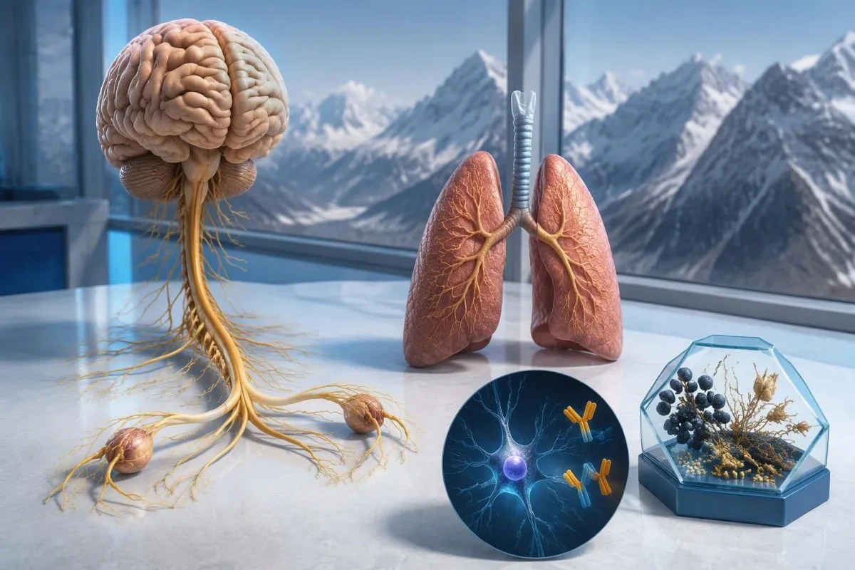
Ένα θετικό αποτέλεσμα αντισωμάτων anti-Hu εγείρει ανησυχία για νευρολογική ανοσολογική αντίδραση σχετιζόμενη με καρκίνο, ιδίως με μικροκυτταρικό καρκίνο του πνεύμονα, αλλά δεν διαγιγνώσκει καρκίνο. Οι γιατροί αξιολογούν τα συμπτώματα, επιβεβαιώνουν το εργαστηριακό εύρημα και διενεργούν έλεγχο για καρκίνο· μπορεί να χρειαστεί επανάληψη του ελέγχου ακόμη και όταν οι αρχικές απεικονιστικές εξετάσεις είναι αρνητικές.



- Anti-Hu / ANNA-1 είναι ένα παρανεοπλασματικό αντίσωμα υψηλού κινδύνου, μια κατηγορία που συσχετίζεται με καρκίνο σε ποσοστό άνω του 70% των καθιερωμένων κλινικών σειρών—όχι σε κάθε μη επιβεβαιωμένο αποτέλεσμα προσυμπτωματικού ελέγχου.
- Διάγνωση καρκίνου απαιτεί τεκμήρια υποκείμενης κακοήθειας, συνήθως συμπεριλαμβανομένης της εξέτασης ιστού· ένα θετικό αποτέλεσμα αντισώματος δεν αποτελεί τέτοιο τεκμήριο.
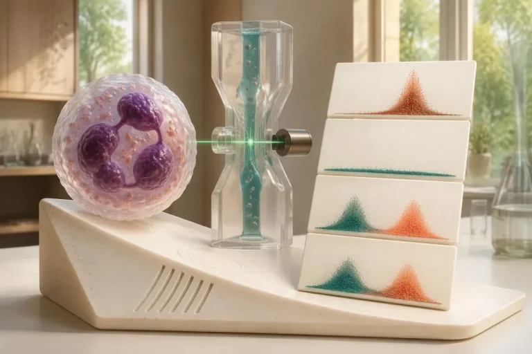
- Επιβεβαίωση της μεθόδου ανάλυσης συχνά συνδυάζει 2 συμπληρωματικές προσεγγίσεις: ένα χαρακτηριστικό πρότυπο χρώσης ιστού και μια εξέταση ειδική για το αντιγόνο.
- Νευρολογικά συμπτώματα όπως η προοδευτική απώλεια αισθητικότητας, η διαταραχή της ισορροπίας, οι επιληπτικές κρίσεις ή η αυτόνομη δυσλειτουργία μεταβάλλουν τη σημασία και τον επείγοντα χαρακτήρα του αποτελέσματος.
- Αρνητική απεικόνιση σημαίνει ότι η συγκεκριμένη εξέταση δεν εντόπισε καρκίνο εκείνη τη χρονική στιγμή· δεν αποκλείει την πιθανότητα ενός μικρού καρκίνου που αρχικά δεν ήταν ανιχνεύσιμος.
- Επαναληπτικός έλεγχος συνιστάται κάθε 4–6 μήνες για 2 χρόνια όταν ένας φαινότυπος υψηλού κινδύνου από το νευρικό σύστημα συνοδεύεται από ένα αντίσωμα υψηλού κινδύνου, σύμφωνα με τα κριτήρια PNS του 2021.
- Μακρύτερη παρακολούθηση μπορεί να είναι κατάλληλη για επιλεγμένους ασθενείς· μια ομάδα ασθενών με anti-Hu του 2023 υποστήριξε την εξέταση του ενδεχομένου ελέγχου για διάστημα μεγαλύτερο των 2 ετών, μερικές φορές έως και 5 χρόνια.
- Επείγοντα συμπτώματα περιλαμβάνουν νέα δυσκολία στην αναπνοή, αδυναμία ασφαλούς κατάποσης, ταχέως επιδεινούμενη αδυναμία, πρώτη επιληπτική κρίση ή αιφνίδια νευρολογικά ελλείμματα.
Τι σημαίνει ένα θετικό αποτέλεσμα αντισωμάτων anti-Hu;
Ενα θετικό για αντίσωμα anti-Hu αποτέλεσμα σημαίνει ότι το εργαστήριο ανίχνευσε αντιδραστικότητα έναντι νευρωνικών πρωτεϊνών Hu· από μόνο του δεν τεκμηριώνει καρκίνο ή νευρολογικό σύνδρομο. Η ερμηνεία έχει 3 σκέλη: αν το αποτέλεσμα είναι γνήσιο, αν η νευρολογική εικόνα είναι συμβατή και αν μπορεί να εντοπιστεί σχετιζόμενος καρκίνος.
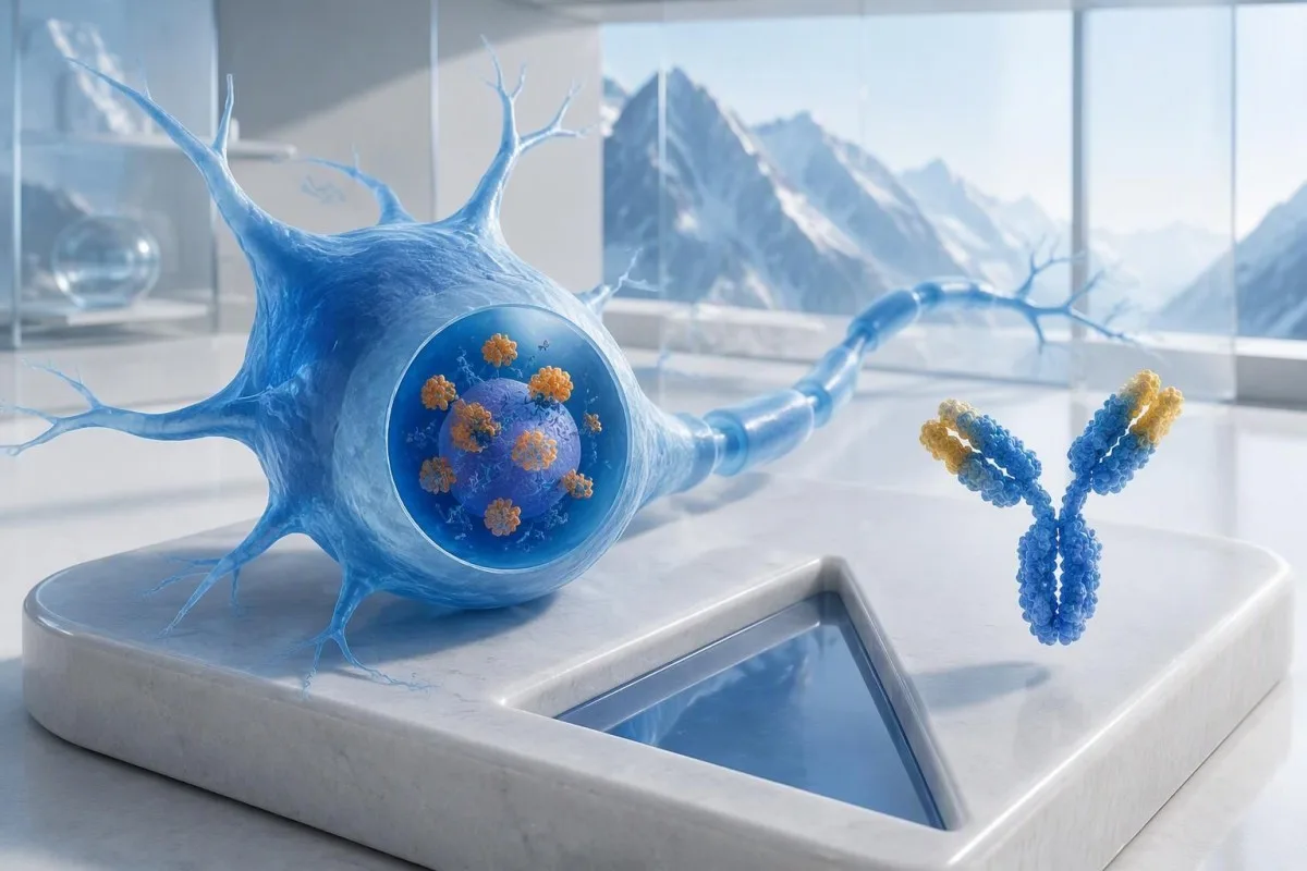
Το anti-Hu ταξινομείται ως αντίσωμα υψηλού κινδύνου που σχετίζεται με καρκίνο, με την κατηγορία να ορίζεται από συσχέτιση με καρκίνο άνω του 70% σε σχετικούς κλινικούς πληθυσμούς (Graus et al., 2021). Αυτό το ποσοστό δεν μπορεί απλώς να αποδοθεί σε ένα κατά τα άλλα υγιές άτομο με ασθενές, μη επιβεβαιωμένο αποτέλεσμα από πάνελ: οι ασθενείς που μελετήθηκαν, η μέθοδος εξέτασης και η νευρολογική εικόνα επηρεάζουν όλα την πιθανότητα.
Ένα υποθετικό άτομο 62 ετών με επιδεινούμενη αστάθεια επί 8 εβδομάδες και ασύμμετρη αισθητική απώλεια χρήζει διαφορετικής αξιολόγησης από κάποιον του οποίου το αντίσωμα εμφανίστηκε τυχαία σε ένα ευρύ πάνελ εξετάσεων για κόπωση. Σε κανένα από τα δύο άτομα δεν πρέπει να ειπωθεί “Έχετε καρκίνο” μόνο με βάση το εργαστηριακό αποτέλεσμα. Η εξήγησή μας για θετικά ευρήματα αντισωμάτων βοηθά να διαχωριστεί ένας ανοσολογικός δείκτης από μια ολοκληρωμένη διάγνωση.
Το Καντέστι είναι ένα Αναλυτής εξέτασης αίματος AI που μπορεί να βοηθήσει στην εξήγηση της διατύπωσης μιας έκθεσης anti-Hu, αλλά η εργαστηριακή επιβεβαίωση και η αξιολόγηση για καρκίνο ανήκουν στους θεράποντες ιατρούς σας. Ως Thomas Klein, MD, Chief Medical Officer στην Kantesti LTD, η πρώτη μου ερώτηση θα ήταν τι οδήγησε στη διενέργεια της συγκεκριμένης εξέτασης — όχι πόσο ανησυχητική φαίνεται η προειδοποιητική ένδειξη· η οργανισμός και η κλινική μας αποστολή εξηγούν αυτή τη διάκριση.
Είναι το ANNA-1 το ίδιο αντίσωμα με το anti-Hu;
αντίσωμα ANNA-1 και αντίσωμα anti-Hu αναφέρονται γενικά στην ίδια οικογένεια αντισωμάτων. Το ANNA-1 σημαίνει αντινευρωνικό πυρηνικό αντίσωμα τύπου 1· ο χαρακτηρισμός Hu αναφέρεται στις ενδοκυττάριες νευρωνικές πρωτεΐνες που αναγνωρίζονται από την ανοσολογική απόκριση και όχι σε ξεχωριστή νόσο ή στάδιο καρκίνου.
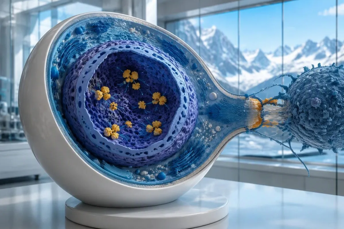
Οι πρωτεΐνες Hu είναι ενδοκυττάριες πρωτεΐνες που δεσμεύουν RNA, και το anti-Hu είναι προτιμότερο να γίνεται αντιληπτό ως δείκτης μιας ανοσολογικής απόκρισης στην οποία συχνά συμμετέχουν κυτταροτοξικά Τ κύτταρα. Αυτό διαφέρει από τις διαταραχές στις οποίες τα αντισώματα δεσμεύονται σε προσβάσιμες πρωτεΐνες της νευρωνικής επιφάνειας. Η διάκριση εξηγεί γιατί η απλή μείωση των επιπέδων των αντισωμάτων δεν αναστρέφει αξιόπιστα την ήδη εγκατεστημένη νευρολογική βλάβη από anti-Hu, ακόμη και όταν 2 εκθέσεις δείχνουν σημαντικά διαφορετικούς τίτλους.
Μια έκθεση που αναφέρει “ανιχνεύθηκε ANNA-1” δεν είναι το ίδιο με ένα συνηθισμένο θετικό ANA, παρά τα παρόμοια γράμματα. ΑΝΑ συνήθως σημαίνει αντιπυρηνικά αντισώματα που χρησιμοποιούνται στη διερεύνηση συστηματικών αυτοάνοσων νοσημάτων, ενώ το ANNA-1 προσδιορίζει ένα πρότυπο νευρωνικού αντισώματος. Ξεχωρίζετε αυτές τις 2 συντομογραφίες όταν συζητάτε τα αποτελέσματά σας· ο οδηγός ορολογίας ANA εξηγεί την πιο γνωστή εξέταση.
Το anti-Hu μπορεί να ανιχνευθεί σε ορισμένα άτομα με μικροκυτταρικό καρκίνο του πνεύμονα που δεν έχουν αναγνωρίσιμο νευρολογικό σύνδρομο. Αντίστροφα, τα νευρολογικά συμπτώματα μπορεί να οδηγήσουν στην ανίχνευση αντισωμάτων προτού γίνει ορατός ο καρκίνος. Πρόκειται για 2 διαφορετικές κλινικές πορείες: από μόνο του, το αντίσωμα δεν δείχνει στον γιατρό αν προκαλείται τώρα νευρική βλάβη ή πού μπορεί να βρίσκεται ένας υποκείμενος καρκίνος.
Πώς πρέπει να διαβαστεί η έκθεση της εξέτασης αντισωμάτων anti-Hu;
An anti-Hu antibody test has no universal numerical cutoff that can be applied across laboratories. Read the specimen type, assay method, result category, laboratory reference interval and confirmation comments together; a dilution titer, band intensity and numerical index are 3 different ways of reporting reactivity.
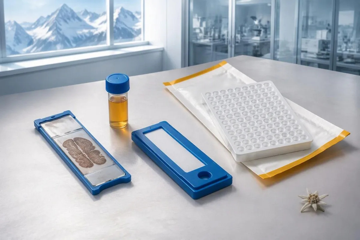
A hypothetical titer of 1:240 describes detectable reactivity after a particular dilution; it is not a cancer probability of 240, nor does it establish cancer stage. A numerical index from another method may use entirely different calibration. Our guide to ποιοτικά έναντι ποσοτικών αποτελεσμάτων explains why “positive” and “how much” answer different laboratory questions.
Weak or borderline reactivity deserves method-specific interpretation, particularly when a broad panel was ordered without a convincing neurological indication. Ask whether the laboratory observed the characteristic neuronal staining pattern and whether an antigen-specific confirmation was performed. Two tests using the same underlying strip technology may reproduce the same analytical problem; agreement is more informative when the methods are genuinely complementary.
Bring the complete report, not just a screenshot of the word positive. The 5 most useful details are the specimen, collection date, assay name, reported strength or titer and laboratory interpretation. Also list recent intravenous immunoglobulin and other immune treatments, because passive antibodies or treatment effects can complicate some serological results; your laboratory can advise whether that issue plausibly affects this particular finding.
Πότε χρειάζεται επιβεβαίωση ενός θετικού αποτελέσματος anti-Hu;
Unexpected, weak or clinically discordant anti-Hu results should be reviewed for confirmation, preferably with an experienced neuroimmunology laboratory. The 2021 PNS criteria emphasize interpreting neuronal antibody testing alongside the phenotype and using appropriate confirmatory methods rather than accepting an isolated commercial-panel result as a diagnosis.

Tissue-based immunohistochemistry or immunofluorescence and an antigen-specific assay provide different information. The first identifies a characteristic distribution of neuronal staining; the second tests reactivity against the relevant antigen. Graus et al. (2021) describe these complementary approaches, with specialist reassessment particularly useful when 1 method gives a result that does not fit the clinical picture.
Repeating a sample can address collection errors or a transient analytical concern, but repeating the same kit is not always enough. In a hypothetical 45-year-old with no objective neurological findings and a faint isolated band, I would want the laboratory’s explanation before treating the report as established anti-Hu autoimmunity. Our GAD65 interpretation guide illustrates another antibody for which context and assay characteristics change meaning.
Confirmation and urgent assessment can happen in parallel. A person who develops seizures, substantial walking difficulty or progressive swallowing impairment should not wait several weeks for a reference-laboratory answer before receiving neurological care. The practical question is not “repeat or investigate?” but which of those 2 tasks can safely proceed together, given the symptoms and the initial laboratory evidence.
Ποια νευρολογικά συμπτώματα καθιστούν το anti-Hu πιο ανησυχητικό;
Progressive sensory neuronopathy, encephalomyelitis, limbic encephalitis and marked autonomic dysfunction are recognized anti-Hu presentations. Symptoms can involve more than 1 part of the nervous system, so the pattern and speed of change are usually more informative than a single symptom such as tingling or fatigue.

Sensory neuronopathy can cause asymmetric numbness, impaired position sense and sensory ataxia, sometimes involving the hands or trunk early rather than following a simple feet-first pattern. A patient may lose balance in the dark because vision had been compensating for reduced position feedback. That is different from the focal finger distribution discussed in our carpal tunnel symptom guide, although neither pattern can be diagnosed from description alone.
Anti-Hu autonomic involvement can affect gut movement, blood-pressure regulation and bladder function. Severe constipation, unexplained intestinal pseudo-obstruction or dizziness on standing becomes more relevant when accompanied by objective neurological abnormalities. Recording symptoms over 7 days can reveal combinations that otherwise get presented as separate stomach, balance and urinary complaints; sudden severe abdominal symptoms still require assessment for non-neurological emergencies.
New memory impairment, behavioral change or seizures may indicate central nervous system involvement, while rapid weakness has a broader emergency differential. The επείγοντα προειδοποιητικά σημεία αδυναμίας in our neurological safety guide apply regardless of the antibody label. Call emergency services for new breathing difficulty, unsafe swallowing, a first seizure or sudden stroke-like symptoms rather than waiting for a scheduled follow-up.
Ποιες νευρολογικές εξετάσεις μπορεί να ακολουθήσουν ένα θετικό αποτέλεσμα;
Neurological follow-up may include examination, nerve conduction studies, MRI and cerebrospinal fluid testing, selected according to the symptoms. There is no mandatory battery for every anti-Hu-positive person; the purpose is to document which structures are affected and exclude other causes, not merely to collect additional positive tests.
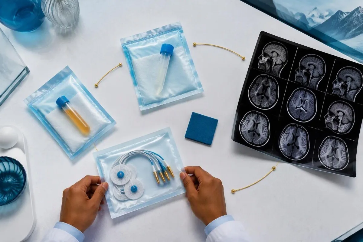
Nerve conduction studies can support a sensory neuronopathy pattern by showing reduced sensory responses across distributions that do not fit a single compressed nerve. Brain or spinal MRI addresses different questions, including structural disease and some inflammatory changes. A normal MRI does not exclude peripheral sensory ganglion involvement; these are 2 distinct anatomical compartments with different diagnostic tools.
Cerebrospinal fluid may show increased cells, protein or evidence of intrathecal immune activity, but none of those findings is specific to anti-Hu. A lumbar puncture is most useful when the neurological syndrome warrants it, rather than as an automatic response to 1 weak serum band. Our discussion of ερμηνεία των ολιγοκλωνικών ζωνών στο ΕΝΥ explains why immune activity in spinal fluid does not identify a single disease.
Basic investigations still matter: glucose control, vitamin status, renal function and serum protein assessment can uncover treatable contributors to neuropathy. A clinician may request 2 or more of these alongside specialist testing, depending on the examination. The για τον έλεγχο των πρωτεϊνών ορού provides background, but normal routine blood work neither confirms nor excludes an anti-Hu-associated neurological process.
Επιβεβαιώνει η θετικότητα στο anti-Hu ένα παρανεοπλασματικό σύνδρομο;
A positive antibody alone does not confirm anti-Hu paraneoplastic syndrome. The PNS-Care approach combines the neurological phenotype, antibody risk category, cancer findings and follow-up; clinicians also check for direct cancer effects, treatment toxicity and unrelated neurological conditions before attributing symptoms to a paraneoplastic immune response.
Definite, probable and possible PNS are diagnostic categories, not cancer stages. The 2021 criteria use a structured scoring system rather than allowing 1 antibody to determine the whole diagnosis (Graus et al., 2021). A confirmed anti-Hu result with a strongly compatible syndrome remains clinically significant even before a cancer is identified, but clinicians should state clearly which parts of the assessment remain unresolved.
An older patient can have anti-Hu reactivity and a separate cause of numbness, such as diabetes, nutritional deficiency or a monoclonal protein disorder. Coexistence is possible; either-or reasoning can miss treatable disease. If serum protein studies reveal a band, our immunofixation result guide explains why that finding requires its own interpretation rather than being automatically folded into the anti-Hu diagnosis.
Το Καντέστι είναι ένα πλατφόρμα ερμηνείας εξετάσεων αίματος AI that can help organize these distinctions in a laboratory explanation, but it cannot assign a reliable PNS-Care diagnosis from a report alone. Our πλαίσιο κλινικής επικύρωσης separates technical evaluation from clinical conclusions. In practice, I would document 3 separate judgments: confidence in the antibody, confidence in the neurological syndrome and the current evidence for cancer.
Πώς αναζητούν οι γιατροί έναν σχετιζόμενο καρκίνο;
Cancer evaluation usually prioritizes the chest because small-cell lung cancer is the strongest adult anti-Hu association. Doctors commonly begin with appropriate chest CT and may add FDG-PET/CT or targeted investigations if suspicion remains; the exact plan depends on age, symptoms, examination, smoking history and previous imaging.
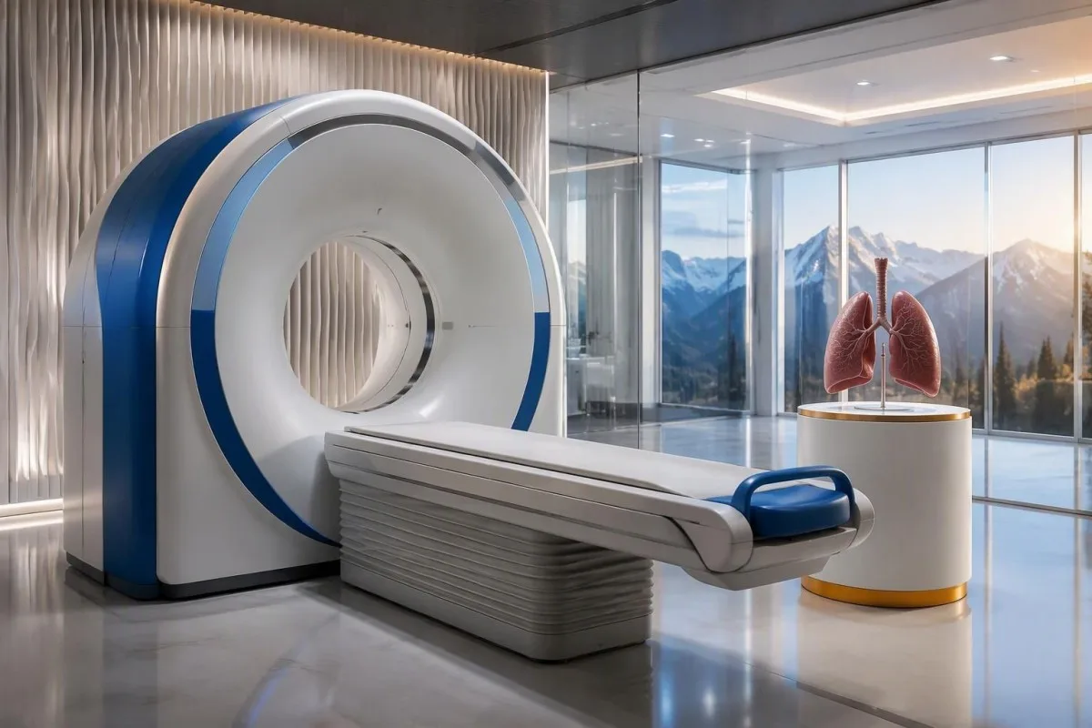
The cancer association is substantial in established anti-Hu clinical cohorts. Villagrán-García et al. (2023) reported identified cancer in 349 of 466 patients, approximately 75%, in a specialist anti-Hu cohort. This is valuable evidence for taking confirmed, clinically relevant anti-Hu seriously; it is not a personalized prediction for an asymptomatic person with an unconfirmed positive screening strip.
A chest X-ray is not equivalent to a chest CT, and routine low-dose screening is not necessarily the correct examination for a suspected paraneoplastic syndrome. The clinician chooses the protocol and body regions based on the diagnostic question. Likewise, neurological symptoms do not automatically mean cancer has spread to the brain; our brain imaging warning-sign guide distinguishes structural investigation from immune-mediated disease.
A suspicious scan often leads to tissue sampling through a method chosen by respiratory or oncology specialists, such as bronchoscopic assessment of an accessible area. Imaging suspicion and cancer confirmation are 2 separate steps. Never-smoking status lowers the baseline probability of small-cell lung cancer but does not cancel a well-confirmed anti-Hu finding with a convincing neurological presentation; other cancers and non-cancer explanations still need consideration.
Γιατί μπορεί οι αρχικές απεικονιστικές εξετάσεις για καρκίνο να είναι αρνητικές;
Initial imaging can be negative because an associated cancer is still too small to detect, lies outside the examined area or is not readily visible with that technique. Neurological autoimmunity may precede cancer detection, so a negative scan reduces concern without necessarily ending follow-up in a high-risk clinical presentation.
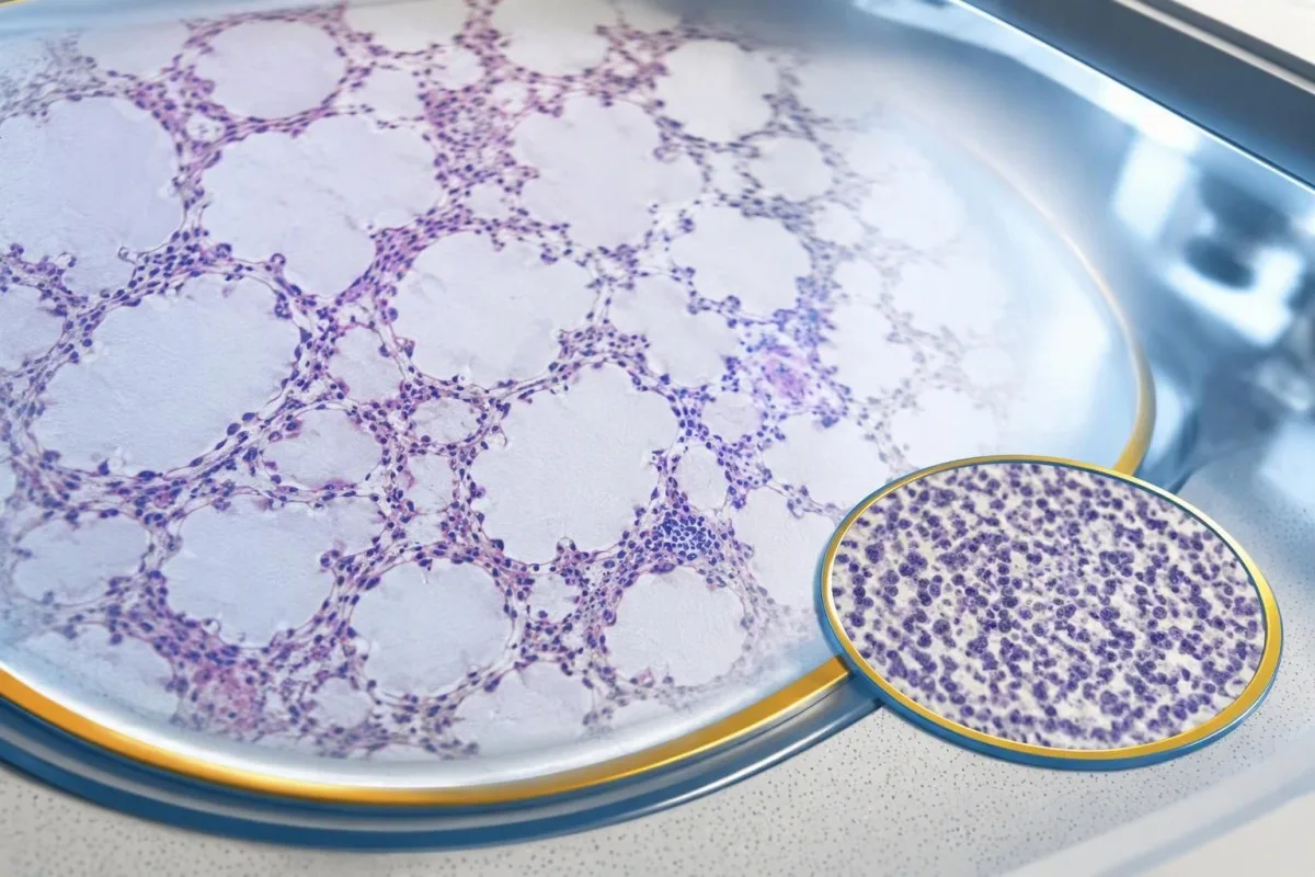
A negative scan is time-specific and method-specific. A normal chest CT performed on 1 date answers whether that examination identified a suspicious chest finding then; it does not guarantee that every possible associated cancer is absent indefinitely. Ask what was actually imaged, whether the protocol was suitable and whether the specialist recommends PET/CT or targeted investigation despite the initial result.
Villagrán-García et al. (2023) described cancers detected beyond the first 2 years in a minority of their anti-Hu cohort, supporting individualized longer surveillance. Their findings also support considering PET-based investigation when initial CT is unrevealing in an appropriate clinical context. This is an escalation decision for the treating team, not a reason to independently purchase repeated whole-body scans without weighing radiation, incidental findings and cost.
Neurological deterioration after negative imaging still needs reassessment. Cancer surveillance and neurological monitoring are 2 parallel tracks; one reassuring scan does not explain new loss of function. A marker such as neurofilament light chain may indicate neuronal injury in selected settings, but our NfL interpretation guide explains why it cannot establish anti-Hu disease, locate cancer or replace examination.
Για πόσο καιρό πρέπει να συνεχίζεται ο έλεγχος για καρκίνο;
The 2021 PNS criteria recommend repeat cancer screening every 4–6 months for 2 years when a high-risk neurological phenotype accompanies a high-risk antibody. Longer surveillance may be appropriate for selected anti-Hu patients; the interval and duration should reflect confirmation, clinical evolution, initial investigations and individual cancer risk.

The 2-year schedule is not an instruction to scan every positive antibody result repeatedly. Graus et al. (2021) link that recommendation to the combination of high-risk phenotype and high-risk antibody. An incidental, weak result without a matching syndrome needs confirmation and individualized assessment first; repeating imaging mechanically can create incidental findings and unnecessary procedures without resolving the original uncertainty.
Older European guidance recommended longer surveillance. The EFNS task force advised repeating screening after 3–6 months and then every 6 months for up to 4 years in relevant antibody-positive paraneoplastic presentations (Titulaer et al., 2011). The 2023 anti-Hu cohort supported considering screening up to 5 years in selected patients, so a specialist may reasonably extend follow-up beyond the newer general 2-year framework.
Keep a dated plan stating the next examination, the clinician responsible and what symptoms should trigger earlier contact. Kantesti explanations can help you understand terminology, but the platform should not be treated as a scan-reminder service or substitute for that plan. Our guide to risk-based cancer screening addresses a different setting: routine prevention is not the same as investigating a possible paraneoplastic syndrome.
Ποια θεραπεία βοηθά στη νευρολογική νόσο anti-Hu;
Treatment focuses on an identified cancer, selected immune therapy and symptom-directed rehabilitation. Anti-Hu-associated neurological disease often responds less predictably to immunotherapy than neuronal surface-antibody disorders because intracellular targets and T-cell-mediated injury are involved; treatment may aim to stabilize function rather than promise complete recovery.

Treating an associated cancer is a central part of care, but established neurological injury may persist even when cancer treatment succeeds. Neurology and oncology teams may consider corticosteroids, intravenous immunoglobulin, plasma exchange or other immune treatments in selected situations. There is no single evidence-based dose or 1 universal regimen appropriate for everyone with anti-Hu positivity, especially without a confirmed clinical syndrome.
An antibody titer is not a dependable treatment target on its own. A lower number does not necessarily mean restored nerve function, and a persistent positive result does not by itself prove cancer progression. This differs from some surface-antibody disorders; our MOG antibody diagnosis guide explains another immune neurological condition, but its diagnostic rules and treatment expectations should not be transferred to anti-Hu.
Immune checkpoint cancer therapies deserve an explicit discussion when anti-Hu neurological disease is present, because they can provoke or worsen paraneoplastic neurological autoimmunity. This is a specialist risk-benefit decision, not an automatic prohibition based on 1 laboratory result. Tell the oncologist about previous neurological symptoms and confirmed neuronal antibodies before treatment decisions are finalized; coordinating the teams early can prevent avoidable misunderstanding.
Τι μπορείτε να κάνετε με ασφάλεια όσο συνεχίζονται οι διερευνήσεις;
While investigations continue, protect mobility, swallowing and medication safety rather than trying to lower anti-Hu antibodies with supplements. Use the neurological symptoms—not the appearance of the laboratory flag—to decide whether ordinary follow-up is sufficient or urgent care is needed; a new functional loss warrants contact before the next planned scan.

Position-sense loss increases fall risk, particularly in poor lighting or on uneven ground. A physiotherapist may recommend a suitable walking aid and individualized balance strategies; occupational therapy can address hand coordination and home hazards. A short 7-day record of falls, near-falls and walking changes is often more useful at review than the vague statement “my balance is worse,” but stop activities that feel unsafe.
Excess vitamin B6 can itself cause sensory neuropathy, making unsupervised high-dose nerve supplements particularly unhelpful during this assessment. Review every supplement and its daily dose in mg, including overlapping multivitamins and combination products. Our οδηγός ασφαλείας για σύμπλεγμα Β explains that problem; correcting a documented deficiency is different from taking progressively larger doses in the hope of treating anti-Hu autoimmunity.
Vitamin B12 deficiency can contribute to sensory and gait problems and may need investigation even when anti-Hu is confirmed. There is no universally interchangeable B12 cutoff: results in pg/mL και pmol/L require their own laboratory context, sometimes with functional testing. Our οδηγός μας για παρακολούθηση οριακής Β12 explains those next steps; new choking, breathing difficulty or rapid weakness should be assessed urgently instead of managed with vitamins.
Πώς μπορεί η ΤΝ να βοηθήσει στην ασφαλή επεξήγηση μιας έκθεσης anti-Hu;
AI can help explain report terminology and organize questions, but it cannot confirm anti-Hu specificity, diagnose paraneoplastic syndrome or rule out cancer. Safe interpretation preserves the original assay details and separates 3 issues: what the laboratory reported, what the result may mean and what a clinician has actually established.

Το Καντέστι είναι ένα Εργαλείο ανάλυσης αιματολογικών εξετάσεων με AI-powered that can provide a plain-language explanation of uploaded laboratory information, including the distinction between an antibody screen and a confirmed clinical diagnosis. A rapid explanation—often around 60 seconds for report processing—is not a reference-laboratory assay. Our οδηγός τεχνολογίας AI describes the interpretation workflow without making anti-Hu-specific diagnostic guarantees.
The complete source report should remain available for verification. An explanation should not silently replace “weakly reactive” with “confirmed,” drop the specimen type or invent a universal reference interval. Our guide to έλεγχο ιατρικών εξηγήσεων από AI describes practical source checks; for this antibody, compare at least the assay wording, confirmation comment and collection date against the original document.
Kantesti cannot examine sensation, review tissue staining directly through a report explanation or establish that negative imaging has excluded a tiny cancer. Those are 3 different clinical capabilities, not features that follow from understanding laboratory language. Share the explanation with the treating team as a discussion aid, and do not delay urgent assessment while uploading documents or waiting for a second automated interpretation.
Το επόμενο ραντεβού σας και η διαφάνεια της έρευνας
Bring the complete antibody report, a dated symptom history, previous imaging reports and a current medication list to your next appointment. Ask the clinician to clarify antibody confirmation, the neurological diagnosis and cancer surveillance separately; those 3 answers may have different levels of certainty and different follow-up dates.
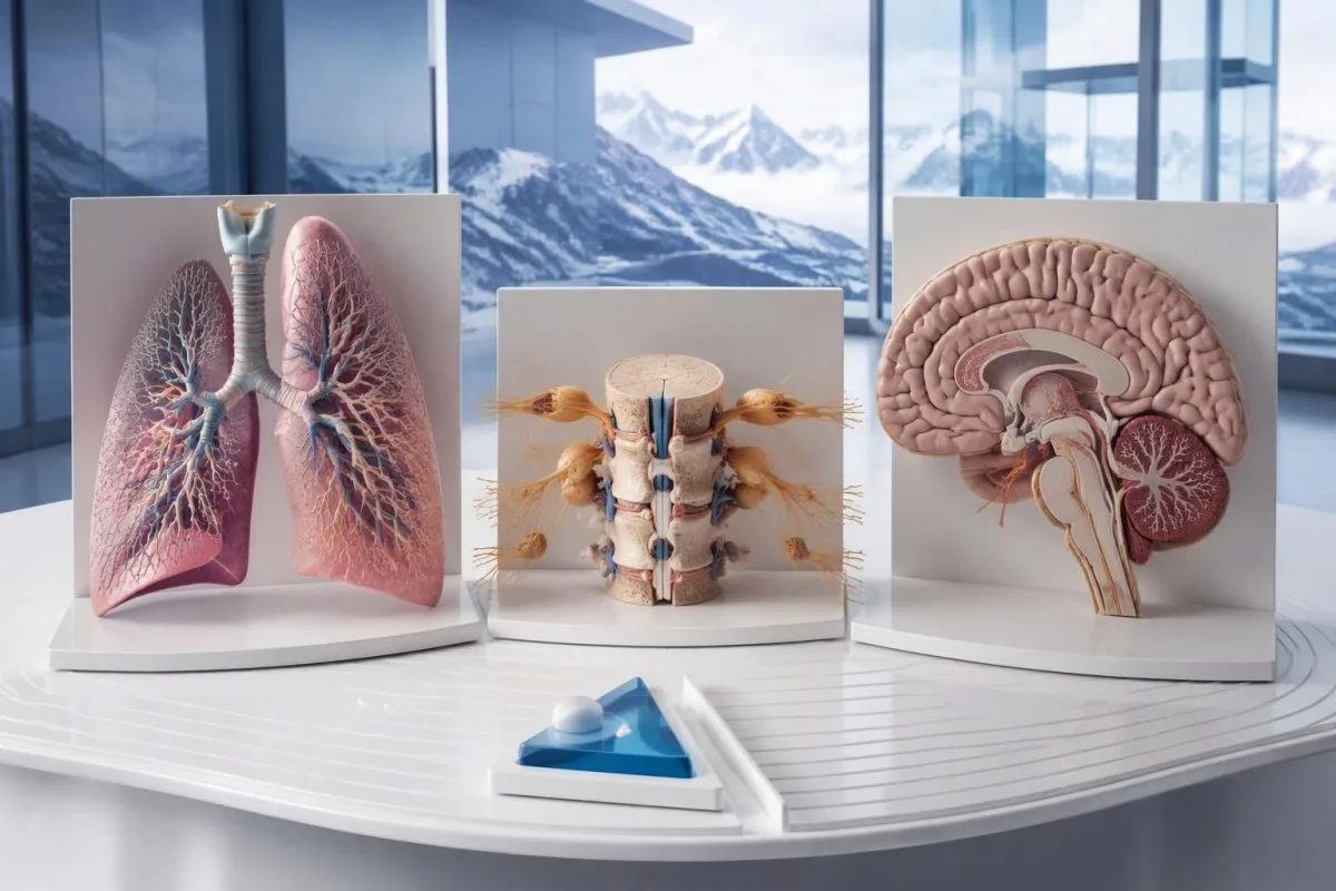
A useful 1-page symptom history records onset, progression, sensory distribution, walking changes, memory problems and autonomic symptoms. Include prior cancer treatment and any recent immunoglobulin therapy, then ask who will coordinate neurology and oncology if both are involved. As Thomas Klein, MD, I would rather see a clear timeline than 20 disconnected screenshots; it makes discrepancies between the laboratory result and clinical course easier to spot.
Medical authorship is not a substitute for an individual neurological examination. Μας πληροφορίες ιατρικής συμβουλευτικής επιτροπής describes the clinicians associated with our work, while your own specialist determines the appropriate next investigation. As of 25 Οκτωβρίου 2026, the recommendations discussed here draw on the 2021 international PNS criteria, the 2011 EFNS screening guidance and the 2023 anti-Hu cohort—not an invented 2026 guideline.
The 2 Figshare publications below are platform-related research references, not evidence validating anti-Hu diagnosis or cancer exclusion. Their supplied titles describe hantavirus triage deployment and a synthetic-case technical benchmark; neither title establishes anti-Hu-specific clinical performance. Because publication authors and dates were not supplied for verification, the citations use title-first APA formatting with “n.d.”; the ResearchGate and Academia.edu links are searches, not claims of verified publication copies.
Συχνές Ερωτήσεις
Σημαίνει ένα θετικό αποτέλεσμα αντισωμάτων anti-Hu ότι έχω καρκίνο;
Ένα θετικό αποτέλεσμα για αντισώματα anti-Hu δεν αρκεί από μόνο του για τη διάγνωση καρκίνου. Τα anti-Hu είναι αντισώματα υψηλού κινδύνου που σχετίζονται με τον καρκίνο και καρκίνος εντοπίστηκε σε 349 από τους 466 ασθενείς μιας εξειδικευμένης ομάδας ασθενών με anti-Hu το 2023, αλλά αυτό το ποσοστό δεν καθορίζει τον κίνδυνο ενός τυχαίου, μη επιβεβαιωμένου αποτελέσματος προσυμπτωματικού ελέγχου. Οι γιατροί αξιολογούν την κλινική εικόνα από το νευρικό σύστημα, επιβεβαιώνουν τα μη αναμενόμενα εργαστηριακά ευρήματα και διερευνούν την πιθανότητα υποκείμενου καρκίνου, δίνοντας συνήθως προτεραιότητα στον θώρακα στους ενήλικες. Η διάγνωση καρκίνου γενικά απαιτεί ξεχωριστά κλινικά, απεικονιστικά και ιστολογικά ευρήματα.
Είναι το anti-Hu και το ANNA-1 το ίδιο πράγμα;
Τα Anti-Hu και ANNA-1 συνήθως προσδιορίζουν την ίδια οικογένεια νευρωνικών αντισωμάτων. Το ANNA-1 σημαίνει αντινευρωνικό πυρηνικό αντίσωμα τύπου 1, ενώ το Hu αναφέρεται στις ενδοκυττάριες νευρωνικές πρωτεΐνες που αναγνωρίζει το αντίσωμα. Το ANNA-1 δεν είναι το ίδιο με μια εξέταση ANA ρουτίνας που χρησιμοποιείται στον έλεγχο συστηματικών αυτοάνοσων νοσημάτων. Ελέγξτε την ακριβή εξέταση και τη διατύπωση επιβεβαίωσης, καθώς τα εργαστήρια μπορεί να αναφέρουν την ίδια οικογένεια αντισωμάτων με διαφορετικούς τρόπους.
Μπορεί μια εξέταση αντισωμάτων anti-Hu να είναι ψευδώς θετική;
Μια εξέταση αντισωμάτων anti-Hu μπορεί να δώσει παραπλανητική ή μη ειδική αντιδραστικότητα, ιδίως όταν ένα ασθενώς θετικό αποτέλεσμα διαλογής δεν συνάδει με την κλινική νευρολογική εικόνα. Η επιβεβαίωση γίνεται συνήθως με 2 συμπληρωματικές προσεγγίσεις: ένα χαρακτηριστικό πρότυπο χρώσης ιστού και μια ειδική για το αντιγόνο δοκιμασία, η οποία ερμηνεύεται από έμπειρο εργαστήριο. Η επανάληψη της ίδιας εμπορικής μεθόδου μπορεί να αναπαράγει το ίδιο αναλυτικό πρόβλημα αντί να το επιλύσει. Τα ταχέως εξελισσόμενα νευρολογικά συμπτώματα εξακολουθούν να απαιτούν έγκαιρη αξιολόγηση εν αναμονή της επιβεβαίωσης.
Πόσο συχνά πρέπει να επαναλαμβάνονται οι απεικονιστικές εξετάσεις αν το anti-Hu είναι θετικό;
Τα κριτήρια PNS του 2021 συνιστούν επανάληψη του προσυμπτωματικού ελέγχου για καρκίνο κάθε 4–6 μήνες επί 2 χρόνια όταν ένας νευρολογικός φαινότυπος υψηλού κινδύνου συνυπάρχει με ένα αντίσωμα υψηλού κινδύνου. Αυτό το πρόγραμμα δεν εφαρμόζεται αυτομάτως σε κάθε τυχαίο θετικό αποτέλεσμα anti-Hu. Οι παλαιότερες κατευθυντήριες οδηγίες της EFNS συνιστούσαν παρακολούθηση έως και για 4 χρόνια, ενώ μια μελέτη κοόρτης του 2023 για το anti-Hu υποστήριξε ότι μπορεί να εξεταστεί παρακολούθηση έως και για 5 χρόνια σε επιλεγμένους ασθενείς. Ο ειδικός σας θα πρέπει να καθορίσει τη μέθοδο απεικόνισης, το μεσοδιάστημα, τη διάρκεια και τα συμπτώματα που απαιτούν νωρίτερη επανεκτίμηση.
Μπορεί το anti-Hu να είναι θετικό όταν οι αξονικές και οι τομογραφίες PET είναι φυσιολογικές;
0 Τα αντισώματα anti-Hu μπορεί να παραμένουν θετικά όταν η απεικονιστική εξέταση με αξονική τομογραφία ή PET δεν έχει εντοπίσει καρκίνο. Η νευρολογική αυτοανοσία μπορεί να προηγείται της ανίχνευσης, ενώ μια αρνητική εξέταση αντικατοπτρίζει τις περιοχές που εξετάστηκαν, την τεχνική και την ημερομηνία της εξέτασης και όχι μια μόνιμη εγγύηση ότι δεν υπάρχει καρκίνος. Για μια συμβατή κλινική εικόνα υψηλού κινδύνου, τα κριτήρια του 2021 συνιστούν επανάληψη του ελέγχου κάθε 4–6 μήνες για 2 χρόνια, με εξατομικευμένη παρακολούθηση μεγαλύτερης διάρκειας σε επιλεγμένους ασθενείς με anti-Hu. Θα πρέπει επίσης να επανεξετάζονται η μη επιβεβαιωμένη αντιδραστικότητα αντισωμάτων και οι μη καρκινικές εξηγήσεις, αντί να θεωρείται δεδομένη η παρουσία ενός αόρατου καρκίνου.
Σημαίνει η μείωση του επιπέδου των αντισωμάτων anti-Hu ότι η θεραπεία είναι αποτελεσματική;
Η μείωση του επιπέδου των αντισωμάτων anti-Hu δεν αποδεικνύει αξιόπιστα νευρολογική αποκατάσταση ή επιτυχή θεραπεία του καρκίνου. Τα anti-Hu αναγνωρίζουν ενδοκυττάριες πρωτεΐνες και η σχετιζόμενη νευρολογική βλάβη συχνά περιλαμβάνει μηχανισμούς που διαμεσολαβούνται από Τ-κύτταρα και όχι μόνο επιδράσεις αντισωμάτων. Δύο τίτλοι από διαφορετικά εργαστήρια μπορεί επίσης να μην είναι συγκρίσιμοι, επειδή οι μέθοδοι διαφέρουν. Οι κλινικοί αξιολογούν τη λειτουργικότητα, τα ευρήματα της εξέτασης, τις σχετικές διερευνήσεις και την ανταπόκριση του καρκίνου, αντί να χρησιμοποιούν τον αριθμό των αντισωμάτων ως αυτοτελή θεραπευτικό στόχο.
Λάβετε σήμερα ανάλυση εξετάσεων αίματος με AI
Εγγραφείτε σε πάνω από 2 εκατομμύρια χρήστες παγκοσμίως που εμπιστεύονται το Kantesti για άμεση, ακριβή ανάλυση εργαστηριακών εξετάσεων. Ανεβάστε τα αποτελέσματα εξετάσεων αίματος και λάβετε ολοκληρωμένη ερμηνεία βιοδεικτών του 15,000+ μέσα σε δευτερόλεπτα.
📚 Παραπομπές σε δημοσιεύσεις έρευνας
Klein, T., Mitchell, S., & Weber, H. (2026). Πολύγλωσση AI με Υποστήριξη Κλινικής Απόφασης για Πρώιμο Τριαζ Αποκλεισμού του Ιού της Χανταβίρωσης: Σχεδιασμός, Μηχανική Επικύρωση και Πραγματική Ανάπτυξη σε 50.000 Ερμηνευμένες Αναφορές Εξετάσεων Αίματος. Ιατρική έρευνα του Kantesti με AI.
Klein, T., Mitchell, S., & Weber, H. (2026). Μια Προ-Εγγεγραμμένη, Αυτοματοποιημένη Τεχνική Δοκιμή Βάσει Ρουμπρίκας του Μηχανισμού Ερμηνείας Αιματολογικών Εξετάσεων Kantesti σε 100.000 Συνθετικές Περιπτώσεις Δοκιμής. Ιατρική έρευνα του Kantesti με AI.
📖 Εξωτερικές ιατρικές αναφορές
Graus F et al. (2021). Updated Diagnostic Criteria for Paraneoplastic Neurologic Syndromes. Neurology: Neuroimmunology & Neuroinflammation.
Titulaer MJ et al. (2011). Screening for tumours in paraneoplastic syndromes: report of an EFNS Task Force. European Journal of Neurology.
Villagrán-García D et al. (2023). Revisiting anti-Hu paraneoplastic autoimmunity: phenotypic characterization and cancer diagnosis. Brain Communications.
⚕️ Ιατρική αποποίηση ευθύνης
Αυτό το άρθρο προορίζεται μόνο για εκπαιδευτικούς σκοπούς και δεν αποτελεί ιατρική συμβουλή. Συμβουλεύεστε πάντα έναν κατάλληλο επαγγελματία υγείας για αποφάσεις σχετικά με τη διάγνωση και τη θεραπεία.
Σήματα εμπιστοσύνης E-E-A-T
Εμπειρία
Κλινική ανασκόπηση από ιατρό για τις ροές εργασίας ερμηνείας εργαστηριακών αποτελεσμάτων.
Πραγματογνωμοσύνη
Εστίαση στην εργαστηριακή ιατρική στο πώς συμπεριφέρονται οι βιοδείκτες στο κλινικό πλαίσιο.
Αυθεντικότητα
Γραμμένο από τον Δρ. Thomas Klein με ανασκόπηση από την Δρ. Sarah Mitchell και τον Καθ. Dr. Hans Weber.
Αξιοπιστία
Ερμηνεία βασισμένη σε τεκμηριωμένα δεδομένα με σαφείς οδούς παρακολούθησης για τη μείωση του συναγερμού.
