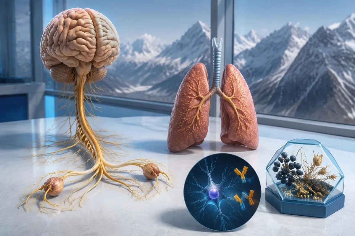
Pozitivan nalaz antitela anti-Hu pobuđuje sumnju na neurološki imunološki odgovor povezan sa karcinomom, naročito karcinomom malih ćelija pluća, ali ne postavlja dijagnozu karcinoma. Lekari procenjuju simptome, potvrđuju laboratorijski nalaz i organizuju ispitivanje na karcinom; možda će biti potrebno ponavljati skrining čak i kada su prvi snimci negativni.



- Anti-Hu / ANNA-1 je visokorizično paraneoplastično antitelo, kategorija povezana sa karcinomom kod više od 70%% ispitanika u potvrđenim kliničkim kohortama — ne sa svakim nepotvrđenim rezultatom skrininga.
- Dijagnoza karcinoma zahteva dokaze o osnovnom malignitetu, obično uključujući pregled tkiva; jedan pozitivan nalaz antitela nije takav dokaz.
- Potvrda testa često obuhvata 2 komplementarna pristupa: karakterističan obrazac bojenja tkiva i test specifičan za antigen.
- Neurološki simptomi kao što su progresivni gubitak senzibiliteta, poremećaj ravnoteže, epileptički napadi ili autonomna disfunkcija menjaju značaj i hitnost nalaza.
- Negativan nalaz snimanja znači da tim pregledom u tom trenutku nije utvrđen karcinom; ne isključuje mogućnost malog karcinoma koji se u početku ne može otkriti.
- Ponovljeni skrining preporučuje se svakih 4–6 meseci tokom 2 godine kada se visokorizičnom neurološkom fenotipu pridruži visokorizično antitelo, prema kriterijumima PNS iz 2021. godine.
- Duže praćenje može biti prikladno za odabrane pacijente; kohortna studija anti-Hu iz 2023. godine podržala je razmatranje skrininga dužeg od 2 godine, ponekad do 5 godina.
- Хитни симптоми obuhvataju nove teškoće s disanjem, nemogućnost bezbednog gutanja, brzo pogoršanje slabosti, prvi epileptični napad ili iznenadne neurološke ispade.
Šta znači pozitivan nalaz antitela anti-Hu?
Један pozitivan nalaz anti-Hu antitela znači da je laboratorija otkrila reaktivnost na Hu neuronalne proteine; sam po sebi ne potvrđuje postojanje karcinoma niti neurološkog sindroma. Tumačenje se sastoji iz 3 dela: da li je rezultat stvaran, da li neurološka slika odgovara i da li se može pronaći povezani karcinom.
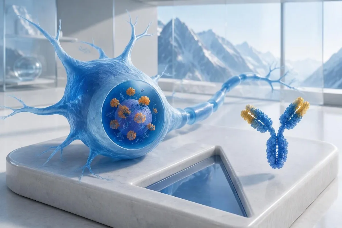
Anti-Hu je klasifikovan kao antitelo visokog rizika povezano s karcinomom, pri čemu je kategorija definisana povezanošću s karcinomom većom od 70% u relevantnim kliničkim populacijama (Graus et al., 2021). Taj procenat ne može se tek tako pripisati osobi bez tegoba koja ima slabo pozitivan, nepotvrđen rezultat panela: ispitivani pacijenti, metoda testiranja i neurološka slika utiču na verovatnoću.
Hipotetički slučaj osobe od 62 godine sa 8 nedelja pogoršanja ravnoteže i asimetričnog gubitka senzibiliteta zahteva drugačiju procenu od slučaja osobe kod koje je antitelo slučajno otkriveno tokom opsežnog panela za umor. Nijednoj osobi ne treba reći: “Imate karcinom” samo na osnovu laboratorijskog nalaza. Naše objašnjenje pozitivne nalaze antitela pomaže da se imunološki marker razlikuje od postavljene dijagnoze.
Кантести је АИ анализатор крви što može pomoći u objašnjenju formulacije nalaza anti-Hu, ali potvrda laboratorijskog rezultata i procena karcinoma treba da budu u nadležnosti vaših lekara. Kao Thomas Klein, MD, glavni medicinski rukovodilac u Kantesti LTD, prvo bih pitao šta je bio razlog za ovaj konkretan test — a ne koliko zastrašujuće deluje crvena zastavica; naš organizacija i klinička misija objašnjavaju tu razliku.
Da li je ANNA-1 isto antitelo kao anti-Hu?
antineuronalno antitelo ANNA-1 i anti-Hu antitelo uglavnom se odnose na istu porodicu antitela. ANNA-1 je skraćenica za antineuronalno nuklearno antitelo tipa 1; oznaka Hu odnosi se na intracelularne neuronalne proteine koje prepoznaje imunski odgovor, a ne na zasebnu bolest ili stadijum karcinoma.
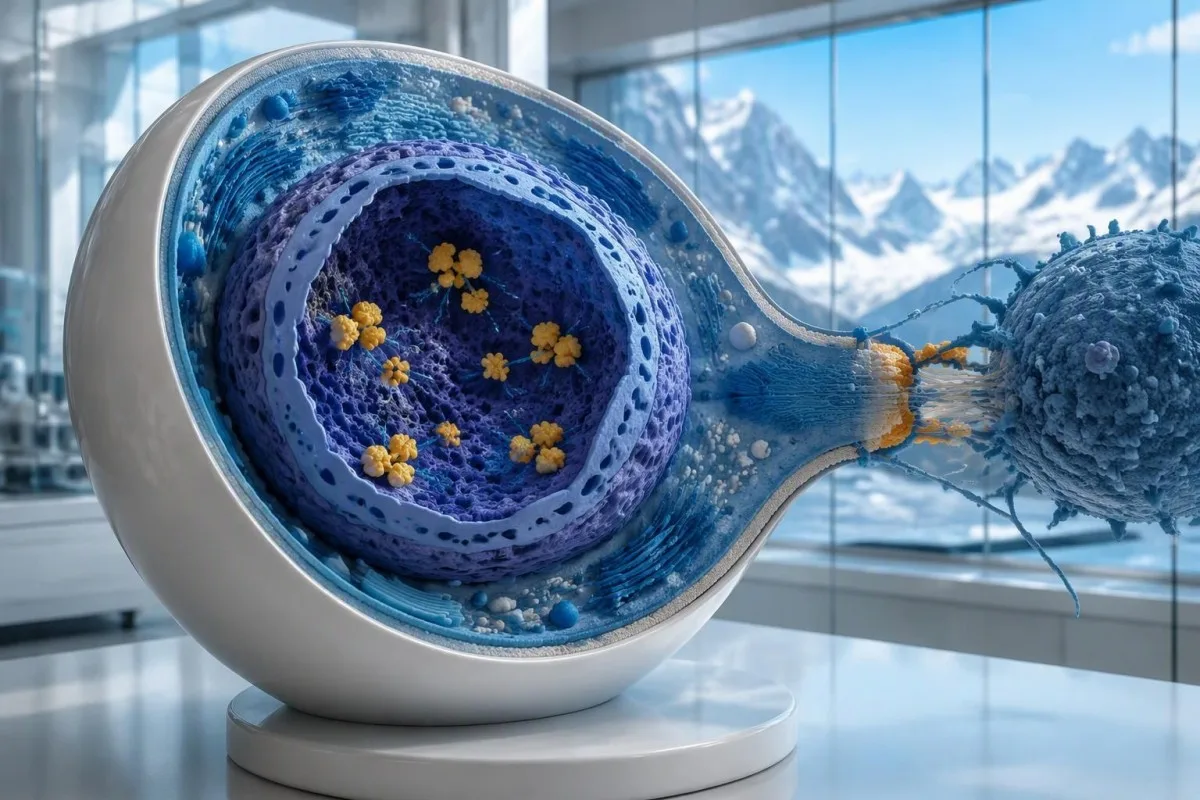
Hu proteini su intracelularni proteini koji vezuju RNK, a anti-Hu je najbolje razumeti kao marker imunskog odgovora koji često uključuje citotoksične T ćelije. To se razlikuje od poremećaja kod kojih se antitela vezuju za dostupne proteine na površini neurona. Ta razlika objašnjava zašto samo snižavanje nivoa antitela ne dovodi pouzdano do povlačenja već nastalog neurološkog oštećenja uzrokovanog anti-Hu, čak i kada 2 nalaza pokazuju znatno različite titre.
Nalaz u kojem piše “ANNA-1 detektovan” nije isto što i rutinski pozitivan ANA, uprkos sličnim slovima. АНА obično označava antinuklearna antitela koja se koriste u proceni sistemskih autoimunskih bolesti, dok ANNA-1 označava obrazac neuronalnih antitela. Kada razgovarate o svojim rezultatima, razlikujte te 2 skraćenice; naš vodič za terminologiju ANA objašnjava poznatiji test.
Anti-Hu se može otkriti kod nekih osoba sa sitnoćelijskim karcinomom pluća koje nemaju prepoznatljiv neurološki sindrom. Nasuprot tome, neurološki simptomi mogu dovesti do otkrivanja antitela pre nego što karcinom postane vidljiv. Reč je o 2 različita klinička toka: samo na osnovu antitela lekar ne može da utvrdi da li su nervi trenutno oštećeni niti gde bi se osnovni karcinom mogao nalaziti.
Kako treba tumačiti izveštaj o testu na antitela anti-Hu?
An anti-Hu antibody test has no universal numerical cutoff that can be applied across laboratories. Read the specimen type, assay method, result category, laboratory reference interval and confirmation comments together; a dilution titer, band intensity and numerical index are 3 different ways of reporting reactivity.
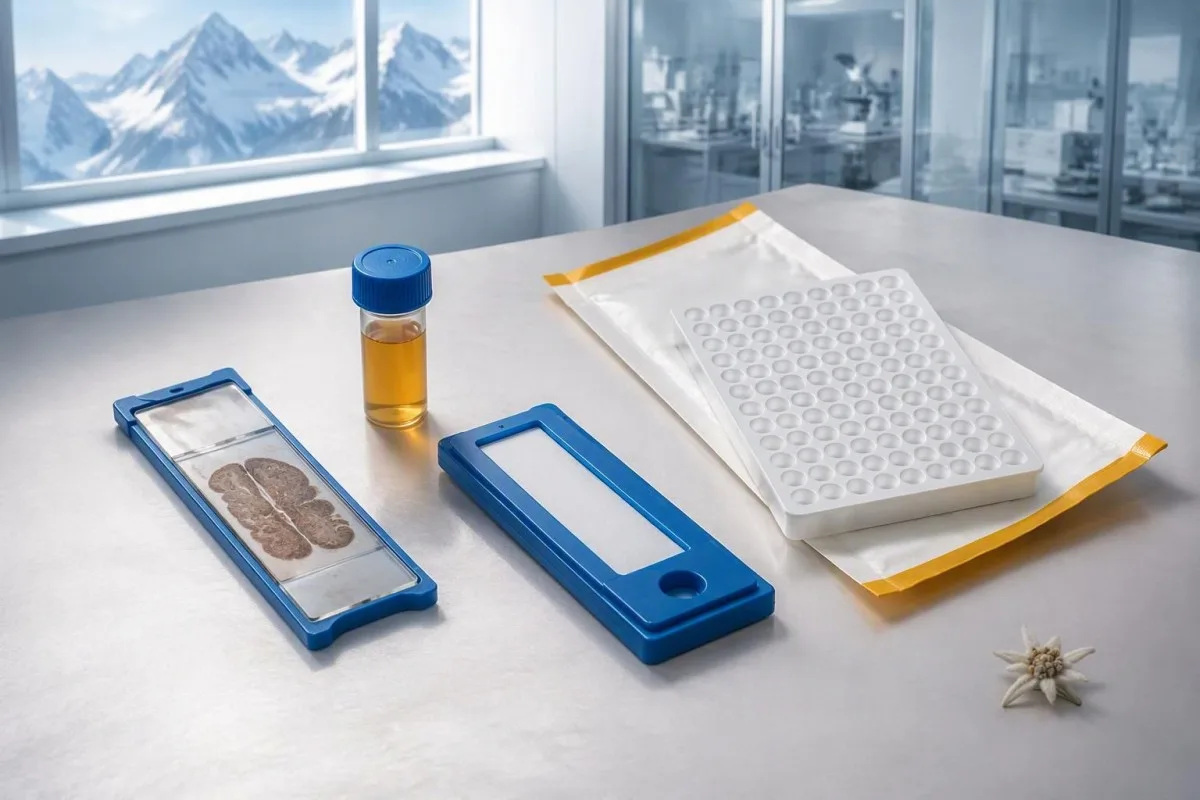
A hypothetical titer of 1:240 describes detectable reactivity after a particular dilution; it is not a cancer probability of 240, nor does it establish cancer stage. A numerical index from another method may use entirely different calibration. Our guide to kvalitativni u odnosu na kvantitativne rezultate explains why “positive” and “how much” answer different laboratory questions.
Weak or borderline reactivity deserves method-specific interpretation, particularly when a broad panel was ordered without a convincing neurological indication. Ask whether the laboratory observed the characteristic neuronal staining pattern and whether an antigen-specific confirmation was performed. Two tests using the same underlying strip technology may reproduce the same analytical problem; agreement is more informative when the methods are genuinely complementary.
Bring the complete report, not just a screenshot of the word positive. The 5 most useful details are the specimen, collection date, assay name, reported strength or titer and laboratory interpretation. Also list recent intravenous immunoglobulin and other immune treatments, because passive antibodies or treatment effects can complicate some serological results; your laboratory can advise whether that issue plausibly affects this particular finding.
Kada je potrebno potvrditi pozitivan nalaz anti-Hu?
Unexpected, weak or clinically discordant anti-Hu results should be reviewed for confirmation, preferably with an experienced neuroimmunology laboratory. The 2021 PNS criteria emphasize interpreting neuronal antibody testing alongside the phenotype and using appropriate confirmatory methods rather than accepting an isolated commercial-panel result as a diagnosis.
Tissue-based immunohistochemistry or immunofluorescence and an antigen-specific assay provide different information. The first identifies a characteristic distribution of neuronal staining; the second tests reactivity against the relevant antigen. Graus et al. (2021) describe these complementary approaches, with specialist reassessment particularly useful when 1 method gives a result that does not fit the clinical picture.
Repeating a sample can address collection errors or a transient analytical concern, but repeating the same kit is not always enough. In a hypothetical 45-year-old with no objective neurological findings and a faint isolated band, I would want the laboratory’s explanation before treating the report as established anti-Hu autoimmunity. Our GAD65 interpretation guide illustrates another antibody for which context and assay characteristics change meaning.
Confirmation and urgent assessment can happen in parallel. A person who develops seizures, substantial walking difficulty or progressive swallowing impairment should not wait several weeks for a reference-laboratory answer before receiving neurological care. The practical question is not “repeat or investigate?” but which of those 2 tasks can safely proceed together, given the symptoms and the initial laboratory evidence.
Zbog kojih neuroloških simptoma anti-Hu više zabrinjava?
Progressive sensory neuronopathy, encephalomyelitis, limbic encephalitis and marked autonomic dysfunction are recognized anti-Hu presentations. Symptoms can involve more than 1 part of the nervous system, so the pattern and speed of change are usually more informative than a single symptom such as tingling or fatigue.
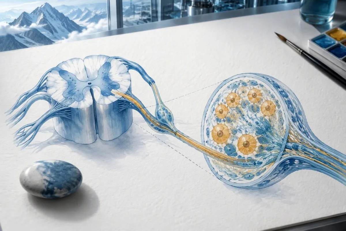
Sensory neuronopathy can cause asymmetric numbness, impaired position sense and sensory ataxia, sometimes involving the hands or trunk early rather than following a simple feet-first pattern. A patient may lose balance in the dark because vision had been compensating for reduced position feedback. That is different from the focal finger distribution discussed in our carpal tunnel symptom guide, although neither pattern can be diagnosed from description alone.
Anti-Hu autonomic involvement can affect gut movement, blood-pressure regulation and bladder function. Severe constipation, unexplained intestinal pseudo-obstruction or dizziness on standing becomes more relevant when accompanied by objective neurological abnormalities. Recording symptoms over 7 days can reveal combinations that otherwise get presented as separate stomach, balance and urinary complaints; sudden severe abdominal symptoms still require assessment for non-neurological emergencies.
New memory impairment, behavioral change or seizures may indicate central nervous system involvement, while rapid weakness has a broader emergency differential. The upozoravajuće znake hitne slabosti in our neurological safety guide apply regardless of the antibody label. Call emergency services for new breathing difficulty, unsafe swallowing, a first seizure or sudden stroke-like symptoms rather than waiting for a scheduled follow-up.
Koji neurološki testovi mogu uslediti nakon pozitivnog nalaza?
Neurological follow-up may include examination, nerve conduction studies, MRI and cerebrospinal fluid testing, selected according to the symptoms. There is no mandatory battery for every anti-Hu-positive person; the purpose is to document which structures are affected and exclude other causes, not merely to collect additional positive tests.
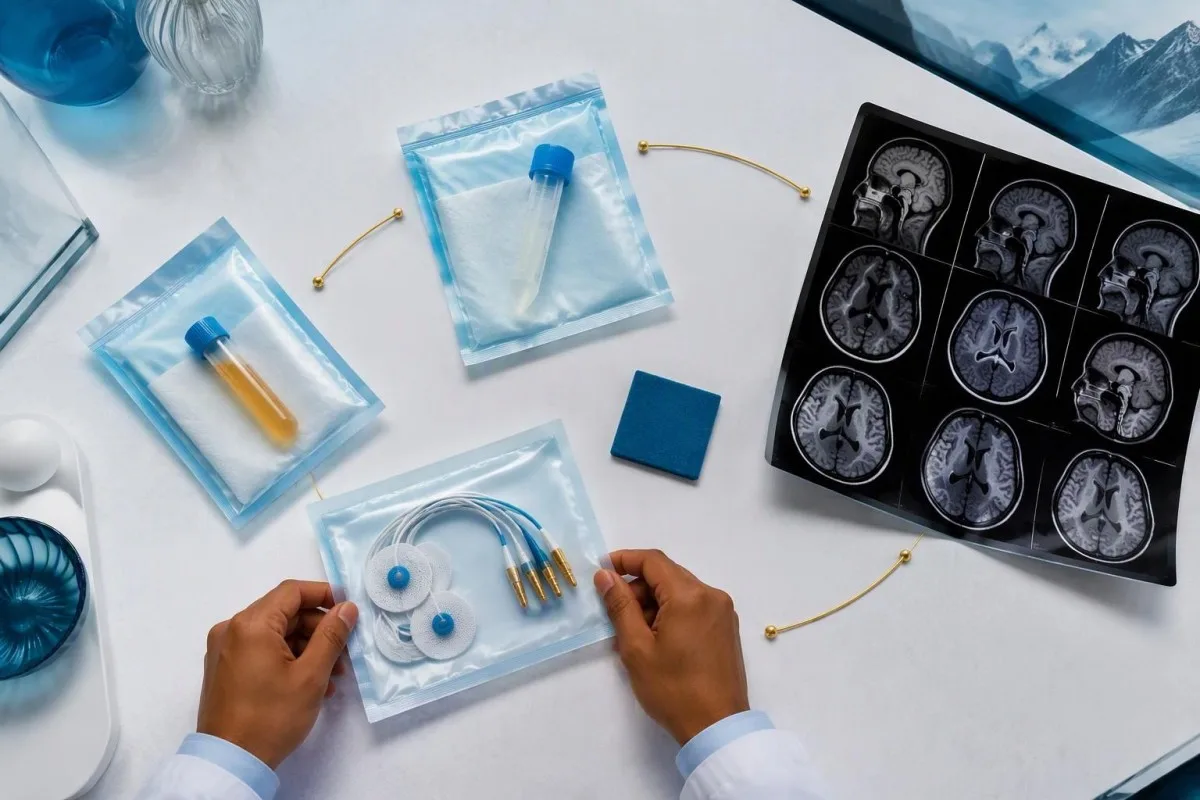
Nerve conduction studies can support a sensory neuronopathy pattern by showing reduced sensory responses across distributions that do not fit a single compressed nerve. Brain or spinal MRI addresses different questions, including structural disease and some inflammatory changes. A normal MRI does not exclude peripheral sensory ganglion involvement; these are 2 distinct anatomical compartments with different diagnostic tools.
Cerebrospinal fluid may show increased cells, protein or evidence of intrathecal immune activity, but none of those findings is specific to anti-Hu. A lumbar puncture is most useful when the neurological syndrome warrants it, rather than as an automatic response to 1 weak serum band. Our discussion of тумачењу олигоклоналних трака у ликвору explains why immune activity in spinal fluid does not identify a single disease.
Basic investigations still matter: glucose control, vitamin status, renal function and serum protein assessment can uncover treatable contributors to neuropathy. A clinician may request 2 or more of these alongside specialist testing, depending on the examination. The водич за процену серумских протеина provides background, but normal routine blood work neither confirms nor excludes an anti-Hu-associated neurological process.
Da li pozitivnost na anti-Hu potvrđuje paraneoplastični sindrom?
A positive antibody alone does not confirm anti-Hu paraneoplastic syndrome. The PNS-Care approach combines the neurological phenotype, antibody risk category, cancer findings and follow-up; clinicians also check for direct cancer effects, treatment toxicity and unrelated neurological conditions before attributing symptoms to a paraneoplastic immune response.
Definite, probable and possible PNS are diagnostic categories, not cancer stages. The 2021 criteria use a structured scoring system rather than allowing 1 antibody to determine the whole diagnosis (Graus et al., 2021). A confirmed anti-Hu result with a strongly compatible syndrome remains clinically significant even before a cancer is identified, but clinicians should state clearly which parts of the assessment remain unresolved.
An older patient can have anti-Hu reactivity and a separate cause of numbness, such as diabetes, nutritional deficiency or a monoclonal protein disorder. Coexistence is possible; either-or reasoning can miss treatable disease. If serum protein studies reveal a band, our immunofixation result guide explains why that finding requires its own interpretation rather than being automatically folded into the anti-Hu diagnosis.
Кантести је AI платформа за тумачење крвне слике that can help organize these distinctions in a laboratory explanation, but it cannot assign a reliable PNS-Care diagnosis from a report alone. Our клинички оквир за валидацију separates technical evaluation from clinical conclusions. In practice, I would document 3 separate judgments: confidence in the antibody, confidence in the neurological syndrome and the current evidence for cancer.
Kako lekari traže povezani karcinom?
Cancer evaluation usually prioritizes the chest because small-cell lung cancer is the strongest adult anti-Hu association. Doctors commonly begin with appropriate chest CT and may add FDG-PET/CT or targeted investigations if suspicion remains; the exact plan depends on age, symptoms, examination, smoking history and previous imaging.
The cancer association is substantial in established anti-Hu clinical cohorts. Villagrán-García et al. (2023) reported identified cancer in 349 of 466 patients, approximately 75%, in a specialist anti-Hu cohort. This is valuable evidence for taking confirmed, clinically relevant anti-Hu seriously; it is not a personalized prediction for an asymptomatic person with an unconfirmed positive screening strip.
A chest X-ray is not equivalent to a chest CT, and routine low-dose screening is not necessarily the correct examination for a suspected paraneoplastic syndrome. The clinician chooses the protocol and body regions based on the diagnostic question. Likewise, neurological symptoms do not automatically mean cancer has spread to the brain; our brain imaging warning-sign guide distinguishes structural investigation from immune-mediated disease.
A suspicious scan often leads to tissue sampling through a method chosen by respiratory or oncology specialists, such as bronchoscopic assessment of an accessible area. Imaging suspicion and cancer confirmation are 2 separate steps. Never-smoking status lowers the baseline probability of small-cell lung cancer but does not cancel a well-confirmed anti-Hu finding with a convincing neurological presentation; other cancers and non-cancer explanations still need consideration.
Zašto prvi snimci za otkrivanje karcinoma mogu biti negativni?
Initial imaging can be negative because an associated cancer is still too small to detect, lies outside the examined area or is not readily visible with that technique. Neurological autoimmunity may precede cancer detection, so a negative scan reduces concern without necessarily ending follow-up in a high-risk clinical presentation.
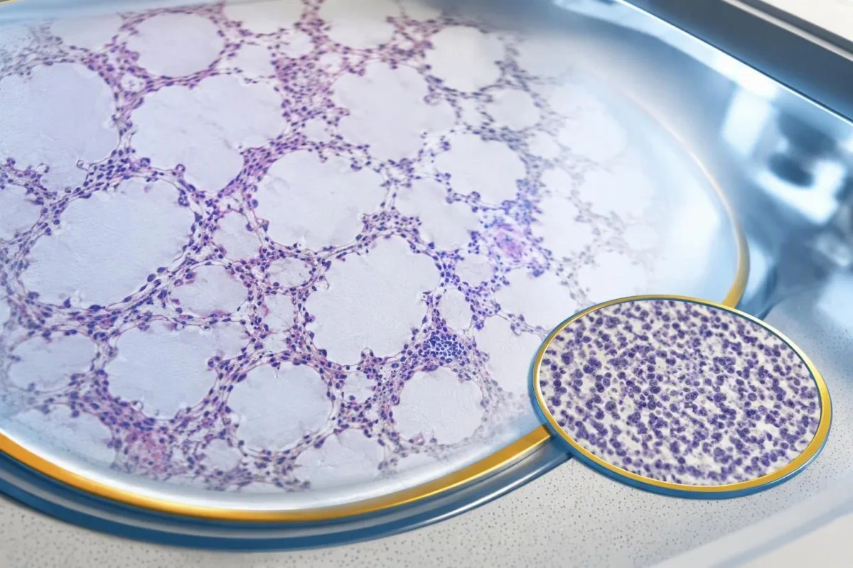
A negative scan is time-specific and method-specific. A normal chest CT performed on 1 date answers whether that examination identified a suspicious chest finding then; it does not guarantee that every possible associated cancer is absent indefinitely. Ask what was actually imaged, whether the protocol was suitable and whether the specialist recommends PET/CT or targeted investigation despite the initial result.
Villagrán-García et al. (2023) described cancers detected beyond the first 2 years in a minority of their anti-Hu cohort, supporting individualized longer surveillance. Their findings also support considering PET-based investigation when initial CT is unrevealing in an appropriate clinical context. This is an escalation decision for the treating team, not a reason to independently purchase repeated whole-body scans without weighing radiation, incidental findings and cost.
Neurological deterioration after negative imaging still needs reassessment. Cancer surveillance and neurological monitoring are 2 parallel tracks; one reassuring scan does not explain new loss of function. A marker such as neurofilament light chain may indicate neuronal injury in selected settings, but our NfL interpretation guide explains why it cannot establish anti-Hu disease, locate cancer or replace examination.
Koliko dugo treba nastaviti skrining na karcinom?
The 2021 PNS criteria recommend repeat cancer screening every 4–6 months for 2 years when a high-risk neurological phenotype accompanies a high-risk antibody. Longer surveillance may be appropriate for selected anti-Hu patients; the interval and duration should reflect confirmation, clinical evolution, initial investigations and individual cancer risk.

The 2-year schedule is not an instruction to scan every positive antibody result repeatedly. Graus et al. (2021) link that recommendation to the combination of high-risk phenotype and high-risk antibody. An incidental, weak result without a matching syndrome needs confirmation and individualized assessment first; repeating imaging mechanically can create incidental findings and unnecessary procedures without resolving the original uncertainty.
Older European guidance recommended longer surveillance. The EFNS task force advised repeating screening after 3–6 months and then every 6 months for up to 4 years in relevant antibody-positive paraneoplastic presentations (Titulaer et al., 2011). The 2023 anti-Hu cohort supported considering screening up to 5 years in selected patients, so a specialist may reasonably extend follow-up beyond the newer general 2-year framework.
Keep a dated plan stating the next examination, the clinician responsible and what symptoms should trigger earlier contact. Kantesti explanations can help you understand terminology, but the platform should not be treated as a scan-reminder service or substitute for that plan. Our guide to risk-based cancer screening addresses a different setting: routine prevention is not the same as investigating a possible paraneoplastic syndrome.
Koje lečenje pomaže kod neurološke bolesti povezane sa anti-Hu?
Treatment focuses on an identified cancer, selected immune therapy and symptom-directed rehabilitation. Anti-Hu-associated neurological disease often responds less predictably to immunotherapy than neuronal surface-antibody disorders because intracellular targets and T-cell-mediated injury are involved; treatment may aim to stabilize function rather than promise complete recovery.

Treating an associated cancer is a central part of care, but established neurological injury may persist even when cancer treatment succeeds. Neurology and oncology teams may consider corticosteroids, intravenous immunoglobulin, plasma exchange or other immune treatments in selected situations. There is no single evidence-based dose or 1 universal regimen appropriate for everyone with anti-Hu positivity, especially without a confirmed clinical syndrome.
An antibody titer is not a dependable treatment target on its own. A lower number does not necessarily mean restored nerve function, and a persistent positive result does not by itself prove cancer progression. This differs from some surface-antibody disorders; our MOG antibody diagnosis guide explains another immune neurological condition, but its diagnostic rules and treatment expectations should not be transferred to anti-Hu.
Immune checkpoint cancer therapies deserve an explicit discussion when anti-Hu neurological disease is present, because they can provoke or worsen paraneoplastic neurological autoimmunity. This is a specialist risk-benefit decision, not an automatic prohibition based on 1 laboratory result. Tell the oncologist about previous neurological symptoms and confirmed neuronal antibodies before treatment decisions are finalized; coordinating the teams early can prevent avoidable misunderstanding.
Šta možete bezbedno da radite dok se ispitivanja nastavljaju?
While investigations continue, protect mobility, swallowing and medication safety rather than trying to lower anti-Hu antibodies with supplements. Use the neurological symptoms—not the appearance of the laboratory flag—to decide whether ordinary follow-up is sufficient or urgent care is needed; a new functional loss warrants contact before the next planned scan.
Position-sense loss increases fall risk, particularly in poor lighting or on uneven ground. A physiotherapist may recommend a suitable walking aid and individualized balance strategies; occupational therapy can address hand coordination and home hazards. A short 7-day record of falls, near-falls and walking changes is often more useful at review than the vague statement “my balance is worse,” but stop activities that feel unsafe.
Excess vitamin B6 can itself cause sensory neuropathy, making unsupervised high-dose nerve supplements particularly unhelpful during this assessment. Review every supplement and its daily dose in mg, including overlapping multivitamins and combination products. Our Vodič za bezbednost B-kompleksa explains that problem; correcting a documented deficiency is different from taking progressively larger doses in the hope of treating anti-Hu autoimmunity.
Vitamin B12 deficiency can contribute to sensory and gait problems and may need investigation even when anti-Hu is confirmed. There is no universally interchangeable B12 cutoff: results in пг/мЛ и pmol/L require their own laboratory context, sometimes with functional testing. Our vodič za praćenje graničnog B12 explains those next steps; new choking, breathing difficulty or rapid weakness should be assessed urgently instead of managed with vitamins.
Kako AI može bezbedno da pomogne u objašnjavanju izveštaja o anti-Hu?
AI can help explain report terminology and organize questions, but it cannot confirm anti-Hu specificity, diagnose paraneoplastic syndrome or rule out cancer. Safe interpretation preserves the original assay details and separates 3 issues: what the laboratory reported, what the result may mean and what a clinician has actually established.

Кантести је Алат за анализу крвних тестова уз помоћ AI that can provide a plain-language explanation of uploaded laboratory information, including the distinction between an antibody screen and a confirmed clinical diagnosis. A rapid explanation—often around 60 seconds for report processing—is not a reference-laboratory assay. Our Vodič kroz AI tehnologiju describes the interpretation workflow without making anti-Hu-specific diagnostic guarantees.
The complete source report should remain available for verification. An explanation should not silently replace “weakly reactive” with “confirmed,” drop the specimen type or invent a universal reference interval. Our guide to proveru objašnjenja o medicini dobijenih od veštačke inteligencije describes practical source checks; for this antibody, compare at least the assay wording, confirmation comment and collection date against the original document.
Kantesti cannot examine sensation, review tissue staining directly through a report explanation or establish that negative imaging has excluded a tiny cancer. Those are 3 different clinical capabilities, not features that follow from understanding laboratory language. Share the explanation with the treating team as a discussion aid, and do not delay urgent assessment while uploading documents or waiting for a second automated interpretation.
Vaš naredni pregled i transparentnost istraživanja
Bring the complete antibody report, a dated symptom history, previous imaging reports and a current medication list to your next appointment. Ask the clinician to clarify antibody confirmation, the neurological diagnosis and cancer surveillance separately; those 3 answers may have different levels of certainty and different follow-up dates.
A useful 1-page symptom history records onset, progression, sensory distribution, walking changes, memory problems and autonomic symptoms. Include prior cancer treatment and any recent immunoglobulin therapy, then ask who will coordinate neurology and oncology if both are involved. As Thomas Klein, MD, I would rather see a clear timeline than 20 disconnected screenshots; it makes discrepancies between the laboratory result and clinical course easier to spot.
Medical authorship is not a substitute for an individual neurological examination. Наше informacije savetodavnog odbora describes the clinicians associated with our work, while your own specialist determines the appropriate next investigation. As of 25. oktobar 2026., the recommendations discussed here draw on the 2021 international PNS criteria, the 2011 EFNS screening guidance and the 2023 anti-Hu cohort—not an invented 2026 guideline.
The 2 Figshare publications below are platform-related research references, not evidence validating anti-Hu diagnosis or cancer exclusion. Their supplied titles describe hantavirus triage deployment and a synthetic-case technical benchmark; neither title establishes anti-Hu-specific clinical performance. Because publication authors and dates were not supplied for verification, the citations use title-first APA formatting with “n.d.”; the ResearchGate and Academia.edu links are searches, not claims of verified publication copies.
Често постављана питања
Da li pozitivan nalaz anti-Hu antitela znači da imam rak?
0 Anti-Hu pozitivan rezultat na antitela sam po sebi ne dijagnostikuje rak. Anti-Hu je antitelo povezano sa visokim rizikom od raka, a rak je utvrđen kod 349 od 466 pacijenata u specijalističkoj kohorti anti-Hu pacijenata iz 2023. godine, ali taj podatak ne određuje rizik od slučajnog, nepotvrđenog rezultata skrininga. Lekari procenjuju neurološku kliničku sliku, potvrđuju neočekivane laboratorijske nalaze i ispituju postojanje osnovnog karcinoma, obično dajući prednost pregledu grudnog koša kod odraslih. Za dijagnozu raka su uglavnom potrebni posebni klinički, slikovni i patohistološki dokazi.
Da li su anti-Hu i ANNA-1 isto?
0 Anti-Hu i ANNA-1 uglavnom označavaju istu porodicu neuronskih antitela. ANNA-1 znači antineuronsko nuklearno antitelo tipa 1, dok se Hu odnosi na unutarćelijske neuronske proteine koje antitelo prepoznaje. ANNA-1 nije isto što i rutinski ANA koji se koristi u testiranju na sistemske autoimunske bolesti. Proverite tačan test i formulaciju potvrde jer laboratorije mogu izveštavati o istoj porodici antitela u različitim formatima.
Može li test na anti-Hu antitela biti lažno pozitivan?
Test na anti-Hu antitela može dati obmanjujuću ili nespecifičnu reaktivnost, naročito kada se slab rezultat skrininga ne uklapa u neurološku kliničku sliku. Potvrda se obično zasniva na 2 komplementarna pristupa: karakterističnom obrascu bojenja tkiva i testu specifičnom za antigen, čije rezultate tumači iskusna laboratorija. Ponavljanje iste komercijalne metode može ponovo dovesti do istog analitičkog problema, umesto da ga reši. Neurološki simptomi koji brzo napreduju i dalje zahtevaju pravovremenu procenu dok se čeka potvrda.
Koliko često treba ponavljati snimanja ako je anti-Hu pozitivan?
PNS kriterijumi iz 2021. preporučuju ponavljanje skrininga na karcinom svakih 4–6 meseci tokom 2 godine kada je visokorizični neurološki fenotip udružen sa visokorizičnim antitelom. Taj raspored se ne primenjuje automatski na svaki slučajno otkriven pozitivan rezultat na anti-Hu. Starije EFNS smernice preporučivale su nadzor u trajanju do 4 godine, a kohortna studija o anti-Hu iz 2023. podržala je razmatranje nadzora u trajanju do 5 godina kod odabranih pacijenata. Vaš specijalista treba da navede metodu snimanja, interval, trajanje i simptome zbog kojih je potreban raniji pregled.
Može li anti-Hu biti pozitivan kada su CT i PET skeniranja uredni?
Anti-Hu može ostati pozitivan i kada CT ili PET snimanje ne otkrije rak. Neurološka autoimunost može prethoditi otkrivanju, a negativan nalaz snimanja odražava pregledane regije, tehniku i datum, a ne trajnu garanciju da raka nema. Kod kompatibilne visokorizične kliničke slike, kriterijumi iz 2021. godine preporučuju ponavljanje skrininga svakih 4–6 meseci tokom 2 godine, uz individualizovano duže praćenje kod odabranih pacijenata sa anti-Hu antitelima. Takođe treba ponovo razmotriti nepotvrđenu reaktivnost antitela i objašnjenja koja nisu povezana sa rakom, umesto da se pretpostavi da je nevidljivi rak definitivno prisutan.
Da li snižavanje nivoa anti-Hu antitela znači da terapija deluje?
Opadanje nivoa anti-Hu antitela ne dokazuje pouzdano neurološki oporavak niti uspešno lečenje karcinoma. Anti-Hu prepoznaje unutarćelijske proteine, a povezano neurološko oštećenje često uključuje mehanizme posredovane T-ćelijama, a ne samo dejstvo antitela. Dva titra iz različitih laboratorija takođe možda nisu uporediva zbog razlika u metodama. Kliničari procenjuju funkcionalno stanje, nalaze pregleda, relevantna ispitivanja i odgovor karcinoma, umesto da broj antitela koriste kao samostalni cilj lečenja.
Укључите AI анализу крвне слике данас
Придружите се више од 2 милиона корисника широм света који верују Kantesti-у за тренутну и прецизну анализу лабораторијских тестова. Отпремите своје резултате крвне слике и добијте свеобухватно тумачење 15,000+ биомаркера за неколико секунди.
📚 Референциране научне публикације
Klajn, T., Mičel, S., i Veber, H. (2026). Višejezična AI podrška kliničkom odlučivanju za ranu trijažu Hantavirusa: Dizajn, inženjerska validacija i primena u realnom svetu na preko 50.000 interpretiranih izveštaja o analizi krvi. Kantesti AI Medical Research.
Klajn, T., Mičel, S., i Veber, H. (2026). Претходно регистровани, рубрик-вођени аутоматизовани технички бенчмарк тумачења крвних тестова за Kantesti Blood-Test Interpretation Engine на 100.000 синтетичких тест случајева. Kantesti AI Medical Research.
📖 Спољне медицинске референце
Graus F et al. (2021). Updated Diagnostic Criteria for Paraneoplastic Neurologic Syndromes. Neurology: Neuroimmunology & Neuroinflammation.
Titulaer MJ et al. (2011). Screening for tumours in paraneoplastic syndromes: report of an EFNS Task Force. European Journal of Neurology.
Villagrán-García D et al. (2023). Revisiting anti-Hu paraneoplastic autoimmunity: phenotypic characterization and cancer diagnosis. Brain Communications.
⚕️ Медицинска одрицање одговорности
Овај чланак је само у образовне сврхе и не представља медицински савет. За одлуке о дијагнози и лечењу увек се консултујте са квалификованим здравственим радником.
Е-Е-А-Т сигнали поверења
Искуство
Клиничка ревизија процеса тумачења лабораторијских налаза коју води лекар.
Експертиза
Фокус на лабораторијску медицину: како се биомаркери понашају у клиничком контексту.
Ауторитативност
Написао др Томас Клајн, уз рецензију др Саре Мичел и проф. др Ханса Вебера.
Поузданост
Тумачење засновано на доказима, са јасним путевима праћења како би се смањила узнемиреност.
