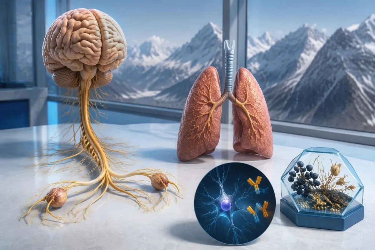
Ett positivt testresultat för anti-Hu-antikropp väcker misstanke om en cancerassocierad neurologisk immunreaktion, särskilt i samband med småcellig lungcancer, men det diagnostiserar inte cancer. Läkare bedömer symtomen, bekräftar laboratoriefyndet och ordnar en cancerutredning; upprepad screening kan behövas även när de första bilddiagnostiska undersökningarna är negativa.



- Anti-Hu / ANNA-1 är en paraneoplastisk antikropp med hög risk, en kategori som är förknippad med cancer i mer än 70% av etablerade kliniska kohorter – inte vid varje obekräftat screeningresultat.
- Cancerdiagnos kräver belägg för en underliggande malignitet, vanligen inklusive vävnadsundersökning; ett enda positivt antikroppsresultat utgör inte sådana belägg.
- Bekräftelse av analysresultat kombinerar ofta två kompletterande metoder: ett karakteristiskt vävnadsfärgningsmönster och ett antigenspecifikt test.
- Neurologiska symtom som progressiv känselnedsättning, balanssvårigheter, epileptiska anfall eller autonom dysfunktion förändrar resultatets betydelse och hur brådskande det är.
- Negativ bilddiagnostik innebär att ingen cancer påvisades vid den undersökningen och vid den tidpunkten; det utesluter inte möjligheten av en liten cancer som ännu inte går att upptäcka.
- Upprepad screening rekommenderas var 4–6 månad i 2 år när en neurologisk fenotyp med hög risk förekommer tillsammans med en antikropp med hög risk, enligt PNS-kriterierna från 2021.
- Längre övervakning kan vara lämplig för utvalda patienter; en anti-Hu-kohort från 2023 stödde övervägande av screening efter 2 år, ibland i upp till 5 år.
- Akutsymtom omfattar nya andningssvårigheter, oförmåga att svälja säkert, snabbt förvärrad svaghet, ett första epileptiskt anfall eller plötsliga neurologiska bortfall.
Vad innebär ett positivt testresultat för anti-Hu-antikropp?
En anti-Hu-antikroppspositivt resultat innebär att laboratoriet har påvisat reaktivitet mot neuronala Hu-proteiner; det fastställer inte i sig cancer eller ett neurologiskt syndrom. Tolkningen har 3 delar: om resultatet är äkta, om den neurologiska symtombilden stämmer och om en associerad cancer kan påvisas.
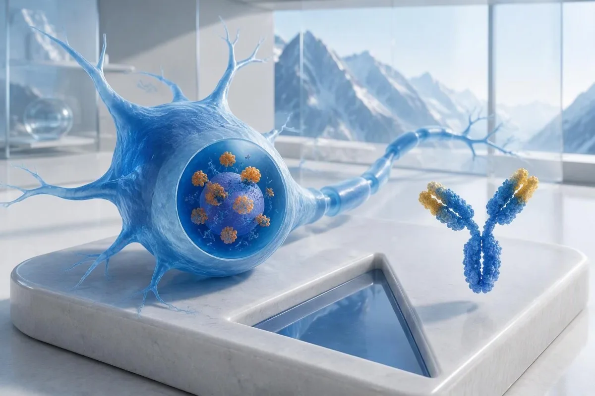
Anti-Hu klassificeras som en cancerassocierad antikropp med hög risk, där kategorin definieras av en cancerassociation på över 70% i relevanta kliniska populationer (Graus et al., 2021). Den procentsatsen kan inte utan vidare tillskrivas en i övrigt frisk person med ett svagt, obekräftat resultat från en antikroppspanel: de studerade patienterna, testmetoden och den neurologiska symtombilden påverkar alla sannolikheten.
En hypotetisk 62-åring med 8 veckors tilltagande balanssvårigheter och asymmetrisk känselnedsättning förtjänar en annan bedömning än någon vars antikropp påträffades av en slump vid en bred utredning av trötthet. Ingen av personerna bör få höra “Du har cancer” enbart utifrån laboratoriesvaret. Vår förklaring av positiva antikroppsfynd hjälper till att skilja en immunmarkör från en fastställd diagnos.
Kantesti är en AI blodprovsanalysator som kan hjälpa till att förklara formuleringen i ett anti-Hu-svar, men laboratoriebekräftelse och cancerbedömning hör hemma hos dina behandlande läkare. Som Thomas Klein, MD, medicinsk chef på Kantesti LTD, skulle min första fråga vara vad som föranledde just detta test – inte hur alarmerande den röda flaggan ser ut; vår organisation och kliniska uppdrag förklarar den skillnaden.
Är ANNA-1 samma antikropp som anti-Hu?
ANNA-1-antikropp och anti-Hu-antikropp avser i allmänhet samma antikroppsfamilj. ANNA-1 står för antineuronal nukleär antikropp typ 1; beteckningen Hu syftar på de intracellulära neuronala proteiner som immunsvaret känner igen, inte på en separat sjukdom eller ett cancerstadium.
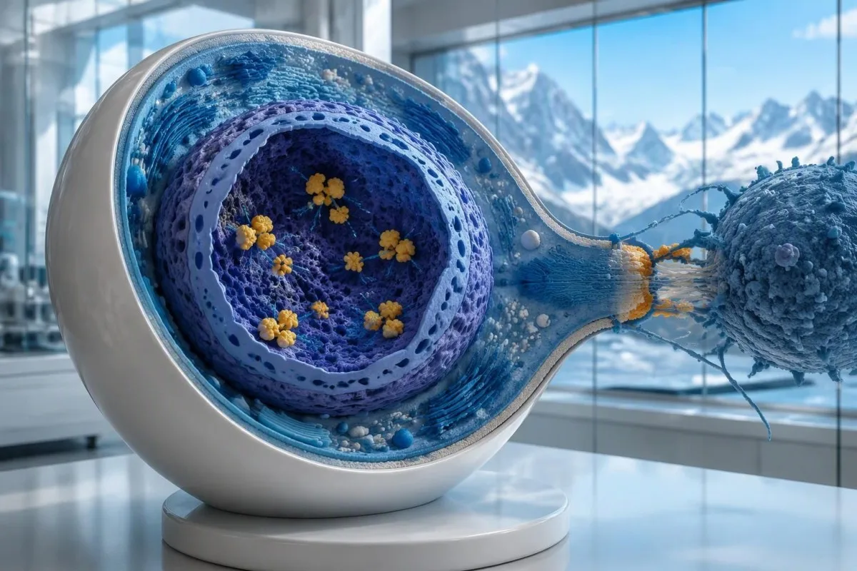
Hu-proteiner är intracellulära RNA-bindande proteiner, och anti-Hu förstås bäst som en markör för ett immunsvar som ofta involverar cytotoxiska T-celler. Detta skiljer sig från sjukdomar där antikroppar binder till åtkomliga proteiner på neuronernas yta. Skillnaden förklarar varför enbart en sänkning av antikroppsnivåerna inte på ett tillförlitligt sätt reverserar etablerad anti-Hu-neurologisk skada, även när 2 svar visar markant olika titrar.
Ett svar med formuleringen “ANNA-1 påvisad” är inte detsamma som ett rutinmässigt positivt ANA, trots de liknande bokstäverna. ANA betyder vanligtvis antinukleära antikroppar som används vid utredning av systemisk autoimmunitet, medan ANNA-1 betecknar ett neuronalt antikroppsmönster. Håll isär dessa 2 förkortningar när du diskuterar dina resultat; vår guide till ANA-terminologi förklarar det mer välbekanta testet.
Anti-Hu kan påvisas hos vissa personer med småcellig lungcancer som inte har något igenkännbart neurologiskt syndrom. Omvänt kan neurologiska symtom leda till att antikroppar påvisas innan cancern blir synlig. Det är 2 olika kliniska förlopp: antikroppen säger inte i sig läkaren om nerverna skadas just nu eller var en bakomliggande cancer kan finnas.
Hur ska provsvaret för anti-Hu-antikropp tolkas?
An anti-Hu antibody test has no universal numerical cutoff that can be applied across laboratories. Read the specimen type, assay method, result category, laboratory reference interval and confirmation comments together; a dilution titer, band intensity and numerical index are 3 different ways of reporting reactivity.
A hypothetical titer of 1:240 describes detectable reactivity after a particular dilution; it is not a cancer probability of 240, nor does it establish cancer stage. A numerical index from another method may use entirely different calibration. Our guide to kvalitativa kontra kvantitativa resultat explains why “positive” and “how much” answer different laboratory questions.
Weak or borderline reactivity deserves method-specific interpretation, particularly when a broad panel was ordered without a convincing neurological indication. Ask whether the laboratory observed the characteristic neuronal staining pattern and whether an antigen-specific confirmation was performed. Two tests using the same underlying strip technology may reproduce the same analytical problem; agreement is more informative when the methods are genuinely complementary.
Bring the complete report, not just a screenshot of the word positive. The 5 most useful details are the specimen, collection date, assay name, reported strength or titer and laboratory interpretation. Also list recent intravenous immunoglobulin and other immune treatments, because passive antibodies or treatment effects can complicate some serological results; your laboratory can advise whether that issue plausibly affects this particular finding.
När behöver ett positivt anti-Hu-resultat bekräftas?
Unexpected, weak or clinically discordant anti-Hu results should be reviewed for confirmation, preferably with an experienced neuroimmunology laboratory. The 2021 PNS criteria emphasize interpreting neuronal antibody testing alongside the phenotype and using appropriate confirmatory methods rather than accepting an isolated commercial-panel result as a diagnosis.
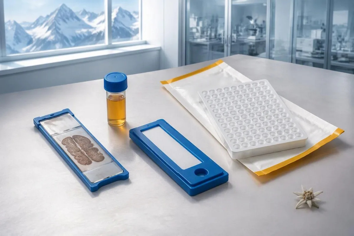
Tissue-based immunohistochemistry or immunofluorescence and an antigen-specific assay provide different information. The first identifies a characteristic distribution of neuronal staining; the second tests reactivity against the relevant antigen. Graus et al. (2021) describe these complementary approaches, with specialist reassessment particularly useful when 1 method gives a result that does not fit the clinical picture.
Repeating a sample can address collection errors or a transient analytical concern, but repeating the same kit is not always enough. In a hypothetical 45-year-old with no objective neurological findings and a faint isolated band, I would want the laboratory’s explanation before treating the report as established anti-Hu autoimmunity. Our GAD65 interpretation guide illustrates another antibody for which context and assay characteristics change meaning.
Confirmation and urgent assessment can happen in parallel. A person who develops seizures, substantial walking difficulty or progressive swallowing impairment should not wait several weeks for a reference-laboratory answer before receiving neurological care. The practical question is not “repeat or investigate?” but which of those 2 tasks can safely proceed together, given the symptoms and the initial laboratory evidence.
Vilka neurologiska symtom gör anti-Hu mer oroande?
Progressive sensory neuronopathy, encephalomyelitis, limbic encephalitis and marked autonomic dysfunction are recognized anti-Hu presentations. Symptoms can involve more than 1 part of the nervous system, so the pattern and speed of change are usually more informative than a single symptom such as tingling or fatigue.

Sensory neuronopathy can cause asymmetric numbness, impaired position sense and sensory ataxia, sometimes involving the hands or trunk early rather than following a simple feet-first pattern. A patient may lose balance in the dark because vision had been compensating for reduced position feedback. That is different from the focal finger distribution discussed in our carpal tunnel symptom guide, although neither pattern can be diagnosed from description alone.
Anti-Hu autonomic involvement can affect gut movement, blood-pressure regulation and bladder function. Severe constipation, unexplained intestinal pseudo-obstruction or dizziness on standing becomes more relevant when accompanied by objective neurological abnormalities. Recording symptoms over 7 days can reveal combinations that otherwise get presented as separate stomach, balance and urinary complaints; sudden severe abdominal symptoms still require assessment for non-neurological emergencies.
New memory impairment, behavioral change or seizures may indicate central nervous system involvement, while rapid weakness has a broader emergency differential. The varningstecken på akut svaghet in our neurological safety guide apply regardless of the antibody label. Call emergency services for new breathing difficulty, unsafe swallowing, a first seizure or sudden stroke-like symptoms rather than waiting for a scheduled follow-up.
Vilka neurologiska undersökningar kan följa efter ett positivt resultat?
Neurological follow-up may include examination, nerve conduction studies, MRI and cerebrospinal fluid testing, selected according to the symptoms. There is no mandatory battery for every anti-Hu-positive person; the purpose is to document which structures are affected and exclude other causes, not merely to collect additional positive tests.
Nerve conduction studies can support a sensory neuronopathy pattern by showing reduced sensory responses across distributions that do not fit a single compressed nerve. Brain or spinal MRI addresses different questions, including structural disease and some inflammatory changes. A normal MRI does not exclude peripheral sensory ganglion involvement; these are 2 distinct anatomical compartments with different diagnostic tools.
Cerebrospinal fluid may show increased cells, protein or evidence of intrathecal immune activity, but none of those findings is specific to anti-Hu. A lumbar puncture is most useful when the neurological syndrome warrants it, rather than as an automatic response to 1 weak serum band. Our discussion of tolkning av oligoklonala band i cerebrospinalvätska explains why immune activity in spinal fluid does not identify a single disease.
Basic investigations still matter: glucose control, vitamin status, renal function and serum protein assessment can uncover treatable contributors to neuropathy. A clinician may request 2 or more of these alongside specialist testing, depending on the examination. The guide om analys av serumproteiner provides background, but normal routine blood work neither confirms nor excludes an anti-Hu-associated neurological process.
Bekräftar anti-Hu-positivitet ett paraneoplastiskt syndrom?
A positive antibody alone does not confirm anti-Hu paraneoplastic syndrome. The PNS-Care approach combines the neurological phenotype, antibody risk category, cancer findings and follow-up; clinicians also check for direct cancer effects, treatment toxicity and unrelated neurological conditions before attributing symptoms to a paraneoplastic immune response.
Definite, probable and possible PNS are diagnostic categories, not cancer stages. The 2021 criteria use a structured scoring system rather than allowing 1 antibody to determine the whole diagnosis (Graus et al., 2021). A confirmed anti-Hu result with a strongly compatible syndrome remains clinically significant even before a cancer is identified, but clinicians should state clearly which parts of the assessment remain unresolved.
An older patient can have anti-Hu reactivity and a separate cause of numbness, such as diabetes, nutritional deficiency or a monoclonal protein disorder. Coexistence is possible; either-or reasoning can miss treatable disease. If serum protein studies reveal a band, our immunofixation result guide explains why that finding requires its own interpretation rather than being automatically folded into the anti-Hu diagnosis.
Kantesti är en AI-plattform för tolkning av blodprov that can help organize these distinctions in a laboratory explanation, but it cannot assign a reliable PNS-Care diagnosis from a report alone. Our kliniskt valideringsramverk separates technical evaluation from clinical conclusions. In practice, I would document 3 separate judgments: confidence in the antibody, confidence in the neurological syndrome and the current evidence for cancer.
Hur letar läkare efter en associerad cancer?
Cancer evaluation usually prioritizes the chest because small-cell lung cancer is the strongest adult anti-Hu association. Doctors commonly begin with appropriate chest CT and may add FDG-PET/CT or targeted investigations if suspicion remains; the exact plan depends on age, symptoms, examination, smoking history and previous imaging.
The cancer association is substantial in established anti-Hu clinical cohorts. Villagrán-García et al. (2023) reported identified cancer in 349 of 466 patients, approximately 75%, in a specialist anti-Hu cohort. This is valuable evidence for taking confirmed, clinically relevant anti-Hu seriously; it is not a personalized prediction for an asymptomatic person with an unconfirmed positive screening strip.
A chest X-ray is not equivalent to a chest CT, and routine low-dose screening is not necessarily the correct examination for a suspected paraneoplastic syndrome. The clinician chooses the protocol and body regions based on the diagnostic question. Likewise, neurological symptoms do not automatically mean cancer has spread to the brain; our brain imaging warning-sign guide distinguishes structural investigation from immune-mediated disease.
A suspicious scan often leads to tissue sampling through a method chosen by respiratory or oncology specialists, such as bronchoscopic assessment of an accessible area. Imaging suspicion and cancer confirmation are 2 separate steps. Never-smoking status lowers the baseline probability of small-cell lung cancer but does not cancel a well-confirmed anti-Hu finding with a convincing neurological presentation; other cancers and non-cancer explanations still need consideration.
Varför kan de första cancerundersökningarna vara negativa?
Initial imaging can be negative because an associated cancer is still too small to detect, lies outside the examined area or is not readily visible with that technique. Neurological autoimmunity may precede cancer detection, so a negative scan reduces concern without necessarily ending follow-up in a high-risk clinical presentation.
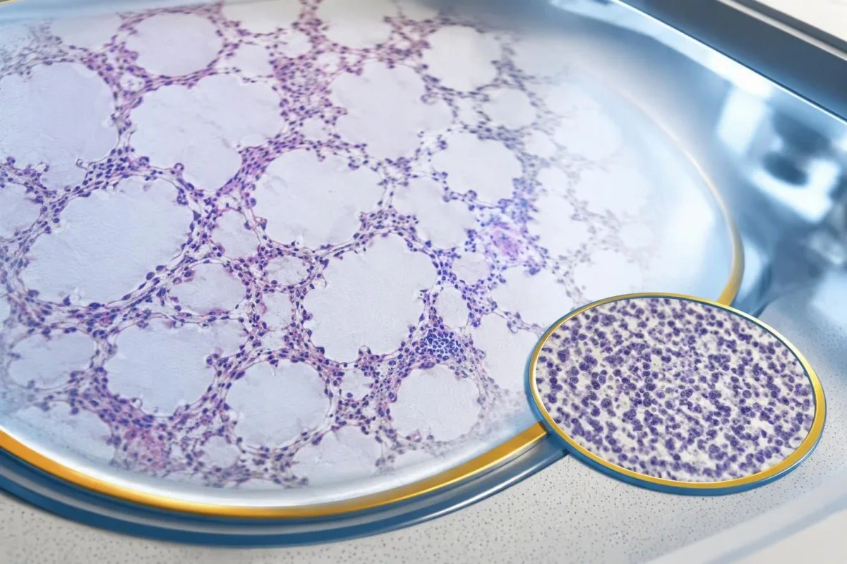
A negative scan is time-specific and method-specific. A normal chest CT performed on 1 date answers whether that examination identified a suspicious chest finding then; it does not guarantee that every possible associated cancer is absent indefinitely. Ask what was actually imaged, whether the protocol was suitable and whether the specialist recommends PET/CT or targeted investigation despite the initial result.
Villagrán-García et al. (2023) described cancers detected beyond the first 2 years in a minority of their anti-Hu cohort, supporting individualized longer surveillance. Their findings also support considering PET-based investigation when initial CT is unrevealing in an appropriate clinical context. This is an escalation decision for the treating team, not a reason to independently purchase repeated whole-body scans without weighing radiation, incidental findings and cost.
Neurological deterioration after negative imaging still needs reassessment. Cancer surveillance and neurological monitoring are 2 parallel tracks; one reassuring scan does not explain new loss of function. A marker such as neurofilament light chain may indicate neuronal injury in selected settings, but our NfL interpretation guide explains why it cannot establish anti-Hu disease, locate cancer or replace examination.
Hur länge bör cancerscreeningen fortsätta?
The 2021 PNS criteria recommend repeat cancer screening every 4–6 months for 2 years when a high-risk neurological phenotype accompanies a high-risk antibody. Longer surveillance may be appropriate for selected anti-Hu patients; the interval and duration should reflect confirmation, clinical evolution, initial investigations and individual cancer risk.

The 2-year schedule is not an instruction to scan every positive antibody result repeatedly. Graus et al. (2021) link that recommendation to the combination of high-risk phenotype and high-risk antibody. An incidental, weak result without a matching syndrome needs confirmation and individualized assessment first; repeating imaging mechanically can create incidental findings and unnecessary procedures without resolving the original uncertainty.
Older European guidance recommended longer surveillance. The EFNS task force advised repeating screening after 3–6 months and then every 6 months for up to 4 years in relevant antibody-positive paraneoplastic presentations (Titulaer et al., 2011). The 2023 anti-Hu cohort supported considering screening up to 5 years in selected patients, so a specialist may reasonably extend follow-up beyond the newer general 2-year framework.
Keep a dated plan stating the next examination, the clinician responsible and what symptoms should trigger earlier contact. Kantesti explanations can help you understand terminology, but the platform should not be treated as a scan-reminder service or substitute for that plan. Our guide to risk-based cancer screening addresses a different setting: routine prevention is not the same as investigating a possible paraneoplastic syndrome.
Vilken behandling hjälper vid anti-Hu-neurologisk sjukdom?
Treatment focuses on an identified cancer, selected immune therapy and symptom-directed rehabilitation. Anti-Hu-associated neurological disease often responds less predictably to immunotherapy than neuronal surface-antibody disorders because intracellular targets and T-cell-mediated injury are involved; treatment may aim to stabilize function rather than promise complete recovery.

Treating an associated cancer is a central part of care, but established neurological injury may persist even when cancer treatment succeeds. Neurology and oncology teams may consider corticosteroids, intravenous immunoglobulin, plasma exchange or other immune treatments in selected situations. There is no single evidence-based dose or 1 universal regimen appropriate for everyone with anti-Hu positivity, especially without a confirmed clinical syndrome.
An antibody titer is not a dependable treatment target on its own. A lower number does not necessarily mean restored nerve function, and a persistent positive result does not by itself prove cancer progression. This differs from some surface-antibody disorders; our MOG antibody diagnosis guide explains another immune neurological condition, but its diagnostic rules and treatment expectations should not be transferred to anti-Hu.
Immune checkpoint cancer therapies deserve an explicit discussion when anti-Hu neurological disease is present, because they can provoke or worsen paraneoplastic neurological autoimmunity. This is a specialist risk-benefit decision, not an automatic prohibition based on 1 laboratory result. Tell the oncologist about previous neurological symptoms and confirmed neuronal antibodies before treatment decisions are finalized; coordinating the teams early can prevent avoidable misunderstanding.
Vad kan du göra på ett säkert sätt medan utredningen pågår?
While investigations continue, protect mobility, swallowing and medication safety rather than trying to lower anti-Hu antibodies with supplements. Use the neurological symptoms—not the appearance of the laboratory flag—to decide whether ordinary follow-up is sufficient or urgent care is needed; a new functional loss warrants contact before the next planned scan.
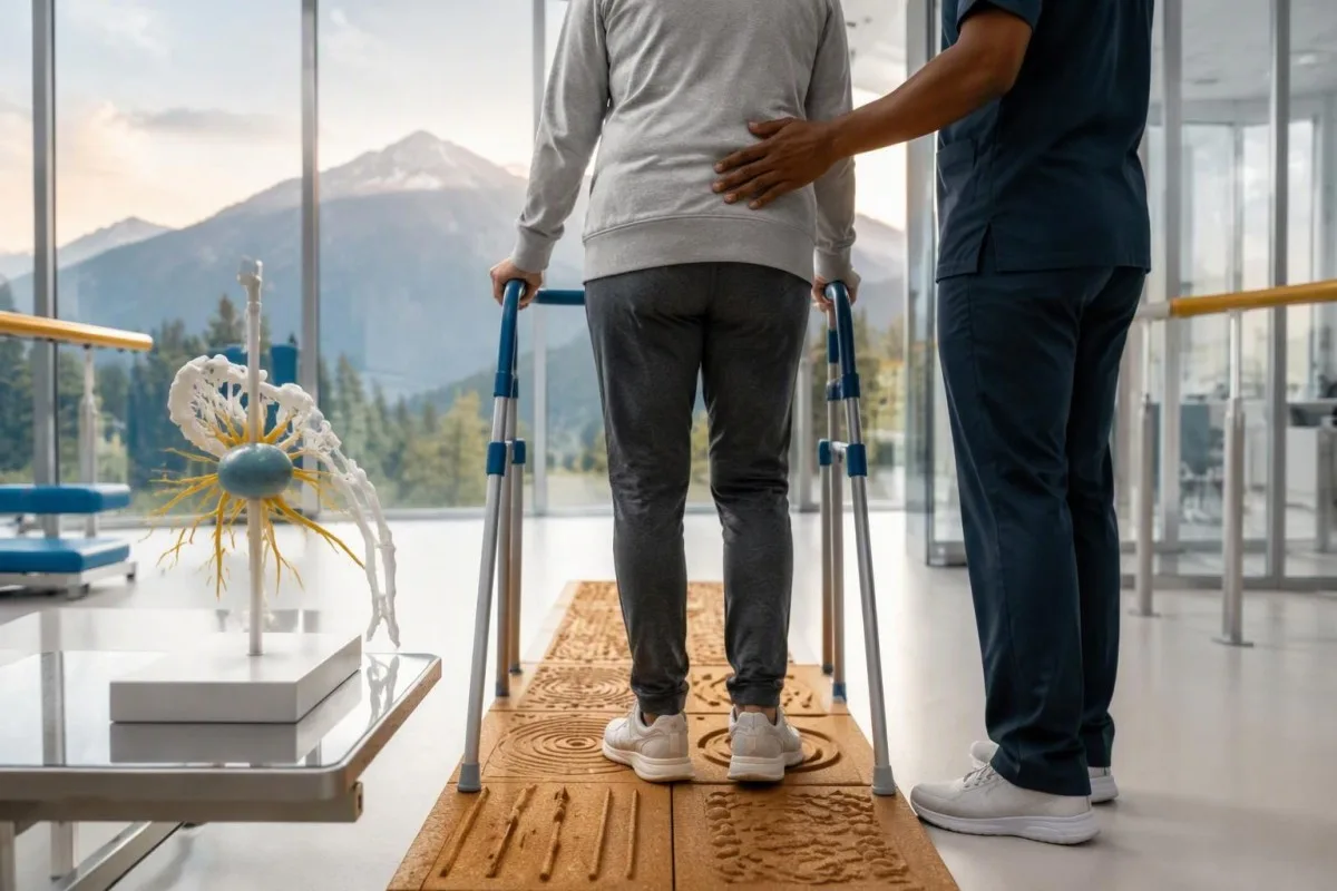
Position-sense loss increases fall risk, particularly in poor lighting or on uneven ground. A physiotherapist may recommend a suitable walking aid and individualized balance strategies; occupational therapy can address hand coordination and home hazards. A short 7-day record of falls, near-falls and walking changes is often more useful at review than the vague statement “my balance is worse,” but stop activities that feel unsafe.
Excess vitamin B6 can itself cause sensory neuropathy, making unsupervised high-dose nerve supplements particularly unhelpful during this assessment. Review every supplement and its daily dose in mg, including overlapping multivitamins and combination products. Our B-komplex säkerhetsguide explains that problem; correcting a documented deficiency is different from taking progressively larger doses in the hope of treating anti-Hu autoimmunity.
Vitamin B12 deficiency can contribute to sensory and gait problems and may need investigation even when anti-Hu is confirmed. There is no universally interchangeable B12 cutoff: results in pg/mL och pmol/L require their own laboratory context, sometimes with functional testing. Our vägledning för uppföljning av gränsfall för B12 explains those next steps; new choking, breathing difficulty or rapid weakness should be assessed urgently instead of managed with vitamins.
Hur kan AI hjälpa till att förklara ett anti-Hu-provresultat på ett säkert sätt?
AI can help explain report terminology and organize questions, but it cannot confirm anti-Hu specificity, diagnose paraneoplastic syndrome or rule out cancer. Safe interpretation preserves the original assay details and separates 3 issues: what the laboratory reported, what the result may mean and what a clinician has actually established.

Kantesti är en AI-baserat analysverktyg för blodprov that can provide a plain-language explanation of uploaded laboratory information, including the distinction between an antibody screen and a confirmed clinical diagnosis. A rapid explanation—often around 60 seconds for report processing—is not a reference-laboratory assay. Our guide för AI-teknik describes the interpretation workflow without making anti-Hu-specific diagnostic guarantees.
The complete source report should remain available for verification. An explanation should not silently replace “weakly reactive” with “confirmed,” drop the specimen type or invent a universal reference interval. Our guide to att granska AI-förklaringar av medicinsk information describes practical source checks; for this antibody, compare at least the assay wording, confirmation comment and collection date against the original document.
Kantesti cannot examine sensation, review tissue staining directly through a report explanation or establish that negative imaging has excluded a tiny cancer. Those are 3 different clinical capabilities, not features that follow from understanding laboratory language. Share the explanation with the treating team as a discussion aid, and do not delay urgent assessment while uploading documents or waiting for a second automated interpretation.
Ditt nästa läkarbesök och öppenhet kring forskningen
Bring the complete antibody report, a dated symptom history, previous imaging reports and a current medication list to your next appointment. Ask the clinician to clarify antibody confirmation, the neurological diagnosis and cancer surveillance separately; those 3 answers may have different levels of certainty and different follow-up dates.
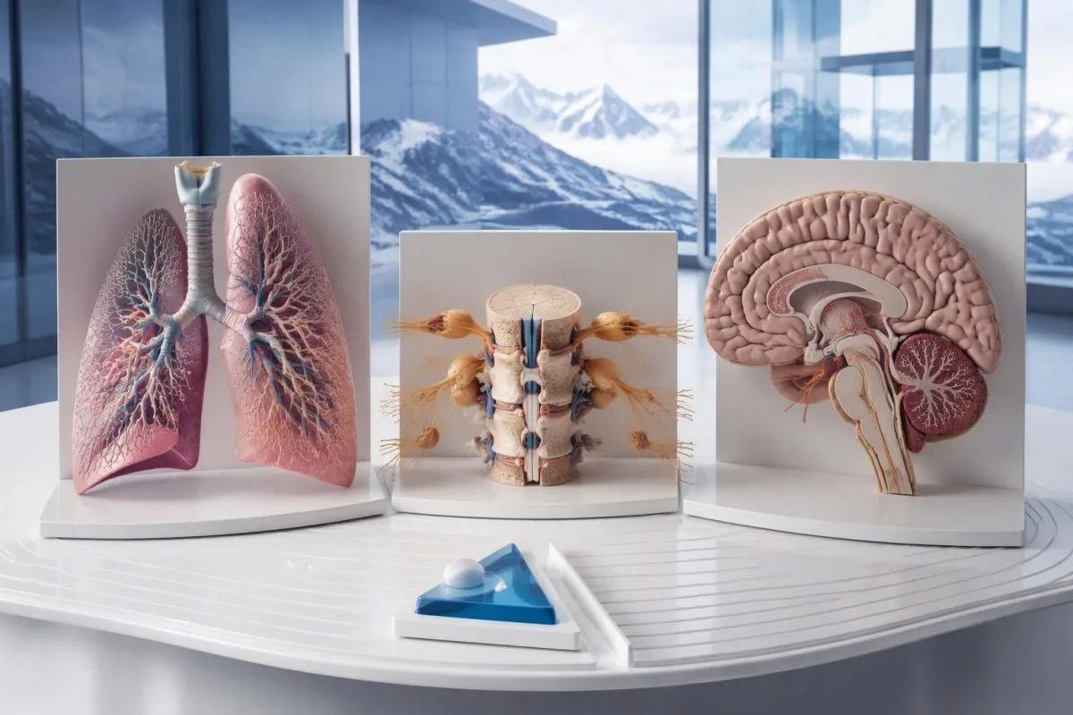
A useful 1-page symptom history records onset, progression, sensory distribution, walking changes, memory problems and autonomic symptoms. Include prior cancer treatment and any recent immunoglobulin therapy, then ask who will coordinate neurology and oncology if both are involved. As Thomas Klein, MD, I would rather see a clear timeline than 20 disconnected screenshots; it makes discrepancies between the laboratory result and clinical course easier to spot.
Medical authorship is not a substitute for an individual neurological examination. Vår information från medicinska rådgivningsnämnden describes the clinicians associated with our work, while your own specialist determines the appropriate next investigation. As of 25 oktober 2026, the recommendations discussed here draw on the 2021 international PNS criteria, the 2011 EFNS screening guidance and the 2023 anti-Hu cohort—not an invented 2026 guideline.
The 2 Figshare publications below are platform-related research references, not evidence validating anti-Hu diagnosis or cancer exclusion. Their supplied titles describe hantavirus triage deployment and a synthetic-case technical benchmark; neither title establishes anti-Hu-specific clinical performance. Because publication authors and dates were not supplied for verification, the citations use title-first APA formatting with “n.d.”; the ResearchGate and Academia.edu links are searches, not claims of verified publication copies.
Vanliga frågor
Betyder ett positivt anti-Hu-antikroppstest att jag har cancer?
0 Ett positivt anti-Hu-antikroppsresultat innebär inte i sig att cancer har diagnostiserats. Anti-Hu är en antikropp med hög risk och koppling till cancer, och cancer identifierades hos 349 av 466 patienter i en specialistkohort med anti-Hu-fall från 2023, men den siffran avgör inte risken vid ett tillfälligt, obekräftat screeningresultat. Läkare bedömer den neurologiska bilden, bekräftar oväntade laboratoriefynd och utreder om det finns en underliggande cancer, vanligtvis med fokus på bröstkorgen hos vuxna. En cancerdiagnos kräver i regel separata kliniska fynd, bilddiagnostik och vävnadsprov.
Är anti-Hu och ANNA-1 samma sak?
Anti-Hu och ANNA-1 betecknar i allmänhet samma familj av neuronala antikroppar. ANNA-1 står för antineuronal kärnantikropp typ 1, medan Hu syftar på de intracellulära neuronala proteiner som antikroppen känner igen. ANNA-1 är inte samma sak som ett rutinmässigt ANA som används vid systemisk autoimmunitetsutredning. Kontrollera det exakta testet och bekräftelseformuleringen eftersom laboratorier kan rapportera samma antikroppsfamilj i olika format.
Kan ett anti-Hu-antikroppstest ge ett falskt positivt resultat?
Ett anti-Hu-antikroppstest kan ge missvisande eller ospecifik reaktivitet, särskilt när ett svagt screeningresultat inte stämmer överens med den neurologiska symtombilden. Bekräftelse sker vanligtvis med två kompletterande metoder: ett karakteristiskt vävnadsfärgningsmönster och en antigenspecifik analys som tolkas av ett erfaret laboratorium. Att upprepa samma kommersiella metod kan ge samma analytiska problem igen i stället för att lösa det. Snabbt progredierande neurologiska symtom kräver fortfarande en skyndsam bedömning medan bekräftelse inväntas.
Hur ofta bör undersökningar upprepas om anti-Hu är positivt?
PNS-kriterierna från 2021 rekommenderar upprepad cancerscreening var 4–6:e månad i 2 år när en högriskneurologisk fenotyp förekommer tillsammans med en högriskantikropp. Det schemat gäller inte automatiskt för varje tillfälligt anti-Hu-positivt fynd. Äldre EFNS-vägledning rekommenderade övervakning i upp till 4 år, och en anti-Hu-kohort från 2023 gav stöd för att överväga upp till 5 år för utvalda patienter. Din specialist bör specificera bilddiagnostisk metod, intervall, varaktighet och symtom som motiverar en tidigare bedömning.
Kan anti-Hu vara positivt när CT- och PET-undersökningar är normala?
0 Anti-Hu kan förbli positivt även när CT- eller PET-undersökningar inte har påvisat cancer. Neurologisk autoimmunitet kan föregå upptäckten, och ett negativt undersökningsresultat speglar de undersökta områdena, tekniken och datumet snarare än en permanent garanti för att cancer saknas. Vid en förenlig högriskbild rekommenderar 2021 års kriterier upprepad screening var 4–6:e månad i 2 år, med individuellt anpassad längre uppföljning hos vissa patienter med anti-Hu. Obekräftad antikroppsreaktivitet och andra förklaringar än cancer bör också omprövas i stället för att utgå från att det definitivt finns en osynlig cancer.
Betyder en sänkning av anti-Hu-antikroppsnivån att behandlingen fungerar?
En sjunkande nivå av anti-Hu-antikroppar bevisar inte tillförlitligt neurologisk återhämtning eller framgångsrik cancerbehandling. Anti-Hu känner igen intracellulära proteiner, och den associerade neurologiska skadan involverar ofta T-cellsmedierade mekanismer snarare än enbart antikroppseffekter. Två titrar från olika laboratorier behöver inte heller vara jämförbara eftersom metoderna skiljer sig åt. Kliniker bedömer funktion, undersökningsfynd, relevanta utredningar och cancersvaret i stället för att använda antikroppsvärdet som ett fristående behandlingsmål.
Få AI-drivna analyser av blodprov redan idag
Gå med i över 2 miljoner användare världen över som litar på Kantesti för snabb och korrekt analys av blodprover. Ladda upp dina blodprovsresultat och få en heltäckande tolkning av 15,000+-biomarkörer på sekunder.
📚 Refererade forskningspublikationer
Klein, T., Mitchell, S., & Weber, H. (2026). Flerspråkigt AI-assisterat kliniskt beslutsstöd för tidig hantavirus-triage: Design, teknisk validering och verklig implementering över 50 000 tolkade blodprovsrapporter. Kantesti AI Medical Research.
Klein, T., Mitchell, S., & Weber, H. (2026). En förregistrerad, rubrikbaserad automatisk teknisk benchmark av Kantesti blodprovs-tolkningsmotorn på 100 000 syntetiska testfall. Kantesti AI Medical Research.
📖 Externa medicinska referenser
Graus F et al. (2021). Updated Diagnostic Criteria for Paraneoplastic Neurologic Syndromes. Neurology: Neuroimmunology & Neuroinflammation.
Titulaer MJ et al. (2011). Screening for tumours in paraneoplastic syndromes: report of an EFNS Task Force. European Journal of Neurology.
Villagrán-García D et al. (2023). Revisiting anti-Hu paraneoplastic autoimmunity: phenotypic characterization and cancer diagnosis. Brain Communications.
⚕️ Medicinsk ansvarsfriskrivning
Den här artikeln är endast avsedd för utbildningsändamål och utgör inte medicinsk rådgivning. Rådgör alltid med en behörig vårdgivare för beslut om diagnos och behandling.
E-E-A-T förtroendesignaler
Uppleva
Läkarledd klinisk granskning av arbetsflöden för laboratorietolkning.
Expertis
Laboratoriemedicinskt fokus på hur biomarkörer beter sig i kliniskt sammanhang.
Auktoritet
Skrivet av Dr. Thomas Klein med granskning av Dr. Sarah Mitchell och Prof. Dr. Hans Weber.
Trovärdighet
Evidensbaserad tolkning med tydliga uppföljningsspår för att minska larm.
