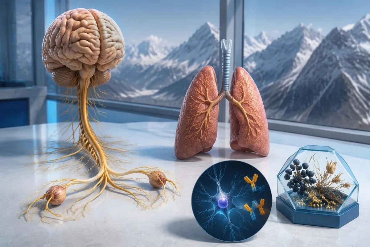
Anti-Hu အင်တီဘော်ဒီ စစ်ဆေးချက်အပေါင်းရလဒ်သည် အထူးသဖြင့် အဆုတ်အသေးစားဆဲလ်ကင်ဆာနှင့် ဆက်စပ်သည့် ကင်ဆာဆိုင်ရာ အာရုံကြောကိုယ်ခံအားတုံ့ပြန်မှု ဖြစ်နိုင်ခြေကို စိုးရိမ်စရာဖြစ်စေသော်လည်း ကင်ဆာရောဂါကို အတည်ပြုသတ်မှတ်ပေးခြင်း မဟုတ်ပါ။ ဆရာဝန်များသည် ရောဂါလက္ခဏာများကို အကဲဖြတ်ခြင်း၊ ဓာတ်ခွဲခန်းတွေ့ရှိချက်ကို အတည်ပြုခြင်းနှင့် ကင်ဆာစစ်ဆေးမှုများ စီစဉ်ခြင်းတို့ကို ပြုလုပ်ကြသည်။ ပထမဆုံး ပုံရိပ်စစ်ဆေးမှုများတွင် မတွေ့ရသော်လည်း ထပ်မံစစ်ဆေးရန် လိုအပ်နိုင်သည်။.



- Anti-Hu / ANNA-1 သည် အန္တရာယ်မြင့် ကင်ဆာနှင့်ဆက်စပ်သော အင်တီဘော်ဒီဖြစ်ပြီး၊ အတည်ပြုပြီးသော ဆေးဘက်ဆိုင်ရာလူနာအုပ်စုများတွင် 70% ထက်ပို၍ ကင်ဆာနှင့် ဆက်စပ်နေသော အမျိုးအစားဖြစ်သည်—အတည်မပြုရသေးသော စစ်ဆေးမှုရလဒ်တိုင်းကို ဆိုလိုခြင်းမဟုတ်ပါ။.
- ကင်ဆာရောဂါ အတည်ပြုခြင်း သည် အရင်းခံကင်ဆာရှိကြောင်း အထောက်အထား လိုအပ်ပြီး၊ များသောအားဖြင့် တစ်ရှူးစစ်ဆေးမှု ပါဝင်သည်။ အင်တီဘော်ဒီအပေါင်းရလဒ်တစ်ခုတည်းသည် ထိုအထောက်အထား မဟုတ်ပါ။.
- စစ်ဆေးမှုရလဒ် အတည်ပြုခြင်း တွင် မကြာခဏ အပြန်အလှန်ဖြည့်စွက်ပေးသည့် နည်းလမ်း 2 ခုကို ပေါင်းစပ်အသုံးပြုသည်—လက္ခဏာရပ်ရှိသော တစ်ရှူးအရောင်စွဲပုံစံနှင့် သီးခြားအန်တီဂျင်ကို စစ်ဆေးသည့် စမ်းသပ်မှု။.
- အာရုံကြောဆိုင်ရာလက္ခဏာများ ဥပမာ တဖြည်းဖြည်းဆိုးရွားလာသော အာရုံခံစားမှုဆုံးရှုံးခြင်း၊ ဟန်ချက်မညီခြင်း၊ တက်ခြင်း သို့မဟုတ် အလိုအလျောက်အာရုံကြောစနစ် ချို့ယွင်းခြင်းတို့သည် ရလဒ်၏ အဓိပ္ပာယ်နှင့် အရေးတကြီးဆောင်ရွက်ရန် လိုအပ်မှုကို ပြောင်းလဲစေသည်။.
- ပုံရိပ်စစ်ဆေးမှုတွင် မတွေ့ရှိခြင်း ဆိုသည်မှာ ထိုအချိန်က ထိုစစ်ဆေးမှုတွင် ကင်ဆာကို မတွေ့ရှိခဲ့ခြင်းဖြစ်သည်။ သို့သော် သေးငယ်ပြီး အစပိုင်းတွင် မတွေ့ရှိနိုင်သေးသော ကင်ဆာဖြစ်နိုင်ခြေကို ပယ်ဖျက်ခြင်း မဟုတ်ပါ။.
- ထပ်မံစစ်ဆေးခြင်း 2021 PNS စံသတ်မှတ်ချက်များအရ အန္တရာယ်မြင့် အာရုံကြောဆိုင်ရာ လက္ခဏာပုံစံနှင့် အန္တရာယ်မြင့် ပဋိပစ္စည်း တွဲဖက်တွေ့ရှိပါက ၂ နှစ်ကြာအောင် ၄–၆ လတစ်ကြိမ် ထပ်မံစစ်ဆေးရန် အကြံပြုထားသည်။.
- ပိုမိုကြာရှည်သော စောင့်ကြည့်စစ်ဆေးမှု ကို ရွေးချယ်ထားသော လူနာများအတွက် သင့်လျော်နိုင်သည်။ 2023 anti-Hu လူနာအုပ်စုလေ့လာမှုအရ ၂ နှစ်ကျော်၊ တစ်ခါတစ်ရံ ၅ နှစ်အထိ စစ်ဆေးရန် စဉ်းစားသင့်ကြောင်း ထောက်ခံထားသည်။.
- အရေးပေါ် လက္ခဏာများ အသက်ရှူရခက်ခဲမှုအသစ်ဖြစ်ပေါ်ခြင်း၊ ဘေးကင်းစွာ မျိုချနိုင်ခြင်းမရှိခြင်း၊ အားနည်းမှု လျင်မြန်စွာပိုဆိုးလာခြင်း၊ ပထမဆုံး တက်ခြင်း သို့မဟုတ် အာရုံကြောဆိုင်ရာ လုပ်ဆောင်ချက်များ ရုတ်တရက်ချို့ယွင်းခြင်းတို့ ပါဝင်သည်။.
Anti-Hu အင်တီဘော်ဒီ စစ်ဆေးချက်အပေါင်းရလဒ်က ဘာကိုဆိုလိုသလဲ။
တစ်ခု anti-Hu ပဋိပစ္စည်း ပေါင်းစစ်ရလဒ် ဆိုသည်မှာ ဓာတ်ခွဲခန်းတွင် Hu အာရုံကြောဆဲလ် ပရိုတင်းများအပေါ် တုံ့ပြန်မှုကို တွေ့ရှိခဲ့သည်ဟု ဆိုလိုသည်။ ၎င်းတစ်ခုတည်းဖြင့် ကင်ဆာရှိကြောင်း သို့မဟုတ် အာရုံကြောဆိုင်ရာ ရောဂါလက္ခဏာစုရှိကြောင်း အတည်မပြုနိုင်ပါ။ အဓိပ္ပာယ်ဖော်ရာတွင် အပိုင်း ၃ ပိုင်းရှိသည်—ရလဒ်အစစ်အမှန် ဟုတ်မဟုတ်၊ အာရုံကြောဆိုင်ရာ လက္ခဏာတင်ပြမှုနှင့် ကိုက်ညီမှုရှိမရှိ၊ ဆက်စပ်ကင်ဆာကို ရှာတွေ့နိုင်မနိုင်တို့ဖြစ်သည်။.
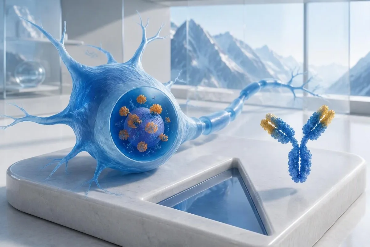
Anti-Hu ကို ကင်ဆာနှင့်ဆက်စပ်မှု အန္တရာယ်မြင့် ပဋိပစ္စည်းအဖြစ် သတ်မှတ်ထားသည်, ။ သက်ဆိုင်ရာ ဆေးဘက်ဆိုင်ရာ လူနာအုပ်စုများတွင် ကင်ဆာနှင့်ဆက်စပ်မှု 70% ထက်ကျော်လွန်ခြင်းကို အမျိုးအစားသတ်မှတ်ချက်အဖြစ် အသုံးပြုထားသည် (Graus et al., 2021)။ ထိုရာခိုင်နှုန်းကို အခြားအခြေအနေများအားလုံး ကောင်းမွန်နေပြီး အားနည်းကာ အတည်မပြုရသေးသော panel ရလဒ်သာရှိသူတစ်ဦးထံ ရိုးရိုးတန်းတန်း သတ်မှတ်၍မရပါ။ လေ့လာခဲ့သော လူနာများ၊ စမ်းသပ်နည်းနှင့် အာရုံကြောဆိုင်ရာ လက္ခဏာတင်ပြမှုတို့က ဖြစ်နိုင်ခြေအပေါ် သက်ရောက်မှုရှိသည်။.
ဟန်ချက်မညီမှု ၈ ပတ်ကြာ ပိုဆိုးလာပြီး အာရုံခံစားမှု တစ်ဖက်နှင့်တစ်ဖက် မညီမညာလျော့နည်းလာသော အသက် ၆၂ နှစ်အရွယ် စိတ်ကူးယဉ်လူနာတစ်ဦးကို၊ ပင်ပန်းနွမ်းနယ်မှုဆိုင်ရာ စမ်းသပ်မှုအမျိုးမျိုး ပြုလုပ်ရာမှ ပဋိပစ္စည်းကို မတော်တဆတွေ့ရှိသူတစ်ဦးနှင့် မတူညီသောနည်းဖြင့် အကဲဖြတ်သင့်သည်။ ဓာတ်ခွဲခန်းစာမျက်နှာတစ်ခုတည်းကိုကြည့်ပြီး မည်သူ့ကိုမျှ “သင့်မှာ ကင်ဆာရှိတယ်” ဟု မပြောသင့်ပါ။ ကျွန်ုပ်တို့၏ ရှင်းလင်းချက် အင်တီဘော်ဒီ အပေါင်းလက္ခဏာတွေ့ရှိချက်များအကြောင်း လမ်းညွှန် သည် ကိုယ်ခံအားအမှတ်အသားနှင့် ပြည့်စုံပြီးသော ရောဂါရှာဖွေသတ်မှတ်ချက်ကို ခွဲခြားနားလည်စေရန် ကူညီပေးသည်။.
ကန်တက်စတီသည် AI သွေးစမ်းသပ်မှုခွဲခြမ်းစိတ်ဖြာ သည် anti-Hu အစီရင်ခံစာပါ အသုံးအနှုန်းများကို ရှင်းပြရာတွင် အထောက်အကူဖြစ်နိုင်သော်လည်း ဓာတ်ခွဲခန်းအတည်ပြုချက်နှင့် ကင်ဆာအကဲဖြတ်မှုတို့ကို သင့်ကုသပေးနေသော ဆရာဝန်များနှင့် ဆောင်ရွက်ရမည်။ Kantesti LTD ၏ Chief Medical Officer ဖြစ်သူ Thomas Klein, MD အနေဖြင့် ကျွန်ုပ် ပထမဆုံးမေးမည့်အချက်မှာ ဤစစ်ဆေးမှုကို အဘယ်ကြောင့် ပြုလုပ်ခဲ့သနည်းဆိုတာဖြစ်မည်—အန္တရာယ်အချက်ပြပုံက ဘယ်လောက်ထိတ်လန့်စရာကောင်းသလဲဆိုတာ မဟုတ်ပါ။ ကျွန်ုပ်တို့၏ အဖွဲ့အစည်းနှင့် ဆေးဘက်ဆိုင်ရာ လုပ်ငန်း ထိုကွာခြားချက်ကို ရှင်းပြပါသည်။.
ANNA-1 နှင့် anti-Hu သည် အင်တီဘော်ဒီတစ်မျိုးတည်းလား။
ANNA-1 ပဋိပစ္စည်းနှင့် anti-Hu ပဋိပစ္စည်းသည် ယေဘုယျအားဖြင့် ပဋိပစ္စည်းမိသားစုတစ်ခုတည်းကို ရည်ညွှန်းသည်။. ANNA-1 သည် antineuronal nuclear antibody type 1 ၏ အတိုကောက်ဖြစ်သည်။ Hu ဟူသောအမည်မှာ သီးခြားရောဂါတစ်ခု သို့မဟုတ် ကင်ဆာအဆင့်တစ်ခုကို ရည်ညွှန်းခြင်းမဟုတ်ဘဲ ကိုယ်ခံအားတုံ့ပြန်မှုက မှတ်မိတုံ့ပြန်သည့် အတွင်းဆဲလ်ပိုင်းရှိ အာရုံကြောဆဲလ်ပရိုတင်းများကို ရည်ညွှန်းသည်။.
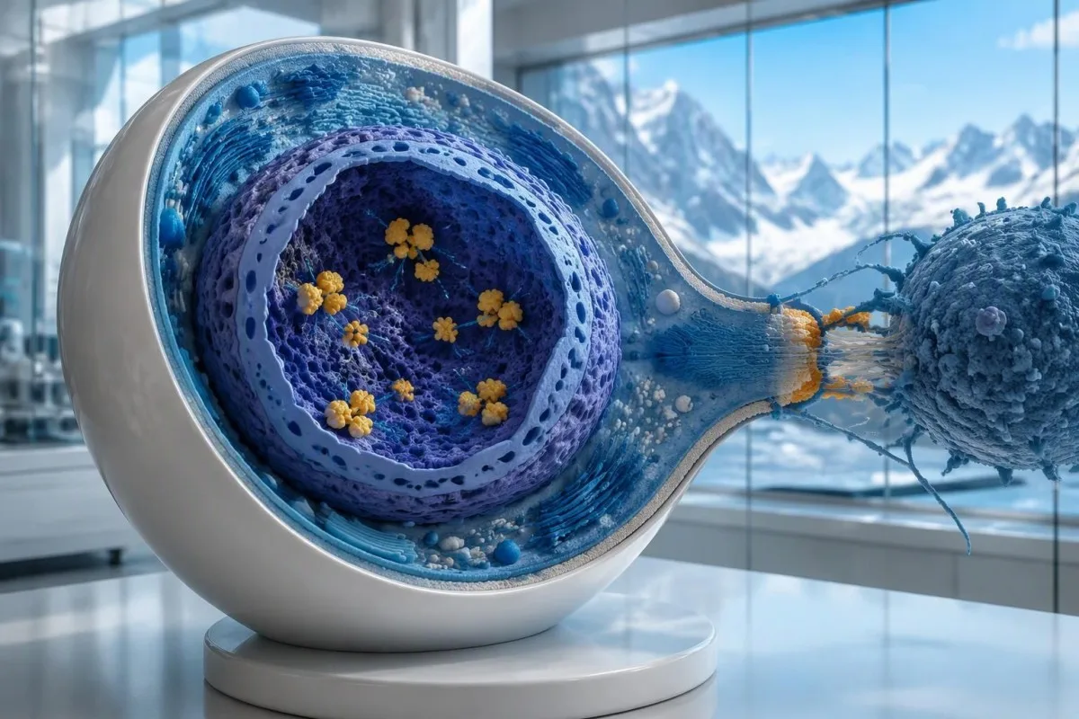
Hu ပရိုတင်းများသည် ဆဲလ်အတွင်းရှိ RNA နှင့် ချိတ်ဆက်သော ပရိုတင်းများဖြစ်သည်, ၊ anti-Hu ကို အဆိပ်သင့်စေသော T ဆဲလ်များ ပါဝင်ပတ်သက်တတ်သည့် ကိုယ်ခံအားတုံ့ပြန်မှု၏ အမှတ်အသားအဖြစ် အကောင်းဆုံးနားလည်နိုင်သည်။ ၎င်းသည် အာရုံကြောဆဲလ်မျက်နှာပြင်ပေါ်ရှိ လက်လှမ်းမီသော ပရိုတင်းများနှင့် ပဋိပစ္စည်းများ ချိတ်ဆက်သည့် ရောဂါများနှင့် ကွာခြားသည်။ ဤကွာခြားချက်ကြောင့် ပဋိပစ္စည်းပမာဏကို လျှော့ချရုံဖြင့် ဖြစ်ပေါ်ပြီးသား anti-Hu အာရုံကြောထိခိုက်မှုကို ယုံကြည်စိတ်ချစွာ ပြန်လည်ကောင်းမွန်စေနိုင်မည်မဟုတ်ကြောင်း ရှင်းပြနိုင်သည်။ အစီရင်ခံစာ ၂ ခုတွင် တိုင်းတာချက်အဆင့်များ သိသိသာသာကွာခြားနေသည့်တိုင် ထိုအချက်မှာ မှန်ကန်နေဆဲဖြစ်သည်။.
“ANNA-1 detected” ဟု ဖော်ပြထားသော အစီရင်ခံစာသည် စာလုံးများဆင်တူသော်လည်း ပုံမှန် ANA ပေါင်းစစ်ရလဒ်နှင့် မတူပါ။. အာနာ သည် ယေဘုယျအားဖြင့် ကိုယ်ခန္ဓာတစ်ခုလုံးဆိုင်ရာ ကိုယ်ခံအားရောဂါများကို အကဲဖြတ်ရာတွင် အသုံးပြုသော antinuclear antibodies ကို ဆိုလိုပြီး၊ ANNA-1 သည် အာရုံကြောဆိုင်ရာ ပဋိပစ္စည်းပုံစံတစ်မျိုးကို ဖော်ထုတ်ပေးသည်။ သင့်ရလဒ်များအကြောင်း ဆွေးနွေးရာတွင် ထိုအတိုကောက် ၂ ခုကို သီးခြားခွဲထားပါ။ ကျွန်ုပ်တို့၏ ANA ဝေါဟာရလမ်းညွှန် သည် ပိုမိုရင်းနှီးကျွမ်းဝင်သော စစ်ဆေးမှုကို ရှင်းပြထားသည်။.
အာရုံကြောဆိုင်ရာ ရောဂါလက္ခဏာစုကို သိသာစွာ မပြသသူအချို့တွင်လည်း anti-Hu ကို တွေ့ရှိနိုင်ပြီး၊ ၎င်းတို့တွင် အဆုတ်၏ small-cell ကင်ဆာရှိနိုင်သည်။ အပြန်အလှန်အားဖြင့် ကင်ဆာကို မြင်တွေ့နိုင်ခြင်းမရှိမီ အာရုံကြောဆိုင်ရာ လက္ခဏာများကြောင့် ပဋိပစ္စည်းကို တွေ့ရှိလာနိုင်သည်။ ၎င်းတို့သည် မတူညီသော ဆေးဘက်ဆိုင်ရာ ဖြစ်စဉ်လမ်းကြောင်း ၂ ခုဖြစ်သည်။ ပဋိပစ္စည်းတစ်ခုတည်းကပင် အာရုံကြောများ လက်ရှိထိခိုက်နေခြင်းရှိမရှိ သို့မဟုတ် အရင်းခံကင်ဆာသည် မည်သည့်နေရာတွင် ရှိနိုင်သည်ကို ဆရာဝန်အား မပြောပြနိုင်ပါ။.
Anti-Hu အင်တီဘော်ဒီ စစ်ဆေးချက်အစီရင်ခံစာကို မည်သို့ဖတ်ရှုသင့်သလဲ။
An anti-Hu antibody test has no universal numerical cutoff that can be applied across laboratories. Read the specimen type, assay method, result category, laboratory reference interval and confirmation comments together; a dilution titer, band intensity and numerical index are 3 different ways of reporting reactivity.
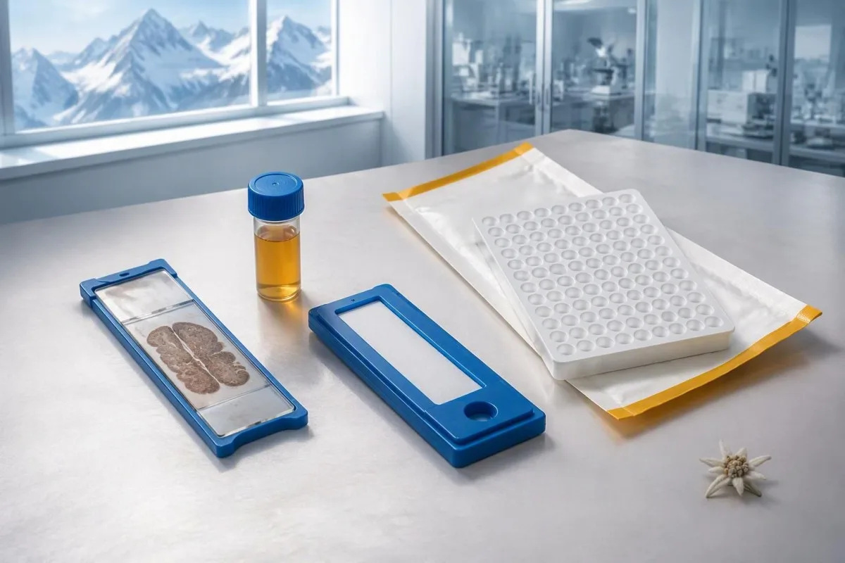
A hypothetical titer of 1:240 describes detectable reactivity after a particular dilution; it is not a cancer probability of 240, nor does it establish cancer stage. A numerical index from another method may use entirely different calibration. Our guide to အရည်အသွေး နှင့် အရေအတွက်ဆိုင်ရာ ရလဒ်များ explains why “positive” and “how much” answer different laboratory questions.
Weak or borderline reactivity deserves method-specific interpretation, particularly when a broad panel was ordered without a convincing neurological indication. Ask whether the laboratory observed the characteristic neuronal staining pattern and whether an antigen-specific confirmation was performed. Two tests using the same underlying strip technology may reproduce the same analytical problem; agreement is more informative when the methods are genuinely complementary.
Bring the complete report, not just a screenshot of the word positive. The 5 most useful details are the specimen, collection date, assay name, reported strength or titer and laboratory interpretation. Also list recent intravenous immunoglobulin and other immune treatments, because passive antibodies or treatment effects can complicate some serological results; your laboratory can advise whether that issue plausibly affects this particular finding.
အပေါင်းရလဒ်ရရှိသောအခါ မည်သည့်အခြေအနေတွင် အတည်ပြုစစ်ဆေးရန် လိုအပ်သလဲ။
Unexpected, weak or clinically discordant anti-Hu results should be reviewed for confirmation, preferably with an experienced neuroimmunology laboratory. The 2021 PNS criteria emphasize interpreting neuronal antibody testing alongside the phenotype and using appropriate confirmatory methods rather than accepting an isolated commercial-panel result as a diagnosis.
Tissue-based immunohistochemistry or immunofluorescence and an antigen-specific assay provide different information. The first identifies a characteristic distribution of neuronal staining; the second tests reactivity against the relevant antigen. Graus et al. (2021) describe these complementary approaches, with specialist reassessment particularly useful when 1 method gives a result that does not fit the clinical picture.
Repeating a sample can address collection errors or a transient analytical concern, but repeating the same kit is not always enough. In a hypothetical 45-year-old with no objective neurological findings and a faint isolated band, I would want the laboratory’s explanation before treating the report as established anti-Hu autoimmunity. Our GAD65 interpretation guide illustrates another antibody for which context and assay characteristics change meaning.
Confirmation and urgent assessment can happen in parallel. A person who develops seizures, substantial walking difficulty or progressive swallowing impairment should not wait several weeks for a reference-laboratory answer before receiving neurological care. The practical question is not “repeat or investigate?” but which of those 2 tasks can safely proceed together, given the symptoms and the initial laboratory evidence.
မည်သည့် အာရုံကြောဆိုင်ရာလက္ခဏာများရှိလျှင် anti-Hu ရလဒ်ကို ပိုမိုစိုးရိမ်သင့်သလဲ။
Progressive sensory neuronopathy, encephalomyelitis, limbic encephalitis and marked autonomic dysfunction are recognized anti-Hu presentations. Symptoms can involve more than 1 part of the nervous system, so the pattern and speed of change are usually more informative than a single symptom such as tingling or fatigue.
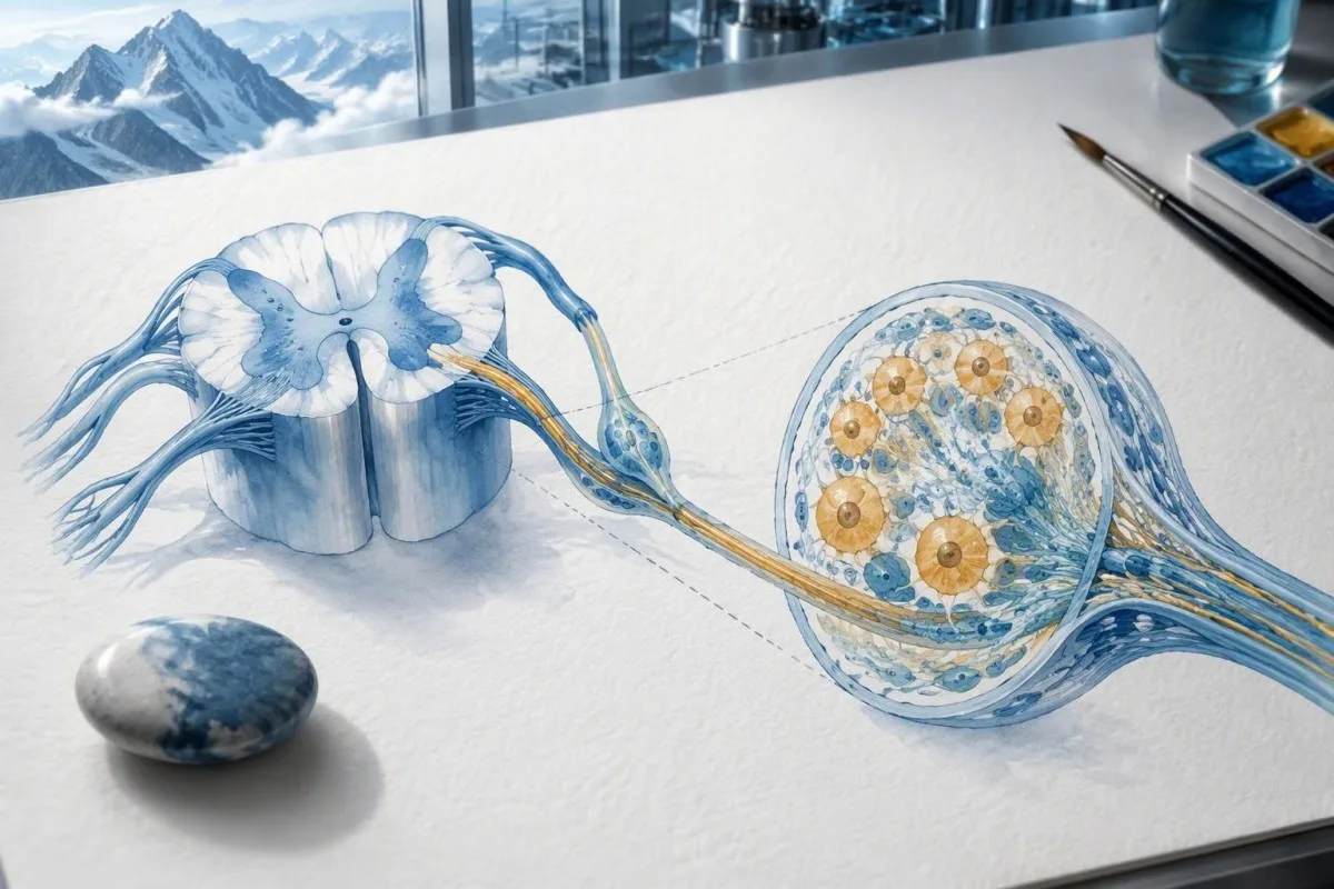
Sensory neuronopathy can cause asymmetric numbness, impaired position sense and sensory ataxia, sometimes involving the hands or trunk early rather than following a simple feet-first pattern. A patient may lose balance in the dark because vision had been compensating for reduced position feedback. That is different from the focal finger distribution discussed in our carpal tunnel symptom guide, although neither pattern can be diagnosed from description alone.
Anti-Hu autonomic involvement can affect gut movement, blood-pressure regulation and bladder function. Severe constipation, unexplained intestinal pseudo-obstruction or dizziness on standing becomes more relevant when accompanied by objective neurological abnormalities. Recording symptoms over 7 days can reveal combinations that otherwise get presented as separate stomach, balance and urinary complaints; sudden severe abdominal symptoms still require assessment for non-neurological emergencies.
New memory impairment, behavioral change or seizures may indicate central nervous system involvement, while rapid weakness has a broader emergency differential. The အားနည်းမှုနှင့်ပတ်သက်သည့် အရေးပေါ်သတိပေးလက္ခဏာများ in our neurological safety guide apply regardless of the antibody label. Call emergency services for new breathing difficulty, unsafe swallowing, a first seizure or sudden stroke-like symptoms rather than waiting for a scheduled follow-up.
အပေါင်းရလဒ်ရရှိပြီးနောက် မည်သည့် အာရုံကြောဆိုင်ရာ စစ်ဆေးမှုများ ပြုလုပ်နိုင်သလဲ။
Neurological follow-up may include examination, nerve conduction studies, MRI and cerebrospinal fluid testing, selected according to the symptoms. There is no mandatory battery for every anti-Hu-positive person; the purpose is to document which structures are affected and exclude other causes, not merely to collect additional positive tests.
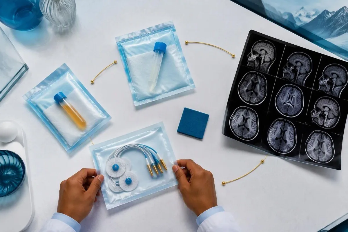
Nerve conduction studies can support a sensory neuronopathy pattern by showing reduced sensory responses across distributions that do not fit a single compressed nerve. Brain or spinal MRI addresses different questions, including structural disease and some inflammatory changes. A normal MRI does not exclude peripheral sensory ganglion involvement; these are 2 distinct anatomical compartments with different diagnostic tools.
Cerebrospinal fluid may show increased cells, protein or evidence of intrathecal immune activity, but none of those findings is specific to anti-Hu. A lumbar puncture is most useful when the neurological syndrome warrants it, rather than as an automatic response to 1 weak serum band. Our discussion of ဦးနှောက်နှင့်ကျောရိုးအရည် အိုလီဂိုကလိုနယ် ဘန် ရလဒ်ဖော်ထုတ်ရေး လမ်းညွှန် explains why immune activity in spinal fluid does not identify a single disease.
Basic investigations still matter: glucose control, vitamin status, renal function and serum protein assessment can uncover treatable contributors to neuropathy. A clinician may request 2 or more of these alongside specialist testing, depending on the examination. The သွေးရည်ကြည်ပရိုတင်း စစ်ဆေးမှုလမ်းညွှန် provides background, but normal routine blood work neither confirms nor excludes an anti-Hu-associated neurological process.
Anti-Hu အပေါင်းဖြစ်ခြင်းက ကင်ဆာနှင့်ဆက်စပ်သော ရောဂါလက္ခဏာစုကို အတည်ပြုသလား။
A positive antibody alone does not confirm anti-Hu paraneoplastic syndrome. The PNS-Care approach combines the neurological phenotype, antibody risk category, cancer findings and follow-up; clinicians also check for direct cancer effects, treatment toxicity and unrelated neurological conditions before attributing symptoms to a paraneoplastic immune response.
Definite, probable and possible PNS are diagnostic categories, not cancer stages. The 2021 criteria use a structured scoring system rather than allowing 1 antibody to determine the whole diagnosis (Graus et al., 2021). A confirmed anti-Hu result with a strongly compatible syndrome remains clinically significant even before a cancer is identified, but clinicians should state clearly which parts of the assessment remain unresolved.
An older patient can have anti-Hu reactivity and a separate cause of numbness, such as diabetes, nutritional deficiency or a monoclonal protein disorder. Coexistence is possible; either-or reasoning can miss treatable disease. If serum protein studies reveal a band, our immunofixation result guide explains why that finding requires its own interpretation rather than being automatically folded into the anti-Hu diagnosis.
ကန်တက်စတီသည် AI သွေးစစ်ဆေးမှု ရလဒ် ပလက်ဖောင်း that can help organize these distinctions in a laboratory explanation, but it cannot assign a reliable PNS-Care diagnosis from a report alone. Our ဆေးဘက်ဆိုင်ရာ validation framework separates technical evaluation from clinical conclusions. In practice, I would document 3 separate judgments: confidence in the antibody, confidence in the neurological syndrome and the current evidence for cancer.
ဆရာဝန်များသည် ဆက်စပ်ကင်ဆာကို မည်သို့ရှာဖွေကြသလဲ။
Cancer evaluation usually prioritizes the chest because small-cell lung cancer is the strongest adult anti-Hu association. Doctors commonly begin with appropriate chest CT and may add FDG-PET/CT or targeted investigations if suspicion remains; the exact plan depends on age, symptoms, examination, smoking history and previous imaging.
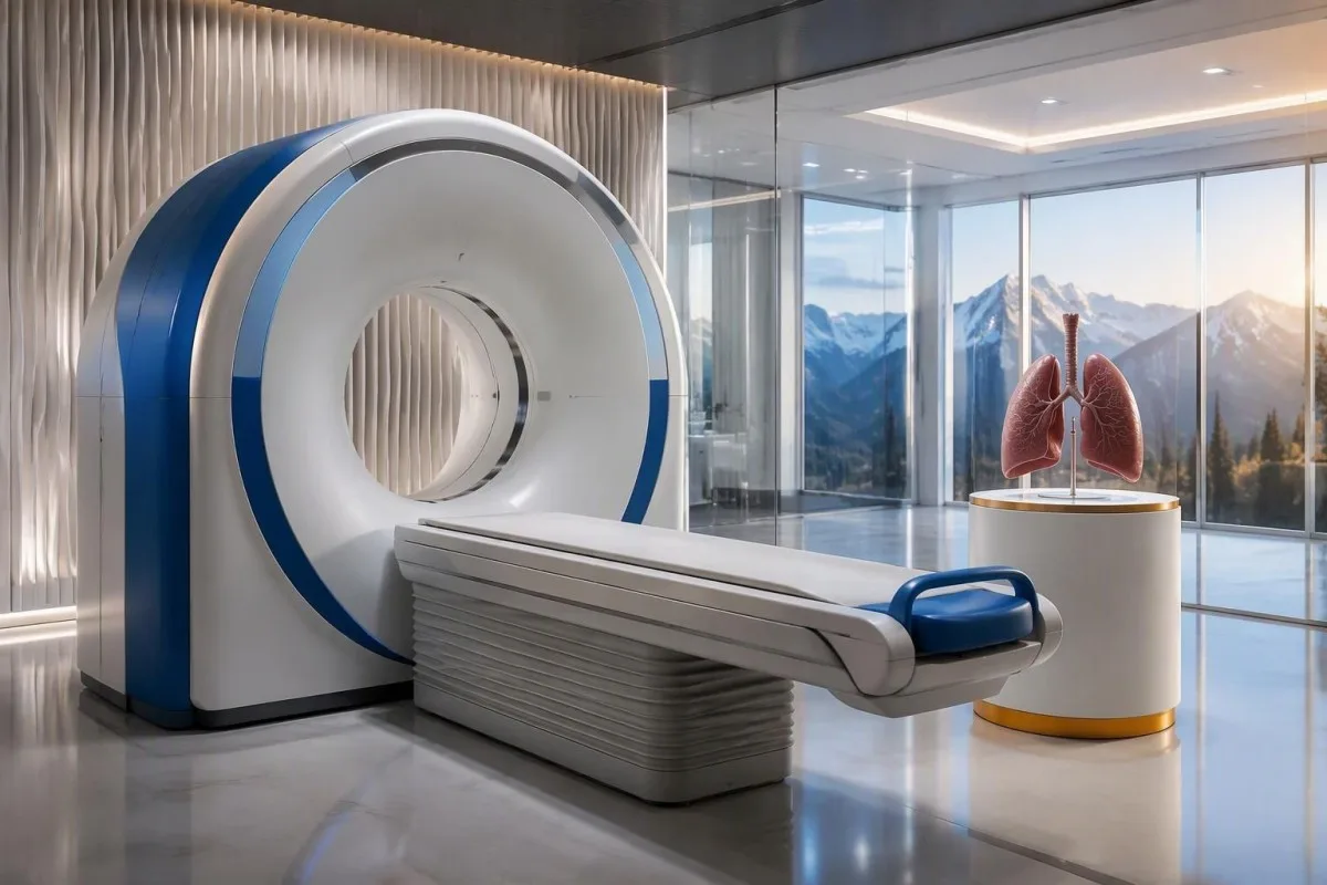
The cancer association is substantial in established anti-Hu clinical cohorts. Villagrán-García et al. (2023) reported identified cancer in 349 of 466 patients, approximately 75%, in a specialist anti-Hu cohort. This is valuable evidence for taking confirmed, clinically relevant anti-Hu seriously; it is not a personalized prediction for an asymptomatic person with an unconfirmed positive screening strip.
A chest X-ray is not equivalent to a chest CT, and routine low-dose screening is not necessarily the correct examination for a suspected paraneoplastic syndrome. The clinician chooses the protocol and body regions based on the diagnostic question. Likewise, neurological symptoms do not automatically mean cancer has spread to the brain; our brain imaging warning-sign guide distinguishes structural investigation from immune-mediated disease.
A suspicious scan often leads to tissue sampling through a method chosen by respiratory or oncology specialists, such as bronchoscopic assessment of an accessible area. Imaging suspicion and cancer confirmation are 2 separate steps. Never-smoking status lowers the baseline probability of small-cell lung cancer but does not cancel a well-confirmed anti-Hu finding with a convincing neurological presentation; other cancers and non-cancer explanations still need consideration.
ပထမဆုံး ကင်ဆာပုံရိပ်စစ်ဆေးမှုများတွင် ဘာကြောင့် မတွေ့နိုင်သလဲ။
Initial imaging can be negative because an associated cancer is still too small to detect, lies outside the examined area or is not readily visible with that technique. Neurological autoimmunity may precede cancer detection, so a negative scan reduces concern without necessarily ending follow-up in a high-risk clinical presentation.
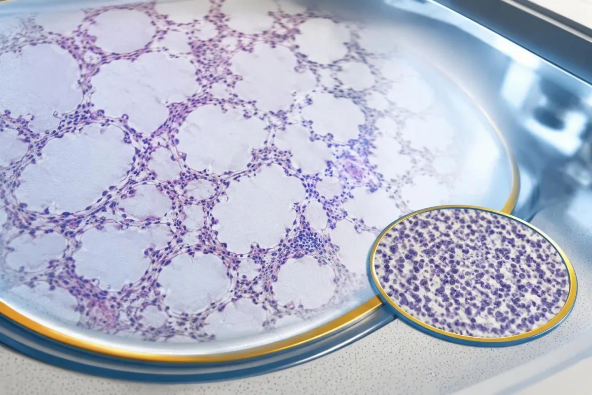
A negative scan is time-specific and method-specific. A normal chest CT performed on 1 date answers whether that examination identified a suspicious chest finding then; it does not guarantee that every possible associated cancer is absent indefinitely. Ask what was actually imaged, whether the protocol was suitable and whether the specialist recommends PET/CT or targeted investigation despite the initial result.
Villagrán-García et al. (2023) described cancers detected beyond the first 2 years in a minority of their anti-Hu cohort, supporting individualized longer surveillance. Their findings also support considering PET-based investigation when initial CT is unrevealing in an appropriate clinical context. This is an escalation decision for the treating team, not a reason to independently purchase repeated whole-body scans without weighing radiation, incidental findings and cost.
Neurological deterioration after negative imaging still needs reassessment. Cancer surveillance and neurological monitoring are 2 parallel tracks; one reassuring scan does not explain new loss of function. A marker such as neurofilament light chain may indicate neuronal injury in selected settings, but our NfL interpretation guide explains why it cannot establish anti-Hu disease, locate cancer or replace examination.
ကင်ဆာစစ်ဆေးမှုကို မည်မျှကြာ ဆက်လက်ပြုလုပ်သင့်သလဲ။
The 2021 PNS criteria recommend repeat cancer screening every 4–6 months for 2 years when a high-risk neurological phenotype accompanies a high-risk antibody. Longer surveillance may be appropriate for selected anti-Hu patients; the interval and duration should reflect confirmation, clinical evolution, initial investigations and individual cancer risk.

The 2-year schedule is not an instruction to scan every positive antibody result repeatedly. Graus et al. (2021) link that recommendation to the combination of high-risk phenotype and high-risk antibody. An incidental, weak result without a matching syndrome needs confirmation and individualized assessment first; repeating imaging mechanically can create incidental findings and unnecessary procedures without resolving the original uncertainty.
Older European guidance recommended longer surveillance. The EFNS task force advised repeating screening after 3–6 months and then every 6 months for up to 4 years in relevant antibody-positive paraneoplastic presentations (Titulaer et al., 2011). The 2023 anti-Hu cohort supported considering screening up to 5 years in selected patients, so a specialist may reasonably extend follow-up beyond the newer general 2-year framework.
Keep a dated plan stating the next examination, the clinician responsible and what symptoms should trigger earlier contact. Kantesti explanations can help you understand terminology, but the platform should not be treated as a scan-reminder service or substitute for that plan. Our guide to risk-based cancer screening addresses a different setting: routine prevention is not the same as investigating a possible paraneoplastic syndrome.
Anti-Hu ကြောင့်ဖြစ်သော အာရုံကြောဆိုင်ရာရောဂါအတွက် မည်သည့်ကုသမှုက အထောက်အကူပြုသလဲ။
Treatment focuses on an identified cancer, selected immune therapy and symptom-directed rehabilitation. Anti-Hu-associated neurological disease often responds less predictably to immunotherapy than neuronal surface-antibody disorders because intracellular targets and T-cell-mediated injury are involved; treatment may aim to stabilize function rather than promise complete recovery.

Treating an associated cancer is a central part of care, but established neurological injury may persist even when cancer treatment succeeds. Neurology and oncology teams may consider corticosteroids, intravenous immunoglobulin, plasma exchange or other immune treatments in selected situations. There is no single evidence-based dose or 1 universal regimen appropriate for everyone with anti-Hu positivity, especially without a confirmed clinical syndrome.
An antibody titer is not a dependable treatment target on its own. A lower number does not necessarily mean restored nerve function, and a persistent positive result does not by itself prove cancer progression. This differs from some surface-antibody disorders; our MOG antibody diagnosis guide explains another immune neurological condition, but its diagnostic rules and treatment expectations should not be transferred to anti-Hu.
Immune checkpoint cancer therapies deserve an explicit discussion when anti-Hu neurological disease is present, because they can provoke or worsen paraneoplastic neurological autoimmunity. This is a specialist risk-benefit decision, not an automatic prohibition based on 1 laboratory result. Tell the oncologist about previous neurological symptoms and confirmed neuronal antibodies before treatment decisions are finalized; coordinating the teams early can prevent avoidable misunderstanding.
စစ်ဆေးမှုများ ဆက်လက်လုပ်ဆောင်နေစဉ် ဘေးကင်းစွာ ဘာလုပ်နိုင်သလဲ။
While investigations continue, protect mobility, swallowing and medication safety rather than trying to lower anti-Hu antibodies with supplements. Use the neurological symptoms—not the appearance of the laboratory flag—to decide whether ordinary follow-up is sufficient or urgent care is needed; a new functional loss warrants contact before the next planned scan.

Position-sense loss increases fall risk, particularly in poor lighting or on uneven ground. A physiotherapist may recommend a suitable walking aid and individualized balance strategies; occupational therapy can address hand coordination and home hazards. A short 7-day record of falls, near-falls and walking changes is often more useful at review than the vague statement “my balance is worse,” but stop activities that feel unsafe.
Excess vitamin B6 can itself cause sensory neuropathy, making unsupervised high-dose nerve supplements particularly unhelpful during this assessment. Review every supplement and its daily dose in mg, including overlapping multivitamins and combination products. Our பி-காம்ப்ளக்ஸ் பாதுகாப்பு வழிகாட்டி explains that problem; correcting a documented deficiency is different from taking progressively larger doses in the hope of treating anti-Hu autoimmunity.
Vitamin B12 deficiency can contribute to sensory and gait problems and may need investigation even when anti-Hu is confirmed. There is no universally interchangeable B12 cutoff: results in pg/mL နှင့် pmol/L require their own laboratory context, sometimes with functional testing. Our အကန့်အသတ်ရှိသော B12 နောက်ဆက်တွဲ လမ်းညွှန် explains those next steps; new choking, breathing difficulty or rapid weakness should be assessed urgently instead of managed with vitamins.
Anti-Hu အစီရင်ခံစာကို ဘေးကင်းစွာ ရှင်းပြရာတွင် AI က မည်သို့ကူညီနိုင်သလဲ။
AI can help explain report terminology and organize questions, but it cannot confirm anti-Hu specificity, diagnose paraneoplastic syndrome or rule out cancer. Safe interpretation preserves the original assay details and separates 3 issues: what the laboratory reported, what the result may mean and what a clinician has actually established.

ကန်တက်စတီသည် AI-powered သွေးစစ်ဆေးမှု ခွဲခြမ်းစိတ်ဖြာ ကိရိယာ that can provide a plain-language explanation of uploaded laboratory information, including the distinction between an antibody screen and a confirmed clinical diagnosis. A rapid explanation—often around 60 seconds for report processing—is not a reference-laboratory assay. Our AI နည်းပညာ လမ်းညွှန် describes the interpretation workflow without making anti-Hu-specific diagnostic guarantees.
The complete source report should remain available for verification. An explanation should not silently replace “weakly reactive” with “confirmed,” drop the specimen type or invent a universal reference interval. Our guide to AI ဆေးဘက်ဆိုင်ရာ ရှင်းလင်းချက်များကို စစ်ဆေးနည်းလမ်းညွှန် describes practical source checks; for this antibody, compare at least the assay wording, confirmation comment and collection date against the original document.
Kantesti cannot examine sensation, review tissue staining directly through a report explanation or establish that negative imaging has excluded a tiny cancer. Those are 3 different clinical capabilities, not features that follow from understanding laboratory language. Share the explanation with the treating team as a discussion aid, and do not delay urgent assessment while uploading documents or waiting for a second automated interpretation.
သင်၏နောက်ချိန်းဆိုမှုနှင့် သုတေသနဆိုင်ရာ ပွင့်လင်းမြင်သာမှု
Bring the complete antibody report, a dated symptom history, previous imaging reports and a current medication list to your next appointment. Ask the clinician to clarify antibody confirmation, the neurological diagnosis and cancer surveillance separately; those 3 answers may have different levels of certainty and different follow-up dates.
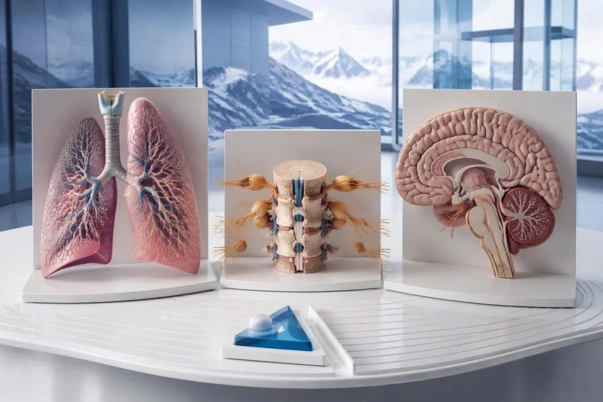
A useful 1-page symptom history records onset, progression, sensory distribution, walking changes, memory problems and autonomic symptoms. Include prior cancer treatment and any recent immunoglobulin therapy, then ask who will coordinate neurology and oncology if both are involved. As Thomas Klein, MD, I would rather see a clear timeline than 20 disconnected screenshots; it makes discrepancies between the laboratory result and clinical course easier to spot.
Medical authorship is not a substitute for an individual neurological examination. ကျွန်တော်တို့ရဲ့ medical advisory board
information describes the clinicians associated with our work, while your own specialist determines the appropriate next investigation. As of ၂၀၂၆ ခုနှစ် အောက်တိုဘာလ ၂၅ ရက်, the recommendations discussed here draw on the 2021 international PNS criteria, the 2011 EFNS screening guidance and the 2023 anti-Hu cohort—not an invented 2026 guideline.
The 2 Figshare publications below are platform-related research references, not evidence validating anti-Hu diagnosis or cancer exclusion. Their supplied titles describe hantavirus triage deployment and a synthetic-case technical benchmark; neither title establishes anti-Hu-specific clinical performance. Because publication authors and dates were not supplied for verification, the citations use title-first APA formatting with “n.d.”; the ResearchGate and Academia.edu links are searches, not claims of verified publication copies.
အမေးများသောမေးခွန်းများ
Anti-Hu အန်တီဘော်ဒီ အပေါင်းလက္ခဏာရလဒ် ရှိတာက ကျွန်ုပ်မှာ ကင်ဆာရှိတယ်လို့ ဆိုလိုပါသလား။
0 Anti-Hu ပဋိပစ္စည်း စစ်ဆေးမှုရလဒ် အပေါင်းဖြစ်ခြင်းတစ်ခုတည်းဖြင့် ကင်ဆာရှိကြောင်း ရောဂါသတ်မှတ်၍ မရပါ။ Anti-Hu သည် ကင်ဆာနှင့် ဆက်စပ်မှုမြင့်မားသော ပဋိပစ္စည်းဖြစ်ပြီး၊ ၂၀၂၃ ခုနှစ်က Anti-Hu အထူးကုလူနာအုပ်စုတွင် လူနာ ၄၆၆ ဦးအနက် ၃၄၉ ဦး၌ ကင်ဆာတွေ့ရှိခဲ့သည်။ သို့သော် ထိုကိန်းဂဏန်းသည် အတည်မပြုရသေးသော ပုံမှန်မဟုတ်သည့် စစ်ဆေးမှုရလဒ်တစ်ခုကြောင့် မတော်တဆ တွေ့ရှိလာသည့်အခါရှိနိုင်သော အန္တရာယ်ကို မသတ်မှတ်ပေးပါ။ ဆရာဝန်များသည် အာရုံကြောဆိုင်ရာ လက္ခဏာများကို အကဲဖြတ်ပြီး၊ မမျှော်လင့်ထားသော ဓာတ်ခွဲခန်းရလဒ်များကို အတည်ပြုကာ၊ နောက်ခံကင်ဆာရှိမရှိ စုံစမ်းစစ်ဆေးကြသည်။ အရွယ်ရောက်သူများတွင် ပုံမှန်အားဖြင့် ရင်ဘတ်ကို ဦးစားပေးစစ်ဆေးသည်။ ကင်ဆာရောဂါဟု သတ်မှတ်ရန် ယေဘုယျအားဖြင့် သီးခြားလက်တွေ့အထောက်အထား၊ ပုံရိပ်ဖော်စစ်ဆေးမှုအထောက်အထားနှင့် တစ်ရှူးအထောက်အထားတို့ လိုအပ်သည်။.
anti-Hu နဲ့ ANNA-1 က အတူတူပဲလား?
0] Anti-Hu နှင့် ANNA-1 တို့သည် ယေဘုယျအားဖြင့် တူညီသော အာရုံကြောဆဲလ်ဆိုင်ရာ ပဋိပစ္စည်းအုပ်စုကို ဖော်ပြသည်။ ANNA-1 ဆိုသည်မှာ အာရုံကြောဆဲလ်နျူကလီးယပ်စ်ဆိုင်ရာ ပဋိပစ္စည်း အမျိုးအစား ၁ ကို ဆိုလိုပြီး Hu ဆိုသည်မှာ ပဋိပစ္စည်းက အသိအမှတ်ပြုသော ဆဲလ်အတွင်းရှိ အာရုံကြောဆဲလ်ပရိုတင်းများကို ရည်ညွှန်းသည်။ ANNA-1 သည် စနစ်တစ်ခုလုံးဆိုင်ရာ ကိုယ်ခံအားရောဂါ စစ်ဆေးရာတွင် ပုံမှန်အသုံးပြုသော ANA နှင့် မတူပါ။ ဓာတ်ခွဲခန်းများသည် တူညီသော ပဋိပစ္စည်းအုပ်စုကို ပုံစံအမျိုးမျိုးဖြင့် အစီရင်ခံနိုင်သောကြောင့် စမ်းသပ်မှုအမျိုးအစားနှင့် အတည်ပြုချက်ဆိုင်ရာ စကားရပ်အတိအကျကို စစ်ဆေးပါ။.
Anti-Hu အင်တီဘော်ဒီ စစ်ဆေးမှုတွင် မှားယွင်းသော အပေါင်းရလဒ် ထွက်နိုင်ပါသလား။
Anti-Hu antibody စစ်ဆေးမှုတွင် လှည့်ဖြားနိုင်သော သို့မဟုတ် အတိအကျမဟုတ်သော တုံ့ပြန်မှုများ ဖြစ်ပေါ်နိုင်သည်။ အထူးသဖြင့် အားနည်းသော စစ်ဆေးမှုရလဒ်သည် အာရုံကြောဆိုင်ရာ လက္ခဏာများနှင့် မကိုက်ညီသည့်အခါ ဖြစ်နိုင်သည်။ အတည်ပြုရန် ပုံမှန်အားဖြင့် အပြန်အလှန် ဖြည့်စွက်ပေးသော နည်းလမ်း 2 ခုကို အသုံးပြုသည်- ထူးခြားသော တစ်ရှူးအရောင်စွန်းပုံစံနှင့် အတွေ့အကြုံရှိ ဓာတ်ခွဲခန်းမှ အဓိပ္ပာယ်ဖော်ထားသော အန်တီဂျင်သီးသန့် စစ်ဆေးမှုတို့ဖြစ်သည်။ တူညီသော စီးပွားဖြစ်နည်းလမ်းကို ထပ်မံအသုံးပြုခြင်းသည် ပြဿနာကို ဖြေရှင်းမည့်အစား တူညီသော ခွဲခြမ်းစိတ်ဖြာမှုဆိုင်ရာ ပြဿနာကိုပင် ထပ်မံဖြစ်ပေါ်စေနိုင်သည်။ အတည်ပြုချက်ကို စောင့်ဆိုင်းနေစဉ်တွင်လည်း လျင်မြန်စွာ ပိုမိုဆိုးရွားလာသော အာရုံကြောဆိုင်ရာ လက္ခဏာများကို အချိန်မီ စစ်ဆေးအကဲဖြတ်ရန် လိုအပ်သည်။.
Anti-Hu ပေါင်းစမ်းသပ်ရလဒ်ဖြစ်ပါက စကင်ဖတ်စစ်ဆေးမှုများကို မည်မျှမကြာခဏ ပြန်လည်ပြုလုပ်သင့်သနည်း။
အန္တရာယ်မြင့် အာရုံကြောဆိုင်ရာလက္ခဏာပုံစံနှင့် အန္တရာယ်မြင့် ပဋိပစ္စည်းတို့ တွဲဖက်တွေ့ရှိပါက ၂ နှစ်ကြာ ၄–၆ လတစ်ကြိမ် ကင်ဆာစစ်ဆေးမှုကို ထပ်မံပြုလုပ်ရန် 2021 PNS စံနှုန်းများက အကြံပြုထားသည်။ ထိုအချိန်ဇယားသည် မတော်တဆတွေ့ရှိသည့် anti-Hu-positive ရလဒ်တိုင်းနှင့် အလိုအလျောက် သက်ဆိုင်သည်မဟုတ်ပါ။ ယခင် EFNS လမ်းညွှန်ချက်များက ၄ နှစ်အထိ စောင့်ကြည့်စစ်ဆေးရန် အကြံပြုခဲ့ပြီး၊ 2023 anti-Hu လူနာအုပ်စုလေ့လာမှုတစ်ခုက ရွေးချယ်ထားသော လူနာများတွင် ၅ နှစ်အထိ စဉ်းစားရန် ထောက်ခံခဲ့သည်။ မည်သည့်ပုံရိပ်ဖော်နည်းကို အသုံးပြုမည်၊ မည်သည့်အချိန်ခြား၍ စစ်ဆေးမည်၊ မည်မျှကြာ စောင့်ကြည့်မည်နှင့် မည်သည့်လက္ခဏာများရှိပါက စောစီးစွာ ပြန်လည်စစ်ဆေးရန် လိုအပ်မည်ကို သင့်အထူးကုဆရာဝန်က သတ်မှတ်ပေးသင့်သည်။.
CT နှင့် PET စကင်များ ပုံမှန်ဖြစ်နေသည့်အခါ anti-Hu စစ်ဆေးချက် အပေါင်းလက္ခဏာ ပြနိုင်ပါသလား။
CT သို့မဟုတ် PET ပုံရိပ်စစ်ဆေးမှုတွင် ကင်ဆာကို မတွေ့ရှိသော်လည်း Anti-Hu ပဋိပစ္စည်းသည် ဆက်လက်အပြုသဘောဖြစ်နေနိုင်သည်။ အာရုံကြောစနစ်အပေါ် ကိုယ်ခံအားစနစ်၏ တိုက်ခိုက်မှုသည် ကင်ဆာကို မတွေ့ရှိမီ ဖြစ်ပေါ်လာနိုင်ပြီး၊ စစ်ဆေးမှုအဖြေ အနုတ်လက္ခဏာဖြစ်ခြင်းသည် စစ်ဆေးထားသော ကိုယ်အင်္ဂါအစိတ်အပိုင်းများ၊ အသုံးပြုသည့်နည်းလမ်းနှင့် စစ်ဆေးသည့်ရက်စွဲကိုသာ ရည်ညွှန်းပြီး ကင်ဆာလုံးဝမရှိကြောင်း အမြဲတမ်းအာမခံခြင်း မဟုတ်ပါ။ ကိုက်ညီသော အန္တရာယ်မြင့် လက္ခဏာများရှိပါက 2021 စံသတ်မှတ်ချက်များအရ ၂ နှစ်ကြာသည်အထိ ၄–၆ လတစ်ကြိမ် ထပ်မံစစ်ဆေးရန် အကြံပြုထားပြီး၊ Anti-Hu လူနာအချို့တွင် တစ်ဦးချင်းအခြေအနေအလိုက် ပိုမိုကြာရှည်စွာ စောင့်ကြည့်နိုင်သည်။ အတည်မပြုရသေးသော ပဋိပစ္စည်းဓာတ်ပြုမှုနှင့် ကင်ဆာမဟုတ်သော အကြောင်းရင်းများကိုလည်း မမြင်နိုင်သော ကင်ဆာတစ်ခု ရှိနေသည်ဟု အတိအကျယူဆမည့်အစား ပြန်လည်စဉ်းစားသင့်သည်။.
0. Anti-Hu ပဋိပစ္စည်းပမာဏ လျော့ကျလာခြင်းသည် ကုသမှု ထိရောက်နေသည်ဟု ဆိုလိုပါသလား။
0 အန်တီ-ဟူ ပဋိပစ္စည်းအဆင့် ကျဆင်းခြင်းသည် အာရုံကြောဆိုင်ရာ ပြန်လည်ကောင်းမွန်လာမှု သို့မဟုတ် ကင်ဆာကုသမှု အောင်မြင်မှုကို ယုံကြည်စိတ်ချစွာ သက်သေမပြနိုင်ပါ။ အန်တီ-ဟူသည် ဆဲလ်အတွင်းရှိ ပရိုတင်းများကို မှတ်မိတုံ့ပြန်ပြီး၊ ဆက်စပ်အာရုံကြောထိခိုက်မှုတွင် ပဋိပစ္စည်းသက်ရောက်မှုတစ်ခုတည်းထက် T-ဆဲလ် ယန္တရားများ ပါဝင်လေ့ရှိသည်။ ဓာတ်ခွဲခန်းမတူများမှ တိုင်းတာထားသည့် တိုင်တာနှစ်ခုသည် အသုံးပြုသောနည်းလမ်းများ ကွဲပြားသောကြောင့် နှိုင်းယှဉ်၍မရနိုင်ပါ။ ဆရာဝန်များသည် ပဋိပစ္စည်းကိန်းဂဏန်းကို တစ်ခုတည်းသော ကုသမှုပစ်မှတ်အဖြစ် အသုံးမပြုဘဲ လုပ်ဆောင်နိုင်စွမ်း၊ စမ်းသပ်စစ်ဆေးတွေ့ရှိချက်များ၊ သက်ဆိုင်ရာ စစ်ဆေးမှုများနှင့် ကင်ဆာတုံ့ပြန်မှုတို့ကို အကဲဖြတ်ကြသည်။.
AI ပါဝါသုံး သွေးစစ်ဆေးမှု ရလဒ်ကို ယနေ့ စတင် ခွဲခြမ်းစိတ်ဖြာပါ
Kantesti ကို အချိန်မီ၊ တိကျသော ဓာတ်ခွဲခန်း စစ်ဆေးမှု ရလဒ်ခွဲခြမ်းစိတ်ဖြာမှုအတွက် ယုံကြည်သည့် ကမ္ဘာတစ်ဝန်း အသုံးပြုသူ 2 သန်းကျော်နှင့် ပူးပေါင်းပါ။ သင့် သွေးစစ်ဆေးမှု ရလဒ်များကို တင်ပြီး စက္ကန့်ပိုင်းအတွင်း 15,000+ biomarker များ၏ ပြည့်စုံသော အဓိပ္ပာယ်ဖတ်ချက်ကို ရယူပါ။.
📚 ကိုးကားထားသော သုတေသန ထုတ်ဝေမှုများ
Klein, T., Mitchell, S., & Weber, H. (2026). ဘာသာစကားများစွာဖြင့် AI အကူအညီဖြင့် ဆေးပညာဆိုင်ရာ ဆုံးဖြတ်ချက်ပံ့ပိုးမှု- အစောပိုင်း Hantavirus ခွဲခြားခြင်း- ဒီဇိုင်း၊ အင်ဂျင်နီယာ- အတည်ပြုချက်၊ နှင့် လက်တွေ့-ကမ္ဘာ-တွင်-အကောင်-အထည်-ဖော်-မှု- 50,000-ကျော်-အနက်-ဖွင့်-ဆို-ထား-သော-သွေး-စစ်-အစီရင်ခံစာ-များ-တစ်-လျှောက်. Kantesti AI Medical Research.
Klein, T., Mitchell, S., & Weber, H. (2026). 100,000 Synthetic Test Cases များပေါ်တွင် Kantesti Blood-Test Interpretation Engine ကို အလိုအလျောက် နည်းပညာဆိုင်ရာ စံနှုန်းတင် (benchmark) ပြုလုပ်ခြင်း—Pre-Registered, Rubric-Based. Kantesti AI Medical Research.
📖 ပြင်ပ ဆေးဘက်ဆိုင်ရာ ကိုးကားချက်များ
Graus F et al. (2021). Updated Diagnostic Criteria for Paraneoplastic Neurologic Syndromes.။ Neurology: Neuroimmunology & Neuroinflammation.
Titulaer MJ et al. (2011). Screening for tumours in paraneoplastic syndromes: report of an EFNS Task Force. European Journal of Neurology.
Villagrán-García D et al. (2023). Revisiting anti-Hu paraneoplastic autoimmunity: phenotypic characterization and cancer diagnosis. Brain Communications.
⚕️ ဆေးဘက်ဆိုင်ရာ ရှင်းလင်းချက်
ဤဆောင်းပါးသည် ပညာရေးဆိုင်ရာ ရည်ရွယ်ချက်အတွက်သာ ဖြစ်ပြီး ဆေးဘက်ဆိုင်ရာ အကြံဉာဏ် မဟုတ်ပါ။ ရောဂါရှာဖွေခြင်းနှင့် ကုသမှု ဆုံးဖြတ်ချက်များအတွက် အရည်အချင်းပြည့်မီသော ကျန်းမာရေးစောင့်ရှောက်မှုပေးသူနှင့် အမြဲတမ်း တိုင်ပင်ပါ။.
E-E-A-T ယုံကြည်မှု အချက်ပြများ
အတွေ့အကြုံ
ဆရာဝန်ဦးဆောင်သည့် ဓာတ်ခွဲခန်း အဓိပ္ပာယ်ဖတ်ခြင်း လုပ်ငန်းစဉ်များကို ပြန်လည်သုံးသပ်ခြင်း။.
ကျွမ်းကျင်မှု
biomarker များသည် ဆေးဘက်ဆိုင်ရာ အခြေအနေတွင် မည်သို့ ပြုမူနေသည်ကို အာရုံစိုက်သည့် ဓာတ်ခွဲခန်း ဆေးပညာ။.
အခွင့်အာဏာရှိခြင်း
ဒေါက်တာ Thomas Klein မှ ရေးသားပြီး ဒေါက်တာ Sarah Mitchell နှင့် ပါမောက္ခ ဒေါက်တာ Hans Weber တို့က ပြန်လည်သုံးသပ်ထားသည်။.
ယုံကြည်စိတ်ချရမှု
စိတ်ပူစရာများကို လျော့ချရန်အတွက် ရှင်းလင်းသော နောက်ဆက်တွဲ လမ်းကြောင်းများပါဝင်သည့် သက်သေအထောက်အထားအခြေပြု အဓိပ္ပာယ်ဖတ်ချက်။.
