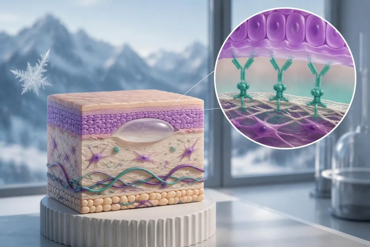
A positive BP180 result supports an autoimmune skin disorder, but the number cannot establish the diagnosis alone. Symptoms, the assay used, and correctly collected skin samples determine what happens next.

Key takeaways
- BP180 antibody positive means the assay detected antibodies against its BP180 target; it does not independently confirm bullous pemphigoid.
- Laboratory cutoffs differ: some BP180 assays use approximately 9 U/mL, while others use approximately 20 U/mL. Read your own laboratory's reference interval.
- Low-positive results without itching, rash, or blisters may represent incidental antibody detection or a false-positive assay result—not established skin disease.
- Nonbullous pemphigoid can cause persistent itching without blisters; 74 of 343 pemphigoid cases in a specialist diagnostic study were nonbullous.
- BP230 antibodies provide complementary evidence, but negative BP230 antibodies do not exclude bullous pemphigoid.
- Direct immunofluorescence examines antibody deposits in skin; linear IgG and/or C3 along the basement membrane supports a pemphigoid disorder.
- Two skin samples are often collected: an affected-area sample for routine microscopy and a nearby, appropriately selected sample for direct immunofluorescence.
- Urgent symptoms include eye pain, vision changes, difficulty swallowing, fever with extensive skin changes, or rapidly spreading blistering; do not wait for another antibody result.
Does a positive BP180 result confirm bullous pemphigoid?
No—a BP180 antibody positive result does not confirm bullous pemphigoid by itself. It supports the diagnosis when symptoms and skin findings fit, but a low positive without blisters may be incidental; dermatologists often need skin biopsy with direct immunofluorescence, and sometimes additional antibody testing, to establish the cause.
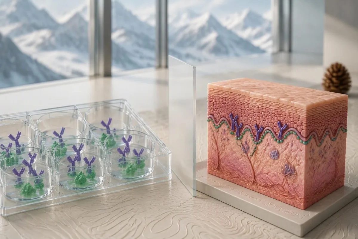
Bullous pemphigoid diagnosis requires clinical and immunopathological agreement, not simply one flagged laboratory value. The 2022 EADV guideline describes diagnosis through the clinical picture and appropriate tissue and circulating-antibody investigations (Borradori et al., 2022); the practical question is whether the detected antibodies explain the person's skin condition.
A positive antibody test and an active autoimmune disease are different findings. For example, a hypothetical result of 12 U/mL may be positive in one laboratory, yet an asymptomatic person with that result has a different diagnostic probability from someone with widespread itchy, tense blisters and the same concentration.
Kantesti is an AI blood test analyzer that helps explain BP180 results alongside the laboratory's cutoff and the information you provide. Our explanation cannot examine your skin or perform direct immunofluorescence; our company background identifies the organization behind this educational support.
Treat the result as a reason to ask a more specific question, rather than as a permanent disease label. Our guide to positive antibody results explains why antibody detection, disease activity, and diagnostic confirmation are three separate concepts.
What does the BP180 antibody test actually measure?
The BP180 antibody test usually measures serum IgG directed against part of BP180, also called type XVII collagen. BP180 is a roughly 180-kDa protein involved in anchoring the epidermis to underlying tissue; many routine assays test its NC16A region rather than the entire protein.
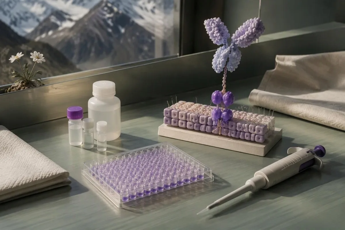
BP180 is a structural skin protein, not a substance whose blood concentration is being measured. The report describes antibodies against that protein, commonly using an enzyme-linked immunosorbent assay, or ELISA; a value of 30 U/mL therefore does not mean that 30 units of skin protein are circulating.
The NC16A domain sits close to the cell membrane on the external portion of BP180 and is a frequent antibody target in classic bullous pemphigoid. Testing this region makes a practical assay possible, but it creates a blind spot: antibodies directed mainly against other regions may escape detection.
A numerical antibody concentration is not interchangeable with an immunofluorescence titer. A result expressed as 1:40 describes reactivity at a dilution, whereas 40 U/mL describes an assay-calibrated concentration; our explanation of quantitative versus qualitative tests helps distinguish these reporting formats.
Check whether the report says BP180 NC16A IgG, another BP180 assay, or a broader pemphigoid panel. These are not automatically equivalent tests, and a negative IgG ELISA does not answer every question about other antibody classes or other basement-membrane targets.
What counts as a low-positive BP180 antibody result?
A low-positive BP180 result is a value just above the cutoff for the specific assay used. There is no universal low-positive range: some laboratories use a threshold near 9 U/mL, while others use one near 20 U/mL, with different calibrations and reporting categories.
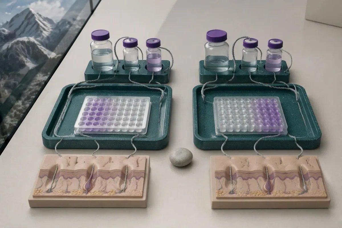
BP180 assay units are not universally standardized across laboratories. A result of 15 U/mL could be positive under a 9-U/mL policy and negative under a 20-U/mL policy, but this does not establish that the second assay missed disease; the methods and calibrators may differ.
Cutoffs balance sensitivity against specificity in the populations used to evaluate the assay. Moving a threshold downward can detect more people with disease while also identifying more people without disease; the cutoff is a laboratory decision boundary, not a biological point where skin suddenly starts blistering.
Kantesti AI can help place a reported BP180 value beside its supplied reference interval, but a cropped image showing only 18 U/mL leaves crucial information missing. Include the test name, units, interpretation categories, and laboratory comments; a generic out-of-range flag cannot substitute for those details.
The examples below illustrate two possible reporting policies rather than a universal severity scale. No BP180 antibody concentration alone defines a medical emergency; extensive skin involvement, dehydration, systemic illness, and eye or airway symptoms determine urgency.
| Level | Range | What it means |
|---|---|---|
| Example assay A: below cutoff | <9 U/mL | Negative under this illustrative laboratory policy; disease is not excluded if clinical suspicion remains high. |
| Example assay A: at or above cutoff | ≥9 U/mL | Positive under this policy; proximity to the cutoff and clinical findings affect interpretation. |
| Example assay B: below cutoff | <20 U/mL | Negative under a different illustrative policy; do not directly convert between assay systems. |
| Example assay B: at or above cutoff | ≥20 U/mL | Positive under this policy; the number does not independently establish diagnosis or severity. |
Why can a positive BP180 result be misleading?
A positive BP180 result is less convincing when the person had little clinical likelihood of pemphigoid before testing. Even a reasonably accurate assay produces false positives, so the meaning of the result changes between an asymptomatic screening population and a specialist clinic evaluating characteristic blisters.
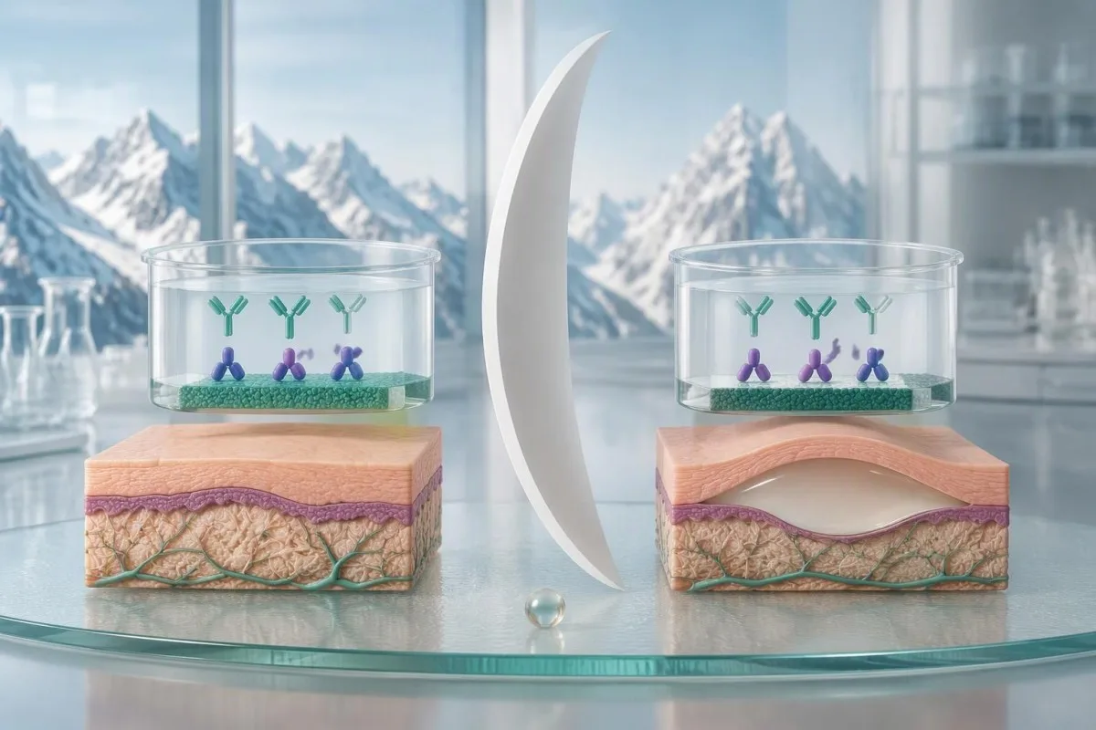
Sensitivity and specificity do not equal the probability that you personally have disease after a positive result. That probability also depends on how common the condition is in the tested group; this distinction matters most when an antibody panel was ordered broadly rather than to investigate a specific skin pattern.
Consider a clearly hypothetical assay with 90% sensitivity and 95% specificity, used in 1,000 people whose disease probability is 1%. It would identify about 9 true positives and 50 false positives, making the positive predictive value roughly 15%; these are teaching assumptions, not performance claims for your BP180 assay.
Use the same hypothetical assay where disease probability is 20%, and approximately 180 true positives and 40 false positives would occur per 1,000 tests. The positive predictive value becomes about 82%—a large change caused by clinical context, not by a different laboratory method.
These examples explain why a dermatologist may request tissue testing despite a positive result, or take characteristic symptoms seriously despite a negative one. Our discussion of MOG antibody interpretation illustrates the same probability principle in another autoimmune condition, although its diagnostic criteria cannot be transferred to pemphigoid.
What if BP180 is positive but you have no blisters?
A low-positive BP180 result without blisters does not establish bullous pemphigoid or reliably predict future blistering. The next distinction is whether you have no skin symptoms at all, or persistent itching and an unexplained rash that could represent a nonbullous pemphigoid presentation.

Incidental BP180 antibody detection can occur without clinically established pemphigoid. Possible explanations include low-level autoreactivity and assay-related false positivity; there is no validated conversion rule that turns an isolated result of 11 U/mL into a percentage risk of developing blisters later.
A hypothetical person with 11 U/mL, no itching, no rash, and no mucosal symptoms usually needs a contextual review rather than automatic immunosuppressive treatment. The clinician should check why the assay was ordered, whether the result was unexpected, and whether a skin examination reveals findings the person had not recognized.
Persistent dryness and itching still deserve assessment because common explanations remain common. Eczema, medication effects, thyroid disease, and other causes may be more likely than pemphigoid; our dry skin testing guide explains why additional blood tests should follow symptoms rather than a blanket panel.
There is no universal repeat-testing interval for an asymptomatic low-positive BP180 result. A clinician may choose observation, a repeat using the same method, or specialist review depending on the history; new persistent itching, urticarial plaques, or blisters should change that plan rather than waiting for a scheduled repeat.
Can bullous pemphigoid begin with itching alone?
Pemphigoid can present with persistent itching, eczema-like patches, urticarial plaques, or scratch-related changes without visible blisters. In a specialist diagnostic study, 74 of 343 pemphigoid cases—21.6%—were nonbullous, showing that the absence of blisters cannot independently exclude the disorder (Meijer et al., 2019).

Nonbullous pemphigoid is not simply every itchy rash with a positive BP180 result. Diagnosis still needs compatible symptoms and appropriate immunopathological evidence; the 21.6% study figure came from a referred population and should not be interpreted as the probability that any person with itching has pemphigoid.
The symptom pattern often matters more than whether one small blister has appeared. Persistent, widespread itching in an older adult, especially with recurrent raised plaques or poor response to ordinary eczema care, warrants a dermatological assessment; our itchy skin laboratory guide separates skin-directed testing from tests for systemic causes.
Eosinophilia can accompany bullous pemphigoid, but it is neither required nor diagnostic. An absolute eosinophil count above approximately 0.5 × 10^9/L is often flagged in adults, yet allergy, medications, and parasites can produce the same finding; our guide to high eosinophil counts explains those alternatives.
A dermatologist does not always need to wait for a blister before collecting a useful sample for direct immunofluorescence. In nonbullous disease, sample selection is tailored to the available rash and laboratory protocol; document the distribution and duration of itching, because a disappearing rash can make later assessment harder.
How do BP230 antibodies complement BP180 testing?
BP230 antibodies provide complementary evidence for pemphigoid but cannot confirm or exclude bullous pemphigoid alone. BP230 is an approximately 230-kDa intracellular anchoring protein, whereas BP180 crosses the cell membrane; their different locations and antibody targets help explain why the two results can disagree.
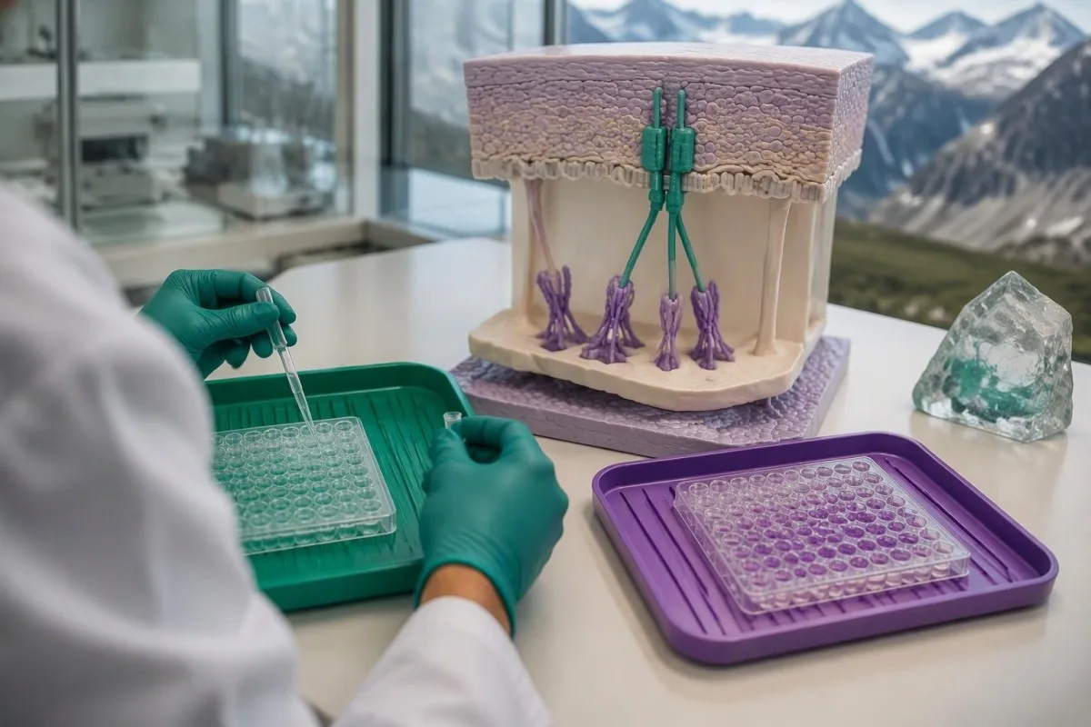
A negative BP230 antibody result does not cancel a positive BP180 result. Some people with confirmed pemphigoid have antibodies detectable against only one tested antigen, and each assay has its own sensitivity; interpreting the pair as a two-test pass-or-fail rule creates avoidable diagnostic errors.
A hypothetical BP180 result of 45 U/mL with negative BP230 antibodies still warrants assessment if tense blisters and severe itching are present. Conversely, isolated low-positive BP230 antibodies without compatible findings are not enough to diagnose pemphigoid; a second positive marker strengthens a pattern only when the overall evidence fits.
Kantesti is an AI biomarker interpretation platform that can explain BP180 and BP230 antibodies as complementary findings rather than interchangeable scores. This is similar to the reasoning used for antibody panels in connective disease, but skin-specific tissue findings remain central to pemphigoid assessment.
Do not add BP180 and BP230 concentrations together, even if both are expressed in U/mL. They represent different antigen-specific measurements with different calibration systems; BP180 levels generally have a better-established relationship with disease activity than BP230 levels, but neither replaces examination.
Why might you need skin biopsy with direct immunofluorescence?
Skin biopsy with direct immunofluorescence can show whether antibodies or complement are deposited at the skin's basement membrane. Dermatologists often collect two separate specimens because routine microscopy and direct immunofluorescence answer different questions and require different sample handling.
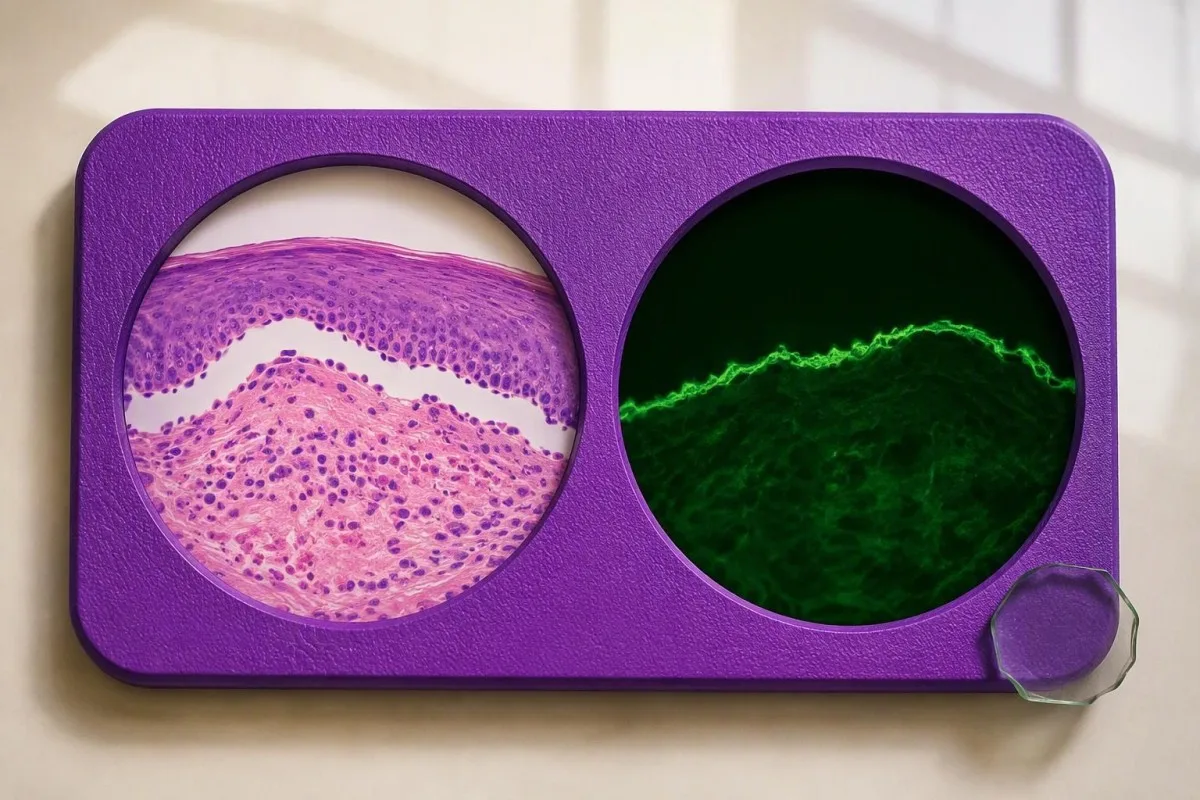
Routine histology examines tissue structure and inflammatory cells. A sample from a fresh blister or suitable affected area may show separation beneath the epidermis and eosinophils, but these features are not unique to bullous pemphigoid; the second sample looks for the immune-deposit pattern that helps classify the disorder.
For classic blistering presentations, the direct-immunofluorescence sample is commonly taken from appropriately selected skin near a fresh blister rather than its eroded center. A small 3–4-mm punch is often used, although technique and location depend on the case; sampling heavily damaged tissue can remove the structures the laboratory needs to examine.
The specimen for direct immunofluorescence must not be routinely placed in formalin. Laboratories generally require a dedicated transport medium or another explicitly agreed handling method, while the routine histology specimen commonly goes into formalin; ask whether the two containers have been prepared for their separate purposes.
In Meijer et al.'s 2019 diagnostic cohort, direct immunofluorescence had a sensitivity of 88.3%, so even this valuable test was not infallible. Kantesti's clinical validation information concerns our interpretation methodology—not a claim that AI can reproduce skin immunofluorescence or establish its findings.
What do positive immunofluorescence and salt-split tests mean?
Linear IgG and/or C3 along the basement membrane on direct immunofluorescence supports a pemphigoid-spectrum disorder, but the pattern is not unique to bullous pemphigoid. Indirect immunofluorescence on salt-split skin can add evidence by showing where circulating antibodies bind relative to an experimentally created tissue separation.
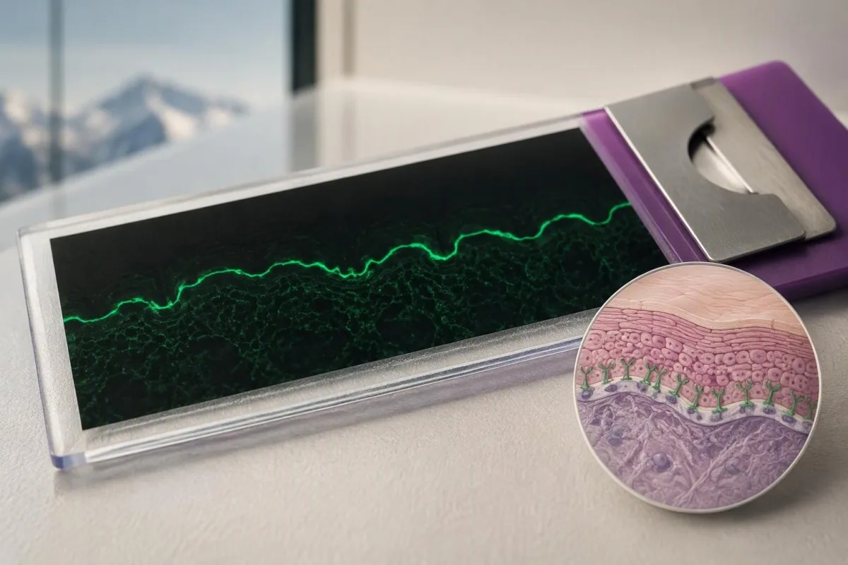
Direct and indirect immunofluorescence use different materials. Direct testing examines deposits already present in the person's skin, while indirect testing applies the person's serum to a laboratory substrate; a positive result in one test and a negative result in the other is therefore possible without a reporting error.
On salt-split skin, bullous pemphigoid antibodies commonly bind the epidermal side, often called the roof. Antibodies binding the dermal side, or floor, raise consideration of other disorders such as epidermolysis bullosa acquisita or anti-p200 pemphigoid, but binding location alone does not fully classify every case.
Meijer et al. reported 99.9% specificity for indirect immunofluorescence on salt-split skin in their 2019 diagnostic cohort. That high figure applies to that study and testing pathway; a negative result still cannot exclude pemphigoid because specificity describes false positives, not how many cases the test misses.
C3 detected in skin is not the same measurement as a serum C3 concentration. A normal circulating complement result does not negate local deposits at the basement membrane; our complement testing explanation helps prevent this common cross-test misunderstanding.
What if BP180, the skin biopsy, and symptoms disagree?
Discordant BP180 and skin-test results require a review of the clinical picture, assay target, sampling site, and specimen handling. A positive ELISA with negative direct immunofluorescence can reflect incidental antibodies or an uninformative skin sample; a negative NC16A ELISA can miss disease directed against other targets.
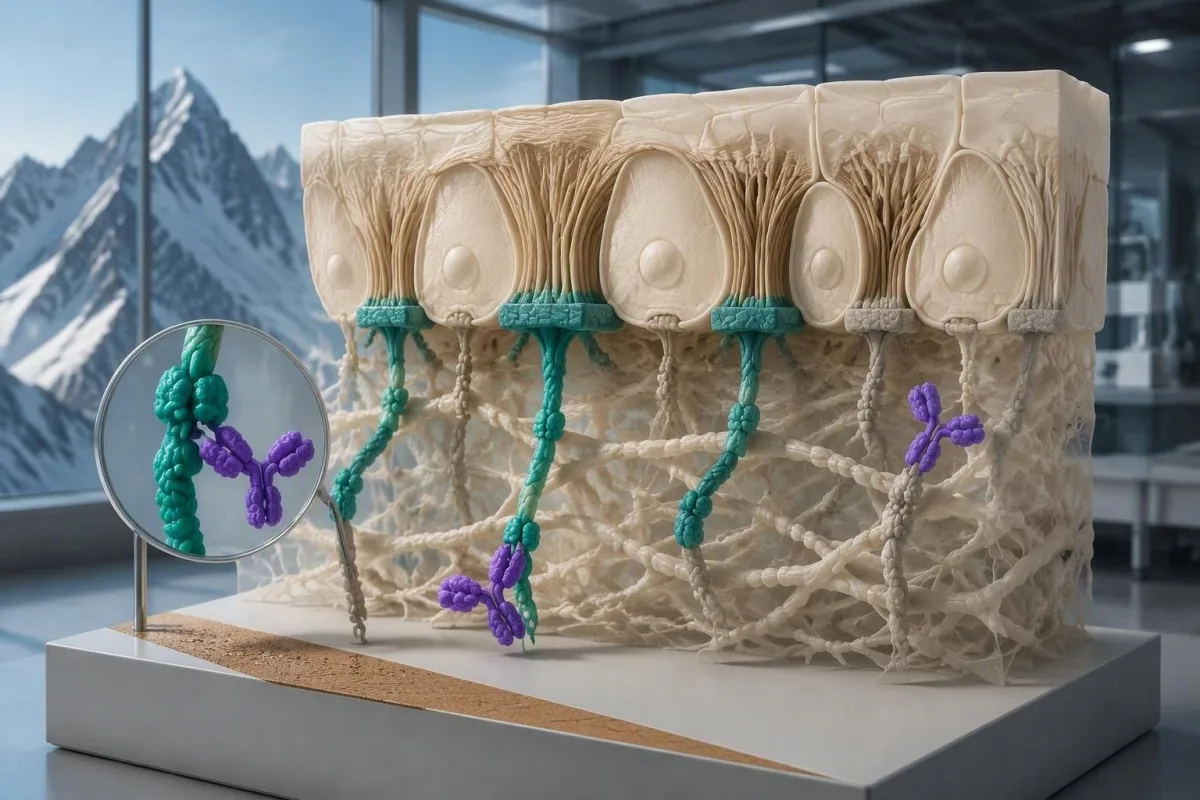
A negative direct-immunofluorescence result is less reassuring if the sample was poorly selected or incorrectly transported. Treatment before sampling may also influence findings, so clinicians need to know which creams or systemic medicines were used and where; never stop prescribed treatment solely to make a test more likely to turn positive.
A hypothetical person with characteristic new blisters, BP180 of 80 U/mL, and a negative direct-immunofluorescence specimen from an old eroded site may need repeat sampling. The same antibody result with no compatible skin findings demands a different investigation; the discrepancy does not have one automatic explanation.
A negative BP180 NC16A result does not exclude every form of pemphigoid. Further testing may include BP230 antibodies, indirect immunofluorescence, or specialized assays for other antigens; our guide to negative myasthenia antibody tests illustrates why a limited antigen panel cannot exclude an entire autoimmune disease category.
Repeat testing should address a specific uncertainty rather than create an endless sequence of panels. Ask whether the next investigation is checking the same antigen by another method, examining an additional antigen, or correcting a tissue-sampling problem; each strategy answers a different diagnostic question.
Which other conditions can resemble BP180-positive pemphigoid?
BP180 antibodies can occur in other pemphigoid-spectrum conditions, including mucous membrane pemphigoid and pemphigoid gestationis. Eczema, scabies, and drug-related eruptions can resemble early bullous pemphigoid clinically, while pemphigus involves different adhesion proteins and usually a different level of skin separation.
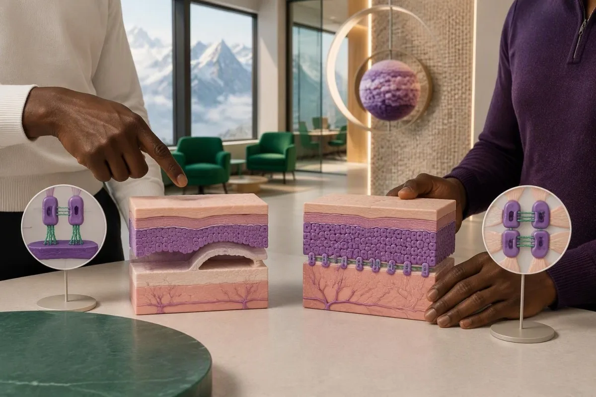
Bullous pemphigoid typically causes tense blisters because separation occurs beneath the epidermis. Pemphigus often produces more fragile blisters or erosions through loss of adhesion within the epidermis; our desmoglein antibody guide explains why BP180 testing cannot substitute for a pemphigus evaluation.
Predominantly mouth, eye, genital, or other mucosal involvement raises concern for mucous membrane pemphigoid, especially when scarring occurs. Eye pain, light sensitivity, or vision changes require urgent assessment because waiting for a repeat antibody concentration can delay treatment of a sight-threatening process.
Pemphigoid gestationis is associated with pregnancy and can produce BP180 antibodies. Pregnancy or recent childbirth therefore changes the interpretation of the same laboratory result; the distinction requires obstetric and dermatological coordination rather than assuming that every BP180-positive blistering disorder is classic older-adult bullous pemphigoid.
Scabies can cause intense itching and may coexist with an unrelated positive antibody result. Household itching, characteristic distribution, and burrows alter the differential; our scabies symptom guide is useful for that comparison, but neither household exposure nor BP180 positivity independently settles the diagnosis.
Do medicines change BP180 interpretation or treatment decisions?
Medication history can change the likelihood and presentation of bullous pemphigoid, but a positive BP180 result alone is not a reason to stop a medicine. DPP-4 inhibitors used for diabetes and immune checkpoint inhibitors are recognized associations; treatment decisions depend on confirmed disease, severity, and the person's other health risks.

DPP-4 inhibitor-associated pemphigoid can have a less typical inflammatory presentation and may show limited NC16A reactivity. This is not universal, and exposure does not prove causation; bring a complete medicine list, including start dates, to a review using our senior medication effects guide as a preparation aid.
The 2022 EADV guideline recommends potent topical corticosteroids as a mainstay when feasible and discusses oral prednisone at approximately 0.5 mg/kg/day as an alternative in suitable circumstances (Borradori et al., 2022). These are clinician-directed treatment options for disease, not instructions to treat an isolated antibody result or begin steroids without assessment.
Systemic corticosteroids can create additional risks that are unrelated to the BP180 concentration. People with relevant residence or travel exposure may need assessment for Strongyloides before significant immunosuppression; our Strongyloides antibody explanation describes why an apparently unrelated parasite history can become a treatment-safety issue.
Kidney function, glucose, infection risk, and the ability to apply topical treatment may influence the choice of therapy more than whether BP180 is 40 or 80 U/mL. Our kidney function interpretation guide covers supportive laboratory assessment, but renal markers do not confirm pemphigoid.
Can BP180 levels monitor disease activity and recovery?
BP180 antibody levels can support monitoring in confirmed bullous pemphigoid, but they do not replace examination or determine treatment alone. Concentrations often track disease activity at a group level; individual patients can remain antibody-positive while their skin is controlled or experience symptoms without a proportionate rise.
A falling BP180 concentration is most useful when it agrees with fewer new blisters and less itching. A hypothetical decline from 120 to 35 U/mL on the same assay supports an improving pattern, but it does not independently establish remission or justify stopping treatment.
An increase from 12 to 14 U/mL near a cutoff may reflect analytical or biological variation rather than a meaningful change in disease. There is no universal percentage increase that proves relapse across all BP180 assays; the method, timing, symptoms, and previous response need to be considered together.
Kantesti is an AI blood test interpretation platform that can organize serial BP180 results without treating a laboratory trend as a diagnosis. Our AI technology explanation describes the interpretation workflow; preserving each report's assay name and reference interval matters when comparing two or more measurements.
Changing laboratories can create an apparent rise or fall that is partly methodological. Our medication-safety trend guide explains why dates, doses, and laboratory context belong beside the numbers; photograph new skin findings and record new-blister counts when your dermatologist requests them.
What should you bring to follow-up, and which evidence applies?
Bring the full BP180 report, any BP230 result, symptom photographs, and your complete medication list to follow-up. These four information groups help a clinician decide whether you need observation, additional serum testing, properly handled skin samples, or urgent assessment rather than another isolated antibody measurement.
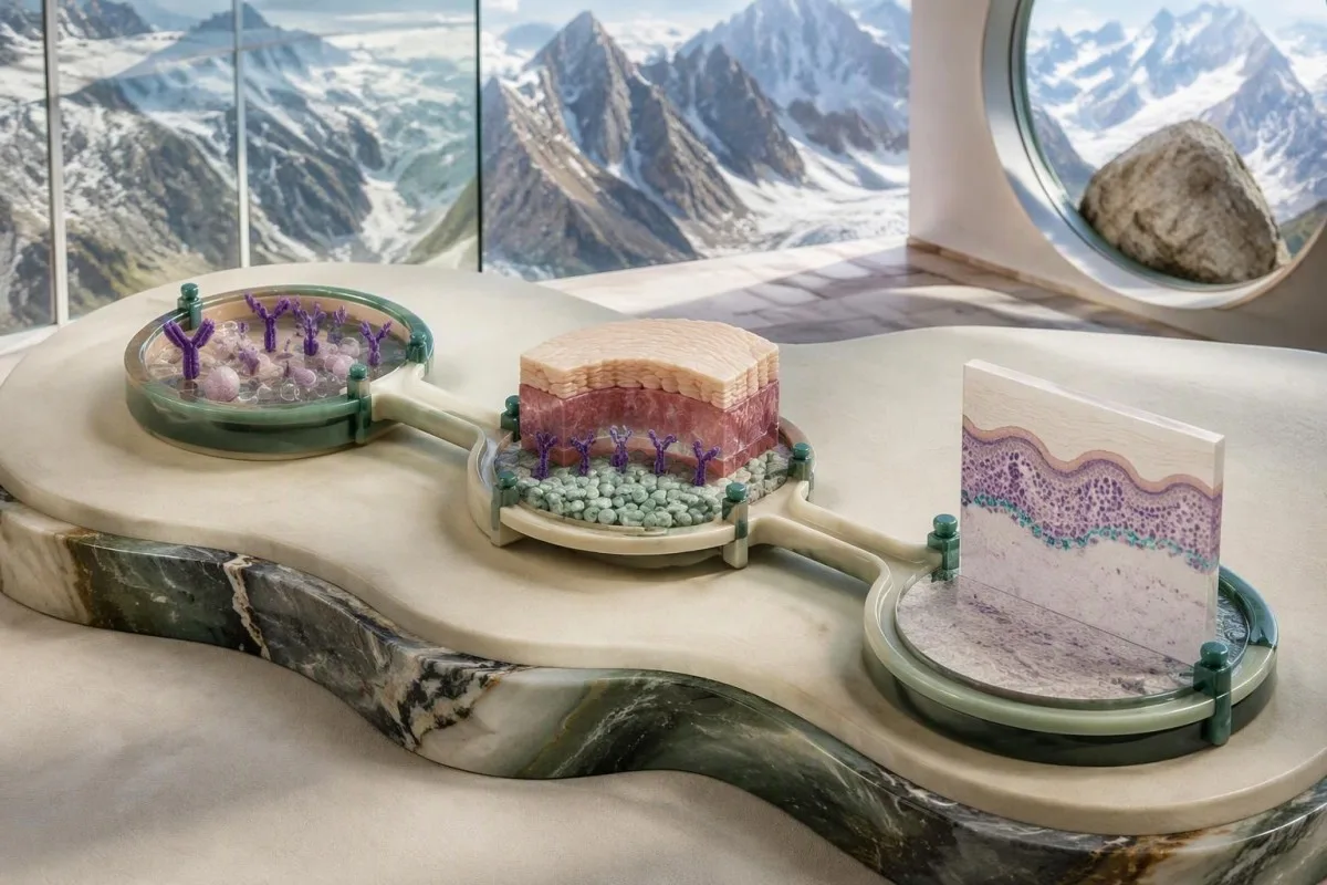
A useful report includes the exact assay name, numerical value, units, cutoff, and collection date. If a hypothetical BP180 result reads 16 U/mL, the missing cutoff may reverse whether it is considered positive; also request the actual histology and direct-immunofluorescence reports rather than relying on the phrase 'skin biopsy normal.'
New eye symptoms, swallowing difficulty, extensive rapidly spreading blisters, fever, or inability to maintain fluids need prompt clinical attention. Do not wait for a scheduled second test when those symptoms appear; supportive urinalysis can answer separate medical questions, as explained in our urinalysis interpretation guide, but it cannot establish a pemphigoid diagnosis.
Kantesti helps explain the distinctions between a flagged antibody result, a probable diagnosis, and confirmed disease. Our responsible AI principles describe the boundaries of educational interpretation; only a clinician with access to your examination and appropriate investigations can decide whether a BP180 antibody positive result represents active pemphigoid.
The directly relevant evidence here comes from the 2022 EADV guideline and the 2019 JAMA Dermatology diagnostic study. The two Zenodo publications listed below concern kidney-function and urinalysis interpretation and are included as background educational publications, not evidence that BP180 confirms bullous pemphigoid or that our AI has validated diagnostic accuracy for this condition.
Frequently asked questions
Does a positive BP180 antibody test mean I definitely have bullous pemphigoid?
A positive BP180 antibody test does not definitely mean you have bullous pemphigoid. Some laboratories use a cutoff near 9 U/mL and others near 20 U/mL, so the exact assay and reference interval matter. Compatible itching or blisters make the result more meaningful, while an isolated low positive may be incidental or falsely positive. Skin biopsy with direct immunofluorescence and additional antibody testing may be needed to establish the diagnosis.
What does a low-positive BP180 result mean if I have no blisters?
A low-positive BP180 result without blisters can reflect incidental antibody detection, assay-related false positivity, or pemphigoid presenting without blisters. For example, 12 U/mL is positive under a 9-U/mL cutoff but cannot predict whether an asymptomatic person will develop disease. Persistent itching or an unexplained rash deserves dermatological assessment even without blisters. There is no universal repeat-testing interval or treatment recommendation based solely on an isolated low-positive result.
Can I have bullous pemphigoid with negative BP180 antibodies?
Bullous pemphigoid can occur despite negative BP180 antibodies on a routine NC16A assay. The assay tests a particular region of the approximately 180-kDa protein and may not detect antibodies directed mainly against other regions. BP230 testing, direct immunofluorescence, and indirect immunofluorescence may provide additional evidence. A negative blood test should not end the evaluation when the skin findings strongly suggest pemphigoid.
Do negative BP230 antibodies rule out bullous pemphigoid?
Negative BP230 antibodies do not rule out bullous pemphigoid. BP230 is an approximately 230-kDa intracellular anchoring protein, and some people with confirmed disease do not have detectable antibodies against it. A positive BP180 result with negative BP230 antibodies can still fit the diagnosis when the clinical and tissue findings agree. The two antibody concentrations should not be added together or treated as a two-test confirmation rule.
Why are two skin biopsies sometimes needed for pemphigoid?
Two skin biopsies are often needed because routine histology and direct immunofluorescence answer different diagnostic questions. A small 3–4-mm punch is commonly used, although the dermatologist chooses the technique and site for the individual presentation. One sample examines tissue structure, while the other looks for immune deposits along the basement membrane. The direct-immunofluorescence specimen requires its own agreed transport method and should not be routinely placed in formalin.
Does a high BP180 level mean my pemphigoid is severe?
A high BP180 level does not independently determine how severe pemphigoid is or whether it is an emergency. A result of 100 U/mL must be interpreted using that assay's cutoff and alongside the extent of skin involvement, itching, new blisters, and general health. Antibody levels often relate to disease activity, but individual results and symptoms can disagree. Eye pain, vision changes, difficulty swallowing, or rapidly spreading extensive blistering require prompt assessment regardless of the antibody concentration.
Should BP180 antibodies become negative before treatment is stopped?
BP180 antibodies do not always need to become negative before a clinician considers reducing or stopping pemphigoid treatment. A hypothetical fall from 120 to 35 U/mL may support improvement, but symptoms, new-blister formation, examination, and relapse history guide the decision. Antibodies can remain detectable when the skin is controlled, and there is no universal stopping cutoff across assays. Do not change prescribed treatment based only on an antibody trend.
Get your blood test results explained
Upload your lab report and Kantesti's AI explains each biomarker in plain language within seconds. Used by more than 2 million people worldwide.
References
- Klein, T., Mitchell, S., & Weber, H. (2026). BUN/Creatinine Ratio Explained: Kidney Function Test Guide. Kantesti AI Medical Research. DOI
- Klein, T., Mitchell, S., & Weber, H. (2026). Urobilinogen in Urine Test: Complete Urinalysis Guide 2026. Kantesti AI Medical Research. DOI
- Borradori L et al. (2022). Updated S2 K guidelines for the management of bullous pemphigoid initiated by the European Academy of Dermatology and Venereology (EADV). Journal of the European Academy of Dermatology and Venereology. DOI
- Meijer JM et al. (2019). Assessment of Diagnostic Strategy for Early Recognition of Bullous and Nonbullous Variants of Pemphigoid. JAMA Dermatology. DOI
- 2M+Tests analysed
- 127+Countries
- 100+Languages
About the medical team


E-E-A-T Trust Signals
Experience
Physician-led clinical review of lab interpretation workflows.
Expertise
Laboratory medicine focus on how biomarkers behave in clinical context.
Authoritativeness
Medically reviewed by Dr. Thomas Klein, with contributions from Dr. Sarah Mitchell and Prof. Dr. Hans Weber.
Trustworthiness
Evidence-based interpretation with clear follow-up pathways to reduce alarm.


Medical disclaimer
This article is for educational purposes only and does not constitute medical advice. Always consult a qualified healthcare provider for diagnosis and treatment decisions.
Published · Contact the editorial team
Published by Kantesti Ltd, registered in England & Wales, company no. 17090423, London, United Kingdom.
