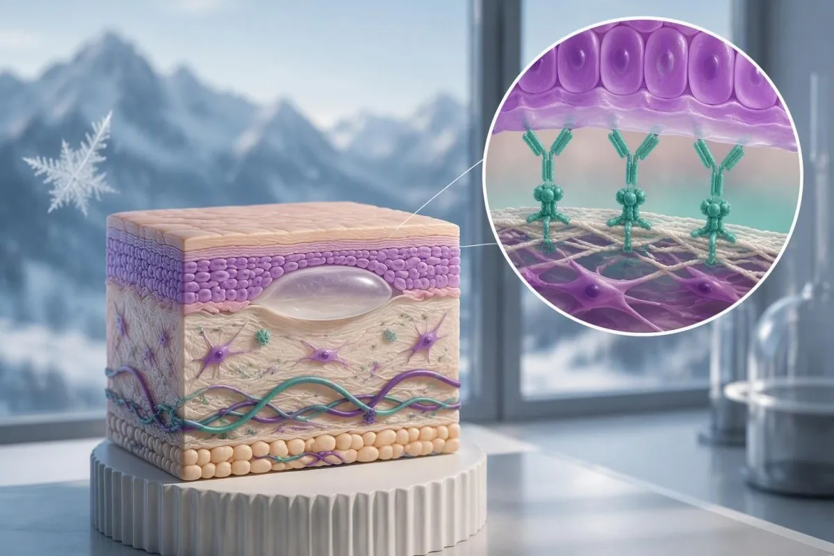
Dodatni wynik BP180 przemŏ za autoimmunologicznym schorzym skōry, ale sama liczba niy może postawić diagnozy. Objawy, użyty test i prawidłowo pobrane próbki skōry decydujō, co bydzie dalij.

Najważniejsze informacje
- Dodatnie przeciwciała BP180 znaczō, że test wykrył przeciwciała skierowane przeciwko celowi BP180; samodzielnie niy potwierdzo to pemfigoidu pęcherzowego.
- Progi laboratoryjne sie rōżniō: niykere testy BP180 używajō wartości ôkoło 9 U/mL, a inksze ôkoło 20 U/mL. Sprawdź zakres referyncyjny swojigo laboratorium.
- Niskie dodatnie wyniki bez świyrbiynio, wysypki abo pęcherzy może ôznaczać przypadkowe wykrycie przeciwciał abo fałszywie dodatni wynik testu — niy potwierdzōne schorzenie skōry.
- Pemfigoid bezpęcherzowy może powodować uporczywe świyrbiynie bez pęcherzy; 74 z 343 przypadkōw pemfigoidu w specjalistycznym badaniu diagnostycznym było bezpęcherzowych.
- Przeciwciała BP230 dostarczajō uzupełniajōcych dowodōw, ale ujemne przeciwciała BP230 niy wykluczajō pemfigoidu pęcherzowego.
- Bezpośrednio immunofluorescyncyjo we ôbie skórne złogi przeciwciał; liniowe IgG i/albo C3 wzdłuż błōny podstawnyj przemŏwiajōm za chorobōm pemfigoidowōm.
- Dwie prōbki skórne sōm czynsto pobiyrane: prōbka z zajyntyj okolicy do rutynowyj mikroskopije i prōbka z pobliskiyj, nojlepi dobranyj okolicy do bezpośrednij immunofluorescyncyje.
- Pilne objawy należōm do nich ból ôka, zmiany widzynio, trudości ze połykaniym, gorōnczka przy rozległych zmianach skórnych abo wartko rozszerzajōnce sie pōmply; niy czekej na kolejny wynik przeciwciał.
Czy dodatni wynik BP180 potwierdzo pemfigoid pęcherzowy?
Niy — pozytywny wynik przeciwciał BP180 niy potwierdzo sam ôd sia pemfigoidu pęcherzowego. Wspiyro diagnoza, kej ôbjawy i znaleziska skórne do sia pasujōm, ale słabo pozytywny wynik bez pōmply może być przipadkowy; dermatolodzy czynsto potrzebujōm biopsyje skórnyj z bezpośredniōm immunofluorescyncyjōm, a czasym tyż dodatkowych badań przeciwciał, coby ustalić przyczynã.
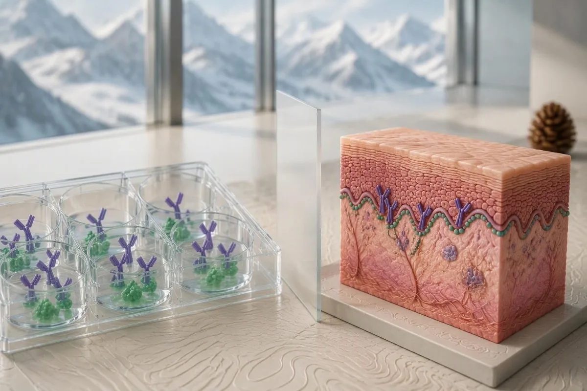
Diagnoza pemfigoidu pęcherzowego wymŏgo zgodności klinicznyj i immunopatologicznyj, a niy ino jednyj ôznaczōnyj wartości laboratoryjnyj. Wytyczne EADV z 2022 roku ôpisujōm diagnoza poprzez ôbraz kliniczny i ôdpowiednie badania tkanki oraz krwi pod kōntem przeciwciał (Borradori et al., 2022); praktyczno kwestijo je, eli wykryte przeciwciała wyjaśniajōm stan skórnyj ôsoby.
Pozytywny test na przeciwciała i aktywno choroba autoimmunologiczno to inksze znaleziska. Na przikłod hipotetyczny wynik 12 U/mL może być pozytywny w jednym laboratorium, ale bezôbjawowo ôsoba z takim wynikiym mo inksze prawdopodobiyństwo diagnostyczne niż ktoś z rozległymi, swyndzōncymi, naprężōnymi pōmplami i takōm samōm koncentracyjōm.
Kantesti je analizatorōm krwi ze sztucznōm inteligyncyjōm, co pōmogo wyjaśnić wyniki BP180 społu z progiym laboratorium i informacyjami, kere podŏwŏsz. Nasze wyjaśniynie niy może ôbejrzeć twojyj skōry ani wykōnać bezpośrednij immunofluorescyncyje; nasze tła firmy ôznaczo ôrganizacyjo, kerŏ stoi za tym wsparciym edukacyjnym.
Traktuj wynik jako powōd, coby zadać bardzij konkretnŏ pytanie, a niy jako stałŏ etykieta choroby. Nasz przewodnik ô pozytywnych wyników przeciwciał wyjaśnio, czamu wykrycie przeciwciał, aktywność choroby i potwierdzynie diagnozy to trzë ôsobne pojyncia.
Co tak właściwie mierzi test na przeciwciała BP180?
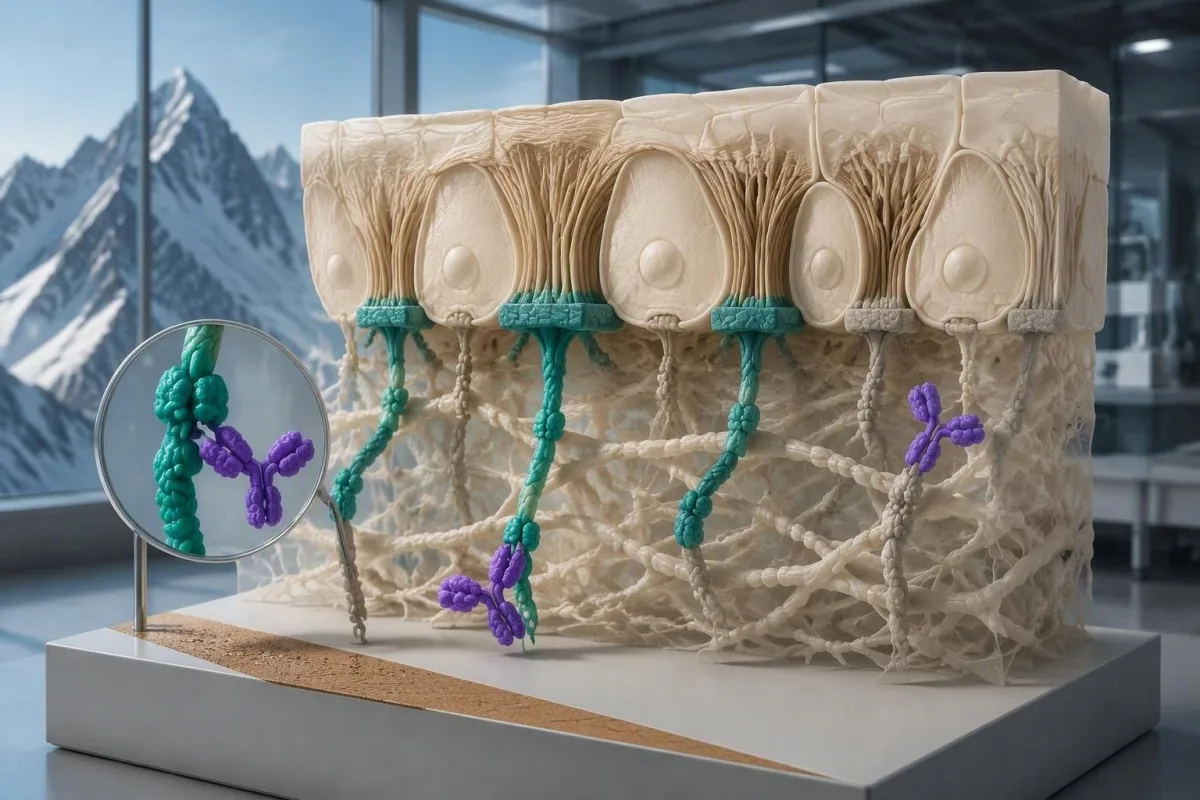
Test na przeciwciała BP180 zwykle mierzi surowicze IgG skierowane przeciwko czynści BP180, zwanego tyż kolagenym typu XVII. BP180 je białkiym ô masie ôkoło 180 kDa , kere bierze udział w kotwiczeniu naskórka do leżōncyj pod nim tkanki; w wielu rutynowych testach bada sie jego region NC16A, a niy cołki białko.
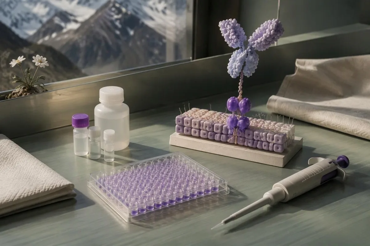
BP180 je strukturalnym białkiym skōry, a niy substancyjōm, kerej stynżynie we krwi sie mierzi. Wynik opisuje przeciwciała przeciwko tymu białku, zwykle z użyciym testu immunoenzymatycznego, czyli ELISA; wartość 30 U/mL niy ôznaczo tōm samym, że we krwi krōnżuje 30 jednostek białka skōry.
Te dōmyna NC16A sii blisko błōny kōrkowyj ôd zewnyntrznyj strōny BP180 i je czynstym cełym prziciwciał we klasycznym pemfigoidzie pyncyrzowym. Badanie tego ôbrzymu robi praktyczny test możebnym, ale ôtwiyro ślepy plac: prziciwciała skierowane głōwnie przeciw inkszym ôbrzymōm mogōm niy być wykryte.
Ilościowe stynżynie prziciwciał niy je wymienialne z mianym immunofluorescyncyjnym. Wynik ôpisany jako 1:40 ôznaczo reaktywność przi rozcieńczyniu, kej 40 U/mL ôznaczo stynżynie skalibrowane do testu; nasze ôbjaśniynie testōw ilościowych a jakościowych pōmogo rozrōżnić te formaty raportowaniŏ.
Sprawdź, eli w raporcie je napisane BP180 NC16A IgG, inkszy test BP180 abo szerszy panel pemfigoidowy. Te testy niy sōm z automatu rōwnowartościowe, a ujemny ELISA IgG niy ôdpowiado na kożde pytanie ô inksze klasy prziciwciał abo inksze cele w błōnie podstawnyj.
Co uznŏwŏ sie za słabo dodatni wynik przeciwciał BP180?
Nisko-dodatni wynik BP180 to wartość ino trocha nad progiym ôdczytu dlŏ użytego testu. Niy ma uniwersalnego zakresu nisko-dodatniego: niykiere laboratoria używajōm progu blisko 9 U/mL, a inksze blisko 20 U/mL, ze ôdmiennymi kalibracyjami a kategoriami raportowaniŏ.
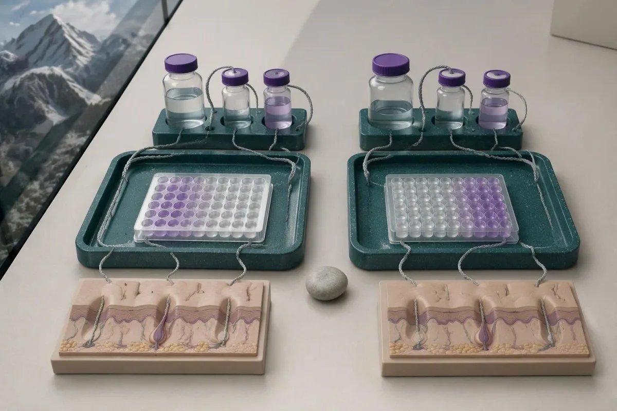
Jednostki testu BP180 niy sōm uniwersalnie standaryzowane miyndzy laboratoriami. Wynik 15 U/mL może być dodatnie przi zasadzie 9-U/mL, a ujemne przi zasadzie 20-U/mL, ale to niy dowodzi, iże drugi test niy wykrył choroby; metody a kalibratory mogōm sie rōżnić.
Progi rōwnoważōm czułość ze swoistościōm we populacyjach użytych do ôceny testu. Ôpuścynie progu może wykryć wiyncyj ôsōb z chorobōm, ale tyż wskŏzać wiyncyj ôsōb bez choroby; prōg je granicōm decyzyjnōm laboratorium, a niy biologicznym punktym, kaj skōra nagle zaczyno sie robić pyncyrzowato.
Kantesti AI może pōmōc zestawić podanōm wartość BP180 z dostarczōnym zakresym referyncyjnym, ale ôbrŏz uciynty tak, iże pokoże ino 18 U/mL ôstawio ważne informacyje. Podaj nazwa testu, jednostki, kategorie interpretacyje a uwagi laboratorium; ôgōlny oznaczenie poza zakresem niy może zastōmpić tych szczegōłōw.
Pōniższe przikłady ôbrŏzujōm dwie możliwe polityki raportowaniŏ, a niy uniwersalnōm skŏlã ciynżkości. Samo stãżynie przeciwciał BP180 niy ôkryślo nagłego przipadku medycznego; rozległe zajyncie skōry, ôdwodniynie, choroba układowŏ oraz ôbjŏwy ôczne abo drōg ôdechowych ôkryślajōm pilność.
| Poziōm | Zakres | Co to znaczy |
|---|---|---|
| Przikładowy test A: pōniżyj progu ôdcinku | <9 U/mL | Wynik ujemny wedle tyj ôbrazowyj polityki laboratoryjnyj; choroba niy je wykluczonŏ, kej kliniczne podejrzynie je durch wysokie. |
| Przikładowy test A: na abo nad progiym ôdcinku | ≥9 U/mL | Wynik dodŏtni wedle tyj polityki; ôdległość ôd progu ôdcinku i wyniki kliniczne wpływajōm na interpretacyjõ. |
| Przikładowy test B: pōniżyj progu ôdcinku | <20 U/mL | Wynik ujemny wedle inkszyj ôbrazowyj polityki; niy przeliczŏj bezpośrednio miyndzy systemami testowymi. |
| Przikładowy test B: na abo nad progiym ôdcinku | ≥20 U/mL | Wynik dodŏtni wedle tyj polityki; ta liczba sama niy ôstŏwia diagnozy ani ciynżkości. |
Czamu dodatni wynik BP180 może mylić?
Dodŏtni wynik BP180 je mynij przekōnywajōncy, kej ta ôsoba miała małõ klinicznõ szansa na pemfigoid przed badaniym. Nawet dość dokładny test daje wyniki fałszywie dodŏtnie, tōż znaczynie wyniku zmiyniŏ sie miyndzy bezobjŏwowo populacyjōm badanōm przesiewowo a poradniōm specjalistycznōm, kaj ôceniajōm charakterystyczne pōcherze.
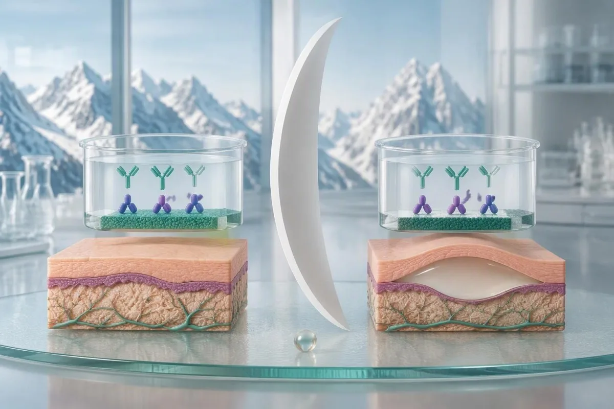
Czułość i swoistość niy sōm tym samym co prawdopodobiyństwo, że ôsobiście mosz choroba po wyniku dodŏtnym. To prawdopodobiyństwo zależy tyż ôd tego, jak czãsto wystympuje ta choroba we badanyj grupie; to rozrōżniynie je nojważniyjsze, kej panel przeciwciał zamōwiyło sie szeroko, a niy po to, coby zbadać konkretny wzōr zmian skōrnych.
Rozważmy jasno hipotetyczny test ze czułościōm 90% i swoistościōm 95%, użyty u 1 000 ôsōb, u kierch prawdopodobiyństwo choroby wynosi 1%. Wykryłby ôn ôkoło 9 prawdziwie dodŏtnich i 50 fałszywie dodŏtnich wynikōw, co dawałoby dodatniōm wartość predykcyjnõ ôkoło 15%; to sōm założynia do nauki, a niy ôświadczenia ô skuteczności twojigo testu BP180.
Użyj tego samego hipotetycznego testu, kej prawdopodobiyństwo choroby wynosi 20%, and approximately 180 true positives and 40 false positives would occur per 1,000 tests. The positive predictive value becomes about 82%—a large change caused by clinical context, not by a different laboratory method.
These examples explain why a dermatologist may request tissue testing despite a positive result, or take characteristic symptoms seriously despite a negative one. Our discussion of interpretacyje przeciwciał MOG illustrates the same probability principle in another autoimmune condition, although its diagnostic criteria cannot be transferred to pemphigoid.
Co, jeśli BP180 je dodatni, ale niy mōsz pęcherzy?
A low-positive BP180 result without blisters does not establish bullous pemphigoid or reliably predict future blistering. The next distinction is whether you have no skin symptoms at all, or persistent itching and an unexplained rash that could represent a nonbullous pemphigoid presentation.

Incidental BP180 antibody detection can occur without clinically established pemphigoid. Possible explanations include low-level autoreactivity and assay-related false positivity; there is no validated conversion rule that turns an isolated result of 11 U/mL into a percentage risk of developing blisters later.
A hypothetical person with 11 U/mL, no itching, no rash, and no mucosal symptoms usually needs a contextual review rather than automatic immunosuppressive treatment. The clinician should check why the assay was ordered, whether the result was unexpected, and whether a skin examination reveals findings the person had not recognized.
Persistent dryness and itching still deserve assessment because common explanations remain common. Eczema, medication effects, thyroid disease, and other causes may be more likely than pemphigoid; our dry skin testing guide explains why additional blood tests should follow symptoms rather than a blanket panel.
Nie ma no universal repeat-testing interval for an asymptomatic low-positive BP180 result. A clinician may choose observation, a repeat using the same method, or specialist review depending on the history; new persistent itching, urticarial plaques, or blisters should change that plan rather than waiting for a scheduled repeat.
Czy pemfigoid pęcherzowy może sie zaczōńć samym świyrbiyniym?
Pemphigoid can present with persistent itching, eczema-like patches, urticarial plaques, or scratch-related changes without visible blisters. In a specialist diagnostic study, 74 of 343 pemphigoid cases—21.6%—were nonbullous, showing that the absence of blisters cannot independently exclude the disorder (Meijer et al., 2019).

Nonbullous pemphigoid is not simply every itchy rash with a positive BP180 result. Diagnosis still needs compatible symptoms and appropriate immunopathological evidence; the 21.6% study figure came from a referred population and should not be interpreted as the probability that any person with itching has pemphigoid.
The symptom pattern often matters more than whether one small blister has appeared. Persistent, widespread itching in an older adult, especially with recurrent raised plaques or poor response to ordinary eczema care, warrants a dermatological assessment; our itchy skin laboratory guide separates skin-directed testing from tests for systemic causes.
Eosinophilia can accompany bullous pemphigoid, but it is neither required nor diagnostic. An absolute eosinophil count above approximately 0,5 × 10^9/l is often flagged in adults, yet allergy, medications, and parasites can produce the same finding; our guide to wysokich liczbach eozynofilów explains those alternatives.
A dermatologist does not always need to wait for a blister before collecting a useful sample for direct immunofluorescence. In nonbullous disease, sample selection is tailored to the available rash and laboratory protocol; document the distribution and duration of itching, because a disappearing rash can make later assessment harder.
Jak przeciwciała BP230 uzupełniajō badanie BP180?
BP230 antibodies provide complementary evidence for pemphigoid but cannot confirm or exclude bullous pemphigoid alone. BP230 is an approximately 230-kDa intracellular anchoring protein, whereas BP180 crosses the cell membrane; their different locations and antibody targets help explain why the two results can disagree.
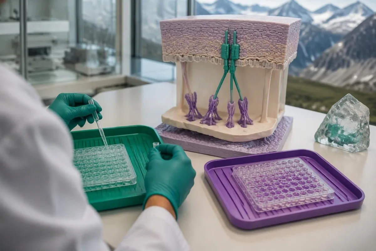
A negative BP230 antibody result does not cancel a positive BP180 result. Some people with confirmed pemphigoid have antibodies detectable against only one tested antigen, and each assay has its own sensitivity; interpreting the pair as a two-test pass-or-fail rule creates avoidable diagnostic errors.
Przikładowo, osoba BP180 result of 45 U/mL with negative BP230 antibodies still warrants assessment if tense blisters and severe itching are present. Conversely, isolated low-positive BP230 antibodies without compatible findings are not enough to diagnose pemphigoid; a second positive marker strengthens a pattern only when the overall evidence fits.
Kantesti is an AI biomarker interpretation platform that can explain BP180 and BP230 antibodies as complementary findings rather than interchangeable scores. This is similar to the reasoning used for antibody panels in connective disease, but skin-specific tissue findings remain central to pemphigoid assessment.
Do not add BP180 and BP230 concentrations together, even if both are expressed in U/mL. They represent different antigen-specific measurements with different calibration systems; BP180 levels generally have a better-established relationship with disease activity than BP230 levels, but neither replaces examination.
Czamu może być potrzebno biyopsjo skōry z bezpośredniō immunofluorescyncjō?
Skin biopsy with direct immunofluorescence can show whether antibodies or complement are deposited at the skin's basement membrane. Dermatologists often collect two separate specimens because routine microscopy and direct immunofluorescence answer different questions and require different sample handling.
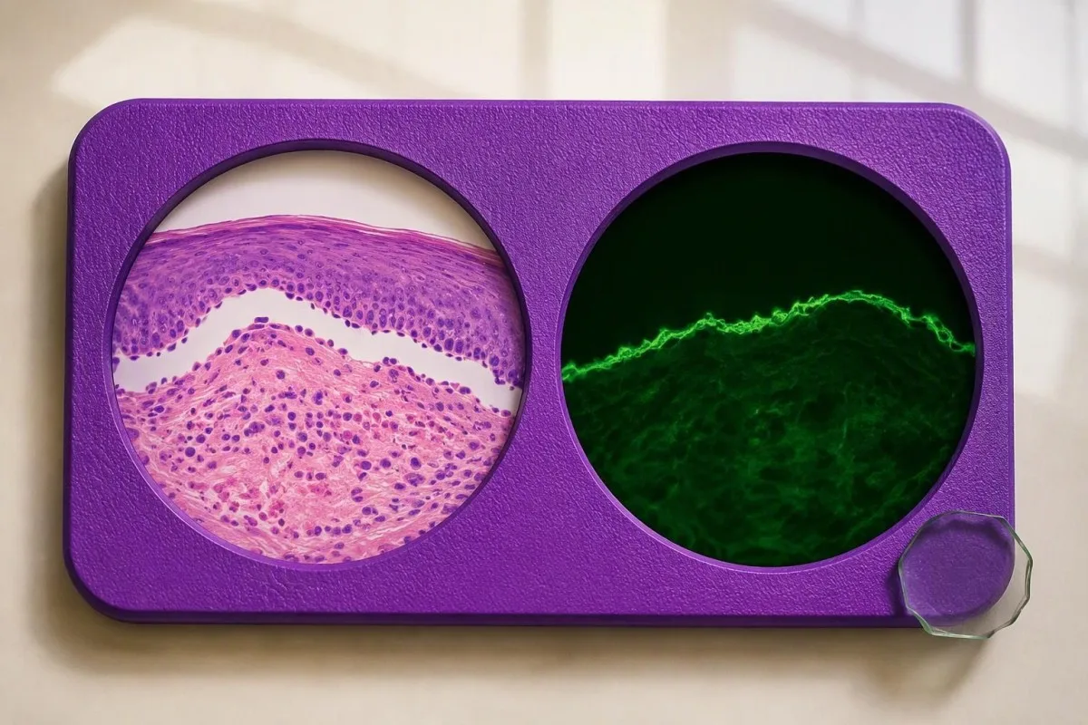
Routine histology examines tissue structure and inflammatory cells. A sample from a fresh blister or suitable affected area may show separation beneath the epidermis and eosinophils, but these features are not unique to bullous pemphigoid; the second sample looks for the immune-deposit pattern that helps classify the disorder.
For classic blistering presentations, the direct-immunofluorescence sample is commonly taken from appropriately selected skin near a fresh blister rather than its eroded center. A small 3–4-mm punch is often used, although technique and location depend on the case; sampling heavily damaged tissue can remove the structures the laboratory needs to examine.
The specimen for direct immunofluorescence must not be routinely placed in formalin. Laboratories generally require a dedicated transport medium or another explicitly agreed handling method, while the routine histology specimen commonly goes into formalin; ask whether the two containers have been prepared for their separate purposes.
In Meijer et al.'s 2019 diagnostic cohort, direct immunofluorescence had a sensitivity of 88.3%, so even this valuable test was not infallible. Kantesti's informacje o walidacji klinicznej concerns our interpretation methodology—not a claim that AI can reproduce skin immunofluorescence or establish its findings.
Co znaczō dodatnie wyniki immunofluorescyncji i testu rozdziału solō?
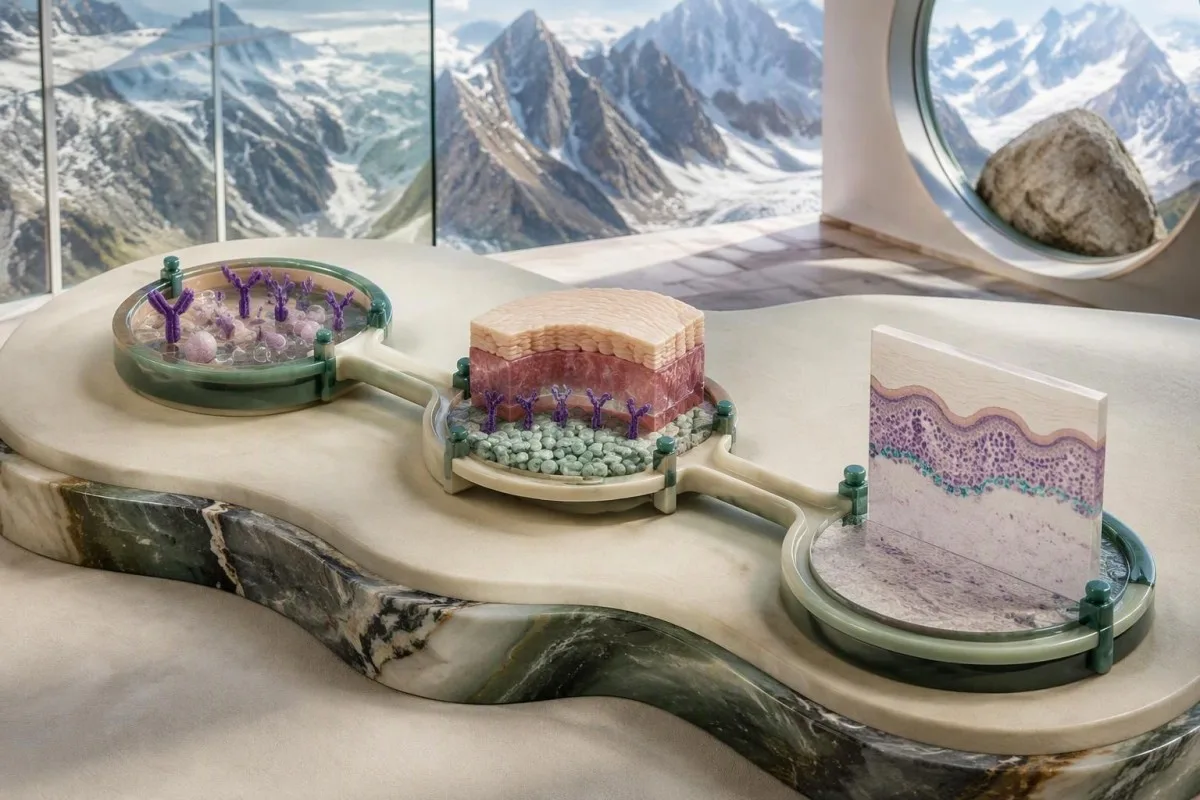
Linear IgG and/or C3 along the basement membrane on direct immunofluorescence supports a pemphigoid-spectrum disorder, but the pattern is not unique to bullous pemphigoid. Indirect immunofluorescence on salt-split skin can add evidence by showing where circulating antibodies bind relative to an experimentally created tissue separation.
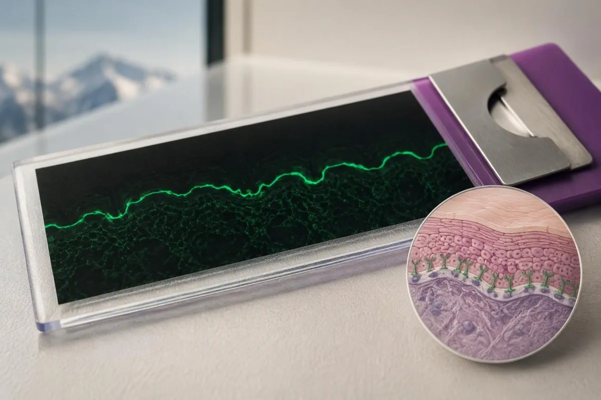
Direct and indirect immunofluorescence use different materials. Direct testing examines deposits already present in the person's skin, while indirect testing applies the person's serum to a laboratory substrate; a positive result in one test and a negative result in the other is therefore possible without a reporting error.
On salt-split skin, bullous pemphigoid antibodies commonly bind the epidermal side, often called the roof. Antibodies binding the dermal side, or floor, raise consideration of other disorders such as epidermolysis bullosa acquisita or anti-p200 pemphigoid, but binding location alone does not fully classify every case.
Meijer et al. reported 99.9% specificity for indirect immunofluorescence on salt-split skin in their 2019 diagnostic cohort. That high figure applies to that study and testing pathway; a negative result still cannot exclude pemphigoid because specificity describes false positives, not how many cases the test misses.
C3 detected in skin is not the same measurement as a serum C3 concentration. A normal circulating complement result does not negate local deposits at the basement membrane; our complement testing explanation helps prevent this common cross-test misunderstanding.
Co, jeśli BP180, biypsjo skōry i objawy sie niy zgadzajō?
Discordant BP180 and skin-test results require a review of the clinical picture, assay target, sampling site, and specimen handling. A positive ELISA with negative direct immunofluorescence can reflect incidental antibodies or an uninformative skin sample; a negative NC16A ELISA can miss disease directed against other targets.
A negative direct-immunofluorescence result is less reassuring if the sample was poorly selected or incorrectly transported. Treatment before sampling may also influence findings, so clinicians need to know which creams or systemic medicines were used and where; never stop prescribed treatment solely to make a test more likely to turn positive.
A hypothetical person with characteristic new blisters, BP180 of 80 U/mL, and a negative direct-immunofluorescence specimen from an old eroded site may need repeat sampling. The same antibody result with no compatible skin findings demands a different investigation; the discrepancy does not have one automatic explanation.
A negative BP180 NC16A result does not exclude every form of pemphigoid. Further testing may include BP230 antibodies, indirect immunofluorescence, or specialized assays for other antigens; our guide to negative myasthenia antibody tests illustrates why a limited antigen panel cannot exclude an entire autoimmune disease category.
Repeat testing should address a specific uncertainty rather than create an endless sequence of panels. Ask whether the next investigation is checking the same antigen by another method, examining an additional antigen, or correcting a tissue-sampling problem; each strategy answers a different diagnostic question.
Ktore inksze schorzenia mogō przypominać pemfigoid z dodatnim BP180?
BP180 antibodies can occur in other pemphigoid-spectrum conditions, including mucous membrane pemphigoid and pemphigoid gestationis. Eczema, scabies, and drug-related eruptions can resemble early bullous pemphigoid clinically, while pemphigus involves different adhesion proteins and usually a different level of skin separation.
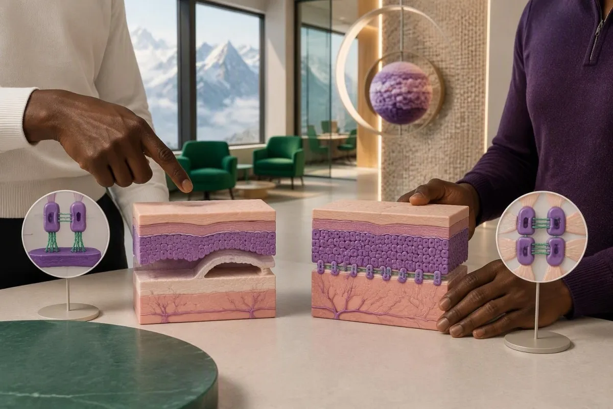
Bullous pemphigoid typically causes tense blisters because separation occurs beneath the epidermis. Pemphigus often produces more fragile blisters or erosions through loss of adhesion within the epidermis; our desmoglein antibody guide explains why BP180 testing cannot substitute for a pemphigus evaluation.
Predominantly mouth, eye, genital, or other mucosal involvement raises concern for mucous membrane pemphigoid, especially when scarring occurs. Eye pain, light sensitivity, or vision changes require urgent assessment because waiting for a repeat antibody concentration can delay treatment of a sight-threatening process.
Pemphigoid gestationis is associated with pregnancy and can produce BP180 antibodies. Pregnancy or recent childbirth therefore changes the interpretation of the same laboratory result; the distinction requires obstetric and dermatological coordination rather than assuming that every BP180-positive blistering disorder is classic older-adult bullous pemphigoid.
Scabies can cause intense itching and may coexist with an unrelated positive antibody result. Household itching, characteristic distribution, and burrows alter the differential; our scabies symptom guide is useful for that comparison, but neither household exposure nor BP180 positivity independently settles the diagnosis.
Czy leki zmieniajō interpretacjo BP180 abo decyzje ô leczeniu?
Medication history can change the likelihood and presentation of bullous pemphigoid, but a positive BP180 result alone is not a reason to stop a medicine. DPP-4 inhibitors used for diabetes and immune checkpoint inhibitors are recognized associations; treatment decisions depend on confirmed disease, severity, and the person's other health risks.

DPP-4 inhibitor-associated pemphigoid can have a less typical inflammatory presentation and may show limited NC16A reactivity. This is not universal, and exposure does not prove causation; bring a complete medicine list, including start dates, to a review using our senior medication effects guide as a preparation aid.
Te 2022 EADV guideline recommends potent topical corticosteroids as a mainstay when feasible and discusses oral prednisone at approximately 0.5 mg/kg/day as an alternative in suitable circumstances (Borradori et al., 2022). These are clinician-directed treatment options for disease, not instructions to treat an isolated antibody result or begin steroids without assessment.
Systemic corticosteroids can create additional risks that are unrelated to the BP180 concentration. People with relevant residence or travel exposure may need assessment for Strongyloides before significant immunosuppression; our Strongyloides antibody explanation describes why an apparently unrelated parasite history can become a treatment-safety issue.
Kidney function, glucose, infection risk, and the ability to apply topical treatment may influence the choice of therapy more than whether BP180 is 40 or 80 U/mL. Nasz poradnik interpretacjeji funkcji nyrōkōw covers supportive laboratory assessment, but renal markers do not confirm pemphigoid.
Czy poziōmy BP180 mogō monitorować aktywność choroby i powrōt do zdrowio?
BP180 antibody levels can support monitoring in confirmed bullous pemphigoid, but they do not replace examination or determine treatment alone. Concentrations often track disease activity at a group level; individual patients can remain antibody-positive while their skin is controlled or experience symptoms without a proportionate rise.
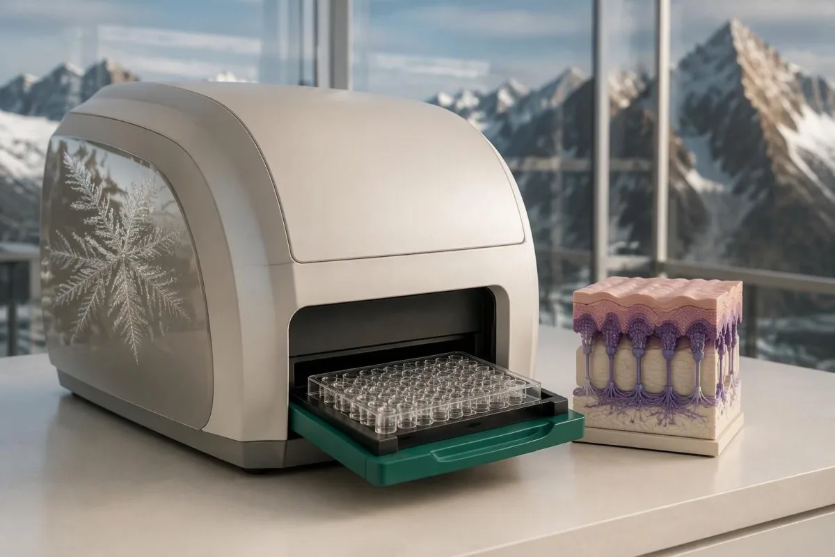
A falling BP180 concentration is most useful when it agrees with fewer new blisters and less itching. A hypothetical decline from 120 to 35 U/mL on the same assay supports an improving pattern, but it does not independently establish remission or justify stopping treatment.
An increase from 12 to 14 U/mL near a cutoff may reflect analytical or biological variation rather than a meaningful change in disease. There is no universal percentage increase that proves relapse across all BP180 assays; the method, timing, symptoms, and previous response need to be considered together.
Kantesti is an AI blood test interpretation platform that can organize serial BP180 results without treating a laboratory trend as a diagnosis. Nasz u wyjasniyniy technologii AI describes the interpretation workflow; preserving each report's assay name and reference interval matters when comparing two or more measurements.
Changing laboratories can create an apparent rise or fall that is partly methodological. Our przewodnik po trendach bezpieczeństwa leków explains why dates, doses, and laboratory context belong beside the numbers; photograph new skin findings and record new-blister counts when your dermatologist requests them.
Co prziniyść na wizyty kontrolne i kero dowody majō zastosowanie?
Bring the full BP180 report, any BP230 result, symptom photographs, and your complete medication list to follow-up. These four information groups help a clinician decide whether you need observation, additional serum testing, properly handled skin samples, or urgent assessment rather than another isolated antibody measurement.
A useful report includes the exact assay name, numerical value, units, cutoff, and collection date. If a hypothetical BP180 result reads 16 U/mL, the missing cutoff may reverse whether it is considered positive; also request the actual histology and direct-immunofluorescence reports rather than relying on the phrase 'skin biopsy normal.'
New eye symptoms, swallowing difficulty, extensive rapidly spreading blisters, fever, or inability to maintain fluids need prompt clinical attention. Do not wait for a scheduled second test when those symptoms appear; supportive urinalysis can answer separate medical questions, as explained in our przewodnik interpretacji badania ogólnego moczu, but it cannot establish a pemphigoid diagnosis.
Kantesti helps explain the distinctions between a flagged antibody result, a probable diagnosis, and confirmed disease. Nasz zasady ôdpowiedzialnego używanio AI describe the boundaries of educational interpretation; only a clinician with access to your examination and appropriate investigations can decide whether a Dodatnie przeciwciała BP180 result represents active pemphigoid.
The directly relevant evidence here comes from the 2022 EADV guideline and the 2019 JAMA Dermatology diagnostic study. The two Zenodo publications listed below concern kidney-function and urinalysis interpretation and are included as background educational publications, not evidence that BP180 confirms bullous pemphigoid or that our AI has validated diagnostic accuracy for this condition.
Często zadawane pytania
Czy dodatni wynik testu na przeciwciała BP180 ôznaczo, że na zicher mom pemfigoid pęcherzowy?
Dodatni wynik testu na przeciwciała BP180 niy ôznaczo na zicher, że mosz pemfigoid pyncyrzowy. Niykere labolatoryje używajōm progu kole 9 U/mL, a inksze kole 20 U/mL, tōż dokładny test i zakres referyncyjny majōm znacziynie. Pasujōnce świyrbienie abo pyncyrze sprawiajōm, że wynik je barzij miarodajny, a ôsobny niski wynik dodatni może być przypadkowy abo fałszywie dodatni. Do postawiyńo diagnozy może być potrzebno badanie wycinka skōry metodōm bezpośredniyj immunofluorescyncyje oraz dodatkowe badania przeciwciał.
Co ônacza niski dodotni wynik BP180, kej niy mom pyncyrzy?
Nisko dodatni wynik BP180 bez pęcherzy może ôznaczać przypadkowe wykrycie przeciwciał, fałszywie dodatni wynik zwiōnzany z testym abo pemfigoid bez pęcherzy. Na przikłod 12 U/mL to wynik dodatni przy progu 9 U/mL, ale niy idzie na tyj podstawie przewidzieć, eli u osoby bez ôbjawōw rozwinie sie choroba. Uporczywe swōndzynie abo niewyjaśniōno wysypka wymago ôceny dermatologa, nawet bez pęcherzy. Niy ma uniwersalnego zalecynio, co do ôdstympu miyndzy powtarzanymi testami ani leczenio, opartego wyłōncznie na pojedyńczym nisko dodatnim wyniku.
Czy moga mieć pemfigoid pęcherzowy przi ujemnych przeciwciałach BP180?
Bullōzny pemfigoid mōże wystympować pōmimo ujemnych przeciwciał BP180 w rutynowym teście NC16A. Test bada ôkryślōny ôbszar białka ô masie ôkoło 180 kDa i mōże niy wykryć przeciwciał skierowanych głōwnie przeciw inkszym ôbszarōm. Badanie BP230, immunofluorescyncyjo bezpośrednio i immunofluorescyncyjo pośrednio mogōm dostarczyć dodatecznych dowodōw. Ujemny wynik badania krwi niy powinien kōńczyć diagnostyki, kej ôbjawy skōrne mocno wskazujōm na pemfigoid.
Czy ujemne przeciwciała BP230 wykluczajō pemfigoid pęcherzowy?
Negatywne antitela BP230 niy wykluczajōm pemfigoidu pōcherzowego. BP230 to wewnōntrzkomōrkowy biołkowy bōndōwacz o masie około 230 kDa, a u niyftorych ôsōb z potwierdzōnōm chorobōm niy idzie wykryć przeciw nim antitel. Dodatni wynik BP180 przi negatywnych antitelach BP230 może dalij pasować do rozpoznanio, kej zgadzajōm sie ôbraz kliniczny i wyniki badania tkanki. Stężeń ôbu antitel niy trza do siebie dodować ani traktować jako reguły potwierdzynio ôpartej na dwōch testach.
Czamu czasym trza zrobić dwie biopsje skóry przi pemfigoidzie?
Często trza pobrać dwie biopsje skóry, bo histologia rutynowo i bezpośrednia immunofluorescyncyjo ôdpowiadajōm na inksze pytania diagnostyczne. Zwykle stosuje sie mały wycinek punch o średnicy 3–4 mm, chocioż dermatolog ôbierŏ technika i miyjsce dlŏ danego ôbrazu klinicznego. Jedna prōbka bada struktura tkanki, a drugo szuko złogōw ôdpornościowych wzdłuż błōny podstawnej. Prōbka do bezpośrednij immunofluorescyncyje wymŏgo ôsobnego, uzgodniōnego sposobu transportu i niy powinno sie jéj rutynowo wkłŏdać do formaliny.
Ôznaczo wysokigo poziōmu BP180, iże mōm ciynżki pemfigoid?
A high BP180 level does not independently determine how severe pemphigoid is or whether it is an emergency. A result of 100 U/mL must be interpreted using that assay's cutoff and alongside the extent of skin involvement, itching, new blisters, and general health. Antibody levels often relate to disease activity, but individual results and symptoms can disagree. Eye pain, vision changes, difficulty swallowing, or rapidly spreading extensive blistering require prompt assessment regardless of the antibody concentration.
Czy przeciwciała przeciw BP180 muszōm sie stać ujemne, zanim leczenie bydzie przerwane?
Anty-BP180 niy zawdy muszōm być niywykrywalne, coby lekarz rozważył zmniyjszynie abo ôdstawiynie leczenio pemfigoidu. Teoretyczny spadek ze 120 do 35 U/mL może wskazywać na poprawa, ale ô decyzji decydujōm ôbjawy, powstawanie nowych pęcherzy, badanie ôraz wywiad nawrotōw. Przeciwciała mogōm być dali wykrywalne, kej stan skōry je pod kōntrolōm, a niy ma jednego, uniwersalnego progu ôdstawienio dlo wszyjstkich testōw. Niy zmieniej przepisanego leczenio ino na podstawie zmian poziōmu przeciwciał.
Dowiydz się, jak czytać wyniki badańo krwi
Wgraj raport laboratoryjny, a AI Kantesti wyjaśni kożdy biomarker prostym jynzykiym w pora sekund. Używane bezma 2 miliony ludzi na cołkim świecie.
Ôdniesiynia
- Klein, T., Mitchell, S., & Weber, H. (2026). Wyjaśnienie wskaźnika BUN/Creatynina: Przewodnik testu funkcji nerek. Badania medyczne AI Kantesti. DOI
- Klein, T., Mitchell, S., & Weber, H. (2026). Test urobilinogenu w moczu: Kompletny przewodnik analizy moczu 2026. Badania medyczne AI Kantesti. DOI
- Borradori L et al. (2022). Updated S2 K guidelines for the management of bullous pemphigoid initiated by the European Academy of Dermatology and Venereology (EADV). Journal of the European Academy of Dermatology and Venereology. DOI
- Meijer JM et al. (2019). Assessment of Diagnostic Strategy for Early Recognition of Bullous and Nonbullous Variants of Pemphigoid. JAMA Dermatology. DOI
- 2M+Ônalyzowane badania
- 127+Kroje
- 100+Jynzyki
Ô zespole medycznym


Sygnały zaufanio E-E-A-T
Doświadczynie
Kliniczny przeglōnd prowadzōny przez lekarza w ramach procydur interpretacyje wynikōw laboratorijnych.
Ekspertyza
Skupiyńce na medycynie laboratorijnej: jak biomarkery zachowujōm sie w klinicznym kontekście.
Autorytetność
Zweryfikowane medycznie przez dr. Thomasa Kleina, przy współudziale dr Sarah Mitchell i prof. dr. Hansa Webera.
Godność
Interpretacyja na bazie dowodōw z jasnymi ścieżkami dalszego postępowania, coby zredukujōć alarm.
Zastrzeżynie medyczne
Ten artykuł je wyłōncznie do celōw edukacyjnych i nie stanowi porady medyczno. Zawsze skonsultuj się z wykwalifikowanym pracownikiem ochrony zdrowio w sprawie decyzji o diagnozie i leczeniu.
Opublikowane · Skontaktuj sie z redakcyjōm
Opublikowane przez Kantesti Ltd, zarejestrowane w Anglii i Walii, numer firmy. 17090423, Londyn, Zjednoczone Królestwo.
