
A disease-causing TTR variant establishes inherited susceptibility to hereditary ATTR amyloidosis—not proof of active disease. An uncertain variant does not establish that susceptibility; symptoms require a separate specialist assessment, and wild-type ATTR develops without a disease-causing inherited TTR variant.

Punts essencials
- Pathogenic TTR variant means inherited susceptibility; 1 genetic finding alone does not establish symptomatic amyloidosis.
- Inheritance risk is usually 50% for each child of a person with 1 disease-causing TTR copy, regardless of the child's sex.
- Uncertain findings belong to 1 of 5 standard variant-classification categories; a VUS should not determine predictive testing or treatment by itself.
- Variant names can differ by 22 amino-acid positions: p.Val50Met and the historical Val30Met name describe the same substitution.
- Wild-type ATTR involves transthyretin without an identified disease-causing TTR variant; hereditary and wild-type deposits cannot reliably be distinguished by symptoms alone.
- Cardiac confirmation may use grade 2 or 3 bone-tracer uptake, but only within the appropriate imaging pathway and after monoclonal protein assessment.
- Carrier surveillance often includes annual assessment, with timing individualized; approximately 10 years before predicted family onset is a planning aid, not a biological deadline.
- Símptomas d’emergéncia such as severe breathlessness, collapse, or chest pain need immediate care—not a wait for the next 6–12-month surveillance visit.
What does a TTR variant actually mean?
A pathogenic or likely pathogenic TTR variant identifies inherited susceptibility to hereditary ATTR amyloidosis, but does not by itself show active amyloid deposition or organ damage. A varianta de significacion incèrta, or VUS, has insufficient evidence for that conclusion; these are separate questions requiring separate clinical decisions.

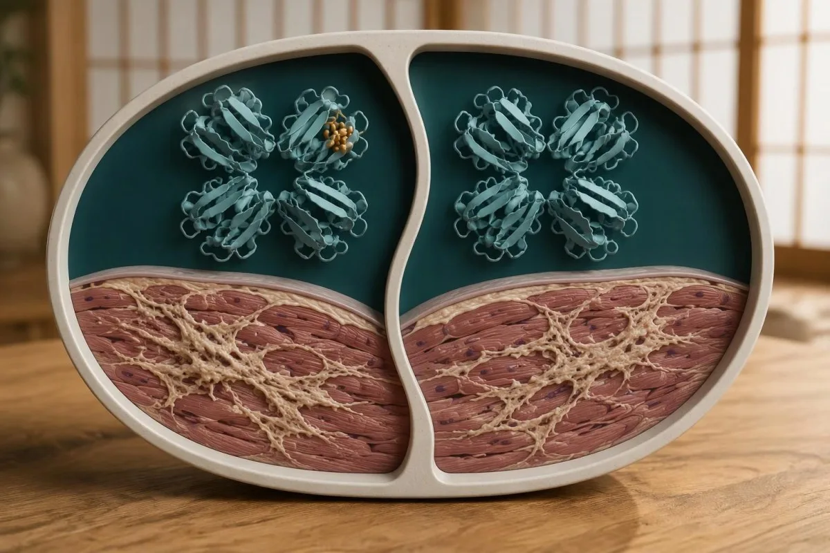
TTR is the gene that encodes transthyretin, a transport protein produced mainly by the liver; its usual circulating structure contains 4 protein subunits. Some variants make that structure less stable, allowing misfolded protein to accumulate as amyloid, particularly in nerves or the heart.
For example, imagine a 46-year-old whose clinical genetic test identifies a known disease-causing TTR variant, but whose neurological examination and cardiac assessment show no disease. The appropriate description may be an asymptomatic variant carrier, not a person with symptomatic amyloidosis; treatment eligibility and follow-up depend on additional findings.
Kantesti is an AI blood test analyzer that helps explain accompanying laboratory results, while keeping genetic susceptibility separate from organ-disease confirmation. Information about nòstre explains our service context; neither a routine laboratory panel nor an AI explanation can replace these 2 specialist assessments.
How do pathogenic TTR findings differ from a VUS?
Pathogenic e probablament patogèna findings have evidence supporting a disease-causing role; a VUS lacks sufficient evidence for either a harmful or harmless classification. Laboratories use 5 standard categories: pathogenic, likely pathogenic, uncertain significance, likely benign, and benign.

The ACMG/AMP sequence-variant framework places likely pathogenic findings above a 90% confidence threshold for pathogenicity, but that percentage describes classification confidence, not an individual's chance of developing symptoms (Richards et al., 2015). Our explanation of inherited cancer test results illustrates the same distinction between a laboratory label and a person's current disease status.
A VUS should not, by itself, trigger disease-specific treatment or routine predictive testing of healthy relatives. Specialists may instead investigate whether the finding tracks with confirmed disease in a family—a segregation study involving 2 or more informative relatives when available—which is different from declaring those relatives genetically affected.
Classification can change when additional family, functional, or population evidence becomes available. Ask who will handle recontact, whether review in 12–24 months would be useful, and whether another laboratory has classified the exact same variant differently; our guide to common genetic variant misconceptions explains why the word mutation is not automatically a diagnosis.
Why can the same TTR variant have two names?
The same TTR variant can have modern and historical protein names because modern numbering includes a 22-amino-acid signal peptide. For example, p.Val50Met is the variant historically called Val30Met, e p.Val142Ile is the variant historically called Val122Ile.

A complete report ideally supplies the gene, transcript reference, DNA-level notation, protein-level notation, zygosity, and classification—not simply a variant nickname. Because a TTR precursor contains 149 amino acids and the mature protein contains 127, comparing reports without noticing the numbering convention can create an apparent disagreement where none exists.
p.Thr80Ala, historically Thr60Ala, can produce a mixed cardiac and neurological phenotype, whereas p.Val142Ile is often associated with predominantly cardiac disease. These are tendencies rather than guarantees: 2 relatives with the same variant may differ in age at onset, affected organs, and severity.
For a hypothetical family, a parent's report saying Val122Ile and an adult child's report saying p.Val142Ile should prompt exact-notation comparison before anyone concludes that there are 2 different familial variants. Similar nomenclature issues arise in enzyme and genetic testing; bring the original reports rather than relying on a handwritten abbreviation.
Does a 50% inheritance risk mean a 50% disease risk?
A 50% inheritance risk is not a 50% risk of symptomatic disease. Hereditary ATTR usually follows autosomal dominant inheritance: a person with 1 disease-causing TTR copy has a 50% chance of passing that copy to each child, but disease expression depends on age-related penetrance and other factors.

Each pregnancy is an independent event; with 2 children, the theoretical probabilities are 25% for neither inheriting the variant, 50% for exactly one, and 25% for both. Those calculations assume one heterozygous parent and describe inheritance only—they do not predict whether or when either child will become ill.
Penetrance means the proportion of variant carriers who develop disease by a stated age, and it varies across TTR variants and populations. The often-quoted 3–4% frequency of p.Val142Ile in some populations of African ancestry is a variant-frequency estimate, not a universal estimate of amyloidosis prevalence or an individual's lifetime disease risk.
An apparently unaffected parent aged 75 can still have transmitted a pathogenic variant, while a negative family history may reflect late onset, small families, or previously unrecognized disease. Our discussion of genetic susceptibility and kidney risk offers a useful comparison, although APOL1 risk architecture should not be substituted for TTR inheritance.
How is hereditary ATTR different from wild-type ATTR?
Hereditary ATTR involves a disease-causing TTR variant; wild-type ATTR involves amyloid formed from transthyretin without an identified disease-causing variant. Both can affect the heart, so neither age nor a cardiac scan alone reliably distinguishes these 2 forms.
TTR sequencing is recommended after ATTR cardiomyopathy is established, including in older adults, because hereditary disease can first become apparent after age 70. The ESC position statement emphasizes genetic counseling and testing to distinguish variant ATTR from wild-type ATTR, rather than assuming that an older patient's disease cannot be inherited (Garcia-Pavia et al., 2021).
A negative result for a known family variant removes that particular inherited susceptibility, but it does not guarantee that the person will never develop wild-type ATTR or another heart condition. Conversely, a VUS does not automatically convert a confirmed ATTR diagnosis into hereditary ATTR; the laboratory classification and amyloid diagnosis still need reconciliation.
ATTR is also different from Amiloïdòsi AL, caused by immunoglobulin light chains, and Amiloïdòsi AA, associated with prolonged inflammatory activity. These are at least 3 distinct amyloid types with different investigations and treatment pathways; an elevated serum amyloid A result does not diagnose TTR-related disease.
Which hereditary amyloidosis symptoms need assessment?
Hereditary amyloidosis symptoms may include progressive numbness or burning feet, weakness, altered bowel function, dizziness on standing, breathlessness, or ankle swelling. These findings are nonspecific, but involvement of 2 systems—such as neuropathy plus unexplained cardiac thickening—raises more concern than either finding alone.

Bilateral carpal tunnel syndrome, particularly when it precedes cardiac symptoms by several years, can be an ATTR clue but is far more commonly explained by other causes. Document whether 1 or both wrists are affected and when symptoms began; our guide to carpal tunnel finger patterns helps distinguish a local median-nerve pattern from broader neuropathy.
Hipotension ortostatica commonly means a sustained fall of at least 20 mmHg systolic or 10 mmHg diastolic within 3 minutes of standing. Amyloid-related autonomic dysfunction is one possible cause, but dehydration and medicines are common alternatives; it is not interchangeable with the heart-rate pattern discussed in POTS diagnostic testing.
Consider a hypothetical 58-year-old carrier who develops numb feet, unintentional weight loss, and new difficulty climbing stairs over 6 months: that combination warrants prompt specialist review rather than waiting for annual screening. Sudden weakness, severe breathlessness, chest pain, or collapse requires urgent care because immediately treatable problems must be assessed before attributing symptoms to ATTR.
Which tests establish active ATTR amyloidosis?
Active ATTR amyloidosis requires evidence of amyloid disease, not just a genetic result. In the appropriate cardiac setting, grade 2 or 3 uptake on a validated bone-tracer scan can establish ATTR cardiomyopathy without biopsy only when monoclonal protein testing is negative and imaging confirms uptake within heart muscle.

The monoclonal screen includes serum free light chains plus serum and urine immunofixation; serum protein electrophoresis alone is insufficient. Gillmore et al.'s 2016 Circulation study evaluated 1,217 patients and established the nonbiopsy cardiac approach under defined conditions; our guide to immunofixation bands and follow-up explains why detecting a monoclonal protein changes the next step.
A monoclonal protein does not prove AL amyloidosis, because an unrelated monoclonal gammopathy can coexist with ATTR, especially in older adults. However, it prevents clinicians from using the simplified scan-only ATTR pathway; interpreting the rapòrt de cadenas leugièras liuras also requires kidney-function context and, where available, assay-specific renal reference intervals.
When tissue examination is needed, Congo red staining establishes amyloid, while reliable protein typing—often mass spectrometry—identifies its source. A negative abdominal fat sample does not exclude ATTR, and grade 1 cardiac uptake is not the same as the grade 2–3 diagnostic criterion; neuropathy-predominant disease may require a different combination of neurological and tissue investigations.
What should TTR genetic counseling cover?
TTR genetic counseling should explain the exact variant, classification, inheritance, limitations of risk prediction, and consequences for relatives before testing or major decisions. For most heterozygous families, the 50% transmission probability is straightforward; predicting age at onset and organ involvement is much less precise.

Counseling distinguishes diagnostic testing in someone with suspected disease from predictive testing in an unaffected relative. A person considering predictive testing should have time to discuss emotional readiness and the right not to know, similar to the consent principles described in predictive neurological genetic testing, although TTR penetrance differs substantially.
Reproductive discussions can include natural conception, prenatal diagnosis, and preimplantation genetic testing where available and legally permitted. Because a 50% transmission risk does not specify future severity, these conversations should be nondirective; confirm the familial pathogenic variant in a clinical laboratory before using it for reproductive testing or other consequential decisions.
Kantesti's explanations of la vida privada e vòstras donadas help readers consider what they share, but genetic information has implications beyond ordinary laboratory values. Before adding reports for 2 or more relatives to a shared account, obtain each person's permission and ask the genetics team about local insurance rules, record access, and secure family communication.
Which relatives should consider targeted TTR testing?
Adult relatives at risk of a confirmed familial pathogenic or likely pathogenic TTR variant should be offered genetic counseling and the option of targeted testing. Each child of a heterozygous carrier has a 50% inheritance probability; a sibling's risk depends on whether a parent carries the variant.

Start with the relative who has confirmed disease whenever possible, then use that person's exact variant to guide cascade testing. Testing all relatives with a broad panel before establishing the family finding may generate additional VUS results; this differs from the recessive inheritance questions in SMA carrier screening, where 2 gene copies have a different meaning.
Predictive testing for adult-onset TTR disease is generally deferred in children younger than 18 when the result would not change childhood care, preserving their future choice. A child with unexplained relevant symptoms needs clinical assessment rather than automatic dismissal; any exceptional testing decision should involve pediatric and genetics specialists, not a routine family screening package.
For example, a 34-year-old who tests negative for a clinically confirmed familial p.Val50Met variant generally does not need surveillance specifically for that family variant. A symptomatic relative still needs assessment for other causes, and a 1-variant consumer screening result cannot substitute for a validated familial test if the relevant variant was not actually examined.
How are asymptomatic TTR carriers followed?
Asymptomatic pathogenic TTR carriers need individualized surveillance, commonly involving annual clinical review, rather than an automatic amyloidosis diagnosis or preventive prescription. Many expert pathways begin structured assessment approximately 10 years before predicted disease onset, but variant-specific evidence and family history may justify earlier or different monitoring.

Baseline assessment may include neurological examination, autonomic history, ECG, echocardiography, kidney function, and cardiac biomarkers; additional tests depend on the variant and symptoms. The ESC position statement outlines regular carrier follow-up (Garcia-Pavia et al., 2021), but a 6–12-month interval should be individualized, especially when new findings appear or expected onset approaches.
A parent's onset at 62 is not a countdown showing that a child's disease will begin at 62, and a normal baseline does not eliminate later risk. Kantesti AI can help explain the difference between a single value and a meaningful laboratory timeline, but specialist decisions also require examination and imaging that routine blood interpretation cannot supply.
Imagine NT-proBNP increasing from 180 to 620 pg/mL while eGFR falls from 62 to 43 mL/min/1.73 m²: cardiac change deserves assessment, but reduced kidney clearance can contribute to the rise. Review intercurrent illness, rhythm, and medicines before interpreting progression; our guida sul rapòrt de foncion renala explains why renal context belongs beside cardiac trends.
What can blood tests reveal—and what can they miss?
Routine blood tests cannot confirm or exclude hereditary ATTR amyloidosis. NT-proBNP, troponin, kidney function, and urine protein can help assess organ involvement or alternatives, but a normal set of results—even across 2 visits—does not establish that a pathogenic TTR carrier will remain disease-free.

Troponina must be interpreted against the assay's 99th-percentile upper reference limit, and NT-proBNP varies with age, kidney function, atrial fibrillation, and body size. A TTR or prealbumin concentration such as 25 mg/dL does not identify a TTR gene mutation or prove amyloid deposition; the test measures circulating protein concentration rather than its genetic sequence.
Neuropathy assessment should also look for treatable alternatives, including diabetes, vitamin B12 deficiency, thyroid disease, and medication effects. A hypothetical B12 result of 210 pg/mL may need additional assessment according to the laboratory and symptoms; our discussion of resultats de vitamina B12 al limit inferior explains why a competing diagnosis should not be overlooked simply because a TTR variant exists.
Kantesti is an AI blood test interpretation platform that explains accompanying laboratory values without treating 1 protein result as an amyloidosis diagnosis. Our guida d'interpretacion de las proteïnas sericas helps separate transport proteins and immunoglobulin findings; neither an ordinary protein panel nor a family-risk feature replaces clinical TTR testing and amyloid typing.
Does a positive TTR genetic result mean treatment should start?
A positive TTR genetic result alone is not a standard indication for disease-modifying treatment. Specialists consider confirmed ATTR disease, the affected organs, functional status, and local approvals; the 2 main medication strategies are stabilizing transthyretin and reducing production of transthyretin.

TTR stabilizers aim to reduce dissociation of the 4-subunit protein, while TTR-lowering therapies reduce production, mainly in the liver. Neither approach edits the inherited DNA, so family transmission considerations remain after treatment; eligibility differs between cardiomyopathy, polyneuropathy, and mixed disease, as do approval and reimbursement rules across countries.
Monitoring depends on the chosen medicine rather than the genetic label alone: some regimens require attention to platelets, kidney function, or vitamin A handling. If vitamin A supplementation is advised during a TTR-lowering regimen, use the prescribed allowance rather than escalating the dose because a laboratory result looks low; 1 abnormal value does not establish a safe replacement dose.
Kantesti does not select amyloidosis medication or establish treatment eligibility from uploaded laboratory results; our validacion tecnica e supervison describe a different task from specialist prescribing. For an asymptomatic carrier, discuss surveillance and research opportunities; there is no single proven preventive prescription that follows automatically from a 50% inheritance-risk calculation.
De qué cal portar a la consulta amb lo especialista?
Bring the original genetic report, symptom timeline, family history, and relevant cardiac or neurological investigations to an amyloidosis or clinical genetics appointment. These 4 information groups help specialists distinguish a confirmed disease-causing variant, an uncertain finding, unrelated symptoms, and early organ involvement.

Record symptom onset by month and year where possible, including numbness, bowel changes, wrist procedures, fainting, breathlessness, and reduced walking tolerance. Include at least 3 generations of family information if available, but distinguish a documented amyloidosis diagnosis from a relative's unexplained heart failure; uncertain family labels should remain visibly uncertain.
Ask 3 focused questions: Is this variant pathogenic, likely pathogenic, or uncertain; what evidence exists for active organ disease; and which relatives should be offered counseling? A written plan should also identify the clinician responsible for follow-up, what changes should trigger earlier contact, and whether a laboratory will notify anyone if the variant classification changes.
Kantesti is an AI-powered blood test analysis tool that can help explain laboratory reports before a consultation, not replace the consultation or infer another relative's genotype. Our guide to consent-based family health tracking covers shared records; our principis d’IA responsabla explain why 1 uncertain genetic result should never become an automated family diagnosis.
Which research supports diagnosis, and which evaluates AI?
Clinical amyloidosis research and AI technical benchmarks answer different questions. The 2016 nonbiopsy diagnostic study and 2021 ESC position statement support specific ATTR assessment pathways; the 2 repository publications listed below describe separate AI projects and do not validate TTR variant classification or diagnose hereditary ATTR amyloidosis.

The clinical evidence cited here addresses 3 distinct tasks: sequence-variant classification, cardiac amyloid confirmation, and specialist management pathways. Congo red staining can reveal amyloid but cannot, by itself, identify a TTR genotype or reliably distinguish all amyloid proteins; the same caution applies to a compelling-looking AI explanation unsupported by the required clinical tests.
Kantesti's repository publications describe early hantavirus triage across 50,000 interpreted reports and a technical benchmark involving 100,000 synthetic test cases. The blood-test engine benchmark provides context for the latter; synthetic-case performance does not establish sensitivity for detecting ATTR in real patients, and a Figshare DOI does not itself demonstrate peer review or regulatory authorization.
For patients and relatives, the next decision should follow the report category: counseling and individualized surveillance for a confirmed pathogenic variant, specialist-led clarification for a VUS, and organ assessment for relevant symptoms. Keep those 3 pathways separate; a genetic finding, an imaging result, and an AI-generated explanation are not interchangeable forms of evidence.
Preguntes més freqüents
Una mutacion del gèn TTR significa que soi portaire de l’amiloïdòsi ATTR ereditària?
Una mutacion patogèna o probablament patogèna del gèn TTR establís una predisposicion ereditària, mas establís pas per ela sola una amiloïdòsi ATTR ereditària activa. Una persona amb una còpia de TTR que causa una malautiá pòt demorar asimptomatica pendent fòrça annadas o jamai desvolopar una malautiá reconeguda. Los especialistas avaloran los simptòmas, las constatacions de l’examen, l’imatjariá e de còps los resultats dels teissuts per determinar se i a una malautiá d’organ. Una variant de significacion incèrta establís pas la meteissa predisposicion ereditària.
Se pòt utilizar una variant de TTR de significacion incèrta per testar ma familha?
0 Una varianta TTR de significacion incèrta deu pas èsser utilizada soleta per de tèsts predicitius de rutina sus de parents en bona santat o per de decisions terapeuticas. Es una de las 5 categorias estandards de classificacion de las variantas e significa que las donadas son insufisentas per classificar la descobèrta coma nociva o inofensiva. Una còla de genetica pòt organizar de tèsts seleccionats dins la familha per estudiar se la varianta es associada a la malautiá confirmada. Aquesta investigacion ajuda a la classificacion; es pas equivalent a qualificar los parents de portaires afectats.
Quina es la probabilitat que mos enfants ereten l'ATTR ereditari?
Cada enfant d’una persona amb una còpia de TTR causanta de malautiá a generalament una probabilitat de 50% d’eretar aquela varianta. La probabilitat es la meteissa pels enfants de quin que siá sèxe, e cada emprenhament es independent. Eretar la varianta permet pas de determinar quand los simptòmas començaràn ni la gravetat de la malautiá. Lo conselh genetic pòt explicar los tèsts predictius e las opcions reproductivas sens considerar una probabilitat de transmission coma un pronostic de la malautiá.
Val30Met e p.Val50Met son la meteissa mutacion de TTR?
Val30Met e p.Val50Met descrivon la meteissa substitucion de la proteïna TTR amb de convencions de numerotacion diferentas. La numerotacion modèrna inclutz un peptid de senhal de 22 aminoacids que la numerotacion istorica de la proteïna madura exclutz. Lo meteis problèma explica Val122Ile contra p.Val142Ile e Thr60Ala contra p.Thr80Ala. Un professional de la genetica deu comparar la notacion completa de l'ADN e de la proteïna quand examina de rapòrts de laboratòris diferents.
Can normal blood tests rule out ATTR amyloidosis?
Normal routine blood tests cannot rule out ATTR amyloidosis or eliminate future disease risk in a pathogenic TTR carrier. Troponin is interpreted using the assay's 99th-percentile limit, while NT-proBNP and kidney results provide context about organ stress rather than amyloid identity. A normal TTR or prealbumin concentration does not exclude a TTR variant or amyloid deposits. Evaluation may still require neurological assessment, cardiac imaging, and selected tissue investigations.
Is wild-type ATTR amyloidosis inherited?
Wild-type ATTR amyloidosis is not attributed to a disease-causing inherited TTR variant, unlike hereditary ATTR. The 2 forms can produce similar cardiac findings, so age and symptoms alone cannot reliably distinguish them. TTR genetic testing is recommended after ATTR cardiomyopathy is established, including in older adults. Relatives do not need targeted testing for a familial variant unless a disease-causing variant has actually been identified.
How often should an asymptomatic TTR carrier see a specialist?
An asymptomatic pathogenic TTR carrier commonly has annual specialist assessment, but follow-up should be individualized rather than fixed for everyone. Some pathways begin structured surveillance approximately 10 years before predicted family onset, with earlier review for symptoms or concerning findings. Neurological examination, ECG, echocardiography, and selected laboratory markers may form part of the plan. New breathlessness, progressive weakness, or fainting should prompt earlier assessment instead of waiting for the scheduled visit.
Fasètz-vos explicar los resultats analisi de sang
Telecargatz vòstre compte rendut de laboratòri e l’IA de Kantesti explica cada biomarcador en lenga simpla en qualques segondas. Utilizat per mai de 2 milions de personas dins lo mond.
Referéncias
- Klein, T., Mitchell, S., & Weber, H. (2026). Ajuda multilingüa a la presa de decisions clinicas amb IA per al triatge precoç de l’hantavirus: concepcion, validacion de l’engenharia e desplegament en condicions realas sus 50,000 rapòrts d’interpretacion d’analisis de sang. Recèrca medicala per IA de Kantesti. DOI
- Klein, T., Mitchell, S., & Weber, H. (2026). Un benchmark tècnic automatitzat preregistrat basat en rúbriques de l’Kantesti Motor d’Interpretació de la Prova de Sang en 100.000 casos de prova sintètics. Recèrca medicala per IA de Kantesti. DOI
- Richards S et al. (2015). Nòrmas e directivas per l’interpretacion de las variants de sequéncia: una recommandacion de consens conjunta del Collègi American de Genetica Medicala e de Genomica e de l’Associacion per la Patologia Moleculara. Genetica en Medecina. DOI PubMed
- Garcia-Pavia P et al. (2021). Diagnosis and treatment of cardiac amyloidosis: a position statement of the ESC Working Group on Myocardial and Pericardial Diseases. Jornal Europèu del Còr. DOI PubMed
- Gillmore JD et al. (2016). Nonbiopsy Diagnosis of Cardiac Transthyretin Amyloidosis. Circulation. DOI PubMed
- 2M+Tèsts analisats
- 127+Païses
- 100+Lengas
A prepaus de l’equipa medicala


Senhals de confiança E-E-A-T
Experiéncia
Revisión clinica menada pel metge de las practicas d’interpretacion de las analisis.
Expertisa
Fòcus sus la medicina de laboratòri sus cossí los biomarcadors se comportan dins un contèxte clinic.
Autoritat
Revisat medicalament pel Dr Thomas Klein, amb de contribucions de la Dr Sarah Mitchell e del Prof. Dr Hans Weber.
Fisança
Interpretacion basada sus d’evidéncias amb de camins de seguiment clars per reduzir l’alarmisme.






Avís medical
Aqueste article es solament per tòcas educatius e constituís pas de conselh medical. Consultatz totjorn un professional de la sant qualificat per las decisions de diagnostica e de tractament.
Publicat · Contactar l’equipa editoriala
Publicat per Kantesti Ltd, enregistrada en Anglatèrra e País de Galas, n° d’entrepresa. 17090423, Londres, Reialme Unit.
