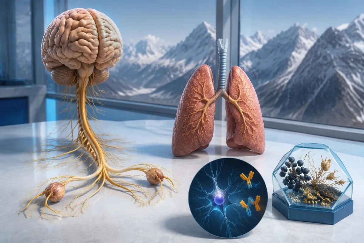
Een positieve anti-Hu-antistofuitslag roept zorgen op over een met kanker geassocieerde neurologische immuunreactie, met name in verband met kleincellige longkanker, maar stelt geen kankerdiagnose. Artsen beoordelen de symptomen, bevestigen de laboratoriumuitslag en regelen kankeronderzoek; herhaalde screening kan nodig zijn, ook als de eerste scans negatief zijn.



- Anti-Hu / ANNA-1 is een hoogrisico-paraneoplastische antistof, een categorie die in meer dan 70% van de gevestigde klinische cohorten met kanker in verband wordt gebracht—maar niet met elke onbevestigde screeningsuitslag.
- Kankerdiagnose vereist bewijs van een onderliggende maligniteit, meestal met inbegrip van weefselonderzoek; één positieve antistofuitslag is daarvoor geen bewijs.
- Bevestiging van de test combineert vaak 2 complementaire benaderingen: een kenmerkend weefselkleuringspatroon en een antigeenspecifieke test.
- Neurologische symptomen zoals progressief gevoelsverlies, evenwichtsproblemen, epileptische aanvallen of autonome disfunctie veranderen de betekenis en urgentie van de uitslag.
- Negatieve beeldvorming betekent dat bij dat onderzoek op dat moment geen kanker is vastgesteld; het sluit de mogelijkheid van een kleine, aanvankelijk niet-detecteerbare kanker niet uit.
- Herhaalde screening wordt volgens de PNS-criteria van 2021 elke 4–6 maanden gedurende 2 jaar aanbevolen wanneer een hoogrisico-neurologisch fenotype samengaat met een hoogrisico-antilichaam.
- Langere surveillance kan passend zijn voor geselecteerde patiënten; een anti-Hu-cohort uit 2023 bood steun voor het overwegen van screening langer dan 2 jaar, soms tot 5 jaar.
- Spoedsymptomen zijn onder meer nieuwe ademhalingsproblemen, niet veilig kunnen slikken, snel verergerende zwakte, een eerste epileptische aanval of plotselinge neurologische uitvalsverschijnselen.
Wat betekent een positieve anti-Hu-antistofuitslag?
Een anti-Hu-antilichaam positief betekent dat het laboratorium reactiviteit tegen neuronale Hu-eiwitten heeft aangetoond; op zichzelf stelt dit geen kanker of neurologisch syndroom vast. De interpretatie bestaat uit 3 onderdelen: of de uitslag echt is, of het neurologische beeld ermee overeenkomt en of er een geassocieerde kanker kan worden gevonden.
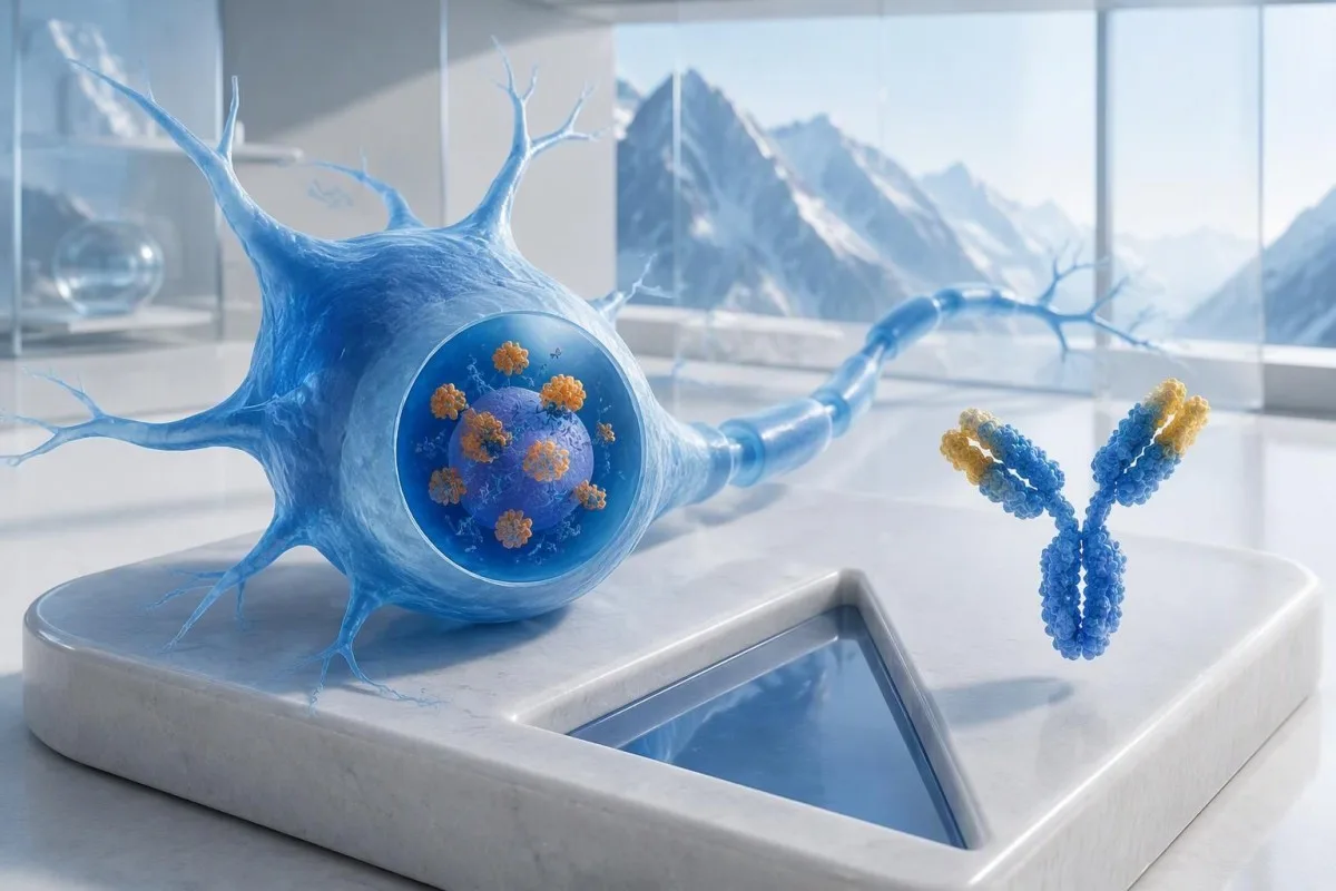
Anti-Hu wordt geclassificeerd als een kankergerelateerd hoogrisico-antilichaam, waarbij de categorie wordt gedefinieerd door een kankerassociatie van meer dan 70% in relevante klinische populaties (Graus et al., 2021). Dat percentage kan niet zonder meer worden toegekend aan een verder gezonde persoon met een zwakke, niet-bevestigde uitslag van een paneltest: de onderzochte patiënten, de testmethode en het neurologische beeld beïnvloeden allemaal de waarschijnlijkheid.
Een hypothetische 62-jarige met 8 weken toenemende evenwichtsproblemen en asymmetrisch sensibiliteitsverlies verdient een andere beoordeling dan iemand bij wie het antilichaam toevallig aan het licht kwam tijdens een breed vermoeidheidspanel. Geen van beiden mag op basis van alleen de laboratoriumuitslag te horen krijgen: “U hebt kanker.” Onze uitleg van positieve antistofbevindingen helpt een immuunmarker te onderscheiden van een afgeronde diagnose.
Kantesti is een AI-bloedtestanalysator die kan helpen de formulering van een anti-Hu-rapport te verklaren, maar laboratoriumbevestiging en kankerbeoordeling vallen onder de verantwoordelijkheid van uw behandelend artsen. Als Thomas Klein, MD, Chief Medical Officer bij Kantesti LTD, zou mijn eerste vraag zijn wat de aanleiding was voor juist deze test—not hoe alarmerend de rode vlag eruitziet; onze organisatie en klinische missie verklaren dat onderscheid.
Is ANNA-1 dezelfde antistof als anti-Hu?
ANNA-1-antilichaam en anti-Hu-antilichaam verwijzen doorgaans naar dezelfde antilichaamfamilie. ANNA-1 staat voor antineuronaal nucleair antilichaam type 1; de aanduiding Hu verwijst naar de intracellulaire neuronale eiwitten die door de immuunrespons worden herkend, en niet naar een afzonderlijke ziekte of een kankerstadium.
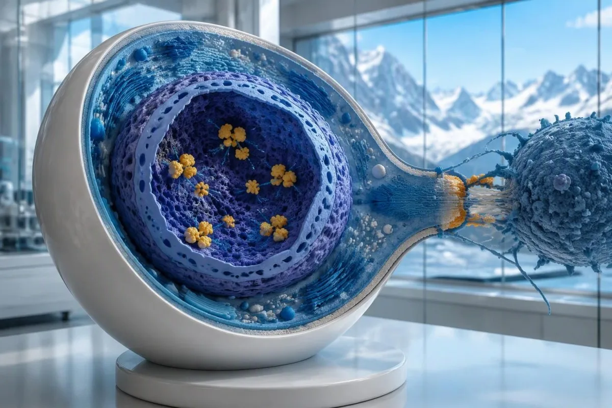
Hu-eiwitten zijn intracellulaire RNA-bindende eiwitten, en anti-Hu kan het best worden opgevat als een marker van een immuunrespons waarbij vaak cytotoxische T-cellen betrokken zijn. Dit verschilt van aandoeningen waarbij antilichamen binden aan toegankelijke eiwitten op het neuronale oppervlak. Dit onderscheid verklaart waarom het simpelweg verlagen van de antilichaamspiegels bestaande anti-Hu-neurologische schade niet betrouwbaar ongedaan maakt, zelfs wanneer 2 rapporten sterk uiteenlopende titers laten zien.
Een rapport met de uitslag “ANNA-1 aangetoond” is niet hetzelfde als een routinematige positieve ANA-uitslag, ondanks de overeenkomstige letters. ANA betekent doorgaans antinucleaire antilichamen die worden gebruikt bij de beoordeling van systemische auto-immuniteit, terwijl ANNA-1 een neuronale antilichaamreactie identificeert. Houd die 2 afkortingen uit elkaar wanneer u uw uitslagen bespreekt; onze ANA-terminologiegids legt de meer bekende test uit.
Anti-Hu kan worden aangetoond bij sommige mensen met kleincellige longkanker die geen herkenbaar neurologisch syndroom hebben. Omgekeerd kunnen neurologische symptomen leiden tot detectie van antilichamen voordat de kanker zichtbaar wordt. Dat zijn 2 verschillende klinische trajecten: het antilichaam vertelt een arts op zichzelf niet of er momenteel zenuwen worden beschadigd of waar een onderliggende kanker zich mogelijk bevindt.
Hoe moet het anti-Hu-antistoftestrapport worden gelezen?
An anti-Hu antibody test has no universal numerical cutoff that can be applied across laboratories. Read the specimen type, assay method, result category, laboratory reference interval and confirmation comments together; a dilution titer, band intensity and numerical index are 3 different ways of reporting reactivity.
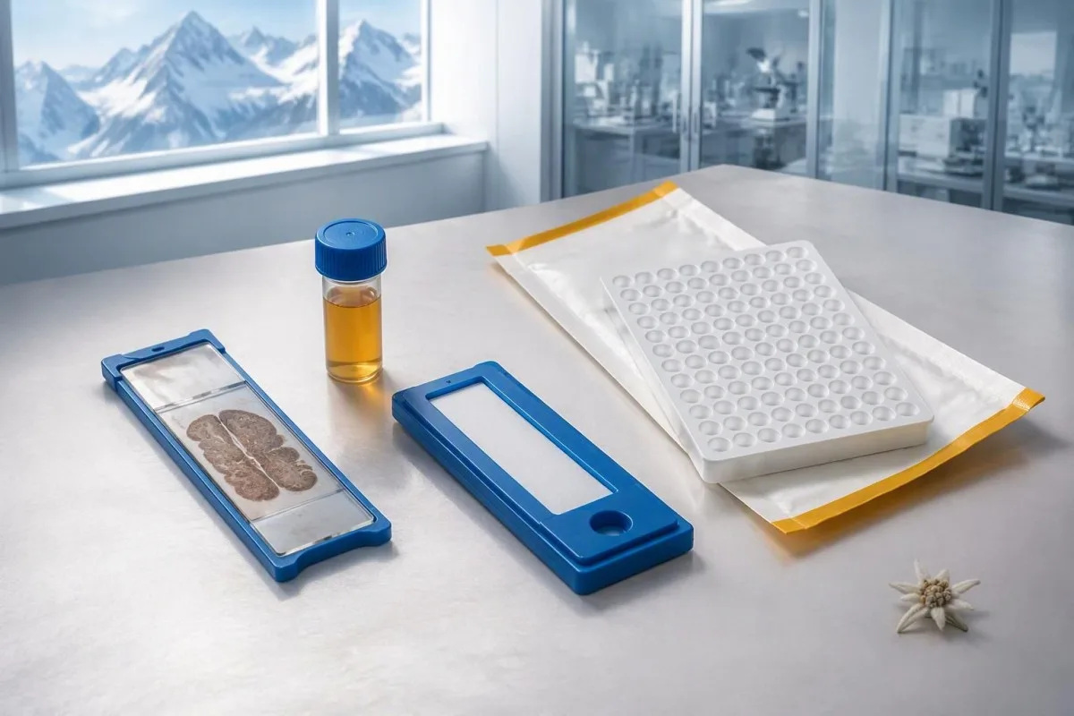
A hypothetical titer of 1:240 describes detectable reactivity after a particular dilution; it is not a cancer probability of 240, nor does it establish cancer stage. A numerical index from another method may use entirely different calibration. Our guide to kwalitatieve versus kwantitatieve resultaten explains why “positive” and “how much” answer different laboratory questions.
Weak or borderline reactivity deserves method-specific interpretation, particularly when a broad panel was ordered without a convincing neurological indication. Ask whether the laboratory observed the characteristic neuronal staining pattern and whether an antigen-specific confirmation was performed. Two tests using the same underlying strip technology may reproduce the same analytical problem; agreement is more informative when the methods are genuinely complementary.
Bring the complete report, not just a screenshot of the word positive. The 5 most useful details are the specimen, collection date, assay name, reported strength or titer and laboratory interpretation. Also list recent intravenous immunoglobulin and other immune treatments, because passive antibodies or treatment effects can complicate some serological results; your laboratory can advise whether that issue plausibly affects this particular finding.
Wanneer moet een positieve anti-Hu-uitslag worden bevestigd?
Unexpected, weak or clinically discordant anti-Hu results should be reviewed for confirmation, preferably with an experienced neuroimmunology laboratory. The 2021 PNS criteria emphasize interpreting neuronal antibody testing alongside the phenotype and using appropriate confirmatory methods rather than accepting an isolated commercial-panel result as a diagnosis.
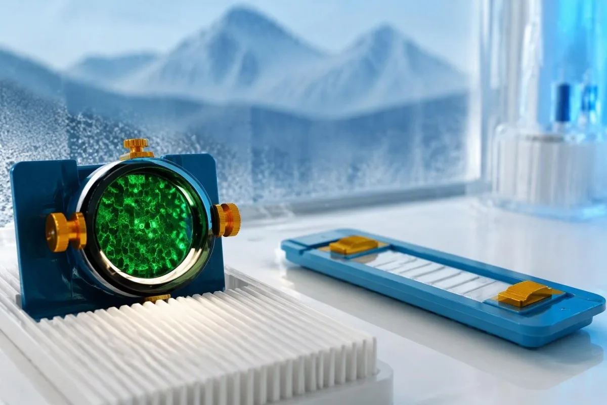
Tissue-based immunohistochemistry or immunofluorescence and an antigen-specific assay provide different information. The first identifies a characteristic distribution of neuronal staining; the second tests reactivity against the relevant antigen. Graus et al. (2021) describe these complementary approaches, with specialist reassessment particularly useful when 1 method gives a result that does not fit the clinical picture.
Repeating a sample can address collection errors or a transient analytical concern, but repeating the same kit is not always enough. In a hypothetical 45-year-old with no objective neurological findings and a faint isolated band, I would want the laboratory’s explanation before treating the report as established anti-Hu autoimmunity. Our GAD65 interpretation guide illustrates another antibody for which context and assay characteristics change meaning.
Confirmation and urgent assessment can happen in parallel. A person who develops seizures, substantial walking difficulty or progressive swallowing impairment should not wait several weeks for a reference-laboratory answer before receiving neurological care. The practical question is not “repeat or investigate?” but which of those 2 tasks can safely proceed together, given the symptoms and the initial laboratory evidence.
Welke neurologische symptomen maken een positieve anti-Hu-uitslag zorgwekkender?
Progressive sensory neuronopathy, encephalomyelitis, limbic encephalitis and marked autonomic dysfunction are recognized anti-Hu presentations. Symptoms can involve more than 1 part of the nervous system, so the pattern and speed of change are usually more informative than a single symptom such as tingling or fatigue.

Sensory neuronopathy can cause asymmetric numbness, impaired position sense and sensory ataxia, sometimes involving the hands or trunk early rather than following a simple feet-first pattern. A patient may lose balance in the dark because vision had been compensating for reduced position feedback. That is different from the focal finger distribution discussed in our carpal tunnel symptom guide, although neither pattern can be diagnosed from description alone.
Anti-Hu autonomic involvement can affect gut movement, blood-pressure regulation and bladder function. Severe constipation, unexplained intestinal pseudo-obstruction or dizziness on standing becomes more relevant when accompanied by objective neurological abnormalities. Recording symptoms over 7 days can reveal combinations that otherwise get presented as separate stomach, balance and urinary complaints; sudden severe abdominal symptoms still require assessment for non-neurological emergencies.
New memory impairment, behavioral change or seizures may indicate central nervous system involvement, while rapid weakness has a broader emergency differential. The waarschuwingssignalen van acute zwakte in our neurological safety guide apply regardless of the antibody label. Call emergency services for new breathing difficulty, unsafe swallowing, a first seizure or sudden stroke-like symptoms rather than waiting for a scheduled follow-up.
Welke neurologische onderzoeken kunnen volgen op een positieve uitslag?
Neurological follow-up may include examination, nerve conduction studies, MRI and cerebrospinal fluid testing, selected according to the symptoms. There is no mandatory battery for every anti-Hu-positive person; the purpose is to document which structures are affected and exclude other causes, not merely to collect additional positive tests.
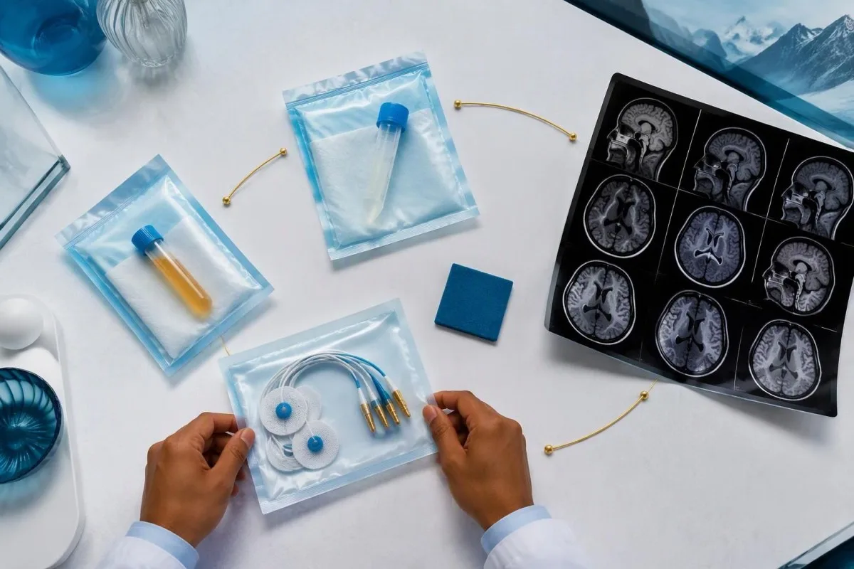
Nerve conduction studies can support a sensory neuronopathy pattern by showing reduced sensory responses across distributions that do not fit a single compressed nerve. Brain or spinal MRI addresses different questions, including structural disease and some inflammatory changes. A normal MRI does not exclude peripheral sensory ganglion involvement; these are 2 distinct anatomical compartments with different diagnostic tools.
Cerebrospinal fluid may show increased cells, protein or evidence of intrathecal immune activity, but none of those findings is specific to anti-Hu. A lumbar puncture is most useful when the neurological syndrome warrants it, rather than as an automatic response to 1 weak serum band. Our discussion of de interpretatie van oligoklonale banden in liquor explains why immune activity in spinal fluid does not identify a single disease.
Basic investigations still matter: glucose control, vitamin status, renal function and serum protein assessment can uncover treatable contributors to neuropathy. A clinician may request 2 or more of these alongside specialist testing, depending on the examination. The gids voor serumproteïneonderzoek provides background, but normal routine blood work neither confirms nor excludes an anti-Hu-associated neurological process.
Bevestigt een positieve anti-Hu-uitslag een paraneoplastisch syndroom?
A positive antibody alone does not confirm anti-Hu paraneoplastic syndrome. The PNS-Care approach combines the neurological phenotype, antibody risk category, cancer findings and follow-up; clinicians also check for direct cancer effects, treatment toxicity and unrelated neurological conditions before attributing symptoms to a paraneoplastic immune response.
Definite, probable and possible PNS are diagnostic categories, not cancer stages. The 2021 criteria use a structured scoring system rather than allowing 1 antibody to determine the whole diagnosis (Graus et al., 2021). A confirmed anti-Hu result with a strongly compatible syndrome remains clinically significant even before a cancer is identified, but clinicians should state clearly which parts of the assessment remain unresolved.
An older patient can have anti-Hu reactivity and a separate cause of numbness, such as diabetes, nutritional deficiency or a monoclonal protein disorder. Coexistence is possible; either-or reasoning can miss treatable disease. If serum protein studies reveal a band, our immunofixation result guide explains why that finding requires its own interpretation rather than being automatically folded into the anti-Hu diagnosis.
Kantesti is een AI-bloedonderzoek uitslagplatform that can help organize these distinctions in a laboratory explanation, but it cannot assign a reliable PNS-Care diagnosis from a report alone. Our klinisch validatiekader separates technical evaluation from clinical conclusions. In practice, I would document 3 separate judgments: confidence in the antibody, confidence in the neurological syndrome and the current evidence for cancer.
Hoe zoeken artsen naar een geassocieerde kanker?
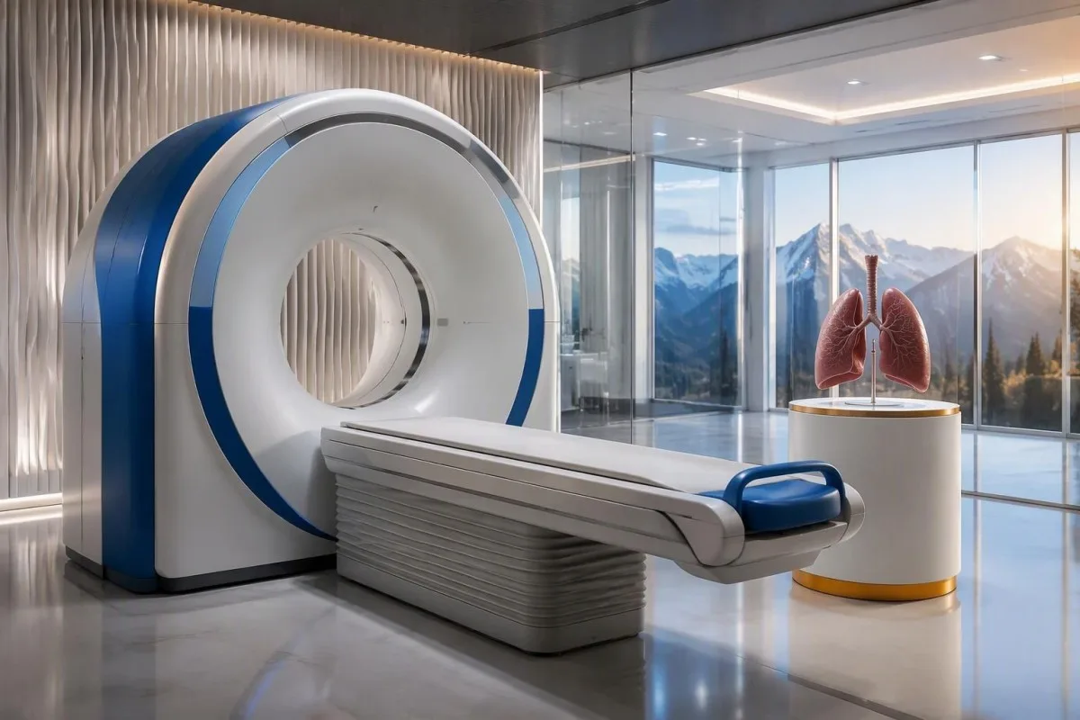
Cancer evaluation usually prioritizes the chest because small-cell lung cancer is the strongest adult anti-Hu association. Doctors commonly begin with appropriate chest CT and may add FDG-PET/CT or targeted investigations if suspicion remains; the exact plan depends on age, symptoms, examination, smoking history and previous imaging.
The cancer association is substantial in established anti-Hu clinical cohorts. Villagrán-García et al. (2023) reported identified cancer in 349 of 466 patients, approximately 75%, in a specialist anti-Hu cohort. This is valuable evidence for taking confirmed, clinically relevant anti-Hu seriously; it is not a personalized prediction for an asymptomatic person with an unconfirmed positive screening strip.
A chest X-ray is not equivalent to a chest CT, and routine low-dose screening is not necessarily the correct examination for a suspected paraneoplastic syndrome. The clinician chooses the protocol and body regions based on the diagnostic question. Likewise, neurological symptoms do not automatically mean cancer has spread to the brain; our brain imaging warning-sign guide distinguishes structural investigation from immune-mediated disease.
A suspicious scan often leads to tissue sampling through a method chosen by respiratory or oncology specialists, such as bronchoscopic assessment of an accessible area. Imaging suspicion and cancer confirmation are 2 separate steps. Never-smoking status lowers the baseline probability of small-cell lung cancer but does not cancel a well-confirmed anti-Hu finding with a convincing neurological presentation; other cancers and non-cancer explanations still need consideration.
Waarom kunnen de eerste scans geen kanker aantonen?
Initial imaging can be negative because an associated cancer is still too small to detect, lies outside the examined area or is not readily visible with that technique. Neurological autoimmunity may precede cancer detection, so a negative scan reduces concern without necessarily ending follow-up in a high-risk clinical presentation.
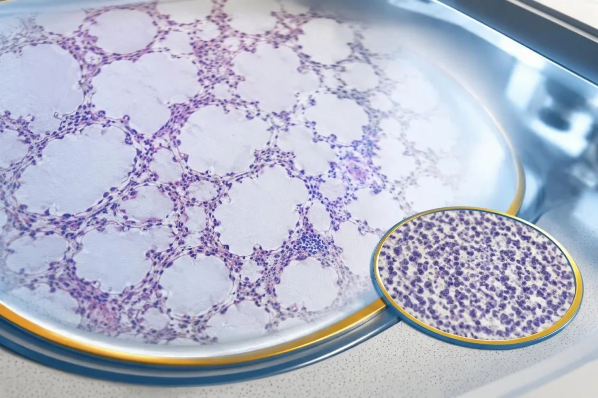
A negative scan is time-specific and method-specific. A normal chest CT performed on 1 date answers whether that examination identified a suspicious chest finding then; it does not guarantee that every possible associated cancer is absent indefinitely. Ask what was actually imaged, whether the protocol was suitable and whether the specialist recommends PET/CT or targeted investigation despite the initial result.
Villagrán-García et al. (2023) described cancers detected beyond the first 2 years in a minority of their anti-Hu cohort, supporting individualized longer surveillance. Their findings also support considering PET-based investigation when initial CT is unrevealing in an appropriate clinical context. This is an escalation decision for the treating team, not a reason to independently purchase repeated whole-body scans without weighing radiation, incidental findings and cost.
Neurological deterioration after negative imaging still needs reassessment. Cancer surveillance and neurological monitoring are 2 parallel tracks; one reassuring scan does not explain new loss of function. A marker such as neurofilament light chain may indicate neuronal injury in selected settings, but our NfL interpretation guide explains why it cannot establish anti-Hu disease, locate cancer or replace examination.
Hoe lang moet de kankerscreening doorgaan?
The 2021 PNS criteria recommend repeat cancer screening every 4–6 months for 2 years when a high-risk neurological phenotype accompanies a high-risk antibody. Longer surveillance may be appropriate for selected anti-Hu patients; the interval and duration should reflect confirmation, clinical evolution, initial investigations and individual cancer risk.

The 2-year schedule is not an instruction to scan every positive antibody result repeatedly. Graus et al. (2021) link that recommendation to the combination of high-risk phenotype and high-risk antibody. An incidental, weak result without a matching syndrome needs confirmation and individualized assessment first; repeating imaging mechanically can create incidental findings and unnecessary procedures without resolving the original uncertainty.
Older European guidance recommended longer surveillance. The EFNS task force advised repeating screening after 3–6 months and then every 6 months for up to 4 years in relevant antibody-positive paraneoplastic presentations (Titulaer et al., 2011). The 2023 anti-Hu cohort supported considering screening up to 5 years in selected patients, so a specialist may reasonably extend follow-up beyond the newer general 2-year framework.
Keep a dated plan stating the next examination, the clinician responsible and what symptoms should trigger earlier contact. Kantesti explanations can help you understand terminology, but the platform should not be treated as a scan-reminder service or substitute for that plan. Our guide to risk-based cancer screening addresses a different setting: routine prevention is not the same as investigating a possible paraneoplastic syndrome.
Welke behandeling helpt bij neurologische aandoeningen door anti-Hu?
Treatment focuses on an identified cancer, selected immune therapy and symptom-directed rehabilitation. Anti-Hu-associated neurological disease often responds less predictably to immunotherapy than neuronal surface-antibody disorders because intracellular targets and T-cell-mediated injury are involved; treatment may aim to stabilize function rather than promise complete recovery.

Treating an associated cancer is a central part of care, but established neurological injury may persist even when cancer treatment succeeds. Neurology and oncology teams may consider corticosteroids, intravenous immunoglobulin, plasma exchange or other immune treatments in selected situations. There is no single evidence-based dose or 1 universal regimen appropriate for everyone with anti-Hu positivity, especially without a confirmed clinical syndrome.
An antibody titer is not a dependable treatment target on its own. A lower number does not necessarily mean restored nerve function, and a persistent positive result does not by itself prove cancer progression. This differs from some surface-antibody disorders; our MOG antibody diagnosis guide explains another immune neurological condition, but its diagnostic rules and treatment expectations should not be transferred to anti-Hu.
Immune checkpoint cancer therapies deserve an explicit discussion when anti-Hu neurological disease is present, because they can provoke or worsen paraneoplastic neurological autoimmunity. This is a specialist risk-benefit decision, not an automatic prohibition based on 1 laboratory result. Tell the oncologist about previous neurological symptoms and confirmed neuronal antibodies before treatment decisions are finalized; coordinating the teams early can prevent avoidable misunderstanding.
Wat kunt u veilig doen terwijl het onderzoek doorgaat?
While investigations continue, protect mobility, swallowing and medication safety rather than trying to lower anti-Hu antibodies with supplements. Use the neurological symptoms—not the appearance of the laboratory flag—to decide whether ordinary follow-up is sufficient or urgent care is needed; a new functional loss warrants contact before the next planned scan.

Position-sense loss increases fall risk, particularly in poor lighting or on uneven ground. A physiotherapist may recommend a suitable walking aid and individualized balance strategies; occupational therapy can address hand coordination and home hazards. A short 7-day record of falls, near-falls and walking changes is often more useful at review than the vague statement “my balance is worse,” but stop activities that feel unsafe.
Excess vitamin B6 can itself cause sensory neuropathy, making unsupervised high-dose nerve supplements particularly unhelpful during this assessment. Review every supplement and its daily dose in mg, including overlapping multivitamins and combination products. Our B-complex veiligheidsgids explains that problem; correcting a documented deficiency is different from taking progressively larger doses in the hope of treating anti-Hu autoimmunity.
Vitamin B12 deficiency can contribute to sensory and gait problems and may need investigation even when anti-Hu is confirmed. There is no universally interchangeable B12 cutoff: results in pg/mL En pmol/L require their own laboratory context, sometimes with functional testing. Our gids voor opvolging van grenswaarde B12 explains those next steps; new choking, breathing difficulty or rapid weakness should be assessed urgently instead of managed with vitamins.
Hoe kan AI helpen een anti-Hu-rapport veilig uit te leggen?
AI can help explain report terminology and organize questions, but it cannot confirm anti-Hu specificity, diagnose paraneoplastic syndrome or rule out cancer. Safe interpretation preserves the original assay details and separates 3 issues: what the laboratory reported, what the result may mean and what a clinician has actually established.

Kantesti is een AI-aangedreven tool voor analyse van bloedtesten that can provide a plain-language explanation of uploaded laboratory information, including the distinction between an antibody screen and a confirmed clinical diagnosis. A rapid explanation—often around 60 seconds for report processing—is not a reference-laboratory assay. Our AI-technologiegids describes the interpretation workflow without making anti-Hu-specific diagnostic guarantees.
The complete source report should remain available for verification. An explanation should not silently replace “weakly reactive” with “confirmed,” drop the specimen type or invent a universal reference interval. Our guide to het controleren van medische uitleg door AI describes practical source checks; for this antibody, compare at least the assay wording, confirmation comment and collection date against the original document.
Kantesti cannot examine sensation, review tissue staining directly through a report explanation or establish that negative imaging has excluded a tiny cancer. Those are 3 different clinical capabilities, not features that follow from understanding laboratory language. Share the explanation with the treating team as a discussion aid, and do not delay urgent assessment while uploading documents or waiting for a second automated interpretation.
Uw volgende afspraak en transparantie over onderzoek
Bring the complete antibody report, a dated symptom history, previous imaging reports and a current medication list to your next appointment. Ask the clinician to clarify antibody confirmation, the neurological diagnosis and cancer surveillance separately; those 3 answers may have different levels of certainty and different follow-up dates.
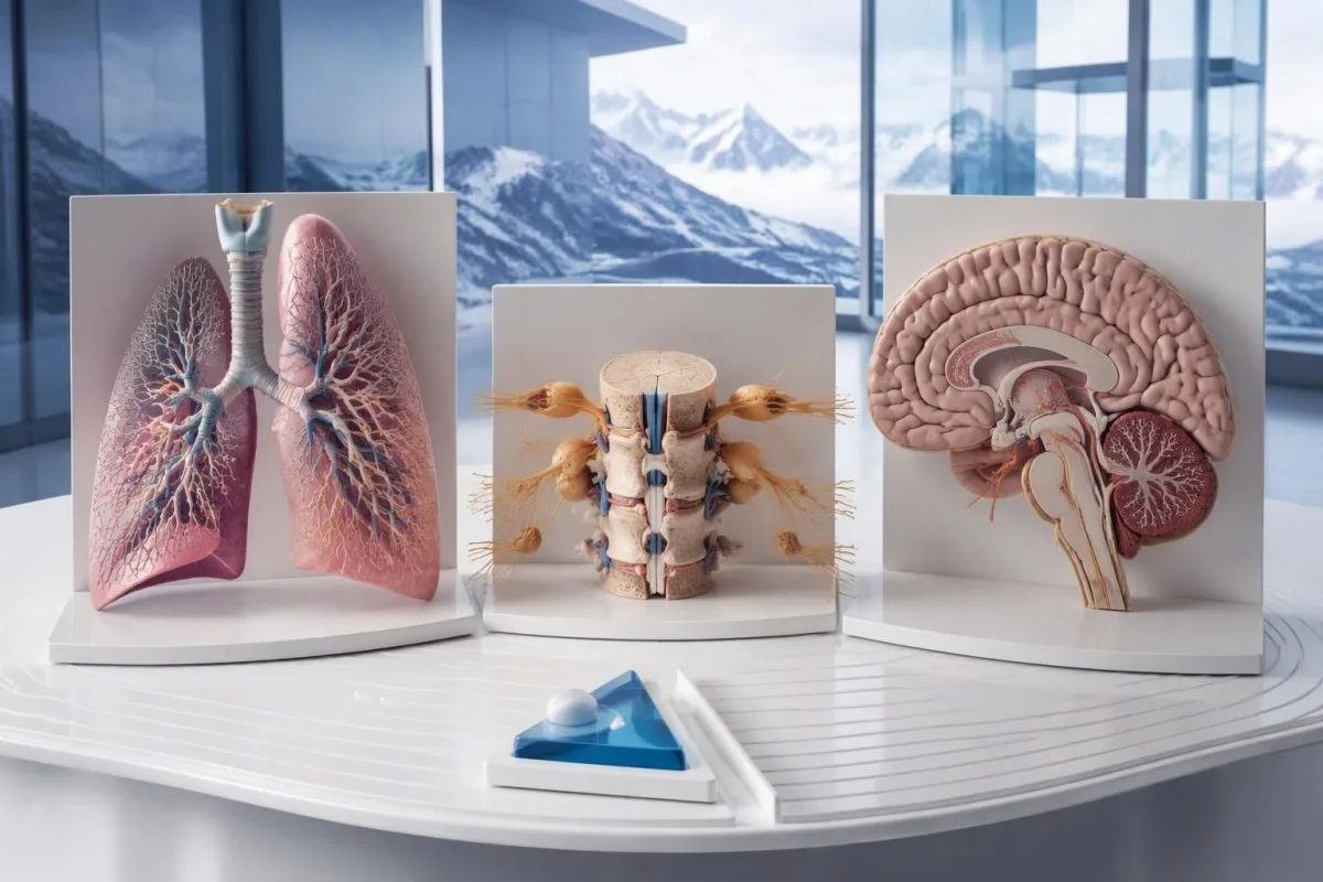
A useful 1-page symptom history records onset, progression, sensory distribution, walking changes, memory problems and autonomic symptoms. Include prior cancer treatment and any recent immunoglobulin therapy, then ask who will coordinate neurology and oncology if both are involved. As Thomas Klein, MD, I would rather see a clear timeline than 20 disconnected screenshots; it makes discrepancies between the laboratory result and clinical course easier to spot.
Medical authorship is not a substitute for an individual neurological examination. Ons informatie van de medische adviesraad describes the clinicians associated with our work, while your own specialist determines the appropriate next investigation. As of 25 oktober 2026, the recommendations discussed here draw on the 2021 international PNS criteria, the 2011 EFNS screening guidance and the 2023 anti-Hu cohort—not an invented 2026 guideline.
The 2 Figshare publications below are platform-related research references, not evidence validating anti-Hu diagnosis or cancer exclusion. Their supplied titles describe hantavirus triage deployment and a synthetic-case technical benchmark; neither title establishes anti-Hu-specific clinical performance. Because publication authors and dates were not supplied for verification, the citations use title-first APA formatting with “n.d.”; the ResearchGate and Academia.edu links are searches, not claims of verified publication copies.
Veelgestelde vragen
Betekent een positieve anti-Hu-antistofuitslag dat ik kanker heb?
0 Een positieve uitslag voor anti-Hu-antistoffen stelt op zichzelf geen diagnose van kanker. Anti-Hu is een antistof die sterk met kanker in verband wordt gebracht; bij 349 van de 466 patiënten in een gespecialiseerde anti-Hu-cohortstudie uit 2023 werd kanker vastgesteld, maar dat cijfer bepaalt niet het risico van een toevallige, onbevestigde screeningsuitslag. Artsen beoordelen de neurologische verschijnselen, bevestigen onverwachte laboratoriumuitslagen en onderzoeken of er sprake is van een onderliggende kanker, waarbij bij volwassenen doorgaans eerst de borstkas wordt onderzocht. Voor een kankerdiagnose zijn over het algemeen afzonderlijke klinische, beeldvormende en weefselbevindingen nodig.
Zijn anti-Hu en ANNA-1 hetzelfde?
0 Anti-Hu en ANNA-1 duiden doorgaans op dezelfde familie van neuronale antilichamen. ANNA-1 staat voor antineuronale nucleaire antistof type 1, terwijl Hu verwijst naar de intracellulaire neuronale eiwitten die door het antilichaam worden herkend. ANNA-1 is niet hetzelfde als een routinematige ANA die wordt gebruikt bij systemisch auto-immuniteitsonderzoek. Controleer de exacte assay en bevestigende formulering, omdat laboratoria dezelfde antilichaamfamilie in verschillende indelingen kunnen rapporteren.
Kan een anti-Hu-antistoftest fout-positief zijn?
0 Een anti-Hu-antistoftest kan misleidende of niet-specifieke reactiviteit opleveren, met name wanneer een zwakke screeningsuitslag niet past bij het neurologische beeld. Ter bevestiging worden vaak 2 complementaire benaderingen gebruikt: een karakteristiek weefselkleuringpatroon en een antigeenspecifieke test die door een ervaren laboratorium wordt geïnterpreteerd. Het herhalen van dezelfde commerciële methode kan hetzelfde analytische probleem opnieuw opleveren in plaats van het op te lossen. Snel progressieve neurologische symptomen vereisen nog steeds tijdige beoordeling terwijl de bevestiging wordt afgewacht.
Hoe vaak moeten scans worden herhaald als anti-Hu positief is?
De PNS-criteria uit 2021 adviseren om bij een hoogrisico-neurologisch fenotype in combinatie met een hoogrisico-antistof elke 4–6 maanden gedurende 2 jaar opnieuw op kanker te screenen. Dat schema geldt niet automatisch voor elke incidentele anti-Hu-positieve uitslag. Oudere EFNS-richtlijnen adviseerden surveillance gedurende maximaal 4 jaar, en een anti-Hu-cohort uit 2023 ondersteunde de overweging om dit bij geselecteerde patiënten tot 5 jaar voort te zetten. Uw specialist moet de beeldvormingsmethode, het interval, de duur en de symptomen die aanleiding geven tot een eerdere controle specificeren.
Kan anti-Hu positief zijn wanneer CT- en PET-scans normaal zijn?
Anti-Hu kan positief blijven wanneer CT- of PET-beeldvorming geen kanker heeft aangetoond. Neurologische auto-immuniteit kan aan de detectie voorafgaan, en een negatieve scan weerspiegelt de onderzochte gebieden, de techniek en de datum, en is geen permanente garantie dat er geen kanker aanwezig is. Bij een passend klinisch beeld met een hoog risico bevelen de criteria van 2021 aan om de screening gedurende 2 jaar elke 4–6 maanden te herhalen, met individueel afgestemde langere follow-up bij geselecteerde anti-Hu-patiënten. Onbevestigde antistofreactiviteit en niet-kankergerelateerde verklaringen moeten ook opnieuw worden overwogen, in plaats van ervan uit te gaan dat er zeker een onzichtbare kanker aanwezig is.
Betekent een verlaging van de anti-Hu-antilichaamspiegel dat de behandeling werkt?
Een dalende anti-Hu-antistofspiegel bewijst niet betrouwbaar dat er neurologisch herstel optreedt of dat de kankerbehandeling succesvol is. Anti-Hu herkent intracellulaire eiwitten, en de daarmee samenhangende neurologische schade is vaak het gevolg van T-celmechanismen in plaats van uitsluitend antistofeffecten. Twee titers van verschillende laboratoria zijn mogelijk ook niet vergelijkbaar, omdat de methoden verschillen. Clinici beoordelen het functioneren, de bevindingen bij onderzoek, relevante onderzoeken en de respons van de kanker in plaats van het antistofgetal als zelfstandig behandeldoel te gebruiken.
Ontvang vandaag nog AI-aangedreven bloedtestanalyse
Sluit je aan bij meer dan 2 miljoen gebruikers wereldwijd die Kantesti vertrouwen voor directe, nauwkeurige analyse van labtests. Upload je bloedwaarden resultaten en ontvang binnen enkele seconden een uitgebreide interpretatie van 15,000+-biomarkers.
📚 Geraadpleegde wetenschappelijke publicaties
Klein, T., Mitchell, S., & Weber, H. (2026). Meertalige AI-ondersteunde klinische besluitvorming voor vroege hantavirus triage: Ontwerp, technische validatie en implementatie in de praktijk van 50.000 geïnterpreteerde bloedtestrapporten. Kantesti AI medisch onderzoek.
Klein, T., Mitchell, S., & Weber, H. (2026). Een vooraf geregistreerde, op rubrics gebaseerde geautomatiseerde technische benchmark van de Kantesti-bloedtestinterpretatie-engine op 100.000 synthetische testcases. Kantesti AI medisch onderzoek.
📖 Externe medische referenties
Graus F et al. (2021). Updated Diagnostic Criteria for Paraneoplastic Neurologic Syndromes. Neurology: Neuroimmunology & Neuroinflammation.
Titulaer MJ et al. (2011). Screening for tumours in paraneoplastic syndromes: report of an EFNS Task Force. European Journal of Neurology.
Villagrán-García D et al. (2023). Revisiting anti-Hu paraneoplastic autoimmunity: phenotypic characterization and cancer diagnosis. Brain Communications.
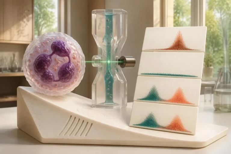
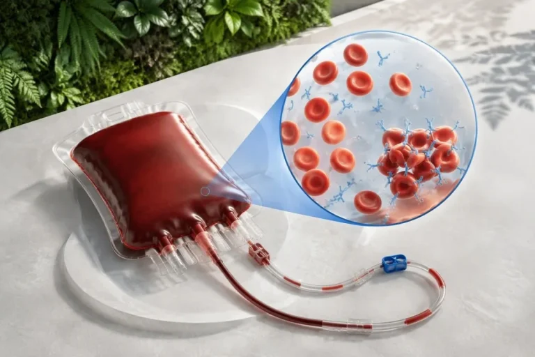
⚕️ Medische disclaimer
Dit artikel is uitsluitend bedoeld voor educatieve doeleinden en vormt geen medisch advies. Raadpleeg altijd een gekwalificeerde zorgverlener voor beslissingen over diagnose en behandeling.
E-E-A-T Vertrouwenssignalen
Ervaring
Klinische beoordeling door artsen van lab-interpretatieworkflows.
Expertise
Laboratoriumgeneeskunde met focus op hoe biomarkers zich gedragen in een klinische context.
Gezag
Geschreven door Dr. Thomas Klein, met review door Dr. Sarah Mitchell en Prof. Dr. Hans Weber.
Betrouwbaarheid
Evidence-based interpretatie met duidelijke vervolgstappen om onrust te verminderen.
