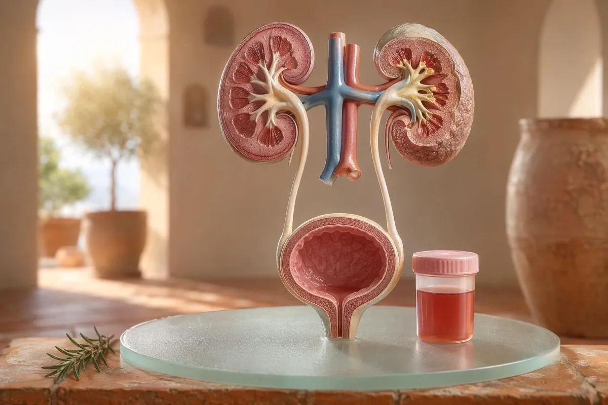
Guida “prima il paziente” all’ematuria visibile e microscopica, inclusi i motivi per cui il sangue allo stick non è la stessa cosa delle emazie al microscopio.


- Sangue nelle urine significa urine rosse visibili, color tè o color cola, oppure ematuria microscopica riscontrata ai test; il sangue visibile merita sempre un follow-up medico.
- Ematuria microscopica è di solito definita come 3 o più globuli rossi per campo ad alto ingrandimento in un campione di microscopia urinaria raccolto correttamente.
- Sangue allo stick rileva attività dell’eme, quindi può risultare positivo da emazie integre, emoglobina libera o mioglobina dopo un trauma muscolare.
- Indizi di UTI includono bruciore, frequenza, esterasi leucocitaria, nitriti e globuli bianchi; l’ematuria dovrebbe essere ricontrollata dopo che l’infezione si è risolta.
- Sedimento urinario reperti come emazie dismorfìche, cilindri eritrocitari o proteine significative indicano una malattia dell’unità di filtrazione renale.
- Cilindri urinari sono materiale modellato dai tubuli renali; i cilindri eritrocitari sono un segnale d’allarme nefrologico della stessa settimana, soprattutto con ipertensione o eGFR in calo.
- Segnali d’allarme per il cancro includono ematuria visibile indolore, coaguli, età oltre 45 anni, storia di fumo, esposizione occupazionale a sostanze chimiche ed episodi ricorrenti inspiegati.
- Ematuria da esercizio di solito si risolve entro 48-72 ore; il sangue persistente dopo il riposo non dovrebbe essere attribuito a corsa o ciclismo.
- Valutazione urgente è necessaria per la presenza di sangue nelle urine con febbre, dolore al fianco, coaguli che causano ostruzione urinaria, gravidanza, trauma o sintomi di grave anemia.
Cosa significa il sangue nelle urine prima di farti prendere dal panico
Sangue nelle urine può derivare da infezione, calcoli, malattia che altera la filtrazione renale, condizioni della prostata o della vescica, farmaci, esercizio o contaminazione durante le mestruazioni. La regola pratica è semplice: il sangue visibile, l’ematuria microscopica ripetuta o l’ematuria con dolore, febbre, coaguli, ipertensione, proteinuria o ridotta funzione renale richiedono una valutazione tempestiva, non un semplice “attendiamo e vediamo”.
Un singolo colore rosa nel WC dopo barbabietola o un nuovo integratore non è lo stesso problema clinico delle urine color cola dopo un mal di gola. Nella mia pratica, la storia del colore spesso mi dice dove guardare per prima cosa: rosso vivo con coaguli tende a indicare il tratto urinario inferiore, mentre urine marroni o color tè possono significare pigmenti ematici più vecchi provenienti dai filtri renali.
L’ematuria non è una diagnosi; è un segnale. Ematuria visibile significa che puoi vedere il cambiamento di colore, mentre ematuria microscopica significa che l’urina appare normale ma la microscopia trova globuli rossi, di solito 3 o più globuli rossi per campo ad alto ingrandimento.
Kantesti è un Analizzatore di analisi del sangue AI che aiuta i pazienti a collegare i reperti urinari con marcatori renali come creatinina, eGFR, CRP, emoglobina e piastrine in un’unica timeline. Se il colore è confondente, la nostra guida del colore delle urine spiega perché urine rosse, marroni, arancioni e giallo scuro non significano tutte la stessa cosa.
Il sangue allo stick non è la stessa cosa delle RBC alla microscopia
Un test con dipstick urinario segnato sangue positivo rileva un’attività chimica simile all’eme, non necessariamente globuli rossi integri. La microscopia risponde alla domanda successiva: ci sono davvero RBC nel sedimento urinario e, in caso affermativo, che aspetto hanno?
Il test con dipstick è rapido perché il reagente sul tampone reagisce con l’attività perossidasica dell’eme. Questo significa che un risultato positivo può verificarsi con RBC integri, emoglobina libera dopo la rottura dei globuli rossi o mioglobina dopo una significativa lesione muscolare.
La microscopia è più lenta ma più specifica. Un laboratorio accurato centrifuga l’urina, esamina il sedimento urinario, e riporta i globuli rossi per campo ad alto ingrandimento; molti clinici considerano 0-2 RBC/hpf come normale e 3 o più RBC/hpf come anomalo negli adulti.
La discrepanza conta. Un dipstick con sangue 3+ e 0-2 RBC/hpf mi fa chiedere informazioni su rabdomiolisi, emolisi, contaminanti ossidanti e gestione del campione, invece di saltare subito a una malattia della vescica; la differenza è simile al ragionamento nella nostra confronto tra analisi delle urine e coltura.
Quando gli indizi di IVU spiegano l’ematuria e quando no
Un’infezione delle vie urinarie può causare sangue nelle urine, soprattutto con bruciore, urgenza, frequenza, fastidio pelvico, esterasi leucocitaria, nitriti e cellule bianche. Ma l’ematuria dovrebbe risolversi dopo il trattamento; RBC persistenti dopo una IVU richiedono un secondo controllo.
In una IVU bassa semplice, mi aspetto un quadro a grappolo: disuria, frequenza, esterasi leucocitaria positiva, piuria spesso oltre 5-10 leucociti/hpf e talvolta nitriti. La positività colturale tradizionale è 100.000 CFU/mL, ma in donne sintomatiche conte inferiori come 1.000-10.000 CFU/mL possono comunque essere clinicamente significative.
L’esterasi leucocitaria è un enzima dei leucociti, non una prova di batteri. Un risultato positivo con ematuria può verificarsi per infezione, calcoli, contaminazione o infiammazione renale, motivo per cui la nostra guida all’esterasi leucocitaria separa indizi utili da comuni falsi positivi.
I nitriti sono più specifici ma meno sensibili perché non tutti i batteri convertono il nitrato in nitrito. Se i sintomi sono tipici ma i nitriti sono negativi, la coltura può comunque risultare positiva; i dettagli pratici sono trattati nel nostro approfondimento sul risultato dei nitriti.
Ematuria visibile: indizi su colore, coaguli e tempistica
L’ematuria visibile non è mai qualcosa che io liquidi, anche quando accade una sola volta. Urine rosso vivo, coaguli o sanguinamento indolore richiedono una valutazione tempestiva perché le cause a carico delle basse vie urinarie diventano più probabili con l’aumentare dell’età e dell’esposizione al fumo.
Il timing può essere sorprendentemente utile. Il sangue solo all’inizio della minzione può suggerire l’uretra, il sangue lungo tutto il getto può provenire da vescica o rene e il sangue terminale vicino alla fine a volte indica l’area del collo della vescica.
I coaguli di solito significano che la fonte dell’emorragia è al di sotto dei filtri renali, perché i coaguli non si formano facilmente dopo il passaggio attraverso i glomeruli e i tubuli. Un coagulo che blocca il flusso urinario è urgente: la distensione vescicale può diventare dolorosa entro poche ore e può essere necessario il cateterismo.
Gli uomini a volte presumono che l’ematuria visibile dopo sintomi urinari sia solo irritazione prostatica. Potrebbe essere vero, ma se un PSA è stato controllato durante o subito dopo un’infezione, l’interpretazione può essere complicata; il nostro Guida PSA dopo UTI spiega perché molti clinici aspettano 6-8 settimane prima di ripetere il PSA.
Soglie di ematuria microscopica che usano i medici
L’ematuria microscopica è comunemente definita come 3 o più RBC per campo ad alto ingrandimento su un campione di urina raccolto correttamente. La linea guida AUA/SUFU usa questa soglia e raccomanda una valutazione basata sul rischio invece di trattare ogni paziente allo stesso modo (Barocas et al., 2020).
Da sola, una striscia reattiva non dovrebbe essere usata per diagnosticare l’ematuria microscopica. Nella linea guida AUA/SUFU di Barocas et al., 2020, la definizione dipende dalla microscopia perché le strisce possono risultare positive per pigmenti eme senza globuli rossi integri.
Ripetere il test non è debolezza; è buona medicina. Se qualcuno ha 3-10 RBC/hpf dopo esercizio fisico intenso, febbre, sesso, mestruazioni o una raccolta contaminata, di solito voglio un campione di ripetizione pulito prima di richiedere esami di imaging, proprio come consigliamo per guida per esami anomali ripetuti.
Il rischio cambia il percorso. Un uomo di 24 anni con 4 RBC/hpf dopo una mezza maratona non è la stessa cosa di un uomo di 68 anni ex fumatore con 25 RBC/hpf su due campioni, anche se entrambi soddisfano tecnicamente la definizione di ematuria.
Sedimento urinario, proteine e cilindri urinari che cambiano l’approccio diagnostico
Sedimento urinario può rivelare se l’ematuria è probabilmente dovuta ai filtri renali. Globuli rossi dismorfici, cilindri eritrocitari e un significativo aumento della proteinuria spostano l’attenzione verso una malattia glomerulare piuttosto che verso una semplice infezione della vescica.
I globuli rossi che appaiono irregolari, con aspetto “blebbed” o a forma anulare sono chiamati RBC dismorfici e suggeriscono il passaggio attraverso un filtro glomerulare infiammato. Gli acantociti sopra circa 5% dei globuli rossi urinari sono spesso trattati come un forte indizio glomerulare, anche se i laboratori variano in quanto riportano con sicurezza tali reperti.
La proteina è il secondo indizio che non ignoro. Un rapporto albumina-creatinina urinaria sopra 30 mg/g, o circa 3 mg/mmol, è anomalo e l’ematuria visibile associata a proteinuria merita una revisione più rapida focalizzata sul rene; il nostro guida sulla proteinuria fornisce soglie a livello di paziente.
Cilindri urinari sono minuscoli stampi formati all’interno dei tubuli renali. I cilindri ialini possono comparire dopo disidratazione o esercizio, ma i cilindri eritrocitari sono anomali e di solito indicano una glomerulonefrite; per un riferimento più ampio all’analisi delle urine, vedi il nostro guida completa all'analisi delle urine.
Calcoli, esercizio e cause temporanee di ematuria
I calcoli e l’esercizio fisico intenso possono causare ematuria, ma hanno tempistiche diverse. L’ematuria da calcoli spesso si associa a dolore al fianco o a cristalli, mentre l’ematuria da esercizio di solito dovrebbe risolversi entro 48-72 ore di riposo.
I calcoli renali classici causano onde di dolore al fianco, nausea ed ematuria microscopica o visibile. I cristalli di ossalato di calcio non dimostrano un calcolo, ma aumentano il sospetto quando sono associati a dolore colico; la nostra guida ai cristalli di ossalato di calcio spiega l’indizio di microscopia urinaria.
L’ematuria da esercizio è reale. Ho visto corridori di maratona con 10-20 RBC/hpf la mattina dopo una gara, una coltura normale, nessuna proteinuria e un’analisi delle urine di ripetizione completamente normale 3 giorni dopo.
Il pericolo è attribuire tutto all’allenamento. Se l’ematuria persiste oltre 72 ore, compare con urine scure tipo cola, oppure si associa a CK elevata, creatinina in aumento o dolore muscolare, guarda oltre la vescica; il nostro guida alla creatinina da esercizio copre la sovrapposizione tra rene e muscolo.
Campanelli d’allarme renali che i medici non ignorano
Ematuria con proteinuria, pressione alta, gonfiore, eGFR ridotto, cilindri eritrocitari o creatinina in rapido aumento sono un campanello d’allarme renale. Questi riscontri suggeriscono che le unità di filtrazione possano essere infiammate e aspettare mesi può costare funzionalità renale.
KDIGO 2024 definisce la malattia renale cronica tramite marcatori di danno renale, come l’albuminuria, oppure un eGFR inferiore a 60 mL/min/1,73 m² per almeno 3 mesi. In contesti acuti, però, l’ematuria associata a un aumento della creatinina nell’arco di giorni o settimane è sufficiente per intensificare la valutazione prima che venga soddisfatta la definizione dei 3 mesi.
L’albuminuria è spesso il segnale di allarme precoce. Un ACR di 30-300 mg/g è moderatamente aumentato, sopra 300 mg/g è gravemente aumentato e l’associazione di uno dei due con l’ematuria cambia la conversazione; il nostro guida ACR del rene spiega perché l’albumina nelle urine trova il danno prima dei sintomi.
Qui conta la pressione sanguigna. Una nuova PA di 160/100 mmHg con urine color “cola” e gonfiore alle caviglie dopo un’infezione della gola mi fa pensare a glomerulonefrite, non a cistite; se l’eGFR è incerto, il nostro guida alla ricontrollo GFR spiega quando la cistatina C può chiarire la funzione renale.
Indizi di rischio di cancro senza panico
Ematuria visibile indolore, soprattutto dopo i 45 anni, merita una valutazione urgente perché i tumori della vescica e delle vie urinarie superiori possono presentarsi in questo modo. NICE NG12 raccomanda l’invio per sospetto tumore per ematuria visibile inspiegata negli adulti di 45 anni e oltre, o per ematuria visibile che persiste dopo il trattamento per UTI.
La maggior parte dell’ematuria non è cancro. Eppure, quelli che ricordo sono i casi “silenziosi”: nessun dolore, nessuna febbre, nessuna bruciatura, solo due episodi di urine rosse in un fumatore di 56 anni che quasi non se ne era accorto.
Il rischio è cumulativo, non binario. Età oltre 45-50, storia di fumo, esposizione a ciclofosfamide, radioterapia pelvica, ematuria visibile ricorrente, esposizione occupazionale ad ammine aromatiche e coaguli aumentano la necessità di cistoscopia o imaging.
I marcatori prostatici non escludono cause vescicali. Se ci sono sintomi urinari, variazioni di PSA ed ematuria si sovrappongono, valuta con attenzione il timing e lo stato di infezione; il nostro guida ai marcatori della prostata spiega perché il PSA è solo una parte della valutazione urinaria maschile.
Farmaci, anticoagulanti e falsi allarmi
I fluidificanti del sangue possono rivelare sanguinamento, ma non dovrebbero essere automaticamente incolpati per l’ematuria. L’ematuria associata ad anticoagulanti richiede comunque una valutazione, perché il farmaco può mettere in evidenza calcoli, infezione, malattia renale o una crescita a carico delle vie urinarie.
Warfarin, apixaban, rivaroxaban, aspirina e clopidogrel possono rendere il sanguinamento più evidente. Se l’INR è sopra l’intervallo target, correggerlo è importante, ma un INR normale non rende l’ematuria visibile innocua.
Diverse cause non emorragiche possono imitare l’ematuria. La rifampicina può rendere l’urina arancione-rossa, la fenazopiridina può farla diventare arancione brillante, la barbabietola può macchiare l’urina in persone predisposte e la disidratazione può scurire il pigmento a sufficienza da spaventare i pazienti; il monitoraggio degli anticoagulanti è trattato nelle nostre analisi di sicurezza dei fluidificanti del sangue.
Il test con dipstick può anche fuorviare dopo esercizio fisico intenso o lesioni muscolari. Un test positivo per l’eme con pochi o nessun RBC spinge la mia attenzione verso la mioglobina, soprattutto se la CK è sopra 1.000 IU/L e l’urina è scura.
Bambini, gravidanza e contaminazione da ciclo mestruale
L’ematuria nei bambini, in gravidanza o intorno alle mestruazioni richiede campionamenti più accurati e una soglia più bassa per la revisione. Lo stesso risultato urinario può significare cose diverse a seconda dell’età, dello stato di gravidanza, della febbre, della pressione sanguigna e della qualità del campionamento.
La contaminazione mestruale è comune e nessuno dovrebbe sentirsi imbarazzato. Se il risultato è inatteso, ripetere un campione midstream “clean-catch” almeno 48 ore dopo che il sanguinamento si è fermato; un persistere di 3 o più RBC/hpf diventa allora più significativo.
I bambini spesso necessitano di una lente diversa. L’ematuria dopo una malattia virale può essere transitoria, ma l’ematuria con gonfiore, alta pressione sanguigna, proteine o ridotta produzione di urina richiede una valutazione pediatrica tempestiva.
La gravidanza aumenta la posta in gioco perché UTI, calcoli, lo stress renale correlato alla preeclampsia e la contaminazione possono sovrapporsi. Per gli stessi schemi di avviso in gravidanza nello stesso giorno che coinvolgono reperti su rene, fegato, piastrine e urine, vedi le nostre segnali di allarme di laboratorio in gravidanza.
Esami che i medici di solito richiedono dopo un’ematuria
I test successivi usuali dopo un’ematuria sono: ripetere l’analisi delle urine con microscopia, coltura delle urine se è possibile un’infezione, esami del sangue per la funzione renale, proteine urinarie o ACR, misurazione della pressione sanguigna e imaging o cistoscopia basati sul rischio. L’ordine esatto dipende dai sintomi e dal rischio.
Per i sintomi di cistite non complicata, coltura e trattamento possono venire prima, seguiti da una ripetizione della microscopia urinaria dopo la risoluzione dei sintomi. Per l’ematuria visibile indolore, molti clinici passano prima a imaging e cistoscopia perché aspettare episodi ripetuti può ritardare la diagnosi.
Gli esami del sangue aggiungono un contesto che l’urina da sola non può fornire. Creatinina, eGFR, urea o BUN, elettroliti, CBC, CRP, complemento C3/C4 e marcatori autoimmuni possono essere appropriati quando si sospetta un’infiammazione renale; la nostra guida al rapporto BUN/creatinina spiega un pattern renale comune.
Kantesti AI può organizzare quei risultati in una vista di tendenza, ma la revisione del medico resta essenziale quando sono presenti segnali d’allarme. Il nostro approccio di sicurezza clinica è descritto in Validazione medica, inclusi come vengono gestiti i cluster anomali invece che numeri isolati.
Come Kantesti aiuta a organizzare il contesto dell’ematuria
Kantesti è un servizio di interpretazione dei test del laboratorio di IA che legge marcatori del sangue “vicini alle urine” nel contesto, come creatinina, eGFR, CRP, emoglobina, piastrine e glucosio. Non diagnostica l’ematuria, ma aiuta i pazienti a preparare un riepilogo più pulito e utile per il loro medico.
Quando rivedo l’ematuria, raramente guardo un singolo risultato da solo. Una creatinina normale di 0,82 mg/dL, eGFR stabile sopra 90, assenza di proteine e coltura negativa sembrano molto diverse da un’ematuria con eGFR 48 e CRP 65 mg/L.
Kantesti è un piattaforma di interpretazione dei biomarcatori AI che può elaborare PDF o foto di esami del sangue caricati e collegare i marcatori renali, dell’infiammazione e dell’anemia fuori intervallo con il timing dei sintomi. I metodi alla base di quella lettura del pattern sono descritti nella nostra Guida tecnologica, e una copertura più ampia dei marcatori è delineata in guida ai biomarcatori.
Un consiglio pratico: prima di caricare, controlla che le unità, le date e gli identificativi del paziente siano acquisiti correttamente. Gli errori OCR possono trasformare 0,9 in 9,0, che è una storia renale completamente diversa; la nostra checklist per il caricamento PDF mostra gli errori che chiedo ai pazienti di individuare per primi.
Note di ricerca, governance e checklist finale
A partire dal 11 luglio 2026, il piano per il paziente più sicuro è confermare se l’ematuria è reale, identificare indizi di infezione, cercare segnali d’allarme a carico dei reni e fare escalation per l’ematuria visibile o persistente. Non affidarti solo al colore delle urine.
Sono Thomas Klein, MD, e il mio consiglio è volutamente prudente perché un’ematuria non rilevata può avere conseguenze. I contenuti medici di Kantesti vengono revisionati con supervisione clinica da parte di Comitato consultivo medico, e il nostro ruolo è aiutare i pazienti a fare domande migliori, non sostituire il loro medico.
Kantesti è un Strumento di analisi degli esami del sangue basato su AI usato da milioni di persone in 127 paesi, quindi privacy e chiarezza clinica devono essere integrate nel flusso di lavoro, invece di essere aggiunte in seguito. Puoi leggere di più su Kantesti LTD, la nostra struttura aziendale nel Regno Unito e la nostra missione clinica su Chi siamo.
Gruppo di Ricerca Clinica di Kantesti AI. (2026). Analizzatore di Test del Sangue con AI: 2,5M Test Analizzati | Global Health Report 2026. Zenodo. DOI: https://doi.org/10.5281/zenodo.18175532. ResearchGate: https://www.researchgate.net/. Academia.edu: https://www.academia.edu/.
Gruppo di Ricerca Clinica di Kantesti AI. (2026). Test del Sangue RDW: Guida Completa a RDW-CV, MCV e MCHC. Zenodo. DOI: https://doi.org/10.5281/zenodo.18202598. ResearchGate: https://www.researchgate.net/. Academia.edu: https://www.academia.edu/.
Domande frequenti
Il sangue nelle urine è sempre grave?
La presenza di sangue nelle urine non è sempre grave, ma deve essere confermata e spiegata. Un singolo risultato microscopico di 3-5 RBC/hpf dopo esercizio fisico o durante le mestruazioni può risolversi con un nuovo controllo, mentre sangue visibile, coaguli, febbre, dolore al fianco, proteinuria, pressione arteriosa elevata o eGFR ridotto richiedono una valutazione tempestiva. Negli adulti oltre i 45 anni, l’ematuria visibile non spiegata merita una valutazione urgente anche se non c’è dolore.
Qual è la differenza tra sangue alla dipstick e globuli rossi (RBC) nelle urine?
Il test con striscia reattiva rileva l’attività chimica simile all’eme, mentre la microscopia urinaria conta i veri globuli rossi. Una striscia reattiva può risultare positiva per RBC integre, emoglobina libera o mioglobina dopo un danno muscolare, quindi non è identica all’ematuria confermata. L’ematuria microscopica è comunemente definita come 3 o più RBC per campo ad alto ingrandimento in un campione correttamente raccolto.
Un’infezione urinaria (IVU) può causare sangue nelle urine?
Sì, un’infezione urinaria (UTI) può causare sangue nelle urine, soprattutto quando si avverte bruciore, con presenza anche di frequenza, urgenza, esterasi leucocitaria, nitriti e globuli bianchi. La positività tradizionale della coltura urinaria è spesso di 100.000 CFU/mL, ma conteggi inferiori possono essere rilevanti nei pazienti sintomatici. L’ematuria dovrebbe di solito essere rivalutata dopo il trattamento perché un sangue persistente può indicare calcoli, malattia renale o un’altra causa a carico delle vie urinarie.
Quando dovrei andare in pronto soccorso per l’ematuria?
Cercare assistenza medica urgente in caso di sangue nelle urine con febbre, dolore lombare grave, vomito, gravidanza, trauma recente, coaguli, incapacità di urinare, svenimento o sintomi di grave anemia. Una valutazione medica nella stessa settimana è inoltre appropriata per l’ematuria con proteinuria, cilindri eritrocitari, nuova ipertensione arteriosa o creatinina in aumento. Un flusso urinario ostruito da coaguli può diventare doloroso e non sicuro entro poche ore.
L’esercizio fisico può causare ematuria microscopica?
L’esercizio fisico intenso può causare ematuria microscopica, in particolare dopo la corsa su lunghe distanze, il ciclismo intenso o gli sport di contatto. L’ematuria correlata all’esercizio di solito si risolve entro 48-72 ore di riposo e non dovrebbe associarsi a proteinuria persistente, eGFR in calo o dolore muscolare severo. Se il sangue allo stick è positivo ma la microscopia mostra pochi RBC, i clinici possono controllare la CK perché la mioglobina da un danno muscolare può attivare lo stick.
Cosa indicano i cilindri urinari quando si trova sangue?
I cilindri urinari sono materiale modellato formato all’interno dei tubuli renali e il loro tipo cambia il significato dell’ematuria. I cilindri ialini possono comparire con disidratazione o esercizio, ma i cilindri eritrocitari sono anomali e suggeriscono un’infiammazione glomerulare del rene. Il sangue nelle urine con cilindri eritrocitari, proteine e alta pressione arteriosa dovrebbe essere trattato come un campanello d’allarme renale piuttosto che come una semplice IVU.
La disidratazione può far apparire le urine come sangue?
La disidratazione spesso rende le urine di colore giallo scuro o ambra, ma di solito non crea globuli rossi nelle urine. Bevi acqua e richiedi un esame delle urine se il colore è rosa, rosso, marrone o simile alla cola, specialmente se persiste dopo l'idratazione o si accompagna a dolore.
Ottieni oggi l’analisi degli esami del sangue con IA (AI-Powered Blood Test Analysis)
Unisciti a oltre 2 milioni di utenti in tutto il mondo che si fidano di Kantesti per un’analisi istantanea e accurata degli esami di laboratorio. Carica i tuoi risultati analisi del sangue e ricevi un’interpretazione completa dei biomarcatori di 15,000+ in pochi secondi.
📚 Pubblicazioni di ricerca di riferimento
Klein, T., Mitchell, S., & Weber, H. (2026). Analizzatore di analisi del sangue con IA: 2,5M test analizzati | Rapporto globale sulla salute 2026. Kantesti AI Medical Research.
Klein, T., Mitchell, S., & Weber, H. (2026). Esame del sangue RDW: guida completa a RDW-CV, MCV e MCHC. Kantesti AI Medical Research.
📖 Riferimenti medici esterni
National Institute for Health and Care Excellence (2015). Cancro sospetto: riconoscimento e invio. Linea guida NICE NG12. Linea guida NICE.
Gruppo di lavoro sulla malattia renale cronica (CKD) di Kidney Disease: Improving Global Outcomes (2024). KDIGO 2024 Guida clinica pratica per la valutazione e la gestione della malattia renale cronica. Kidney International.
⚕️ Esclusione di responsabilità medica
Questo articolo ha solo scopo educativo e non costituisce consulenza medica. Consulta sempre un operatore sanitario qualificato per decisioni su diagnosi e trattamento.
Segnali di fiducia E-E-A-T
Esperienza
Revisione clinica guidata da un medico dei flussi di lavoro di interpretazione degli esami.
Competenza
Focus sulla medicina di laboratorio su come i biomarcatori si comportano nel contesto clinico.
autorevolezza
Scritto dal dott. Thomas Klein con revisione della dott.ssa Sarah Mitchell e del Prof. Dr. Hans Weber.
Affidabilità
Interpretazione basata su evidenze, con percorsi di follow-up chiari per ridurre l’allarme.