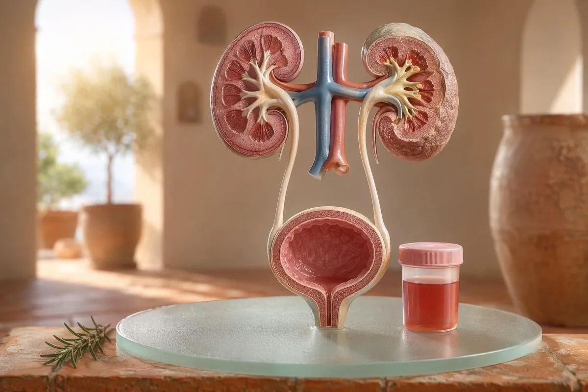
Pacientam pirmajā vietā vērsts ceļvedis redzamai un mikroskopiskai hematūrijai, tostarp par to, kāpēc ar teststrēmeli konstatētās asinis nav tas pats, kas sarkanās asins šūnas mikroskopā.


- Asinis urīnā nozīmē redzami sarkanu, tējas krāsas vai kolas krāsas urīnu, vai mikroskopisku hematūriju, kas konstatēta izmeklēšanā; redzamas asinis vienmēr ir pelnījušas medicīnisku turpmāku izvērtēšanu.
- Mikroskopiska hematūrija parasti tiek definēta kā 3 vai vairāk eritrocīti (RBC) uz viena lieljaudas lauka (high-power field) pareizi savāktā urīna mikroskopijas paraugā.
- Teststrēmeles asinis nosaka hema aktivitāti, tāpēc tās var būt pozitīvas no neskartām sarkanajām asins šūnām, brīva hemoglobīna vai mioglobīna pēc muskuļu traumas.
- UTI norādes ietver dedzināšanu, biežumu, leikocītu esterāzi, nitrītus un baltās asins šūnas; hematūrija jāpārbauda atkārtoti pēc tam, kad infekcija ir izzudusi.
- Urīna nogulsnes tādi atradumi kā dismorfie eritrocīti, eritrocītu cilindri vai nozīmīgs proteīns norāda uz nieru filtrēšanas vienības slimību.
- Urīna cilindri ir veidotas no nieru kanāliņu materiāla; eritrocītu cilindri ir vienas nedēļas laikā parādījusies nefroloģiska “sarkanais karogs”, īpaši, ja ir augsts asinsspiediens vai krītas eGFR.
- Vēža brīdinājuma pazīmes ietver nesāpīgu redzamu hematūriju, recekļus, vecumu virs 45 gadiem, smēķēšanas vēsturi, profesionālu ķīmisku vielu iedarbību un atkārtotas neizskaidrojamas epizodes.
- Fiziska slodzes izraisīta hematūrija parasti norimst 48–72 stundu laikā; pastāvīga asiņu klātbūtne pēc atpūtas nedrīkst tikt vainota skriešanā vai riteņbraukšanā.
- Steidzama izvērtēšana ir nepieciešama, ja urīnā ir asinis kopā ar drudzi, sāpēm sānos, recekļiem, kas izraisa urīna nosprostojumu, grūtniecību, traumu vai smagas anēmijas simptomiem.
Ko nozīmē asinis urīnā, pirms jūs sākat krist panikā
Asinis urīnā var rasties infekcijas, akmeņu, nieru filtrēšanas slimības, prostatas vai urīnpūšļa stāvokļu, medikamentu, fiziskas slodzes dēļ vai arī periodu izraisīta kontaminācija dēļ. Praktiskais noteikums ir vienkāršs: redzamas asinis, atkārtota mikroskopiska hematūrija vai hematūrija ar sāpēm, drudzi, recekļiem, augstu asinsspiedienu, proteīnūriju vai samazinātu nieru funkciju prasa steidzamu izvērtēšanu, nevis novērošanu bez rīcības.
Vienreizējs sārts tualetes poda saturs pēc bietēm vai jauna uztura bagātinātāja nav tāda pati klīniskā problēma kā kolas krāsas urīns pēc iekaisuša kakla. Manā praksē krāsas stāsts bieži pasaka, kur vispirms meklēt: spilgti sarkans ar recekļiem mēdz liecināt par apakšējo urīnceļu problēmu, bet brūns vai tējas krāsas urīns var nozīmēt vecākus hema pigmentus no nieru filtriem.
Hematūrija nav diagnoze; tā ir pazīme. Redzama hematūrija nozīmē, ka jūs redzat krāsas maiņu, savukārt mikroskopiska hematūrija nozīmē, ka urīns izskatās normāls, bet mikroskopijā tiek atrastas sarkanās asins šūnas, parasti pie 3 vai vairāk sarkanajām asins šūnām uz viena lieljaudas redzes lauka.
Kantesti ir AI asins analīžu analizators kas palīdz pacientiem vienā laika līnijā sasaistīt urīna atradnes ar nieru rādītājiem, piemēram, kreatinīnu, eGFR, CRP, hemoglobīnu un trombocītiem. Ja krāsa ir neskaidra, mūsu urīna krāsu ceļvedis izskaidro, kāpēc sarkans, brūns, oranžs un tumši dzeltens urīns ne vienmēr nozīmē vienu un to pašu.
Ar teststrēmeli konstatētās asinis nav tas pats, kas RBC mikroskopijā
Urīna teststrēmele ar atzīmi “asinis pozitīvas” nosaka hemai līdzīgu ķīmisku aktivitāti, ne vienmēr neskartas eritrocītu asins šūnas. Mikroskopija atbild uz nākamo jautājumu: vai urīna nogulumos ir īstas RBC, un ja jā, tad kā tās izskatās?
Teststrēmeļu pārbaude ir ātra, jo reaģenta laukums reaģē ar hema peroksidāzes aktivitāti. Tas nozīmē, ka pozitīvs rezultāts var rasties ar neskartiem RBC, brīvu hemoglobīnu pēc eritrocītu sabrukšanas vai mioglobīnu pēc nozīmīga muskuļu bojājuma.
Mikroskopija ir lēnāka, bet precīzāka. Rūpīga laboratorija centrifugē urīnu, apskata urīna nogulsnes, un ziņo par sarkanajām asins šūnām uz viena lieljaudas redzes lauka; daudzi klīnicisti pieaugušajiem uzskata 0–2 RBC/hpf par normu un 3 vai vairāk RBC/hpf par patoloģisku.
Neatbilstība ir svarīga. Teststrēmele ar 3+ asinīm un 0–2 RBC/hpf liek man jautāt par rabdomiolīzi, hemolīzi, oksidējošiem kontaminantiem un parauga apstrādi, nevis uzreiz lēkt uz urīnpūšļa slimību; atšķirība ir līdzīga loģikai mūsu urīna analīzes un kultūras salīdzinājums.
Kad UTI pazīmes izskaidro hematūriju un kad ne
Urīnceļu infekcija var izraisīt asinis urīnā, īpaši, ja ir dedzināšana, steidzamība, biežums, diskomforts iegurņa apvidū, leikocītu esterase, nitrīti un leikocīti. Taču hematūrijai pēc ārstēšanas vajadzētu izzust; noturīgas sarkanās asins šūnas pēc UTI prasa atkārtotu izvērtējumu.
Vienkāršā apakšējo UTI gadījumā es gaidu šādu “kopu”: dizūrija, biežums, leikocītu esterase pozitīva, piūrija bieži virs 5–10 leikocītiem/hpf un dažreiz nitrīti. Tradicionāla kultūras pozitivitāte ir 100 000 CFU/mL, bet simptomātiskām sievietēm klīniski nozīmīgi var būt arī zemāki rādītāji, piemēram, 1 000–10 000 CFU/mL.
Leikocītu esterase ir leikocītu enzīms, nevis pierādījums baktērijām. Pozitīvs rezultāts ar hematūriju var rasties infekcijas, akmeņu, kontaminācijas vai nieru iekaisuma dēļ, tāpēc mūsu leikocītu esterase ceļvedis atdala noderīgas norādes no biežiem viltus pozitīviem.
Nitrīti ir specifiskāki, bet mazāk jutīgi, jo ne visas baktērijas pārvērš nitrātu par nitrītu. Ja simptomi ir klasiski, bet nitrīti ir negatīvi, kultūra joprojām var būt pozitīva; praktiskās detaļas ir izklāstītas mūsu nitrītu rezultātu skaidrojumā.
Redzama hematūrija: krāsa, recekļi un laika norādes
Redzama hematūrija nekad nav kaut kas, ko es ignorētu, pat ja tā notiek tikai vienreiz. Spilgti sarkans urīns, recekļi vai nesāpīga asiņošana prasa savlaicīgu izvērtējumu, jo, pieaugot vecumam un smēķēšanas iedarbībai, biežāk kļūst iespējami cēloņi no apakšējiem urīnceļiem.
Laiks var būt pārsteidzoši noderīgs. Asinis tikai urinēšanas sākumā var liecināt par urīnizvadkanālu, asinis visā strūklā var nākt no urīnpūšļa vai nieru avotiem, un terminālas asinis tuvu beigām dažkārt norāda uz urīnpūšļa kakliņa apvidu.
Trombi parasti nozīmē, ka asiņošanas avots atrodas zem nieru filtriem, jo trombi viegli neveidojas pēc iziešanas caur glomeruliem un kanāliņiem. Trombs, kas bloķē urīna plūsmu, ir steidzams stāvoklis: urīnpūšļa pārpildīšanās var kļūt sāpīga dažu stundu laikā, un var būt nepieciešama katetra aprūpe.
Vīrieši dažkārt pieņem, ka redzama hematūrija pēc urīnceļu simptomiem ir tikai prostatas kairinājums. Tas var būt patiesi, taču, ja PSA tika pārbaudīts infekcijas laikā vai drīz pēc tās, interpretācija var būt sarežģīta; mūsu PSA pēc UTI vadlīnijas izskaidro, kāpēc daudzi klīnicisti gaida 6–8 nedēļas, pirms atkārto PSA.
Mikroskopiskās hematūrijas sliekšņi, ko izmanto ārsti
Mikroskopiska hematūrija parasti tiek definēta kā 3 vai vairāk RBC uz augstas palielināšanas lauku pareizi savāktā urīna paraugā. AUA/SUFU vadlīnijas izmanto šo slieksni un iesaka uz risku balstītu izvērtēšanu, nevis ārstēt katru pacientu vienādi (Barocas et al., 2020).
Vienīgi teststrēmeli nevajadzētu izmantot, lai diagnosticētu mikroskopisku hematūriju. Barocas et al. 2020 AUA/SUFU vadlīnijās definīcija ir atkarīga no mikroskopijas, jo teststrēmeles var būt pozitīvas hema pigmentu dēļ bez neskartiem eritrocītiem.
Atkārtota pārbaude nav vājums; tā ir laba medicīna. Ja pēc intensīvas slodzes, drudža, dzimumakta, menstruācijām vai piesārņotas savākšanas kādam ir 3–10 RBC/hpf, es parasti gribu tīru atkārtotu paraugu pirms izmeklējumu (scan) nozīmēšanas, līdzīgi kā mēs iesakām atkārtoti neparasti izmeklējumi.
Riska izmaiņas maina ceļu. 24 gadus vecs cilvēks ar 4 RBC/hpf pēc pusmaratona nav tas pats, kas 68 gadus vecs bijušais smēķētājs ar 25 RBC/hpf divos paraugos, pat ja abi tehniski atbilst hematūrijas definīcijai.
Urīna nogulsnes, proteīns un urīna cilindri, kas maina izmeklēšanas plānu
Urīna nogulsnes var atklāt, vai hematūrija, visticamāk, nāk no nieru filtriem. Disemorfi eritrocīti, eritrocītu cilindri un nozīmīga proteīna izmaiņa bažas novirza uz glomerulāru slimību, nevis vienkāršu urīnpūšļa infekciju.
Eritrocīti, kas izskatās nevienmērīgi, ar „bleb” veida izaugumiem vai gredzenveida, tiek saukti par disemorfiem RBC, un tie liecina par iziešanu caur iekaistu glomerulāru filtru. Acanthocytes virs aptuveni 5% urīna eritrocītu bieži ārstē kā spēcīgu glomerulāru norādi, lai gan laboratorijas atšķiras ar to, cik pārliecinoši tās ziņo.
Proteīns ir otrā norāde, ko es neignorēju. Urīna albumīna–kreatinīna attiecība virs 30 mg/g jeb aptuveni 3 mg/mmol ir patoloģiska, un redzama hematūrija kopā ar proteīnu prasa ātrāku nierēm vērstu izvērtējumu; mūsu proteīns urīnā ceļvedis sniedz pacientam balstītus sliekšņus.
Urīna cilindri ir sīki „mold” veida veidojumi, kas veidojas nieru kanāliņos. Hialīnie cilindri var parādīties pēc dehidratācijas vai slodzes, bet eritrocītu cilindri ir patoloģiski un parasti norāda uz glomerulonefrītu; plašākai urīna analīzes atsaucei skatiet mūsu Pilnīga urīna analīzes rokasgrāmata.
Akmeņi, fiziskā slodze un pārejoši hematūrijas cēloņi
Akmeņi un spraiga fiziska slodze var izraisīt hematūriju, taču tām ir atšķirīgi laika grafiki. Hematūrija, kas saistīta ar akmeni, bieži nāk kopā ar sāpēm sānos (flank pain) vai kristāliem, savukārt slodzes izraisītai hematūrijai parasti vajadzētu norimt 48–72 stundu laikā pēc atpūtas.
Nierakmeņi klasiskā veidā izraisa sāpju viļņus sānos, sliktu dūšu un mikroskopisku vai redzamu hematūriju. Kalcija oksalāta kristāli nepierāda akmeni, bet tie palielina aizdomas, ja kopā ar kolikveida sāpēm; mūsu kalcija oksalāta kristālu vadlīnijas izskaidro urīna mikroskopijas norādi.
Slodzes izraisīta hematūrija ir īsta. Esmu redzējis maratonistus ar 10–20 RBC/hpf nākamajā rītā pēc sacensībām, normālu kultūru, bez proteīna un pilnīgi normālu atkārtotu urīna analīzi 3 dienas vēlāk.
Briesmas ir visu vainot treniņos. Ja hematūrija saglabājas ilgāk par 72 stundām, parādās ar tumšu kolas krāsas urīnu vai nāk kopā ar augstu CK, pieaugošu kreatinīnu vai muskuļu sāpēm, skatieties tālāk par urīnpūsli; mūsu vingrinājumu kreatinīna ceļvedis aptver nieru un muskuļu pārklāšanos.
Nieru “sarkanie karogi”, ko ārsti neignorē
Hematūrija ar proteīnu, augsts asinsspiediens, tūska, samazināts eGFR, eritrocītu cilindri vai strauji pieaugošs kreatinīns ir nieru sarkanais karogs. Šie atradumi liecina, ka filtrējošās vienības var būt iekaisušas, un gaidīšana mēnešiem var maksāt nieru funkciju.
KDIGO 2024 hronisku nieru slimību definē pēc nieru bojājuma marķieriem, piemēram, albuminūrijas, vai eGFR zem 60 mL/min/1,73 m² vismaz 3 mēnešus. Tomēr akūtā situācijā hematūrija kopā ar kreatinīna pieaugumu dažu dienu vai nedēļu laikā ir pietiekama, lai eskalētu, pirms tiek sasniegta 3 mēnešu definīcija.
Albuminūrija bieži ir agrīnais brīdinājuma signāls. ACR 30–300 mg/g ir mēreni palielināts, virs 300 mg/g ir izteikti palielināts, un, ja kādu no tiem kombinē ar hematūriju, saruna mainās; mūsu nieru ACR ceļvedis izskaidro, kāpēc urīna albumīns atrod bojājumu pirms simptomiem.
Šeit nozīmīgs ir asinsspiediens. Jauns BP 160/100 mmHg ar kolas krāsas urīnu un potīšu tūsku pēc rīkles infekcijas liek man domāt par glomerulonefrītu, nevis cistītu; ja eGFR ir neskaidrs, mūsu GFR atkārtotas pārbaudes ceļvedis izskaidro, kad cistatīns C var precizēt nieru funkciju.
Vēža riska norādes bez panikas
Nesāpīga redzama hematūrija, īpaši pēc 45 gadu vecuma, ir pelnījusi steidzamu izvērtēšanu, jo urīnpūšļa un augšējo urīnceļu vēži var izpausties šādi. NICE NG12 iesaka aizdomas par vēzi nosūtīšanai pie speciālista pieaugušajiem no 45 gadu vecuma un vecākiem ar neizskaidrojamu redzamu hematūriju vai redzamu hematūriju, kas saglabājas pēc UTI ārstēšanas.
Lielākā daļa hematūrijas nav vēzis. Tomēr gadījumi, ko es atceros, ir tie klusākie: nav sāpju, nav drudža, nav dedzināšanas — tikai divas epizodes ar sarkanu urīnu 56 gadus vecam smēķētājam, kurš gandrīz to ignorēja.
Risks ir kumulatīvs, nevis binārs. Vecums virs 45–50, smēķēšanas vēsture, ciklofosfamīda iedarbība, iegurņa staru terapija, atkārtota redzama hematūrija, profesionāla aromātisko amīnu iedarbība un asins recekļi — visi palielina nepieciešamību pēc cistoskopijas vai attēldiagnostikas.
Prostatas marķieri neizslēdz urīnpūšļa cēloņus. Ja ir urīnceļu simptomi, PSA izmaiņas un hematūrija pārklājas, rūpīgi izvērtējiet laiku un infekcijas statusu; mūsu prostatas marķieru ceļvedis skaidro, kāpēc PSA ir tikai viena daļa no vīrieša urīnceļu izvērtēšanas.
Medikamenti, antikoagulanti un viltus trauksmes
Asins šķidrinātāji var atklāt asiņošanu, taču tos nedrīkst automātiski vainot pie hematūrijas. Antikoagulantu izraisīta hematūrija joprojām ir jāizvērtē, jo zāles var atklāt akmeņus, infekciju, nieru slimību vai urīnceļu sistēmas veidojumu.
Varfarīns, apiksabāns, rivaroksabāns, aspirīns un klopidogrels var padarīt asiņošanu pamanāmāku. Ja INR ir virs mērķa diapazona, tā korekcija ir svarīga, taču normāls INR nepadara redzamu hematūriju nekaitīgu.
Vairāki citi cēloņi, kas neizraisa asiņošanu, var līdzināties hematūrijai. Rifampicīns var iekrāsot urīnu oranži sarkanīgu, fenazopiridīns var padarīt to spilgti oranžu, bietes var iekrāsot urīnu uzņēmīgiem cilvēkiem, un dehidratācija var padarīt pigmentu pietiekami tumšu, lai nobiedētu pacientus; antikoagulantu uzraudzība ir ietverta mūsu asins šķidrinātāju drošības analīzēs.
Teststrēmeles var arī maldināt pēc smagas fiziskas slodzes vai muskuļu traumas. Pozitīvs heme tests ar dažiem vai bez RBC liek man domāt par mioglobīnu, īpaši, ja CK ir virs 1 000 IU/L un urīns ir tumšs.
Bērni, grūtniecība un perioda piesārņojums
Hematūrija bērniem, grūtniecības laikā vai ap menstruācijām prasa tīrāku parauga noņemšanu un zemāku slieksni pārskatīšanai. Tas pats urīna rezultāts var nozīmēt dažādas lietas atkarībā no vecuma, grūtniecības stāvokļa, drudža, asinsspiediena un parauga kvalitātes.
Menstruāciju kontaminācija ir bieža, un neviens nedrīkst justies par to apkaunots. Ja rezultāts ir negaidīts, atkārtojiet vidusplūsmas tīru paraugu vismaz 48 stundas pēc tam, kad asiņošana ir beigusies; pastāvīga 3 vai vairāk RBC/hpf saglabāšanās kļūst nozīmīgāka.
Bērniem bieži vajadzīgs cits skatījums. Hematūrija pēc vīrusa izslimošanas var būt pārejoša, taču hematūrija ar tūsku, augstu asinsspiedienu, proteīnūriju vai samazinātu urīna izdalīšanos prasa steidzamu pediatra izvērtējumu.
Grūtniecība paaugstina likmes, jo UTI, akmeņi, preeklampsijas izraisīts nieru stress un kontaminācija var pārklāties. Par tiem pašas dienas grūtniecības brīdinājuma modeļiem, kas saistīti ar nieru, aknu, trombocītu un urīna atradumiem, skatiet mūsu grūtniecības laboratorijas “sarkanajiem karogiem”.
Izmeklējumi, ko ārsti parasti nozīmē pēc hematūrijas
Parasti nākamie izmeklējumi pēc hematūrijas ir atkārtota urīna analīze ar mikroskopiju, urīna kultūra, ja infekcija ir iespējama, nieru funkcijas asins analīzes, urīna proteīns vai ACR, asinsspiediena mērīšana un riska balstīta attēldiagnostika vai cistoskopija. Precīza secība ir atkarīga no simptomiem un riska.
Ne sarežģītas cistīta simptomu gadījumā vispirms var būt kultūra un ārstēšana, kam seko atkārtota urīna mikroskopija pēc simptomu izzušanas. Ja ir nesāpīga redzama hematūrija, daudzi ārsti pāriet uz attēldiagnostiku un cistoskopiju ātrāk, jo gaidīšana uz atkārtotiem epizodēm var aizkavēt diagnozi.
Asins analīzes pievieno kontekstu, ko urīns viens pats nevar sniegt. Kreatinīns, eGFR, urīnviela vai BUN, elektrolīti, CBC, CRP, komplementa C3/C4 un autoimūnie marķieri var būt piemēroti, ja ir aizdomas par nieru iekaisumu; mūsu BUN/ kreatinīna attiecības ceļvedī skaidro vienu bieži sastopamu nieru modeli.
Kantesti AI var sakārtot šos rezultātus tendences skatā, taču ārsta izvērtējums joprojām ir būtisks, ja ir sarkanie karogi. Mūsu klīniskās drošības pieeja ir aprakstīta Medicīniskā validācija, tostarp kā tiek apstrādāti neparasti klasteri, nevis tikai atsevišķi skaitļi.
Kā Kantesti palīdz sakārtot hematūrijas kontekstu
Kantesti ir AI laboratorijas testa interpretācijas pakalpojumā kas nolasa ar urīnu saistītus asins marķierus kontekstā, piemēram, kreatinīnu, eGFR, CRP, hemoglobīnu, trombocītus un glikozi. Tā nenosaka hematūrijas diagnozi, bet palīdz pacientiem sagatavot tīrāku, noderīgāku kopsavilkumu savam ārstam.
Kad es izvērtēju hematūriju, es reti skatos tikai uz vienu rezultātu. Normāls kreatinīns 0,82 mg/dL, stabils eGFR virs 90, nav proteīna un negatīva kultūra izskatās ļoti atšķirīgi no hematūrijas ar eGFR 48 un CRP 65 mg/L.
Kantesti ir AI biomarķieru interpretācijas platforma kas var apstrādāt augšupielādētus asins analīžu PDF vai fotogrāfijas un sasaistīt ārpus normas esošos nieru, iekaisuma un anēmijas marķierus ar simptomu laiku. Metodes aiz šī modeļa nolasīšanas ir aprakstītas mūsu Tehnoloģiju ceļvedis, un plašāka marķieru aptvere ir izklāstīta biomarķieru ceļvedis.
Viens praktisks padoms: pirms augšupielādes pārbaudiet, vai vienības, datumi un pacienta identifikatori ir fiksēti pareizi. OCR kļūdas var pārvērst 0,9 par 9,0, kas ir pilnīgi cits nieru stāsts; mūsu PDF augšupielādes kontrolsaraksts parāda kļūdas, kuras es lūdzu pacientiem vispirms pamanīt.
Pētniecības piezīmes, pārvaldība un noslēdzošais kontrolsaraksts
No 2026. gada 11. jūlija drošākais pacienta plāns ir apstiprināt, vai hematūrija ir īsta, noteikt infekcijas pazīmes, meklēt nieru “sarkanos karogus” un eskalēt redzamu vai noturīgu hematūriju. Neļaujieties tikai urīna krāsai.
Es esmu Tomass Kleins, MD, un mans padoms ir apzināti piesardzīgs, jo izlaista hematūrija var būt nozīmīga. Kantesti medicīniskais saturs tiek pārskatīts ar klīnisku uzraudzību no mūsu Medicīnas konsultatīvā padome, un mūsu loma ir palīdzēt pacientiem uzdot labākus jautājumus, nevis aizstāt viņu ārstu.
Kantesti ir ar AI balstīts asins analīzes rīks ko izmanto miljoniem cilvēku 127 valstīs, tāpēc privātums un klīniskā skaidrība ir jāiebūvē darba plūsmā, nevis jāpievieno vēlāk. Jūs varat uzzināt vairāk par Kantesti LTD, mūsu Apvienotās Karalistes uzņēmuma struktūru un klīnisko misiju vietnē Par mums.
Kantesti AI Klīnisko pētījumu grupa. (2026). AI asins analīžu analizators: analizēti 2,5 miljoni testu | Globālais veselības ziņojums 2026. Zenodo. DOI: https://doi.org/10.5281/zenodo.18175532. ResearchGate: https://www.researchgate.net/. Academia.edu: https://www.academia.edu/.
Kantesti AI Clinical Research Group. (2026). RDW Blood Test: Complete Guide to RDW-CV, MCV & MCHC. Zenodo. DOI: https://doi.org/10.5281/zenodo.18202598. ResearchGate: https://www.researchgate.net/. Academia.edu: https://www.academia.edu/.
Bieži uzdotie jautājumi
Vai asinis urīnā vienmēr ir nopietni?
Asinis urīnā ne vienmēr ir nopietnas, taču tas ir jāapstiprina un jāizskaidro. Vienreizējs mikroskopisks rezultāts 3–5 RBC/hpf pēc fiziskas slodzes vai menstruācijām var izzust atkārtotā pārbaudē, savukārt redzamas asinis, recekļi, drudzis, sāpes sānos (flank pain), proteīnūrija, paaugstināts asinsspiediens vai samazināts eGFR prasa steidzamu izvērtēšanu. Pieaugušajiem, kas vecāki par 45 gadiem, neizskaidrojama redzama hematūrija ir jāizvērtē steidzami pat tad, ja nav sāpju.
Kāda ir atšķirība starp teststrēmeles (dipstick) asinīm un RBC urīnā?
Dipstika tests nosaka hemei līdzīgu ķīmisku aktivitāti, savukārt urīna mikroskopija skaita faktiskās eritrocītus. Dipstika var būt pozitīva no neskartiem RBC, brīva hemoglobīna vai mioglobīna pēc muskuļu traumas, tāpēc tā nav identiska apstiprinātai hematūrijai. Mikroskopiska hematūrija parasti tiek definēta kā 3 vai vairāk RBC uz lielas palielināšanas lauka (high-power field) pareizi savāktā paraugā.
Vai urīnceļu infekcija var izraisīt asinis urīnā?
Jā, UTI var izraisīt asinis urīnā, īpaši, ja ir dedzināšana, biežāka urinēšana, steidzamība, kā arī ja ir klāt leikocītu esterase, nitrīti un leikocīti. Tradicionālā urīna kultūras pozitivitāte bieži ir 100 000 SV/ml, taču zemāki rādītāji var būt nozīmīgi simptomātiskiem pacientiem. Hematūrija parasti jāpārbauda atkārtoti pēc ārstēšanas, jo pastāvīgas asinis var liecināt par akmeņiem, nieru slimību vai citu urīnceļu iemeslu.
Kad man vajadzētu doties uz neatliekamo aprūpi hematūrijas gadījumā?
Meklējiet neatliekamu medicīnisko palīdzību, ja urīnā ir asinis kopā ar drudzi, stiprām sāpēm sānos (flank pain), vemšanu, grūtniecību, nesenu traumu, asins recekļiem, nespēju izvadīt urīnu, ģīboni vai smagas anēmijas simptomiem. Medicīniska pārbaude tajā pašā nedēļā ir piemērota arī hematūrijai ar proteīnūriju, eritrocītu cilindriem, jaunizveidotu augstu asinsspiedienu vai pieaugošu kreatinīna līmeni. Nosprostota urīna plūsma asins recekļu dēļ var kļūt sāpīga un bīstama dažu stundu laikā.
Vai fiziskā slodze var izraisīt mikroskopisku hematūriju?
Intensīva fiziska slodze var izraisīt mikroskopisku hematūriju, īpaši pēc tālsatiksmes skriešanas, intensīvas riteņbraukšanas vai kontaktsporta veidiem. Ar fizisko slodzi saistīta hematūrija parasti izzūd 48–72 stundu laikā pēc atpūtas un nedrīkst būt saistīta ar pastāvīgu proteīnūriju, krītošu eGFR vai stiprām muskuļu sāpēm. Ja urīna teststrēmeles (dipstick) tests uzrāda asinis, bet mikroskopijā redz tikai dažas RBC, klīnicisti var pārbaudīt CK, jo mioglobīns no muskuļu bojājuma var izraisīt teststrēmeles pozitīvu reakciju.
Ko nozīmē urīna cilindri, ja ir konstatētas asinis?
Urīna cilindri ir veidoti materiāli, kas veidojas nieru kanāliņos, un to veids maina hematūrijas nozīmi. Stiklainie (hialīnie) cilindri var parādīties dehidratācijas vai fiziskas slodzes gadījumā, bet eritrocītu cilindri ir patoloģiski un liecina par glomerulāru nieru iekaisumu. Asinis urīnā kopā ar eritrocītu cilindriem, proteīnu un paaugstinātu asinsspiedienu jāārstē kā nieru sarkanais brīdinājums, nevis kā vienkāršu UTI.
Vai dehidratācija var likt urīnam izskatīties kā asinīm?
Dehidratācija bieži padara urīnu tumši dzeltenu vai dzintara krāsā, taču tā parasti nerada sarkanos asinsķermenīšus urīnā. Dzeriet ūdeni un noformējiet urīna analīzes, ja krāsa ir rozā, sarkana, brūna vai kolas krāsā, īpaši, ja tā saglabājas pēc hidratācijas vai kopā ar sāpēm.
Iegūstiet AI vadītu asins analīžu analīzi jau šodien
Pievienojieties vairāk nekā 2 miljoniem lietotāju visā pasaulē, kuri uzticas Kantesti tūlītējai, precīzai laboratorijas analīžu interpretācijai. Augšupielādējiet savas asins analīzes rezultātus un dažu sekunžu laikā saņemiet visaptverošu 15,000+ biomarķieru interpretāciju.
📚 Atsauces pētniecības publikācijas
Kleins, T., Mičels, S., un Vēbers, H. (2026). AI asins analīžu analizators: analizēti 2,5M testi | Globālais veselības pārskats 2026. Kantesti AI Medicīnas pētījumi.
Kleins, T., Mičels, S., un Vēbers, H. (2026). RDW asins analīze: pilnīgs ceļvedis RDW-CV, MCV un MCHC. Kantesti AI Medicīnas pētījumi.
📖 Ārējās medicīniskās atsauces
Nacionālais veselības un aprūpes izcilības institūts (NICE) (2015). Aizdomas par vēzi: atpazīšana un nosūtīšana. NICE vadlīnija NG12. NICE vadlīnijas.
Nieru slimība: uzlabošanas globālie rezultāti (KDIGO) CKD darba grupa (2024). KDIGO 2024 klīniskā prakses vadlīnija hroniskas nieru slimības izvērtēšanai un ārstēšanas vadībai. Kidney International.
⚕️ Medicīniskā atruna
Šis raksts ir paredzēts tikai izglītojošiem nolūkiem un nav medicīnisks padoms. Vienmēr konsultējieties ar kvalificētu veselības aprūpes speciālistu, lai pieņemtu lēmumus par diagnostiku un ārstēšanu.
E-E-A-T uzticēšanās signāli
Pieredze
Ārstu vadīta klīniskā laboratorijas interpretācijas darbplūsmu pārskatīšana.
Ekspertīze
Laboratorijas medicīnas fokuss uz to, kā biomarķieri uzvedas klīniskā kontekstā.
Autoritāte
Sagatavojis Dr. Thomas Klein, pārskatījusi Dr. Sarah Mitchell un prof. Dr. Hans Weber.
Uzticamība
Uz pierādījumiem balstīta interpretācija ar skaidriem turpmākās rīcības ceļiem, lai mazinātu trauksmi.