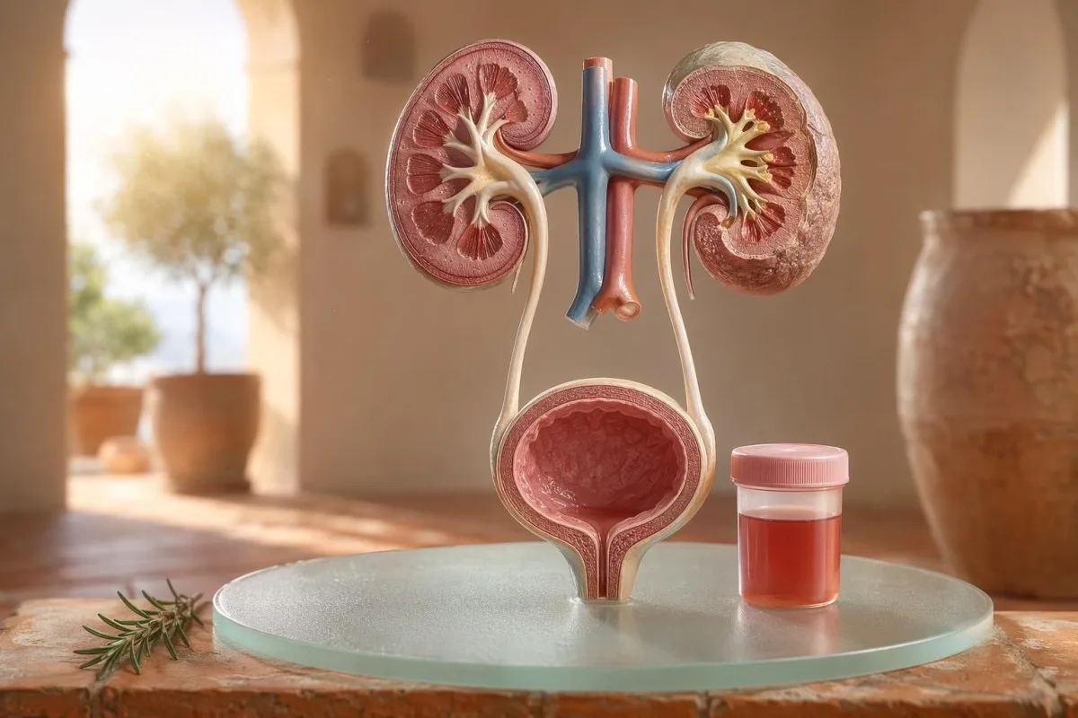
Першачарговае для пацыента кіраўніцтва па бачнай і мікраскапічнай гематурыі, уключаючы тое, чаму кроў на стрып-тэсце не тое самае, што эрытрацыты пад мікраскопам.


- Кроў у мачы азначае бачную чырвоную, колеру гарбаты або колу мачу, альбо мікраскапічную гематурыю, выяўленую пры даследаванні; бачная кроў заўсёды заслугоўвае медыцынскага назірання.
- Мікроскопічна гематурія звычайна вызначаюць як 3 або больш эрытрацытаў на адно поле высокай павелічальнай здольнасці ў належным чынам сабраным узоры мікраскапіі мачы.
- Кроў на стрып-тэсце выяўляе актыўнасць гема, таму можа быць станоўчай як з цэлых эрытрацытаў, так і з свабоднага гемаглабіну або міяглабіну пасля траўмы цягліц.
- Падказкі ІСП уключаюць паленне, пачашчанае мачавыпусканне, лейкацыт-эстэразу, нітрыты і лейкацыты; гематурыю трэба пераправерыць пасля таго, як інфекцыя знікне.
- Асад мачы знаходкі, такія як дысморфныя эрытрацыты, цыліндры з эрытрацытаў або значная протэінурыя, паказваюць на хваробу фільтруючага адзінкі нырак.
- Цыліндры мачы зліплены з матэрыялу з нырачных канальчыкаў; эрытрацытарныя цыліндры — гэта сігнал трывогі ў нефралогіі на працягу аднаго тыдня, асабліва пры высокім артэрыяльным ціску або зніжэнні eGFR.
- Сігналы трывогі пры раку уключаюць бязбольную бачыную гематурыю, згусткі, узрост старэйшы за 45 гадоў, гісторыю курэння, прафесійнае ўздзеянне хімічных рэчываў і паўторныя незразумелыя эпізоды.
- Гематурыя пасля фізічных нагрузак звычайна ўціхае на працягу 48–72 гадзін; пастаянная кроў пасля адпачынку не павінна спісвацца на бег або язду на ровары.
- Неадкладная ацэнка неабходна пры наяўнасці крыві ў мачы з тэмпературай, болем у баку (фланку), згусткамі, якія выклікаюць блакаванне мачавыпускання, цяжарнасці, траўме або сімптомах цяжкай анеміі.
Што азначае кроў у мачы, перш чым вы пачнеце панікаваць
Кроў у мачы можа паходзіць ад інфекцыі, камянёў, хвароб, якія парушаюць фільтрацыю ў нырках, станаў прастаты або мачавой бурбалкі, лекаў, фізічных нагрузак ці кантамінацыі падчас менструацыі. Практычнае правіла простае: бачная кроў, паўторная мікраскапічная гематурыя або гематурыя з болем, тэмпературай, згусткамі, высокім артэрыяльным ціскам, бялком або зніжанай функцыяй нырак патрабуюць неадкладнай ацэнкі, а не назірання без дзеянняў.
А адзінкавы ружовы колер у ўнітазе пасля буракоў або новай дабаўкі — гэта не тая ж клінічная праблема, што мача колеру колы пасля ангіны. У маёй практыцы гісторыя колеру часта падказвае, куды глядзець перш за ўсё: ярка-чырвоная з згусткамі часцей звязана з ніжнімі аддзеламі мочавыдзяляльных шляхоў, тады як карычневая або колеру гарбаты мача можа азначаць больш старыя гемавыя пігменты з нырачных фільтраў.
Гематурыя — гэта не дыягназ; гэта прыкмета. Бачная гематурыя азначае, што вы бачыце змяненне колеру, а мікраскапічная гематурыя азначае, што мача выглядае нармальна, але мікраскапія знаходзіць эрытрацыты, звычайна пры 3 і больш эрытрацытах на адно поле высокай павелічальнасці.
Кантэсці — гэта Аналізатар крыві са штучным інтэлектам гэта дапамагае пацыентам звязаць знаходкі ў мачы з маркерамі нырак, такімі як креатынін, eGFR, CRP, гемаглабін і трамбацыты, у адной часовай шкале. Калі колер незразумелы, наш гід па колеры мачы тлумачыць, чаму чырвоная, карычневая, аранжавая і цёмна-жоўтая мача не азначаюць адно і тое ж.
Кроў на стрып-тэсце не тое самае, што RBC на мікраскапіі
Тэст-палоска для мачы з пазнакай кроў станоўчая выяўляе гемападобную хімічную актыўнасць, а не абавязкова цэлыя эрытрацыты. Мікраскапія адказвае на наступнае пытанне: ці ёсць сапраўдныя RBC у асадку мачы, і калі так — як яны выглядаюць?
Тэставанне тэст-палоскай хуткае, бо рэагентная пляцоўка рэагуе з пероксидазнай актыўнасцю гема. Гэта азначае, што станоўчы вынік можа ўзнікаць пры наяўнасці цэлых RBC, свабоднага гемаглабіну пасля распаду эрытрацытаў або міяглабіну пасля значнага пашкоджання цягліц.
Мікраскапія павольнейшая, але больш спецыфічная. Уважлівая лабараторыя центрыфугуюць мачу, даследуе асадок мачы, і паведамляе колькасць эрытрацытаў на поле высокай павелічальнасці; многія клініцысты лічаць 0–2 RBC/hpf нармальным і 3 і больш RBC/hpf — ненармальным у дарослых.
Нязгода мае значэнне. Тэст-палоска з 3+ крыві і 0–2 RBC/hpf прымушае мяне пытацца пра рабдаміёліз, гемоліз, акісляльныя кантамінанты і апрацоўку ўзору, а не адразу пераходзіць да хваробы мачавой бурбалкі; розніца падобная да разваг у нашай Порівняння аналізу сечі та посіву.
Калі падказкі пра ІСП тлумачаць гематурыю, а калі не
Інфекція сечових шляхів може спричинити кров у сечі, особливо при печінні, ургентності, частому сечовипусканні, дискомфорті в ділянці таза, лейкоцитарній естеразі, нітритах і наявності лейкоцитів. Але гематурія має зникнути після лікування; якщо після ІСШ (UTI) залишаються еритроцити, потрібен повторний огляд.
У типових випадках нижньої ІСШ я очікую кластер: дизурія, часте сечовипускання, позитивна лейкоцитарна естераза, піурія часто понад 5–10 лейкоцитів/впс, а інколи й нітрити. Традиційна позитивність посіву — 100 000 КУО/мл, але в симптомних жінок нижчі показники, як-от 1 000–10 000 КУО/мл, все ще можуть мати клінічне значення.
Лейкоцитарна естераза — це фермент лейкоцитів, а не доказ наявності бактерій. Позитивний результат разом із гематурією може виникати через інфекцію, камені, контамінацію або запалення нирок, тому наш гайд з лейкоцитарної естерази відокремлює корисні підказки від типових хибнопозитивних результатів.
Нітрити більш специфічні, але менш чутливі, бо не всі бактерії перетворюють нітрат на нітрит. Якщо симптоми типові, але нітрити негативні, посів усе одно може бути позитивним; практичні деталі описані в нашому пояснювачі результату щодо нітритів.
Бачная гематурыя: падказкі па колеры, згустках і часе
Видима гематурія ніколи не є тим, що я ігнорую, навіть якщо вона трапляється один раз. Яскраво-червона сеча, згустки або безболісна кровотеча потребують своєчасної оцінки, адже причини з боку нижніх сечових шляхів стають імовірнішими зі зростанням віку та впливу куріння.
Часові рамкі могуць быць нечакана карыснымі. Кроў толькі на пачатку мачавыпускання можа сведчыць пра ўрэтру, кроў на працягу ўсёй струі можа паходзіць з мачавой бурбалкі або ныркі, а тэрмінальная кроў каля канца часам паказвае на вобласць шыйкі мачавой бурбалкі.
Згусткі звычайна азначаюць, што крыніца крывацёку знаходзіцца ніжэй нырачных фільтраў, бо згусткі не лёгка ўтвараюцца пасля праходжання праз клубочкі і канальчыкі. Згустак, які блакуе адток мачы, — тэрміновая сітуацыя: перапаўненне мачавой бурбалкі можа стаць балючым на працягу некалькіх гадзін, і можа спатрэбіцца догляд за катетарам.
Мужчыны часам мяркуюць, што бачная гематурыя пасля сімптомаў з боку мочавыдзяляльнай сістэмы — гэта проста раздражненне прастаты. Гэта можа быць праўдай, але калі PSA быў правераны падчас або неўзабаве пасля інфекцыі, інтэрпрэтацыя можа быць складанай; наша PSA пасля ІСП (UTI) — гайд тлумачыць, чаму многія клініцысты чакаюць 6–8 тыдняў перад паўторным вызначэннем PSA.
Порогі мікраскапічнай гематурыі, якія выкарыстоўваюць лекары
Мікраскапічная гематурыя звычайна вызначаецца як 3 або больш RBC на поле высокай магутнасці у належным чынам сабраным узоры мачы. Кіраўніцтва AUA/SUFU выкарыстоўвае гэты парог і рэкамендуе ацэнку паводле рызыкі, а не лячэнне кожнага пацыента аднолькава (Barocas et al., 2020).
Адна толькі тест-палоска не павінна выкарыстоўвацца для дыягностыкі мікраскапічнай гематурыі. У кіраўніцтве Barocas et al. 2020 AUA/SUFU вызначэнне залежыць ад мікраскапіі, бо тест-палоскі могуць быць станоўчымі з-за гемавых пігментаў без цэлых эрытрацытаў.
Паўторнае тэставанне — не слабасць; гэта добрая медыцына. Калі ў кагосьці пасля інтэнсіўных фізічных нагрузак, ліхаманкі, сэксу, менструацыі або забруджанага збору ёсць 3–10 RBC/hpf, я звычайна хачу чысты паўторны ўзор перад прызначэннем сканаванняў, гэтак жа, як мы раім для даведнік па паўторна анамальных аналізах.
Рызыка змяняе шлях. 24-гадовы з 4 RBC/hpf пасля паўмарафону — гэта не тое самае, што 68-гадовы былы курэц з 25 RBC/hpf у двух узорах, нават калі абодва тэхнічна адпавядаюць вызначэнню гематурыі.
Асад мачы, бялок і цыліндры мачы, якія змяняюць абследаванне
Асад мачы можа паказаць, ці, хутчэй за ўсё, гематурыя паходзіць з нырачных фільтраў. Дысморфныя чырвоныя клеткі, цыліндры з эрытрацытаў і значны зрух у бялку пераносяць занепакоенасць у бок клубочковай хваробы, а не простай інфекцыі мачавой бурбалкі.
Эрытрацыты, якія выглядаюць няроўнымі, «пузырчатымі» (blebbed) або ў форме кольца, называюцца дысморфнымі RBC, і яны сведчаць пра праходжанне праз запалены клубочковы фільтр. Акантацыты вышэй прыкладна 5% з мачавых чырвоных клетак часта разглядаюць як моцную клубочковую падказку, хоць лабараторыі адрозніваюцца ў тым, наколькі ўпэўнена яны паведамляюць пра гэта.
Бялок — другая падказка, якую я не ігнарую. Суадносіны альбумін/крэатынін у мачы вышэй за 30 mg/g, або прыкладна 3 mg/mmol, — ненармальнае, і бачная гематурыя разам з бялком заслугоўвае хутчэйшага агляду, арыентаванага на ныркі; наша гід па бялку ў мачы дае парогі на ўзроўні пацыента.
Цыліндры мачы — гэта мікраскапічныя «плесні», якія ўтвараюцца ўнутры нырачных канальчыкаў. Гіаліновыя цыліндры могуць з’яўляцца пасля абязводжвання або фізічных нагрузак, але цыліндры з эрытрацытаў — ненармальныя і звычайна паказваюць на гломерулонефрыт; для больш шырокай даведкі па агульным аналізе мачы глядзіце наша поўнае кіраўніцтва па аналізе мачы.
Камяні, фізічныя нагрузкі і часовыя прычыны гематурыі
Камяні і інтэнсіўныя фізічныя нагрузкі могуць выклікаць гематурыю, але ў іх розныя тэрміны. Гематурыя, звязаная з камянямі, часта суправаджаецца болем у баку (фланку) або крысталамі, тады як гематурыя пасля нагрузак звычайна павінна супакоіцца на працягу 48–72 гадзін адпачынку.
Нырачныя камяні класічна выклікаюць хвалі болю ў баку, млоснасць і мікраскапічную або бачную гематурыю. Крышталі оксалату кальцыю не даказваюць камень, але павялічваюць падазрэнне, калі спалучаюцца з колікападобным болем; наша гайд па крышталах оксалату кальцыю тлумачыць падказку мікраскапіі мачы.
Гематурыя пасля фізічных нагрузак — рэальная. Я бачыў бегуноў на марафоне з 10–20 RBC/hpf раніцай пасля забегу, з нармальнай культурай, без бялку, і з цалкам нармальным паўторным агульным аналізам мачы праз 3 дні.
Небяспека — звязваць усё з трэніроўкамі. Калі гематурыя захоўваецца пасля 72 гадзін, з’яўляецца разам з цёмнай «кола»-падобнай мачой або суправаджаецца высокім CK, ростам креатыніну ці болем у цягліцах, шукайце прычыны па-за межамі мачавой бурбалкі; наша руководство щодо креатиніну під час фізичних вправ охоплює перетин проблем із нирками та м’язами.
«Чырвоныя сцяжкі» з боку нырак, якія лекары не ігнаруюць
Гематурія з протеїнурією, високий артеріальний тиск, набряки, знижений eGFR, циліндри з еритроцитів або швидке зростання креатиніну — це «нирковий червоний прапорець». Ці знахідки вказують, що фільтраційні одиниці можуть бути запалені, і очікування місяцями може коштувати функції нирок.
KDIGO 2024 визначає хронічну хворобу нирок за маркерами ураження нирок, такими як альбумінурія, або eGFR нижче 60 мл/хв/1,73 м² щонайменше протягом 3 місяців. Однак у гострих ситуаціях гематурія разом із підвищенням креатиніну протягом днів або тижнів достатня, щоб підвищити рівень терміновості ще до того, як буде досягнуто 3-місячне визначення.
Альбумінурія часто є раннім сигналом попередження. ACR 30–300 мг/г — помірно підвищений, понад 300 мг/г — суттєво підвищений, а поєднання будь-якого з них із гематурією змінює розмову; наше кіраўніцтва па ACR для нырак пояснює, чому альбумін у сечі виявляє ураження раніше за симптоми.
Тут важливий артеріальний тиск. Новий АТ 160/100 мм рт. ст. із «колою» сечею та набряком щиколоток після інфекції горла змушує мене думати про гломерулонефрит, а не про цистит; якщо eGFR невизначений, наше керівництво для повторної перевірки GFR пояснює, коли цистатин C може уточнити функцію нирок.
Падказкі рызыкі раку без панікі
Безболісна видима гематурія, особливо після 45 років, потребує термінового оцінювання, оскільки рак сечового міхура та верхніх сечових шляхів може проявлятися так. NICE NG12 рекомендує направлення до спеціаліста з підозрою на рак при нез’ясованій видимій гематурії у дорослих 45 років і старших або при видимій гематурії, що зберігається після лікування інфекції сечових шляхів.
Більшість гематурій — не рак. Та все ж ті випадки, які я пам’ятаю, — тихі: немає болю, немає температури, немає печіння — лише два епізоди червоної сечі в 56-річного курця, який майже проігнорував це.
Ризик накопичується, а не є бінарним. Вік понад 45–50, історія куріння, вплив циклофосфаміду, променева терапія таза, рецидивуюча видима гематурія, професійний вплив ароматичних амінів і згустки — усе це підвищує потребу в цистоскопії або візуалізації.
Маркери простати не виключають причини з боку сечового міхура. Якщо є уринарні симптоми, зміни PSA та гематурія перетинаються, уважно перегляньте час появи та статус інфекції; наше настанови щодо простат-ного маркера пояснює, чому PSA є лише однією частиною оцінювання стану сечової системи у чоловіків.
Лекі, антыкаагулянты і ілжывыя сігналы трывогі
Антикоагулянти можуть виявляти кровотечу, але їх не слід автоматично звинувачувати в гематурії. Гематурія, пов’язана з антикоагулянтами, усе одно потребує обстеження, оскільки ліки можуть виявити камені, інфекцію, хворобу нирок або утворення в сечових шляхах.
Варфарин, апіксабан, ривароксабан, аспірин і клопідогрел можуть зробити кровотечу більш очевидною. Якщо INR вище цільового діапазону, його корекція має значення, але нормальний INR не робить видиму гематурію нешкідливою.
Декілька причин, що не пов’язані з кровотечею, можуть імітувати гематурію. Рифампіцин може забарвлювати сечу в оранжево-червоний колір, феназопіридин — у яскраво-оранжевий, буряк може забарвлювати сечу в чутливих людей, а зневоднення може затемнювати пігмент настільки, що лякає пацієнтів; моніторинг антикоагулянтів висвітлено в нашому лабораторному контролі безпеки для засобів, що розріджують кров.
Тест-смужка також може вводити в оману після інтенсивного фізичного навантаження або травми м’язів. Позитивна реакція на гему при невеликій кількості або відсутності RBC підштовхує мої міркування до міоглобіну, особливо якщо CK > 1,000 IU/L і сеча темна.
Дзеці, цяжарнасць і забруджванне менструальнай крывёй
Гематурія в дітей, під час вагітності або напередодні/під час менструації потребує чистішого забору зразка та нижчого порогу для перегляду. Один і той самий результат сечі може означати різне залежно від віку, статусу вагітності, наявності гарячки, артеріального тиску та якості збору.
Менструальне забруднення є поширеним, і ніхто не має соромитися цього. Якщо результат є несподіваним, повторіть пробу середньої порції «чистий улов» щонайменше через 48 годин після припинення кровотечі; стійкі 3 або більше RBC/hpf стають більш значущими.
Дітям часто потрібен інший підхід. Гематурія після вірусного захворювання може бути тимчасовою, але гематурія зі набряком, високим артеріальним тиском, протеїнурією або зменшенням об’єму сечі потребує негайного педіатричного огляду.
Вагітність підвищує ставки, оскільки УТІ, камені, нирковий стрес, пов’язаний із прееклампсією, та контамінація можуть накладатися. Для попереджувальних патернів щодо вагітності в той самий день, що стосуються нирок, печінки, тромбоцитів і знахідок у сечі, див. наш сігналах трывогі ў лабараторных аналізах пры цяжарнасці.
Аналізы, якія лекары звычайна прызначаюць пасля гематурыі
Зазвичай наступні обстеження після гематурії — це повторний аналіз сечі з мікроскопією, посів сечі, якщо можлива інфекція, аналізи крові для оцінки функції нирок, аналіз білка в сечі або ACR, вимірювання артеріального тиску та візуалізація або цистоскопія залежно від ризику. Точна послідовність залежить від симптомів і ризику.
За симптомів неускладненого циститу першими можуть бути посів і лікування, а потім — повторна мікроскопія сечі після зникнення симптомів. За безболісної видимої гематурії багато лікарів переходять раніше до візуалізації та цистоскопії, оскільки очікування повторних епізодів може відтермінувати діагностику.
Аналізи крові додають контекст, який сама лише сеча не може надати. Креатинін, eGFR, сечовина або BUN, електроліти, CBC, CRP, комплемент C3/C4 та аутоімунні маркери можуть бути доречними, коли підозрюють запалення нирок; наш кіраўніцтве па суадносінах BUN/крэатынін пояснює один поширений нирковий патерн.
Kantesti AI може організувати ці результати в подання тренду, але огляд лікаря залишається критично важливим, коли є «червоні прапорці». Наш підхід до клінічної безпеки описано в Медыцынская праверка, зокрема як обробляють аномальні кластери, а не ізольовані числа.
Як Kantesti дапамагае арганізаваць кантэкст гематурыі
Кантэсці — гэта сервісі інтерпретації тестів ШІ який читає маркери крові, близькі до сечі, в контексті, наприклад креатинін, eGFR, CRP, гемоглобін, тромбоцити та глюкозу. Він не діагностує гематурію, але допомагає пацієнтам підготувати чистіший і корисніший підсумок для їхнього лікаря.
Коли я переглядаю гематурію, я рідко дивлюся лише на один результат. Нормальний креатинін 0.82 мг/дл, стабільний eGFR понад 90, відсутність протеїну та негативний посів відчуваються зовсім інакше, ніж гематурія з eGFR 48 і CRP 65 мг/л.
Кантэсці — гэта платформа інтэрпрэтацыі біямаркераў AI який може обробляти завантажені PDF з аналізами крові або фото та пов’язувати маркери нирок, запалення й анемії поза межами норми з часом появи симптомів. Методи, що лежать в основі такого читання патерну, описано в нашому Кіраўніцтва па тэхналогіях, а шырэйшае ахопленне маркераў выкладзена ў кіраўніцтва па біямаркерах.
Одна практична порада: перед завантаженням перевірте, що одиниці вимірювання, дати та ідентифікатори пацієнта зафіксовані коректно. Помилки OCR можуть перетворити 0.9 на 9.0, що є абсолютно іншою історією щодо нирок; наш чэк-ліст загрузкі PDF паказвае памылкі, якія я прашу пацыентаў заўважыць першымі.
Даследчыя нататкі, кіраванне і канчатковы чэкліст
Па стане на 11 ліпеня 2026 года, самы бяспечны план для пацыента — пацвердзіць, ці сапраўды ёсць гематурыя, вызначыць прыкметы інфекцыі, шукаць «чырвоныя сцягі» з боку нырак і эскалаваць пры наяўнасці бачнай або пастаяннай гематурыі. Не спадзявайцеся толькі на колер мачы.
Я Томас Кляйн, MD, і мая парада наўмысна кансерватыўная, бо прапушчаная гематурыя можа мець значэнне. Медыцынскі змест Kantesti праходзіць агляд з клінічным наглядам ад нашых Медыцынская кансультатыўная рада, і наша роля — дапамагаць пацыентам задаваць лепшыя пытанні, а не замяняць іх лекара.
Кантэсці — гэта Інструмент аналізу вынікаў аналізу крыві з падтрымкай AI выкарыстоўваецца мільёнамі людзей у 127 краінах, таму канфідэнцыяльнасць і клінічная яснасць павінны быць закладзены ў працоўны працэс, а не дадавацца пазней. Вы можаце даведацца больш пра Kantesti LTD, нашу структуру кампаніі ў Вялікабрытаніі і клінічную місію на Пра нас.
AI Clinical Research Group. (2026). AI Blood Test Analyzer: 2.5M Tests Analyzed | Global Health Report 2026. Zenodo. DOI: https://doi.org/10.5281/zenodo.18175532. ResearchGate: https://www.researchgate.net/. Academia.edu: https://www.academia.edu/.
Kantesti AI Clinical Research Group. (2026). RDW Blood Test: Complete Guide to RDW-CV, MCV & MCHC. Zenodo. DOI: https://doi.org/10.5281/zenodo.18202598. ResearchGate: https://www.researchgate.net/. Academia.edu: https://www.academia.edu/.
Часта задаваныя пытанні
Ці заўсёды кроў у мачы з’яўляецца сур’ёзным станам?
Кров у сечі не завжди є небезпечною, але її потрібно підтвердити та пояснити. Одноразовий мікроскопічний результат 3–5 RBC/hpf після фізичних навантажень або менструації може зникнути під час повторного тестування, тоді як видима кров, згустки, гарячка, біль у ділянці попереку, протеїнурія, високий артеріальний тиск або знижений eGFR потребують невідкладного огляду. У дорослих віком понад 45 років необґрунтована видима гематурія заслуговує на термінову оцінку навіть за відсутності болю.
Якая розніца паміж крывёй на тэст-полосцы і RBC у мачы?
Тест-смужка виявляє активність гемоподібних хімічних речовин, тоді як аналіз сечі під мікроскопом підраховує фактичні еритроцити. Тест-смужка може бути позитивною через цілі RBC, вільний гемоглобін або міоглобін після ушкодження м’язів, тож вона не є тотожною підтвердженій гематурії. Мікроскопічна гематурія зазвичай визначається як 3 або більше RBC на поле високого збільшення в належно зібраному зразку.
Ці можа інфекцыя сечових шляхів (ІСШ) спричинити кров у сечі?
Так, ІСШ може спричиняти кров у сечі, особливо коли є печіння, часте сечовипускання, ургентність, а також наявні лейкоцитарна естераза, нітрити та лейкоцити. Традиційна позитивність посіву сечі часто становить 100 000 КУО/мл, але нижчі показники можуть мати значення у симптоматичних пацієнтів. Гематурію зазвичай слід повторно перевірити після лікування, оскільки збереження крові може вказувати на камені, хворобу нирок або іншу причину з боку сечових шляхів.
Калі мне трэба звярнуцца ў неадкладную дапамогу пры гематурыі?
Звярніцеся по невідкладну медичну допомогу при наявності крові в сечі з температурою, сильним болем у ділянці попереку, блюванням, вагітністю, нещодавньою травмою, згустками, неможливістю помочитися, непритомністю або симптомами тяжкої анемії. Огляд упродовж того ж тижня також доцільний при гематурії з протеїнурією, еритроцитарними циліндрами, новою високою артеріальною гіпертензією або підвищенням креатиніну. Закупорений потік сечі через згустки може стати болісним і небезпечним протягом кількох годин.
Ці можа фізічна нагрузка спричинити мікроскопічну гематурію?
Напружаныя фізічныя нагрузкі могуць выклікаць мікраскапічную гематурыю, асабліва пасля працяглага бегу на дыстанцыю, інтэнсіўнага веласіпеднага катання або кантактных відаў спорту. Гематурыя, звязаная з фізічнымі нагрузкамі, звычайна знікае на працягу 48–72 гадзін адпачынку і не павінна суправаджацца стойкай протэінурыяй, зніжэннем eGFR або моцным болем у цягліцах. Калі на тэст-полосцы кроў станоўчая, але мікраскапія паказвае мала RBC, клініцысты могуць праверыць CK, паколькі міяглабін з пашкоджання цягліц можа выклікаць станоўчы вынік тэст-полоскі.
Што азначаюць цыліндры ў мачы, калі выяўлена кроў?
Цилиндри в сечі — це формовані матеріали, утворені всередині ниркових канальців, і їхній тип змінює значення гематурії. Гіалінові циліндри можуть з’являтися при зневодненні або фізичному навантаженні, але еритроцитарні циліндри є патологічними та вказують на запалення клубочків нирок. Наявність крові в сечі з еритроцитарними циліндрами, білком і підвищеним артеріальним тиском слід лікувати як нирковий «червоний прапор», а не як звичайну UTI.
Ці можа абязводжванне зрабіць мачу падобнай да крыві?
Пры абязводжванні мача часта становіцца цёмна-жоўтай або бурштынавай, але звычайна гэта не прыводзіць да з'яўлення эрытрацытаў у мачы. Піце ваду і здайце аналіз мачы, калі яе колер ружовы, чырвоны, карычневы або падобны да колеру колы, асабліва калі ён захоўваецца пасля папаўнення запасаў вадкасці або суправаджаецца болем.
Атрымаць аналіз крыві з AI сёння
Далучайцеся больш чым да 2 мільёнаў карыстальнікаў па ўсім свеце, якія давяраюць Kantesti для імгненнага, дакладнага аналізу лабараторных тэстаў. Загрузіце вынікі аналізу крыві і атрымайце поўную інтэрпрэтацыю біямаркераў 15,000+ за лічаныя секунды.
📚 Публікацыі даследаванняў са спасылкамі
Кляйн, Т., Мітчэл, С., і Вебер, Г. (2026). Аналізатар AI для аналізу крыві: прааналізавана 2,5 млн тэстаў | Глабальная справаздача аб здароўі 2026. Kantesti AI Medical Research.
Кляйн, Т., Мітчэл, С., і Вебер, Г. (2026). Аналіз крыві на RDW: поўнае кіраўніцтва па RDW-CV, MCV і MCHC. Kantesti AI Medical Research.
📖 Знешнія медыцынскія спасылкі
Нацыянальны інстытут аховы здароўя і дасканаласці ў медыцыне (NICE) (2015). Падазрэнне на рак: распазнаванне і накіраванне. Рэкамендацыя NICE NG12. Рэкамендацыі NICE.
Група па хранічнай хваробе нырак (ХХН) Kidney Disease: Improving Global Outcomes CKD Work Group (2024). KDIGO 2024 клінічныя рэкамендацыі па ацэнцы і лячэнні хранічнай хваробы нырак. Kidney International.
⚕️ Медыцынская адмова ад адказнасці
Гэты артыкул прызначаны толькі для адукацыйных мэт і не з’яўляецца медычнай парадай. Заўсёды кансультуйцеся з кваліфікаваным спецыялістам аховы здароўя для прыняцця рашэнняў па дыягностыцы і лячэнні.
Сігналы даверу E-E-A-T
Вопыт
Клінічны агляд, які вядзе лекар, працэсаў інтэрпрэтацыі лабараторных паказчыкаў.
Экспертыза
Фокус лабараторнай медыцыны на тым, як біямаркеры паводзяць сябе ў клінічным кантэксце.
Аўтарытэтнасць
Напісана доктарам Томасам Кляйнам, з аглядам доктара Сара Мітчэл і праф. доктара Ганса Вебера.
Надзейнасць
Інтэрпрэтацыя на аснове доказаў з выразнымі шляхамі далейшых дзеянняў, каб паменшыць трывогу.