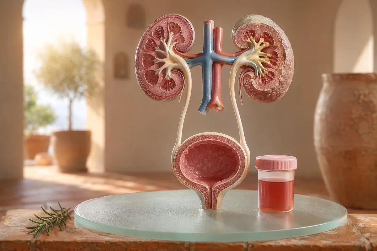
نظر ايندڙ ۽ خوردبيني hematuria لاءِ مريض-پهريون رهنمائي، جنهن ۾ اهو به شامل آهي ته dipstick تي رت هجڻ جو مطلب خوردبيني هيٺ ڳاڙهن سيلن (red cells) سان ساڳيو ڇو نه هوندو آهي.


- پيشاب ۾ رت مطلب نظر ايندڙ ڳاڙهو، چانهه-رنگ يا ڪولا-رنگ پيشاب، يا ٽيسٽ تي مليل خوردبيني hematuria؛ نظر ايندڙ رت هميشه طبي فالو اپ جوڳو آهي.
- Microscopic hematuria عام طور تي صحيح طرح گڏ ڪيل پيشاب جي خوردبيني نموني ۾ في high-power field 3 يا وڌيڪ RBCs طور بيان ڪيو ويندو آهي.
- Dipstick رت heme سرگرمي کي سڃاڻي ٿو، تنهنڪري اهو برقرار ڳاڙهن سيلن، آزاد hemoglobin يا myoglobin کان عضلاتي زخم کان پوءِ به مثبت ٿي سگهي ٿو.
- UTI جا اشارا شامل آهن: جلڻ (burning)، بار بار پيشاب اچڻ (frequency)، leukocyte esterase، nitrites ۽ اڇا سيل (white cells)؛ انفيڪشن ختم ٿيڻ کان پوءِ hematuria ٻيهر چيڪ ڪرڻ گهرجي.
- پيشاب جو sediment اهڙيون ڳالهيون جهڙوڪ dysmorphic red cells، red cell casts يا اهم پروٽين گردي جي فلٽرنگ-يونٽ بيماري ڏانهن اشارو ڪن ٿيون.
- Urine casts اهي ٺهيل مادا گردن جي نلين (kidney tubules) مان ٿين ٿا؛ ڳاڙهي سيل ڪاسٽ (red cell casts) ساڳئي هفتي جي نيڦرولوجي (nephrology) لاءِ هڪ ڳاڙهو جهنڊو (red flag) آهي، خاص طور تي جڏهن بلڊ پريشر وڌيڪ هجي يا eGFR گهٽجي رهيو هجي.
- ڪينسر جون خبرداري جون نشانيون ان ۾ بي درد نظر ايندڙ هيماتوريا (painless visible hematuria)، لوتا (clots)، 45 سالن کان وڌيڪ عمر، سگريٽ نوشي جي تاريخ، پيشه ور ڪيميائي نمائش (occupational chemical exposure) ۽ بار بار اڻڄاتل قسطن (recurrent unexplained episodes) شامل آهن.
- ورزش سبب هيماتوريا (Exercise hematuria) عام طور تي 48-72 ڪلاڪن اندر ٺيڪ ٿي وڃي ٿي؛ آرام کان پوءِ به رت جاري رهي ته ان کي ڊوڙڻ يا سائيڪل هلائڻ تي نه مڙهيو وڃي.
- تڪڙي جاچ پيشاب ۾ رت لاءِ ضرورت آهي جڏهن بخار هجي، پاسن/ڪمر ۾ سور (flank pain) هجي، لوتا پيشاب جي رڪاوٽ ڪن (clots causing urinary blockage)، حمل (pregnancy) هجي، صدمو (trauma) هجي يا شديد انيميا (severe anemia) جون علامتون هجن.
پريشان ٿيڻ کان اڳ پيشاب ۾ رت اچڻ جو مطلب ڇا ٿي سگهي ٿو
پيشاب ۾ رت اهو انفيڪشن (infection)، پٿريون (stones)، گردن جي فلٽرنگ جي بيماري (kidney filtering disease)، پروسٽيٽ (prostate) يا مثاني (bladder) جون حالتون، دوائون (medicines)، ورزش (exercise)، يا دورن (periods) مان آلودگي (contamination) سبب به ٿي سگهي ٿو. عملي قاعدو سادو آهي: نظر ايندڙ رت (visible blood)، بار بار مائڪروسڪوپڪ هيماتوريا (repeated microscopic hematuria)، يا درد، بخار، لوتا، وڌيڪ بلڊ پريشر، پروٽين (protein)، يا گردن جي ڪم ۾ گهٽتائي (reduced kidney function) سان گڏ هيماتوريا کي محتاط انتظار (watchful waiting) بدران فوري جائزو (prompt evaluation) گهرجي.
بيٽروٽ (beetroot) يا نئين سپليمينٽ کان پوءِ ٽوائلٽ ۾ هڪ دفعو گلابي رنگ (pink) اچڻ، ڳلي جي سور (sore throat) کان پوءِ ڪولا جهڙو رنگ (cola-colored urine) اچڻ واري ڪلينڪل مسئلي جهڙو ناهي. منهنجي ڪلينڪ ۾، رنگ جي ڪهاڻي اڪثر ٻڌائي ٿي ته پهرين ڪٿي ڏسڻو آهي: لوتن سان روشن ڳاڙهو (bright red with clots) هيٺين پيشاب واري رستي (lower urinary tract) ڏانهن اشارو ڪري ٿو، جڏهن ته ڀورو (brown) يا چانهه جهڙو (tea-colored) پيشاب گردن جي فلٽرن مان پراڻا هيم (heme) رنگدار جزا (heme pigments) ظاهر ڪري سگهي ٿو.
هيماتوريا هڪ تشخيص (diagnosis) ناهي؛ اها هڪ نشاني (sign) آهي. نظر ايندڙ هيماتوريا (Visible hematuria) مطلب ته توهان رنگ ۾ تبديلي ڏسي سگهو ٿا، جڏهن ته مائڪروسڪوپڪ هيماتوريا (microscopic hematuria) مطلب ته پيشاب عام نظر اچي ٿو پر مائڪروسڪوپي ۾ ڳاڙهي سيل (red cells) ملن ٿا، عام طور تي في هاءِ پاور فيلڊ (high-power field) 3 يا وڌيڪ ڳاڙهي رت جا سيل.
ڪينٽيسٽي هڪ آهي اي آءِ بلڊ ٽيسٽ اينالائيزر اهو مريضن کي هڪ ئي ٽائم لائن ۾ پيشاب جي نتيجن کي گردن جي نشانين (kidney markers) جهڙوڪ ڪريئٽينين (creatinine)، eGFR، CRP، هيموگلوبن (hemoglobin) ۽ پليٽليٽس (platelets) سان ڳنڍڻ ۾ مدد ڪري ٿو. جيڪڏهن رنگ سمجهه کان ٻاهر هجي، اسان پيشاب رنگ گائيڊ (urine color guide) ٻڌائي ٿي ته ڳاڙهو (red)، ڀورو (brown)، نارنگي (orange) ۽ ڳاڙهو پيلو (dark yellow) پيشاب سڀ هڪ جهڙي ڳالهه ڇو نٿا ڪن.
Dipstick رت خوردبيني تي RBCs سان ساڳيو ناهي
پيشاب ڊِپ اسٽڪ (urine dipstick) تي نشان لڳل رت مثبت (blood positive) هيم جهڙي ڪيميائي سرگرمي (heme-like chemical activity) ڳولي ٿو، لازمي طور تي مڪمل (intact) ڳاڙهي رت جا سيل نه. مائڪروسڪوپي ايندڙ سوال جو جواب ڏئي ٿي: ڇا پيشاب جي تلڇٽ (urine sediment) ۾ واقعي RBCs موجود آهن، ۽ جيڪڏهن آهن ته اهي ڪيئن نظر اچن ٿا؟
ڊِپ اسٽڪ ٽيسٽ تيز آهي ڇو ته ري ايجِنٽ پيڊ (reagent pad) هيم جي پيرو آڪسائيڊيز سرگرمي (peroxidase activity) سان ردعمل ڪري ٿو. ان جو مطلب آهي ته مثبت نتيجو مڪمل RBCs سان به ٿي سگهي ٿو، ڳاڙهي سيلن جي ٽٽڻ کان پوءِ آزاد هيموگلوبن (free hemoglobin) سان، يا وڏي عضلاتي زخمي (significant muscle injury) کان پوءِ ميوگلوبن (myoglobin) سان.
مائڪروسڪوپي سست آهي پر وڌيڪ مخصوص (more specific). هڪ محتاط ليب پيشاب کي سينٽريفيوج (centrifuge) ڪري، جانچيندي آهي پيشاب جي تلڇٽ (urine sediment), ، ۽ في هاءِ پاور فيلڊ ڳاڙهي سيلن جو تعداد رپورٽ ڪري ٿي؛ ڪيترائي ڪلينشين بالغن ۾ 0-2 RBC/hpf کي عام (normal) ۽ 3 يا وڌيڪ RBC/hpf کي غير عام (abnormal) سمجهن ٿا.
اها بي ميل (mismatch) اهميت رکي ٿي. هڪ ڊِپ اسٽڪ جنهن تي 3+ رت (3+ blood) ۽ 0-2 RBC/hpf هجي، مون کي مثاني جي بيماري ڏانهن سڌو ٽپو ڏيڻ بدران rhabdomyolysis، hemolysis، آڪسائيڊنگ آلودگين (oxidizing contaminants)، ۽ نموني جي هٿ ڪرڻ (specimen handling) بابت پڇڻ تي مجبور ڪري ٿي؛ فرق اسان جي پيشاب جي تجزيي ۽ ڪلچر جو مقابلو.
جڏهن UTI جا اشارا hematuria بيان ڪن ٿا، ۽ جڏهن نٿا ڪن
پيشاب جي نالي جي انفيڪشن (UTI) سبب ٿي سگهي ٿي پيشاب ۾ رت, ، خاص طور تي جلن، تڪڙ، بار بار پيشاب، پيٽ جي هيٺئين حصي ۾ تڪليف، leukocyte esterase، نائٽريٽس ۽ اڇن رت جي خاني سان. پر UTI جي علاج کان پوءِ هيماتوريا ختم ٿيڻ گهرجي؛ UTI کان پوءِ به مسلسل رت جا سيل موجود هجن ته ٻيهر جائزو وٺڻ ضروري آهي.
سادي هيٺين UTI ۾، مان هڪ گڏيل نمونو اميد ڪريان ٿو: ڊيسوريا، بار بار پيشاب، leukocyte esterase مثبت، پائوريا اڪثر 5-10 کان وڌيڪ اڇا رت جا سيل/hpf، ۽ ڪڏهن ڪڏهن نائٽريٽس. روايتي ڪلچر جي مثبت هجڻ جي حد 100,000 CFU/mL آهي، پر علامتي عورتن ۾ گهٽ ڳڻپون جهڙوڪ 1,000-10,000 CFU/mL به ڪلينڪل طور معنيٰ خيز ٿي سگهن ٿيون.
Leukocyte esterase هڪ اڇي رت جي خاني جو اينزائم آهي، بيڪٽيريا جو ثبوت نه آهي. هيماتوريا سان گڏ مثبت نتيجو انفيڪشن، پٿريون، آلودگي، يا گردن جي سوزش سبب به ٿي سگهي ٿو، جنهن ڪري اسان جي leukocyte esterase گائيڊ عام غلط مثبتن مان ڪارآمد اشارا الڳ ڪري ٿي.
نائٽريٽس وڌيڪ مخصوص آهن پر گهٽ حساس، ڇاڪاڻ ته سڀئي بيڪٽيريا نائٽريٽ کي نائٽريٽ ۾ تبديل نٿا ڪن. جيڪڏهن علامتون عام/کلاسيڪ هجن پر نائٽريٽس منفي هجن ته به ڪلچر مثبت ٿي سگهي ٿو؛ عملي تفصيل اسان جي نائٽريٽ نتيجي جي وضاحت.
نظر ايندڙ hematuria: رنگ، رت جا ڦڙا ۽ وقت جا اشارا
نظر ايندڙ هيماتوريا ڪڏهن به مان نظرانداز نٿو ڪريان، جيتوڻيڪ اهو صرف هڪ ڀيرو ئي ڇو نه ٿئي. روشن ڳاڙهو پيشاب، رت جا لخته، يا بي درد خونريزي کي وقتائتي جائزي جي ضرورت آهي، ڇاڪاڻ ته عمر ۽ سگريٽ نوشي جي نمائش وڌڻ سان هيٺين پيشاب واري نالي جا سبب وڌيڪ ممڪن ٿي ويندا آهن.
وقت جي ترتيب حيرت انگيز طور مفيد ٿي سگهي ٿي. پيشاب ڪرڻ جي شروعات ۾ صرف رت اچڻ يورٿرا (پيشاب جي نالي) ڏانهن اشارو ڪري سگهي ٿو، جڏهن ته وهڪري دوران رت اچڻ مثاني يا گردي جي ذريعن مان ٿي سگهي ٿو، ۽ آخر ۾ ويجهو رت ڪڏهن ڪڏهن مثاني جي ڳچي واري علائقي ڏانهن اشارو ڪندو آهي.
ڪلٽس (رت جا ٺڪاءُ) عام طور مطلب اهو ٿيندو آهي ته رت وهڻ جو ذريعو گردي جي فلٽرن کان هيٺ آهي، ڇاڪاڻتہ ڪلٽس گلو ميرولِي ۽ ٽيوبولز مان گذرڻ کان پوءِ آساني سان ٺهندا ناهن. پيشاب جي وهڪري کي روڪيندڙ ڪلٽ تڪڙي حالت آهي: مثاني جو ڦولجڻ چند ڪلاڪن اندر ڏکوئيندڙ ٿي سگهي ٿو، ۽ ڪيٿٽر جي سنڀال جي ضرورت پئجي سگهي ٿي.
مرد ڪڏهن ڪڏهن سمجهن ٿا ته پيشاب جي علامتن کان پوءِ نظر ايندڙ هيماتوريا صرف پروسٽٽ جي تڪليف (irritation) آهي. اهو ٿي سگهي ٿو، پر جيڪڏهن انفيڪشن دوران يا ان کان ٿورو پوءِ PSA چيڪ ڪيو ويو هجي، ته تشريح پيچيده ٿي سگهي ٿي؛ اسان جو UTI کان پوءِ PSA گائيڊ ٻڌائي ٿو ته ڪيترائي ڪلينيشين PSA ٻيهر چيڪ ڪرڻ کان اڳ 6-8 هفتا ڇو ترسندا آهن.
خوردبيني hematuria جون حدون جيڪي ڊاڪٽر استعمال ڪندا آهن
مائڪروسڪوپڪ هيماتوريا عام طور تي ان طرح بيان ڪئي ويندي آهي في هاءِ پاور فيلڊ 3 يا وڌيڪ RBC صحيح نموني سان گڏ ڪيل پيشاب جي نموني ۾. AUA/SUFU گائيڊ لائن هن حد کي استعمال ڪري ٿي ۽ هر مريض کي ساڳي طرح علاج ڪرڻ بدران خطري جي بنياد تي جائزو وٺڻ جي سفارش ڪري ٿي (Barocas et al., 2020).
صرف ڊِپ اسٽڪ (dipstick) کي استعمال ڪري مائڪروسڪوپڪ هيماتوريا جي تشخيص نه ڪرڻ گهرجي. Barocas et al. 2020 AUA/SUFU گائيڊ لائن ۾ تعريف مائڪروسڪوپي تي دارومدار رکي ٿي، ڇاڪاڻتہ ڊِپ اسٽڪ هيم (heme) رنگن مان مثبت ٿي سگهي ٿو بغير ڳاڙهن رت جي مڪمل خاني (intact red cells) جي.
ٻيهر ٽيسٽ ڪرڻ ڪمزوري ناهي؛ اهو سٺو طبي عمل آهي. جيڪڏهن ڪنهن کي سخت ورزش، بخار، جنسي تعلق، حيض، يا آلوده گڏ ڪيل نموني کان پوءِ 3-10 RBC/hpf هجن، ته مان عام طور تي اسڪين آرڊر ڪرڻ کان اڳ صاف ٻيهر نمونو چاهيندس، بلڪل ائين جيئن اسان لاءِ صلاح ڏيون ٿا غيرمعمولي ليب نتيجن جي ٻيهر جاچ.
خطرو (Risk) رستي (pathway) کي بدلائي ٿو. 24 سالن جو ماڻهو جيڪو اڌ مارٿون کان پوءِ 4 RBC/hpf سان هجي، اهو 68 سالن جي اڳوڻي سگريٽ نوشي ڪندڙ کان ساڳيو ناهي جنهن جي ٻن نمونن ۾ 25 RBC/hpf هجي، جيتوڻيڪ ٻنهي ٽيڪنيڪل طور هيماتوريا جي تعريف پوري ڪئي هجي.
پيشاب جو sediment، پروٽين ۽ urine casts جيڪي جاچ جو رخ بدلائين ٿا
پيشاب جو sediment ظاهر ڪري سگهي ٿو ته هيماتوريا ممڪن طور گردي جي فلٽرن مان اچي رهيو آهي. ڊسمورفڪ ڳاڙها رت جا خانا (dysmorphic red cells)، ڳاڙهن رت جا ڪاسٽس (red cell casts)، ۽ اهم پروٽين ۾ تبديلي ڳڻتي کي گلو ميرولر بيماري ڏانهن وڌيڪ ڌڪين ٿا، نه ڪي سادي مثاني جي انفيڪشن ڏانهن.
ڳاڙها رت جا خانا جيڪي اڻ برابر نظر اچن، ڦڦڙيل/blebbed هجن يا انگوزي جهڙا (ring-shaped) هجن، انهن کي ڊسمورفڪ RBCs چيو ويندو آهي، ۽ اهي سوزش ٿيل گلو ميرولر فلٽر مان گذرڻ جي نشاندهي ڪن ٿا. پيشاب ۾ موجود ڳاڙهن رت جي خانن جي لڳ ڀڳ 5% کان مٿي Acanthocytes اڪثر ڪري مضبوط گلو ميرولر اشارو طور علاج ڪيا ويندا آهن، جيتوڻيڪ ليبز انهن کي ڪيتري پڪ سان رپورٽ ڪن ٿيون، ان ۾ فرق ٿي سگهي ٿو.
پروٽين ٻيو اشارو آهي جنهن کي مان نظرانداز نٿو ڪريان. پيشاب ۾ albumin-creatinine ratio 30 mg/g کان مٿي، يا لڳ ڀڳ 3 mg/mmol، غير معمولي آهي، ۽ نظر ايندڙ هيماتوريا سان گڏ پروٽين کي تيز تر گردي-مرڪوز جائزي جي لائق سمجهيو وڃي ٿو؛ اسان جو پيشاب ۾ پروٽين جي گائيڊ مريض-سطح جون حدون (thresholds) ڏئي ٿو.
Urine casts ننڍڙا مولڊز (tiny molds) آهن جيڪي گردي جي ٽيوبولز اندر ٺهن ٿا. هائيلين ڪاسٽس (hyaline casts) ڊي هائيڊريشن يا ورزش کان پوءِ ظاهر ٿي سگهن ٿا، پر ڳاڙهن رت جا ڪاسٽس غير معمولي آهن ۽ عام طور تي گلو ميرولونيفرائٽس (glomerulonephritis) ڏانهن اشارو ڪن ٿا؛ وسيع پيشاب جي تجزيي (urinalysis) جي حوالي لاءِ ڏسو اسان جو پيشاب جي چڪاس جي مڪمل گائيڊ.
پٿريون، ورزش ۽ hematuria جا عارضي سبب
پٿر (Stones) ۽ سخت ورزش هيماتوريا جو سبب بڻجي سگهن ٿا، پر انهن جا وقت مختلف هوندا آهن. پٿر سان لاڳاپيل هيماتوريا اڪثر پاسي (flank) جو سور يا ڪرسٽلز سان گڏ هوندي آهي، جڏهن ته ورزش واري هيماتوريا عام طور تي آرام جي 48-72 ڪلاڪن اندر ٺيڪ ٿي وڃڻ گهرجي.
گردي جا پٿر ڪلاسيڪي طور پاسي جي لهرن جهڙو سور (waves of flank pain)، متلي (nausea)، ۽ مائڪروسڪوپڪ يا نظر ايندڙ هيماتوريا جو سبب بڻجن ٿا. ڪيلشيم آڪسالٽ ڪرسٽلز (calcium oxalate crystals) پٿر ثابت نٿا ڪن، پر جڏهن اهي ڪوليڪي (colicky) سور سان گڏ هجن ته شڪ وڌائين ٿا؛ اسان جو calcium oxalate crystals گائيڊ پيشاب جي مائڪروسڪوپي واري اشارو (clue) بيان ڪري ٿو.
ورزش واري هيماتوريا حقيقي آهي. مون مارٿون رنرن کي ڏٺو آهي جن ۾ ريس کان پوءِ صبح جو 10-20 RBC/hpf هجن، عام ڪلچر (normal culture) هجي، پروٽين نه هجي، ۽ 3 ڏينهن بعد مڪمل طور تي عام ٻيهر پيشاب جي تجزيي (urinalysis) هجي.
خطرو اهو آهي ته سڀ ڪجهه ٽريننگ تي مڙهجي. جيڪڏهن هيماتوريا 72 ڪلاڪن کان پوءِ به برقرار رهي، ڪاري ڪولا جهڙي پيشاب سان ظاهر ٿئي، يا ان سان گڏ هاءِ CK، ڪريئٽينائن وڌڻ (rising creatinine)، يا عضلاتي سور هجي، ته مثاني کان ٻاهر ڏسو؛ اسان جو ورزش کریٹینین گائیڈ گردے ۽ عضلات جي اوورليپ کي ڍڪي ٿو.
گردي جا ڳاڙها جهنڊا جن کي ڊاڪٽر نظرانداز نٿا ڪن
پروٽين سان گڏ هيماتوريا، وڌيڪ رت جو دٻاءُ، سوڄ، eGFR ۾ گهٽتائي، ڳاڙهي سيل ڪاسٽس يا تيزيءَ سان وڌندڙ کریٹینین گردي لاءِ ڳاڙهو جهنڊو آهي. اهي نتيجا ڏيکارين ٿا ته فلٽرنگ يونٽس ۾ سوزش ٿي سگهي ٿي، ۽ مهينن جو انتظار گردي جي ڪارڪردگي وڃائڻ جو سبب بڻجي سگهي ٿو.
KDIGO 2024 دائمي گردي جي بيماري کي گردي جي نقصان جي نشانين، جهڙوڪ البومينوريا، يا eGFR 60 mL/min/1.73 m² کان گهٽ، گهٽ ۾ گهٽ 3 مهينن لاءِ، سان بيان ڪري ٿو. پر تڪڙي حالتن ۾، هيماتوريا سان گڏ چند ڏينهن يا هفتن ۾ کریٹینین جو وڌڻ 3 مهينن واري تعريف پوري ٿيڻ کان اڳ ئي وڌائڻ لاءِ ڪافي آهي.
البومينوريا اڪثر شروعاتي خبرداري جو سگنل هوندو آهي. ACR 30-300 mg/g وچولي طور وڌيل آهي، 300 mg/g کان مٿي سختيءَ سان وڌيل آهي، ۽ ان مان ڪنهن به هڪ کي هيماتوريا سان گڏ ڪرڻ ڳالهه کي تبديل ڪري ٿو؛ اسان جو گردن جي ACR گائيڊ وضاحت ڪري ٿو ته پيشاب ۾ البومين ڪيئن علامتن کان اڳ نقصان ڳولي ٿو.
هتي رت جو دٻاءُ اهم آهي. ڳلي جي انفيڪشن کان پوءِ ڪولا جهڙو پيشاب ۽ ٽخنن جي سوڄ سان نئون BP 160/100 mmHg مون کي سيسٽائٽس نه، پر گلووميرولونيفرائٽس بابت سوچڻ تي مجبور ڪري ٿو؛ جيڪڏهن eGFR غير يقيني هجي، اسان جو GFR ري چيڪ گائيڊ وضاحت ڪري ٿو ته ڪڏهن سسٽاٽين C گردي جي ڪارڪردگي کي واضح ڪري سگهي ٿو.
ڪينسر جي خطري جا اشارا بغير پريشاني جي
بي درد نظر ايندڙ هيماتوريا، خاص طور تي 45 سالن کان پوءِ، تڪڙي جائزي جي لائق آهي، ڇاڪاڻ ته مثاني ۽ مٿئين پيشاب واري رستي جا ڪينسر هن طريقي سان به ظاهر ٿي سگهن ٿا. NICE NG12 بالغن ۾ 45 ۽ ان کان مٿي عمر لاءِ اڻڄاتل نظر ايندڙ هيماتوريا، يا UTI جي علاج کان پوءِ به جاري رهندڙ نظر ايندڙ هيماتوريا لاءِ suspected-cancer referral جي سفارش ڪري ٿو.
گهڻو ڪري هيماتوريا ڪينسر نه هوندو آهي. پر جيڪي ڪيس مون کي ياد آهن، اهي خاموش وارا هئا: درد ناهي، بخار ناهي، جلڻ ناهي—بس 56 سالن جي سگريٽ نوش ڪندڙ ۾ ڳاڙهي پيشاب جا ٻه واقعا، جنهن تقريباً ان کي نظرانداز ئي ڪري ڇڏيو.
خطرو گڏيل (cumulative) هوندو آهي، نه ته صرف ٻه حالتون (binary). 45-50 کان مٿي عمر، سگريٽ نوشي جي تاريخ، cyclophosphamide جو اثر، pelvic radiotherapy، بار بار نظر ايندڙ هيماتوريا، پيشه ورانه aromatic amine جي نمائش، ۽ ڪلٽس—اهي سڀ cystoscopy يا اميجنگ جي ضرورت وڌائين ٿا.
پروسٽٽ جا مارڪر مثاني جي سببن کي رد نٿا ڪن. جيڪڏهن پيشاب جون علامتون، PSA ۾ تبديليون ۽ هيماتوريا گڏ ٿين، ته وقت ۽ انفيڪشن جي حالت کي احتياط سان جائزو وٺو؛ اسان جو پروسٽيٽ مارڪر گائيڊ بيان ڪري ٿو ته ڇو PSA صرف مردن جي پيشاب جي جائزي جو هڪ حصو آهي.
دوائون، anticoagulants ۽ غلط الارم
رت پتلا ڪندڙ دوائون خونريزي ظاهر ڪري سگهن ٿيون، پر انهن کي خود بخود هيماتوريا جو ذميوار قرار نه ڏيڻ گهرجي. anticoagulant سان لاڳاپيل هيماتوريا کي اڃا به جائزو وٺڻ ضروري آهي، ڇاڪاڻتہ اها دوا پٿريون، انفيڪشن، گردن جي بيماري يا پيشاب جي نالي ۾ واڌ (growth) ظاهر ڪري سگهي ٿي.
Warfarin، apixaban، rivaroxaban، aspirin ۽ clopidogrel خونريزي کي وڌيڪ واضح ڪري سگهن ٿا. جيڪڏهن INR حدف واري رينج کان مٿي آهي ته ان کي درست ڪرڻ اهم آهي، پر عام INR جو مطلب اهو ناهي ته نظر ايندڙ هيماتوريا بي ضرر آهي.
ڪيتريون ئي غير-خونريزي سبب هيماتوريا جهڙا لڳن ٿا. Rifampicin پيشاب کي نارنگي-ڳاڙهو ڪري سگهي ٿي، phenazopyridine ان کي روشن نارنگي ڪري سگهي ٿي، چقندر (beetroot) حساس ماڻهن ۾ پيشاب کي داغدار ڪري سگهي ٿي، ۽ ڊي هائيڊريشن رنگت کي ايترو ڳرو ڪري سگهي ٿي جو مريض ڊڄي وڃن؛ anticoagulant مانيٽرنگ اسان جي رت پتلا ڪندڙن جي حفاظت واري ليبز ۾ شامل آهي.
ڊِپ اسٽڪ به سخت ورزش يا عضلاتي زخمي کان پوءِ گمراهه ڪري سگهي ٿو. ٿوري يا ڪابه RBCs سان گڏ مثبت heme pad منهنجي سوچ کي myoglobin ڏانهن وڌيڪ وٺي وڃي ٿو، خاص طور تي جيڪڏهن CK 1,000 IU/L کان مٿي هجي ۽ پيشاب ڳاڙهو هجي.
ٻار، حمل ۽ دورانيه (period) جي آلودگي
ٻارن ۾، حمل دوران يا حيض جي آس پاس هيماتوريا لاءِ وڌيڪ صاف نموني (cleaner sampling) ۽ جائزي لاءِ گهٽ حد (lower threshold) گهربل آهي. ساڳيو پيشاب جو نتيجو عمر، حمل جي حالت، بخار، بلڊ پريشر ۽ گڏ ڪرڻ جي معيار تي مدار رکي مختلف مطلب رکي سگهي ٿو.
حيض جي آلودگي عام آهي ۽ ڪنهن کي به ان تي شرمندگي محسوس نه ڪرڻ گهرجي. جيڪڏهن نتيجو غير متوقع هجي ته خونريزي بند ٿيڻ کان پوءِ گهٽ ۾ گهٽ 48 ڪلاڪ بعد هڪ ڀيرو ٻيهر midstream clean-catch نمونو وٺو؛ مسلسل 3 يا وڌيڪ RBC/hpf پوءِ وڌيڪ معنيٰ رکي ٿو.
ٻارن کي اڪثر مختلف نظرئي جي ضرورت هوندي آهي. وائرل بيماري کان پوءِ هيماتوريا عارضي ٿي سگهي ٿي، پر هيماتوريا سان گڏ سوڄ، وڌيڪ بلڊ پريشر، پروٽين، يا پيشاب جي پيداوار گهٽجڻ لاءِ فوري طور تي ٻارن جي ڊاڪٽر کان جائزو ضروري آهي.
حمل ۾ خطرو وڌي وڃي ٿو ڇاڪاڻتہ UTI، پٿريون، preeclampsia سان لاڳاپيل گردن جو دٻاءُ ۽ آلودگي (contamination) هڪ ٻئي سان اوورليپ ٿي سگهن ٿا. گردن، جگر، platelet ۽ پيشاب جي نتيجن سان لاڳاپيل ساڳئي ڏينهن حمل جي خبرداري واري نمونن لاءِ، ڏسو اسان جي حمل جي ليب (lab) لاءِ ريڊ فليگز.
ڊاڪٽر عام طور تي hematuria کان پوءِ ڪهڙا ٽيسٽ ڪرائيندا آهن
هيماتوريا کان پوءِ عام طور تي ايندڙ ٽيسٽون آهن: microscopy سان repeat urinalysis، جيڪڏهن انفيڪشن ممڪن هجي ته urine culture، گردن جي ڪارڪردگيءَ جا رت جا ٽيسٽ، پيشاب جو پروٽين يا ACR، بلڊ پريشر جي ماپ، ۽ خطري (risk)-بنياد تصويري جاچ يا cystoscopy. صحيح ترتيب علامتن ۽ خطري تي دارومدار رکي ٿي.
غير پيچيده cystitis جي علامتن لاءِ، ڪلچر ۽ علاج پهريان ٿي سگهن ٿا، پوءِ علامتون ختم ٿيڻ کان پوءِ repeat urine microscopy. بي درد نظر ايندڙ هيماتوريا لاءِ، ڪيترائي ڪلينشين جلد ئي تصويري جاچ ۽ cystoscopy ڏانهن وڌن ٿا، ڇاڪاڻتہ بار بار ٿيندڙ قسطن جو انتظار تشخيص ۾ دير ڪري سگهي ٿو.
رت جا ٽيسٽ اضافي پسمنظر ڏين ٿا جيڪو صرف پيشاب نٿو ڏئي سگهي. Creatinine، eGFR، urea يا BUN، electrolytes، CBC، CRP، complement C3/C4 ۽ autoimmune markers مناسب ٿي سگهن ٿا جڏهن گردن جي سوزش (inflammation) جو شڪ هجي؛ اسان جي BUN/creatinine ratio گائيڊ هڪ عام گردن واري نموني (pattern) بابت بيان ڪري ٿي.
Kantesti AI انهن نتيجن کي trend view ۾ منظم ڪري سگهي ٿي، پر جڏهن red flags موجود هجن ته ڊاڪٽر جي جائزي (physician review) اڃا به ضروري آهي. اسان جو ڪلينڪل حفاظتي طريقو بيان ڪيو ويو آهي طبي تصديق, ، جنهن ۾ ٻڌايو ويو آهي ته غير معمولي ڪلسٽرز (clusters) کي ڪيئن سنڀاليو وڃي ٿو، صرف الڳ الڳ انگن جي بجاءِ.
Kantesti ڪيئن hematuria جي حوالي (context) کي منظم ڪرڻ ۾ مدد ڪري ٿو
ڪينٽيسٽي هڪ آهي AI ليب ٽيسٽ جي تشريح واري سروس جيڪو پيشاب جي ويجهو رت جا مارڪر (urine-adjacent blood markers) پسمنظر سان پڙهي ٿو، جهڙوڪ creatinine، eGFR، CRP، hemoglobin، platelets ۽ glucose. اهو هيماتوريا جي تشخيص نٿو ڪري، پر اهو مريضن کي سندن ڊاڪٽر لاءِ وڌيڪ صاف ۽ ڪارآمد خلاصو تيار ڪرڻ ۾ مدد ڪري ٿو.
جڏهن مان هيماتوريا جو جائزو وٺان ٿو، ته مان گهٽ ئي هڪ ئي نتيجو اڪيلو ڏسندو آهيان. 0.82 mg/dL جو عام creatinine، 90 کان مٿي مستحڪم eGFR، پروٽين نه هجڻ ۽ منفي culture، eGFR 48 ۽ CRP 65 mg/L سان گڏ هيماتوريا کان بلڪل مختلف محسوس ٿئي ٿو.
ڪينٽيسٽي هڪ آهي AI بايو مارڪر تشريحي پليٽ فارم جيڪو اپلوڊ ٿيل رت جي ٽيسٽ PDFs يا تصويرن کي پروسيس ڪري سگهي ٿو ۽ گردن، سوزش ۽ انيميا (anemia) جا out-of-range مارڪر علامتن جي وقت (timing) سان ڳنڍي سگهي ٿو. انهي نموني جي پڙهڻ پويان طريقا اسان جي ٽيڪنالاجي گائيڊ, ۾ بيان ٿيل آهي، ۽ وسيع مارڪر ڪوريج جو خاڪو بايو مارڪرز گائيڊ.
هڪ عملي صلاح: اپلوڊ ڪرڻ کان اڳ چيڪ ڪريو ته units، تاريخون ۽ مريض جا سڃاڻپ ڪندڙ (patient identifiers) صحيح طرح پڪڙيا ويا آهن. OCR جون غلطيون 0.9 کي 9.0 ۾ بدلائي سگهن ٿيون، جيڪو بلڪل مختلف گردن واري ڪهاڻي آهي؛ اسان جي PDF اپلوڊ چيڪ لسٽ انهن غلطين کي ڏيکاري ٿو جيڪي مان مريضن کان پهريان پڪڙڻ لاءِ چوندو آهيان.
تحقيقي نوٽس، governance ۽ آخري چيڪ لسٽ
11 جولاءِ 2026 تائين، سڀ کان محفوظ مريض منصوبو اهو آهي ته پڪ ڪئي وڃي ته هيماتوريا (hematuria) واقعي موجود آهي، انفيڪشن جا نشان سڃاتا وڃن، گردن بابت خطري جا نشان ڳوليا وڃن، ۽ نظر ايندڙ يا مسلسل هيماتوريا کي فوري طور تي اڳتي وڌايو وڃي. صرف پيشاب جي رنگ تي ڀروسو نه ڪريو.
مان ٿامس ڪلين، MD آهيان، ۽ منهنجي صلاح عمدي طور تي محتاط آهي، ڇاڪاڻتہ هيماتوريا (hematuria) ڇٽي وڃڻ اهم ٿي سگهي ٿو. Kantesti جو طبي مواد اسان جي طرفان ڪلينڪل نگراني سان جائزو ورتو وڃي ٿو طبي صلاحڪار بورڊ, ، ۽ اسان جو ڪردار مريضن کي بهتر سوال پڇڻ ۾ مدد ڏيڻ آهي، نه ته سندن ڊاڪٽر جو متبادل بڻجڻ.
ڪينٽيسٽي هڪ آهي AI-powered رت جي ٽيسٽ تجزيي جو اوزار لکين ماڻهن پاران 127 ملڪن ۾ استعمال ڪيو وڃي ٿو، تنهنڪري رازداري ۽ ڪلينڪل وضاحت کي بعد ۾ شامل ڪرڻ بدران ڪم جي وهڪري (workflow) ۾ ئي ٺاهڻو پوندو. توهان Kantesti LTD، اسان جي برطانيه (UK) واري ڪمپني ساخت ۽ ڪلينڪل مشن بابت وڌيڪ پڙهي سگهو ٿا اسان جي باري ۾.
Kantesti AI Clinical Research Group. (2026). AI بلڊ ٽيسٽ تجزيو ڪندڙ: 2.5M ٽيسٽ جو تجزيو ڪيو ويو | گلوبل هيلٿ رپورٽ 2026. Zenodo. DOI: https://doi.org/10.5281/zenodo.18175532. ResearchGate: https://www.researchgate.net/. Academia.edu: https://www.academia.edu/.
Kantesti AI Clinical Research Group. (2026). RDW Blood Test: Complete Guide to RDW-CV, MCV & MCHC. Zenodo. DOI: https://doi.org/10.5281/zenodo.18202598. ResearchGate: https://www.researchgate.net/. Academia.edu: https://www.academia.edu/.
وچان وچان سوال ڪرڻ
ڇا پيشاب ۾ رت اچڻ هميشه سنجيده هوندو آهي؟
پيشاب ۾ رت ھميشه سنجيده نه ھوندو آھي، پر ان جي تصديق ۽ وضاحت ٿيڻ گھرجي. ورزش ڪرڻ يا حيض کان پوءِ 3-5 RBC/hpf جو ھڪ ڀيرو مائڪروسڪوپڪ نتيجو ورجائي جاچ ۾ صاف ٿي سگھي ٿو، جڏهن ته نظر ايندڙ رت، رت جا ڦڙا، بخار، پاسن (flank) جو سور، پروٽين، رت جو دٻاءُ وڌيل ھجڻ يا گھٽيل eGFR کي فوري جائزي جي ضرورت آھي. 45 سالن کان مٿي بالغن ۾، بنا وضاحت جي نظر ايندڙ ھيماتوريا کي تڪڙي جاچ جي ضرورت آھي جيتوڻيڪ سور نه ھجي.
پيشاب ۾ ڊِپ اسٽڪ رت ۽ RBCs (ڳاڙها رت جا جزا) ۾ ڪهڙو فرق آهي؟
ڊِپسٽڪ رت پيشاب ۾ هيم جهڙي ڪيميائي سرگرمي کي ڳولي ٿو، جڏهن ته پيشاب جي مائڪروسڪوپي حقيقي ڳاڙهي رت جي خاني (RBC) کي ڳڻيندي آهي. ڊِپسٽڪ برقرار RBCs، آزاد هيموگلوبن، يا عضلاتي زخم کان پوءِ مائوگلوبن جي موجودگيءَ سبب به مثبت ٿي سگهي ٿو، تنهنڪري اهو تصديق ٿيل هيماتوريا جي برابر نه آهي. مائڪروسڪوپي هيماتوريا عام طور تي صحيح طريقي سان گڏ ڪيل نموني ۾ في هاءِ پاور فيلڊ 3 يا وڌيڪ RBCs طور بيان ڪئي ويندي آهي.
ڇا پيشاب جي نالي جي انفيڪشن (UTI) پيشاب ۾ رت جو سبب بڻجي سگهي ٿي؟
ها، پيشاب جي نالي جي انفيڪشن (UTI) پيشاب ۾ رت جو سبب بڻجي سگهي ٿي، خاص طور تي جڏهن ساڙڻ هجي، بار بار پيشاب اچڻ، تڪڙ، leukocyte esterase، nitrites ۽ اڇا رت جا خليا (white cells) به موجود هجن. روايتي پيشاب ڪلچر ۾ مثبت هجڻ اڪثر 100,000 CFU/mL هوندو آهي، پر گهٽ ڳڻپ به علامتي مريضن ۾ اهم ٿي سگهي ٿي. هيماتوريا (پيشاب ۾ رت) کي علاج کان پوءِ عام طور تي ٻيهر چيڪ ڪرڻ گهرجي، ڇاڪاڻتہ رت جاري رهڻ پٿرن، گردن جي بيماري يا پيشاب جي نالي جي ڪنهن ٻئي سبب ڏانهن اشارو ڪري سگهي ٿو.
مون کي هيماتوريا لاءِ هنگامي سنڀال (urgent care) ڪڏهن وٺڻ گهرجي؟
بخار، شديد کمر/پاسي جو سور، الٽي، حمل، تازو صدمو، رت جا ڦڙا، پيشاب ڪرڻ ۾ ناڪامي، بيهوشي، يا شديد انيميا جي علامتن سان پيشاب ۾ رت هجي ته فوري طبي مدد وٺو. ساڳئي هفتي ۾ طبي جائزو پڻ مناسب آهي جيڪڏهن هيماتوريا سان گڏ پروٽين هجي، ڳاڙهي رت جي سيلن جا ڪاسٽ (red cell casts) هجن، نئون تمام گهڻو بلڊ پريشر هجي يا ڪريئٽينين وڌي رهيو هجي. ڦڙن سبب پيشاب جو وهڪرو بند ٿيڻ ڪجهه ڪلاڪن اندر ڏکوئيندڙ ۽ غير محفوظ ٿي سگهي ٿو.
ڇا ورزش ڪري سگهي ٿي مائڪروسڪوپڪ هيماتوريا جو سبب؟
سخت ورزش ڪري سگھي ٿي مائڪروسڪوپڪ هيماتوريا، خاص طور تي ڊگھي فاصلي واري ڊوڙ کان پوءِ، تيز سائيڪلنگ يا رابطي واري راندين ۾. ورزش سان لاڳاپيل هيماتوريا عام طور تي آرام جي 48-72 ڪلاڪن اندر ختم ٿي ويندي آهي ۽ ان سان گڏ مسلسل پروٽين، گهٽجندڙ eGFR يا شديد عضلاتي سور نه هجڻ گهرجي. جيڪڏهن ڊِپ اسٽڪ تي رت مثبت هجي پر مائڪروسڪوپي ۾ ٿورا RBC نظر اچن، ته ڪلينشين CK چيڪ ڪري سگھن ٿا ڇو ته عضلاتي زخم مان نڪرندڙ مائوگلوبن ڊِپ اسٽڪ کي متحرڪ ڪري سگھي ٿو.
پيشاب جي سلن ۾ رت ملڻ وقت ان جو ڇا مطلب آهي؟
پيشاب جا سلنڊر (casts) ٺهيل مادو آهن جيڪي گردن جي نلين (tubules) اندر ٺهن ٿا، ۽ انهن جو قسم هيماتوريا (hematuria) جي معنيٰ بدلائي ٿو. هائيلين سلنڊر (hyaline casts) ڊي هائيڊريشن يا ورزش سان ظاهر ٿي سگهن ٿا، پر ڳاڙهي رت جي سيلن جا سلنڊر (red cell casts) غير معمولي آهن ۽ گلوميرولر گردن جي سوزش (glomerular kidney inflammation) جو اشارو ڏين ٿا. پيشاب ۾ رت (blood in urine) سان گڏ جيڪڏهن red cell casts، پروٽين (protein) ۽ بلڊ پريشر (high blood pressure) وڌيڪ هجي ته ان کي سادي UTI بدران گردن جو “red flag” سمجهي علاج ڪيو وڃي.
Ma deyrashada ka dhigi kartaa kaadida inay u ekaato dhiig?
Deyrashada inta badan waxay kaadida ka dhigtaa huruud madow ama amber, laakiin guud ahaan ma abuuri karto unugyo cas oo kaadida ah. Cab biyo oo diyaari baadhista kaadida haddii midabku yahay casaan, guduud, bunni, ama midabka cola, gaar ahaan haddii ay sii socoto ka dib qoyaan ama ay la socoto xanuun.
اڄ ئي AI-طاقتور خون جي جاچ جو تجزيو حاصل ڪريو
دنيا ڀر ۾ 2 ملين کان وڌيڪ استعمال ڪندڙن ۾ شامل ٿيو جيڪي فوري ۽ درست ليب ٽيسٽ تجزيو لاءِ Kantesti تي ڀروسو ڪن ٿا. پنهنجا خون جي جاچ جا نتيجا اپلوڊ ڪريو ۽ سيڪنڊن ۾ 15,000+ بائيو مارڪرز جي جامع تشريح حاصل ڪريو.
📚 حوالا ڏنل تحقيقي اشاعتون
ڪلين، ٽي.، مچل، ايس.، ۽ ويبر، ايڇ. (2026). AI خون جي جاچ تجزيو: 2.5M جاچون تجزيو ڪيون ويون | عالمي صحت رپورٽ 2026. Kantesti AI Medical Research.
ڪلين، ٽي.، مچل، ايس.، ۽ ويبر، ايڇ. (2026). RDW Blood Test: RDW-CV، MCV ۽ MCHC لاءِ مڪمل گائيڊ. Kantesti AI Medical Research.
📖 ٻاهرين طبي حوالا
National Institute for Health and Care Excellence (2015). ڪينسر جو شڪ: سڃاڻپ ۽ ريفرل. NICE گائيڊ لائين NG12. NICE گائيڊ لائن.
گردن جي بيماري: بهتر عالمي نتيجا (KDIGO) CKD ورڪ گروپ (2024). KDIGO 2024 ڪلينڪل پريڪٽس گائيڊلائن فار ايواليوئيشن اينڊ مئنيجمينٽ آف ڪرونڪ ڪڊني ڊزيز. Kidney International.
⚕️ طبي دستبرداري
هي آرٽيڪل صرف تعليمي مقصدن لاءِ آهي ۽ طبي مشورو نٿو بڻجي. تشخيص ۽ علاج جي فيصلن لاءِ هميشه ڪنهن قابل صحت فراهم ڪندڙ سان صلاح ڪريو.
E-E-A-T اعتماد جا سگنل
تجربو
ڊاڪٽر جي نگرانيءَ هيٺ ليبارٽري نتيجن جي تشريح واري عمل جو جائزو.
ماهر
ليبارٽري دوائن جو ڌيان ان ڳالهه تي ته بايو مارڪرز ڪلينڪل حوالي سان ڪيئن رويو ڏيکارين ٿا.
اختيار
ڊاڪٽر ٿامس ڪلين لکيو، ۽ ڊاڪٽر ساره مچل ۽ پروف. ڊاڪٽر هانس ويبر طرفان جائزو ورتل.
اعتبار
ثبوتن تي ٻڌل تشريح، جنهن سان خبرداري گهٽائڻ لاءِ واضح پيرويءَ جا رستا موجود هجن.