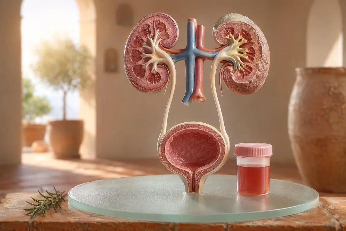
En patientförst-guide till synlig och mikroskopisk hematuri, inklusive varför urinstickans blod inte är samma sak som röda blodkroppar under mikroskopet.


- Blod i urinen betyder synligt rött, te-färgat eller colafärgat urin, eller mikroskopisk hematuri som påvisas vid testning; synligt blod förtjänar alltid medicinsk uppföljning.
- Mikroskopisk hematuri definieras vanligtvis som 3 eller fler röda blodkroppar per högförstoringsfält i ett korrekt insamlat urinmikroskopiprov.
- Urinstickans blod detekterar hemeaktivitet, så det kan vara positivt från intakta röda blodkroppar, fritt hemoglobin eller myoglobin efter muskelskada.
- Ledtrådar vid UVI innefattar sveda, frekvens, leukocytesteras, nitriter och vita celler; hematuri bör kontrolleras på nytt när infektionen har gått över.
- Urinsediment fynd som dysmorfa röda blodkroppar, erytrocytcylindrar eller betydande protein talar för sjukdom i njurens filtreringsenhet.
- Urincylindrar är formade material från njurtubuli; erytrocytcylindrar är en varningssignal för nefrologi samma vecka, särskilt vid högt blodtryck eller sjunkande eGFR.
- Cancer-varningssignaler innefattar smärtfri synlig hematuri, koagel, ålder över 45, rökhistorik, yrkesmässig kemisk exponering och återkommande oförklarade episoder.
- Träningsutlöst hematuri brukar lägga sig inom 48–72 timmar; kvarvarande blod efter vila ska inte skyllas på löpning eller cykling.
- Snar bedömning behövs för blod i urin med feber, flanksmärta, koagel som orsakar urinretention, graviditet, trauma eller symtom på svår anemi.
Vad blod i urinen betyder innan du får panik
Blod i urinen kan komma från infektion, stenar, njurfiltreringssjukdom, prostata- eller blåssjukdomar, läkemedel, träning eller kontamination från menstruation. Den praktiska regeln är enkel: synligt blod, upprepad mikroskopisk hematuri eller hematuri med smärta, feber, koagel, högt blodtryck, protein eller nedsatt njurfunktion kräver snabb utredning snarare än avvaktande.
En enstaka rosa färg i toalettstolen efter rödbetor eller ett nytt kosttillskott är inte samma kliniska problem som urinfärg som liknar cola efter en halsont. I min praktik berättar färghistorien ofta var jag först ska leta: klarrött med koagel talar för nedre urinvägar, medan brun- eller tefärgad urin kan betyda äldre heme-pigment från njurens filter.
Hematuri är inte en diagnos; det är ett tecken. Synlig hematuri betyder att du kan se färgförändringen, medan mikroskopisk hematuri betyder att urinen ser normal ut men att mikroskopi hittar röda blodkroppar, vanligtvis vid 3 eller fler röda blodkroppar per högförstoringsfält.
Kantesti är en AI blodprovsanalysator som hjälper patienter att koppla urinfynd med njurmarkörer som kreatinin, eGFR, CRP, hemoglobin och trombocyter i en och samma tidslinje. Om färgen är förvirrande har vi färgguide för urin som förklarar varför röd, brun, orange och mörkgul urin inte alla betyder samma sak.
Urinstickans blod är inte samma sak som RBC:er vid mikroskopi
Ett urinstickstest som är märkt blodpositivt detekterar hemliknande kemisk aktivitet, inte nödvändigtvis intakta erytrocyter. Mikroskopi besvarar nästa fråga: finns det faktiska RBC i urinsedimentet, och i så fall, hur ser de ut?
Urinstickstest är snabbt eftersom reagensdynan reagerar med peroxidasaktiviteten hos heme. Det betyder att ett positivt svar kan uppstå med intakta RBC, fritt hemoglobin efter nedbrytning av röda blodkroppar, eller myoglobin efter betydande muskelskada.
Mikroskopi går långsammare men är mer specifik. Ett noggrant labb centrifugerar urin, undersöker urinsedimentet, och rapporterar röda blodkroppar per högförstoringsfält; många kliniker använder 0–2 RBC/hpf som normalt och 3 eller fler RBC/hpf som avvikande hos vuxna.
Felmatchningen spelar roll. Ett urinstickstest med 3+ blod och 0–2 RBC/hpf får mig att fråga om rabdomyolys, hemolys, oxiderande kontaminanter och hantering av provet i stället för att hoppa direkt till blåssjukdom; skillnaden liknar resonemanget i vår jämförelse av urinstickprov och odling.
När UVI-ledtrådar förklarar hematuri, och när de inte gör det
En urinvägsinfektion kan orsaka blod i urinen, särskilt vid sveda, trängningar, frekvens, obehag i bäckenet, leukocytesteras, nitriter och vita blodkroppar. Men hematurin bör gå över efter behandling; kvarvarande röda blodkroppar efter en UVI behöver en andra bedömning.
Vid en okomplicerad nedre UVI förväntar jag mig en klunga: dysuri, frekvens, leukocytesteras positivt, pyuri ofta över 5–10 vita blodkroppar/hpf, och ibland nitriter. Traditionell odlingspositivitet är 100 000 CFU/mL, men hos symtomgivande kvinnor kan lägre värden som 1 000–10 000 CFU/mL fortfarande vara kliniskt betydelsefulla.
Leukocytesteras är ett enzym från vita blodkroppar, inte ett bevis på bakterier. Ett positivt fynd tillsammans med hematuri kan ses vid infektion, stenar, kontamination eller njurinflammation, vilket är anledningen till att vår leukocytesterasguide skiljer användbara ledtrådar från vanliga falska positiva fynd.
Nitriter är mer specifika men mindre känsliga eftersom inte alla bakterier omvandlar nitrat till nitrit. Om symtomen är typiska men nitriterna är negativa kan odlingen ändå vara positiv; de praktiska detaljerna finns i vår förklaring av nitritresultat.
Synlig hematuri: färg, koagel och tidsmässiga ledtrådar
Synlig hematuri är aldrig något jag avfärdar, även om det bara händer en gång. Klarröd urin, koagel eller smärtfri blödning behöver en snabb bedömning eftersom orsaker från nedre urinvägar blir mer sannolika när ålder och rökexponering ökar.
Tidsaspekten kan vara förvånansvärt användbar. Blod endast i början av urineringen kan tyda på urinröret, blod genom hela strålen kan komma från urinblåsa eller njurkällor, och terminalt blod nära slutet kan ibland peka mot området vid urinblåsans hals.
Koagel betyder oftast att blödningskällan ligger under njurens filter eftersom koagel inte bildas lätt efter att de passerat glomeruli och tubuli. Ett koagel som blockerar urinflödet är akut: urinblåsans distension kan bli smärtsam inom timmar, och katetervård kan behövas.
Män antar ibland att synlig hematuri efter urinsymtom bara beror på irritation i prostatan. Det kan stämma, men om PSA kontrollerades under eller strax efter en infektion kan tolkningen bli rörig; vår PSA efter UVI-riktlinje förklarar varför många kliniker väntar 6–8 veckor innan de upprepar PSA.
Mikroskopiska hematuritrösklar som läkare använder
Mikroskopisk hematuri definieras vanligen som 3 eller fler RBC per högförstoringsfält i ett korrekt insamlat urinprov. AUA/SUFU-riktlinjen använder denna gräns och rekommenderar riskbaserad utredning i stället för att behandla alla patienter likadant (Barocas et al., 2020).
Ett urinstickstest ensamt bör inte användas för att diagnostisera mikroskopisk hematuri. I Barocas et al. 2020 AUA/SUFU-riktlinjen beror definitionen på mikroskopi eftersom stickor kan vara positiva för hempigment utan intakta röda blodkroppar.
Upprepad provtagning är inte svaghet; det är god medicin. Om någon har 3–10 RBC/hpf efter kraftig träning, feber, sex, menstruation eller en kontaminerad provtagning vill jag vanligtvis ha ett rent upprepat prov innan jag beställer skanningar, ungefär som vi råder för guide för att upprepa avvikande labbvärden.
Risk förändrar vägen. En 24-åring med 4 RBC/hpf efter ett halvmara är inte samma sak som en 68-årig före detta rökare med 25 RBC/hpf i två prov, även om båda tekniskt sett uppfyller definitionen av hematuri.
Urinsediment, protein och urincylindrar som förändrar utredningen
Urinsediment kan avslöja om hematurin sannolikt kommer från njurens filter. Dysmorfa erytrocyter, erytrocytcylindrar och en betydande proteinskiftning gör att oron lutar mot glomerulär sjukdom snarare än en enkel urinblåsinfektion.
Röda blodkroppar som ser ojämna ut, har blebbar eller är ringformade kallas dysmorfa RBC, och de tyder på passage genom ett inflammerat glomerulärt filter. Acanthocyter ovan ungefär 5% av de urinära röda blodkropparna behandlas ofta som en stark glomerulär ledtråd, även om laboratorier varierar i hur säkert de rapporterar dem.
Protein är den andra ledtråden som jag inte ignorerar. Ett urin-albumin-kreatinin-kvot över 30 mg/g, eller cirka 3 mg/mmol, är avvikande, och synlig hematuri tillsammans med protein förtjänar snabbare granskning med fokus på njurarna; vår protein i urin-guide ger tröskelvärden på patientnivå.
Urincylindrar är små mögel-liknande strukturer som bildas inne i njurtubuli. Hyalina cylindrar kan uppträda efter uttorkning eller träning, men erytrocytcylindrar är avvikande och pekar vanligtvis mot glomerulonefrit; för en mer omfattande referens för urinalys, se vår komplett guide för urinanalys.
Stenar, träning och tillfälliga orsaker till hematuri
Stenar och ansträngande träning kan orsaka hematuri, men de har olika tidsförlopp. Hematuri kopplad till sten kommer ofta med flanksmärta eller kristaller, medan träningshematuri vanligtvis bör avta inom 48–72 timmar efter vila.
Njurstenar orsakar klassiskt vågor av flanksmärta, illamående och mikroskopisk eller synlig hematuri. Kalciumoxalatkristaller bevisar inte en sten, men de ökar misstanken när de kombineras med kolikartad smärta; vår kalciumoxalatkristallguide förklarar ledtråden från urins mikroskopi.
Träningshematuri är verklig. Jag har sett maratonlöpare med 10–20 RBC/hpf morgonen efter ett lopp, en normal odling, inget protein och en helt normal upprepad urinalys 3 dagar senare.
Faran är att skylla allt på träning. Om hematurin kvarstår efter mer än 72 timmar, uppträder tillsammans med mörk cola-färgad urin, eller kommer med högt CK, stigande kreatinin eller muskelsmärta, titta bortom urinblåsan; vår träningsguide för kreatinin täcker överlappet mellan njure och muskel.
Njurvarningssignaler som läkare inte ignorerar
Hematuri med protein, högt blodtryck, svullnad, sänkt eGFR, erytrocytcylindrar eller snabbt stigande kreatinin är en njurvarningssignal. Dessa fynd tyder på att filtreringsenheterna kan vara inflammerade, och att vänta i månader kan kosta njurfunktion.
KDIGO 2024 definierar kronisk njuk sjukdom utifrån markörer för njurskada, såsom albuminuri, eller eGFR under 60 mL/min/1,73 m² i minst 3 månader. I akuta situationer räcker dock hematuri tillsammans med en kreatininstegring över dagar eller veckor för att eskalera innan 3-månadersdefinitionen är uppfylld.
Albuminuri är ofta den tidiga varningssignalen. Ett ACR på 30–300 mg/g är måttligt förhöjt, över 300 mg/g är kraftigt förhöjt, och att kombinera något av detta med hematuri ändrar samtalet; vår njur-ACR-guide förklarar varför urin-albumin hittar skada innan symtom.
Blodtrycket spelar roll här. Ett nytt BT på 160/100 mmHg med cola-färgad urin och svullnad runt anklarna efter en halsinfektion får mig att tänka på glomerulonefrit, inte cystit; om eGFR är osäkert, vår GFR-omkontrollguide förklarar när cystatin C kan förtydliga njurfunktionen.
Ledtrådar om cancer-risk utan panik
Smärtfri synlig hematuri, särskilt efter 45 års ålder, förtjänar akut utredning eftersom cancer i urinblåsa och övre urinvägar kan ge sig till känna på detta sätt. NICE NG12 rekommenderar remiss för misstänkt cancer vid oförklarad synlig hematuri hos vuxna 45 år och äldre, eller synlig hematuri som kvarstår efter UVI-behandling.
Det mesta av hematurin är inte cancer. Ändå är de fall jag minns de tysta: ingen smärta, ingen feber, ingen sveda—bara två episoder av röd urin hos en 56-årig rökare som nästan struntade i det.
Risken är kumulativ, inte binär. Ålder över 45–50, rökhistorik, exponering för cyklofosfamid, bäckent strålbehandling, återkommande synlig hematuri, yrkesmässig exponering för aromatiska aminer och koagel ökar alla behovet av cystoskopi eller bilddiagnostik.
Prostatamarkörer utesluter inte orsaker från urinblåsan. Om urinsymtom, PSA-förändringar och hematuri överlappar, granska tidpunkt och infektionsstatus noggrant; vår prostatamarkörguide förklarar varför PSA bara är en del av den manliga urinbedömningen.
Läkemedel, antikoagulantia och falsklarm
Blodförtunnande läkemedel kan avslöja blödning, men de ska inte automatiskt skyllas för hematuri. Hematuri i samband med antikoagulantia behöver fortfarande utredas eftersom läkemedlet kan synliggöra stenar, infektion, njursjukdom eller en tumör i urinvägarna.
Warfarin, apixaban, rivaroxaban, acetylsalicylsyra och klopidogrel kan göra blödning mer uppenbar. Om INR ligger över målintervallet spelar korrigering roll, men ett normalt INR gör inte synlig hematuri ofarlig.
Flera orsaker utan blödning kan efterlikna hematuri. Rifampicin kan göra urinen orange-röd, fenazopyridin kan göra den klarorange, rödbetor kan färga urinen hos mottagliga personer, och dehydrering kan mörka pigmentet så mycket att det skrämmer patienter; antikoagulantmonitorering täcks i vår säkerhetslabbs för blodförtunnande.
Urinstickan kan också vilseleda efter hård träning eller muskelskada. Ett positivt heme-pad med få eller inga RBCs gör att jag tänker mer på myoglobin, särskilt om CK är över 1 000 IU/L och urinen är mörk.
Barn, graviditet och kontamination från menstruation
Hematuri hos barn, under graviditet eller i anslutning till menstruation kräver renare provtagning och en lägre tröskel för granskning. Samma urinresultat kan betyda olika saker beroende på ålder, graviditetsstatus, feber, blodtryck och provtagningskvalitet.
Menstruationskontamination är vanligt och ingen ska behöva skämmas för det. Om resultatet är oväntat, upprepa ett midstream rent fångstprov minst 48 timmar efter att blödningen har upphört; kvarstående 3 eller fler RBC/hpf blir mer meningsfullt.
Barn behöver ofta ett annat perspektiv. Hematuri efter en virusinfektion kan vara övergående, men hematuri med svullnad, högt blodtryck, protein eller minskad urinproduktion kräver snabb barnläkarbedömning.
Graviditet höjer insatserna eftersom UVI, stenar, njurpåverkan vid preeklampsi och kontamination kan överlappa. För varningsmönster samma dag vid graviditet som involverar njure, lever, trombocyter och urinfynd, se vår graviditetslabbsvarningssignaler.
Tester som läkare vanligtvis beställer efter hematuri
De vanliga nästa testerna efter hematuri är upprepad urinalys med mikroskopi, urinodling om infektion är möjlig, blodprover för njurfunktion, urinprotein eller ACR, blodtrycksmätning samt riskbaserad bilddiagnostik eller cystoskopi. Exakt ordningsföljd beror på symtom och risk.
Vid okomplicerad cystit kan odling och behandling komma först, följt av upprepad urinsedimentmikroskopi när symtomen har gått över. Vid smärtfri synlig hematuri går många kliniker tidigare till bilddiagnostik och cystoskopi eftersom väntan på upprepade episoder kan fördröja diagnosen.
Blodprover tillför sammanhang som urin ensam inte kan ge. Kreatinin, eGFR, urea eller BUN, elektrolyter, CBC, CRP, komplement C3/C4 och autoimmuna markörer kan vara lämpliga när man misstänker njurinflammation; vår guide för kvoten BUN/kreatinin förklarar ett vanligt njurmönster.
Kantesti AI kan organisera dessa resultat till en trendvy, men läkargranskning förblir avgörande när varningsflaggor finns. Vårt kliniska säkerhetsupplägg beskrivs i Medicinsk validering, inklusive hur avvikande kluster hanteras i stället för enstaka siffror.
Hur Kantesti hjälper till att organisera hematuri-kontext
Kantesti är en Tolkningstjänst för AI-labbtester som läser urinnära blodmarkörer i sitt sammanhang, såsom kreatinin, eGFR, CRP, hemoglobin, trombocyter och glukos. Den ställer inte diagnos om hematuri, men den hjälper patienter att förbereda en renare och mer användbar sammanfattning för sin läkare.
När jag granskar hematuri tittar jag sällan på ett enda resultat isolerat. Ett normalt kreatinin på 0,82 mg/dL, stabil eGFR över 90, inget protein och en negativ odling känns väldigt annorlunda än hematuri med eGFR 48 och CRP 65 mg/L.
Kantesti är en Plattform för tolkning av AI-biomarkörer som kan bearbeta uppladdade PDF:er för blodprover eller foton och länka markörer för njurpåverkan, inflammation och anemi utanför referensintervall till tidpunkten för symtomen. Metoderna bakom den tolkningen beskrivs i vår Teknikguide, och mer omfattande täckning av markörer beskrivs i den guide för biomarkörer.
Ett praktiskt tips: kontrollera innan du laddar upp att enheter, datum och patientidentifierare fångas korrekt. OCR-fel kan göra 0,9 till 9,0, vilket är en helt annan njurberättelse; vår checklista för PDF-uppladdning visar de misstag jag ber patienter att först fånga upp.
Forskningsanteckningar, styrning och den slutliga checklistan
Den 11 juli 2026 är den säkraste patientplanen att bekräfta om hematuri är verklig, identifiera tecken på infektion, leta efter varningsflaggor från njurarna och eskalera synlig eller ihållande hematuri. Lita inte enbart på urinfärgen.
Jag är Thomas Klein, MD, och mitt råd är medvetet konservativt eftersom missad hematuri kan spela roll. Kantesti:s medicinska innehåll granskas med klinisk tillsyn från vår Medicinsk rådgivande nämnd, och vår roll är att hjälpa patienter att ställa bättre frågor, inte att ersätta deras läkare.
Kantesti är en AI-baserat analysverktyg för blodprov används av miljontals människor i 127 länder, så integritet och klinisk tydlighet måste byggas in i arbetsflödet snarare än läggas till i efterhand. Du kan läsa mer om Kantesti LTD, vår brittiska bolagsstruktur och kliniska uppdrag på Om oss.
Kantesti AI Clinical Research Group. (2026). AI Blood Test Analyzer: 2,5M tester analyserade | Global Health Report 2026. Zenodo. DOI: https://doi.org/10.5281/zenodo.18175532. ResearchGate: https://www.researchgate.net/. Academia.edu: https://www.academia.edu/.
Kantesti AI Clinical Research Group. (2026). RDW Blood Test: Komplett guide till RDW-CV, MCV & MCHC. Zenodo. DOI: https://doi.org/10.5281/zenodo.18202598. ResearchGate: https://www.researchgate.net/. Academia.edu: https://www.academia.edu/.
Vanliga frågor
Är blod i urinen alltid allvarligt?
Blod i urinen är inte alltid allvarligt, men det bör bekräftas och förklaras. Ett enstaka mikroskopiskt fynd på 3–5 RBC/hpf efter träning eller menstruation kan försvinna vid upprepad provtagning, medan synligt blod, koagel, feber, flanksmärta, protein, högt blodtryck eller sänkt eGFR kräver snabb bedömning. Hos vuxna över 45 år förtjänar oförklarad synlig hematuri en akut utredning även om det inte finns någon smärta.
Vad är skillnaden mellan blod på urinsticka och RBC i urin?
Urinstickan påvisar blod genom hemliknande kemisk aktivitet, medan urinsedimentmikroskopi räknar faktiska erytrocyter. En urinsticka kan vara positiv från intakta RBC, fritt hemoglobin eller myoglobin efter muskelskada, så den är inte identisk med verifierad hematuri. Mikroskopisk hematuri definieras vanligen som 3 eller fler RBC per högförstoringsfält i ett korrekt insamlat prov.
Kan en urinvägsinfektion orsaka blod i urinen?
Ja, en urinvägsinfektion (UVI) kan orsaka blod i urinen, särskilt när det svider, samt vid frekvens, trängningar, leukocytesteras, nitriter och vita blodkroppar också är närvarande. Traditionell positiv urindyrkning är ofta 100 000 CFU/ml, men lägre värden kan vara betydelsefulla hos symtomatiska patienter. Hematuri bör vanligtvis kontrolleras på nytt efter behandling eftersom kvarvarande blod kan tyda på stenar, njursjukdom eller en annan orsak från urinvägarna.
När ska jag söka akutvård för hematuri?
Sök akut vård vid blod i urinen med feber, svår flanksmärta, kräkningar, graviditet, nyligen trauma, koagel, oförmåga att kissa, svimning eller symtom på svår anemi. Medicinsk bedömning samma vecka är också lämplig vid hematuri med protein, erytrocytcylindrar, nyuppkommen högt blodtryck eller en stigande kreatinin. En blockerad urinström från koagel kan bli smärtsam och osäker inom timmar.
Kan träning orsaka mikroskopisk hematuri?
Ansträngande träning kan orsaka mikroskopisk hematuri, särskilt efter långdistanslöpning, intensiv cykling eller kontaktsporter. Träningsrelaterad hematuri brukar gå över inom 48–72 timmar efter vila och bör inte åtföljas av kvarstående proteinuri, sjunkande eGFR eller svår muskelsmärta. Om blod på urinsticka är positivt men mikroskopi visar få RBC, kan kliniker kontrollera CK eftersom myoglobin från muskelskada kan utlösa urinstickan.
Vad betyder urincylindrar när blod påträffas?
Urincylindrar är formade material som bildas inne i njurtubuli, och deras typ ändrar innebörden av hematuri. Hyalina cylindrar kan förekomma vid uttorkning eller träning, men erytrocytcylindrar är onormala och tyder på glomerulär njurinflammation. Blod i urinen med erytrocytcylindrar, protein och högt blodtryck bör behandlas som en njurvarningssignal snarare än en enkel UVI.
Kan uttorkning få urinen att se ut som blod?
Uttorkning gör ofta urinen mörkgul eller bärnstensfärgad, men det skapar vanligtvis inte röda blodkroppar i urinen. Drick vatten och boka en urinprovtagning om färgen är rosa, röd, brun eller kolaliknande, särskilt om den kvarstår efter vätskeintag eller åtföljs av smärta.
Få AI-drivna analyser av blodprov redan idag
Gå med i över 2 miljoner användare världen över som litar på Kantesti för snabb och korrekt analys av blodprover. Ladda upp dina blodprovsresultat och få en heltäckande tolkning av 15,000+-biomarkörer på sekunder.
📚 Refererade forskningspublikationer
Klein, T., Mitchell, S., & Weber, H. (2026). AI blodprovsanalys: 2,5M analyserade tester | Global hälsorapport 2026. Kantesti AI Medical Research.
Klein, T., Mitchell, S., & Weber, H. (2026). Blodprov RDW: Komplett guide till RDW-CV, MCV & MCHC. Kantesti AI Medical Research.
📖 Externa medicinska referenser
National Institute for Health and Care Excellence (2015). Misstänkt cancer: igenkänning och remiss. NICE-riktlinje NG12. NICE-riktlinje.
Njursjukdom: Improving Global Outcomes CKD Work Group (2024). KDIGO 2024 Klinisk riktlinje för utvärdering och hantering av kronisk njursjukdom. Kidney International.
⚕️ Medicinsk ansvarsfriskrivning
Den här artikeln är endast avsedd för utbildningsändamål och utgör inte medicinsk rådgivning. Rådgör alltid med en behörig vårdgivare för beslut om diagnos och behandling.
E-E-A-T förtroendesignaler
Uppleva
Läkarledd klinisk granskning av arbetsflöden för laboratorietolkning.
Expertis
Laboratoriemedicinskt fokus på hur biomarkörer beter sig i kliniskt sammanhang.
Auktoritet
Skrivet av Dr. Thomas Klein med granskning av Dr. Sarah Mitchell och Prof. Dr. Hans Weber.
Trovärdighet
Evidensbaserad tolkning med tydliga uppföljningsspår för att minska larm.