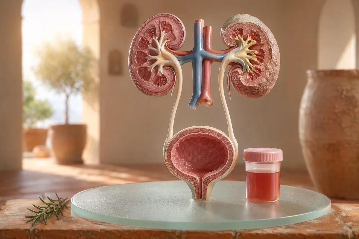
Հիվանդին ուղղված ուղեցույց՝ տեսանելի և միկրոսկոպիկ հեմատուրիայի վերաբերյալ, ներառյալ՝ ինչու դիպսթիքով հայտնաբերված արյունը նույնը չէ, ինչ կարմիր բջիջները՝ միկրոսկոպի տակ։.


- Արյուն մեզում նշանակում է տեսանելի կարմիր, թեյի գույնի կամ կոլայի գույնի մեզ, կամ միկրոսկոպիկ հեմատուրիա՝ թեստավորման ժամանակ հայտնաբերված; տեսանելի արյունը միշտ արժանի է բժշկական հետագա հսկողության։.
- Միկրոսկոպիկ հեմատուրիա սովորաբար սահմանվում է որպես 3 կամ ավելի կարմիր արյան բջիջ մեկ բարձր խոշորացման դաշտում՝ պատշաճ հավաքված մեզի միկրոսկոպիայի նմուշում։.
- Դիպսթիքով հայտնաբերված արյուն հայտնաբերում է հեմի ակտիվությունը, ուստի կարող է դրական լինել՝ ամբողջական կարմիր բջիջներից, ազատ հեմոգլոբինից կամ միոգլոբինից՝ մկանային վնասվածքից հետո։.
- UTI-ի հուշումներ ներառում են այրոց, հաճախամիզություն, լեյկոցիտ էստերազ, նիտրիտներ և սպիտակ բջիջներ. հեմատուրիան պետք է կրկնակի ստուգվի, երբ վարակը մաքրվի։.
- Մեզի նստվածք հայտնաբերումներ, ինչպիսիք են դիսմորֆիկ կարմիր բջիջները, կարմիր բջիջների գլանիկները կամ զգալի սպիտակուցը, ուղղորդում են դեպի երիկամների ֆիլտրող միավորի հիվանդություն։.
- Մեզի գլանիկներ ձևավորված են երիկամային խողովակիկներից. էրիթրոցիտային գլանիկները նույն շաբաթվա ընթացքում նեֆրոլոգիական «կարմիր դրոշ» են, հատկապես բարձր արյան ճնշման կամ ընկնող eGFR-ի դեպքում։.
- Քաղցկեղի «կարմիր դրոշներ» ներառում են ցավազուրկ տեսանելի հեմատուրիա, թրոմբեր, 45 տարեկանից բարձր տարիք, ծխելու պատմություն, մասնագիտական քիմիական ազդեցություն և կրկնվող, չբացատրված դրվագներ։.
- վարժանքային հեմատուրիա սովորաբար հանդարտվում է 48-72 ժամվա ընթացքում. հանգստից հետո պահպանվող արյունը չի կարելի մեղադրել վազքի կամ հեծանվավարության վրա։.
- Շտապ գնահատում անհրաժեշտ է, երբ մեզում արյուն կա՝ ուղեկցվող ջերմությամբ, կողային/գոտկային ցավով, մեզուղիների փակման պատճառ դառնալու թրոմբերով, հղիությամբ, տրավմայով կամ ծանր անեմիայի ախտանիշներով։.
Ի՞նչ է նշանակում մեզում արյունը՝ նախքան խուճապի մատնվելը
Արյուն մեզում կարող է առաջանալ վարակից, քարերից, երիկամի ֆիլտրող հիվանդությունից, շագանակագեղձի կամ միզապարկի վիճակներից, դեղերից, վարժանքից կամ դաշտանային արյունից աղտոտումից։ Գործնական կանոնը պարզ է. տեսանելի արյունը, կրկնվող միկրոսկոպիկ հեմատուրիան կամ հեմատուրիան՝ ցավով, ջերմությամբ, թրոմբերով, բարձր արյան ճնշմամբ, սպիտակուցով կամ երիկամային ֆունկցիայի նվազմամբ, պահանջում է անհապաղ գնահատում՝ այլ ոչ թե սպասողական մոտեցում։.
Մեկ անգամ վարդագույն գույնի զուգարանի աման՝ ճակնդեղ ուտելուց կամ նոր հավելում ընդունելուց հետո, նույն կլինիկական խնդիրը չէ, ինչ կոլայի գույնի մեզը՝ կոկորդի ցավից հետո։ Իմ պրակտիկայում գունային պատմությունը հաճախ ասում է, թե որտեղից սկսել որոնումը. վառ կարմիրը՝ թրոմբերով, սովորաբար վերաբերում է ստորին միզուղիներին, իսկ շագանակագույն կամ թեյանման գույնի մեզը կարող է նշանակել ավելի հին հեմի պիգմենտներ՝ երիկամային ֆիլտրերից։.
Հեմատուրիան ախտորոշում չէ. դա նշան է։. Տեսանելի հեմատուրիա նշանակում է՝ դուք տեսնում եք գույնի փոփոխությունը, մինչդեռ միկրոսկոպիկ հեմատուրիա նշանակում է՝ մեզը նորմալ է թվում, բայց միկրոսկոպիան հայտնաբերում է կարմիր բջիջներ՝ սովորաբար 3 կամ ավելի կարմիր արյան բջիջ/բարձր խոշորացման դաշտ (high-power field)։.
Կանտեստին մի է AI արյան անալիզատոր դա օգնում է հիվանդներին կապել մեզի հայտնաբերումները երիկամային մարկերների հետ՝ ինչպիսիք են կրեատինինը, eGFR-ը, CRP-ն, հեմոգլոբինը և թրոմբոցիտները՝ մեկ ժամանակագրական շղթայում։ Եթե գույնը շփոթեցնող է, մեր մեզի գույնի ուղեցույցը բացատրում է, թե ինչու կարմիր, շագանակագույն, նարնջագույն և մուգ դեղին մեզը բոլորը նույն բանը չեն նշանակում։.
Դիպսթիքով հայտնաբերված արյունը նույնը չէ, ինչ RBC-ները միկրոսկոպիայի ժամանակ
Մեզի դիպսթիկով նշված արյուն դրական հայտնաբերում է հեմանման քիմիական ակտիվություն, ոչ պարտադիր՝ ամբողջական կարմիր արյան բջիջներ։ Միկրոսկոպիան պատասխանում է հաջորդ հարցին. մեզի նստվածքում կա՞ն իրական RBC-ներ, և եթե այո՝ ինչպիսի՞ն են դրանք։
Դիպսթիկով թեստավորումը արագ է, քանի որ ռեագենտային թերթիկը արձագանքում է հեմի պերօքսիդազային ակտիվությանը։ Դա նշանակում է, որ դրական արդյունք կարող է լինել՝ ամբողջական RBC-ների դեպքում, կարմիր բջիջների քայքայումից հետո ազատ հեմոգլոբինի դեպքում կամ զգալի մկանային վնասվածքից հետո միոգլոբինի դեպքում։.
Միկրոսկոպիան ավելի դանդաղ է, բայց ավելի սպեցիֆիկ։ Զգույշ լաբորատորիան մեզը ցենտրիֆուգում է, զննում է մեզի նստվածքը, և հայտնում է կարմիր բջիջները՝ մեկ high-power field-ում. շատ կլինիկոսներ մեծահասակների մոտ նորմալ են համարում 0-2 RBC/hpf-ը, իսկ 3 կամ ավելի RBC/hpf-ը՝ աննորմալ։.
Անհամապատասխանությունը կարևոր է։ Դիպսթիկով 3+ արյուն և 0-2 RBC/hpf՝ ստիպում է ինձ հարցնել ռաբդոմիոլիզի, հեմոլիզի, օքսիդացնող աղտոտիչների և նմուշի մշակման մասին՝ միզապարկի հիվանդությանն անմիջապես անցնելու փոխարեն. տարբերությունը նման է մեր այն տրամաբանությանը, որը մեզի ընդհանուր քննություն և կուլտուրայի համեմատություն.
Երբ UTI-ի նշանները բացատրում են հեմատուրիան և երբ՝ ոչ
Միզուղիների վարակը կարող է առաջացնել արյուն մեզում, հատկապես՝ այրոցով, շտապողականությամբ, հաճախամիզությամբ, կոնքի անհարմարությամբ, լեյկոցիտ էստերազով, նիտրիտներով և սպիտակ բջիջներով։ Սակայն հեմատուրիան պետք է մաքրվի բուժումից հետո. UTI-ից հետո պահպանվող կարմիր բջիջները պահանջում են երկրորդ հայացք։.
Պարզ ստորին UTI-ի դեպքում ես ակնկալում եմ մի խումբ՝ դիզուրիա, հաճախամիզություն, լեյկոցիտ էստերազը դրական, պիուրիան հաճախ՝ ավելի քան 5-10 սպիտակ բջիջ/hpf, և երբեմն՝ նիտրիտներ։ Ավանդական կուլտուրայի դրականությունը 100,000 CFU/mL է, բայց ախտանշաններով կանանց մոտ ավելի ցածր քանակները, օրինակ՝ 1,000-10,000 CFU/mL, կարող են դեռևս կլինիկորեն նշանակալի լինել։.
Լեյկոցիտ էստերազը սպիտակ բջիջների ֆերմենտ է, ոչ թե բակտերիաների ապացույց։ Դրական արդյունքը հեմատուրիայի հետ կարող է առաջանալ վարակից, քարերից, աղտոտումից կամ երիկամի բորբոքումից, այդ իսկ պատճառով մեր լեյկոցիտ էստերազի ուղեցույցը առանձնացնում է օգտակար հուշումները սովորական կեղծ դրականներից։.
Նիտրիտները ավելի սպեցիֆիկ են, բայց՝ ավելի քիչ զգայուն, քանի որ բոլոր բակտերիաները չեն փոխակերպում նիտրատը նիտրիտի։ Եթե ախտանիշները դասական են, բայց նիտրիտները բացասական են, կուլտուրան դեռ կարող է դրական լինել. գործնական մանրամասները ներառված են մեր նիտրիտի արդյունքի բացատրիչում.
Տեսանելի հեմատուրիա. գույն, թրոմբներ և ժամանակային նշաններ
Տեսանելի հեմատուրիան երբեք բան չէ, որ ես անտեսեմ, նույնիսկ եթե դա լինում է մեկ անգամ։ Վառ կարմիր մեզը, թրոմբերը կամ ցավազուրկ արյունահոսությունը պահանջում են ժամանակին գնահատում, քանի որ ստորին միզուղիների պատճառները ավելի հավանական են դառնում տարիքի և ծխելու ազդեցության աճի հետ։.
Ժամանակացույցը կարող է անսպասելիորեն օգտակար լինել։ Մեզի արտազատման սկզբում միայն արյունը կարող է հուշել միզուկի մասին, հոսքի ընթացքում արյունը կարող է գալ միզապարկի կամ երիկամային աղբյուրներից, իսկ վերջում՝ տերմինալ արյունը երբեմն մատնանշում է միզապարկի պարանոցի հատվածը։.
Թրոմբերը սովորաբար նշանակում են, որ արյունահոսության աղբյուրը գտնվում է երիկամի ֆիլտրերի տակ, քանի որ թրոմբերը հեշտությամբ չեն ձևավորվում գլոմերուլներով և խողովակներով անցնելուց հետո։ Արյան թրոմբ, որը խոչընդոտում է մեզի հոսքը, շտապ է․ միզապարկի լցվածությունը կարող է ցավոտ դառնալ մի քանի ժամվա ընթացքում, և կարող է անհրաժեշտ լինել կաթետերի խնամք։.
Տղամարդիկ երբեմն ենթադրում են, որ միզային ախտանիշներից հետո տեսանելի հեմատուրիան պարզապես շագանակագեղձի գրգռում է։ Դա կարող է ճիշտ լինել, բայց եթե PSA-ն ստուգվել է վարակի ընթացքում կամ դրանից անմիջապես հետո, մեկնաբանությունը կարող է բարդ լինել. մեր PSA-ն UTI-ից հետո ուղեցույց բացատրում է, թե ինչու շատ կլինիկիստներ սպասում են 6-8 շաբաթ՝ PSA-ն կրկնելուց առաջ։.
Միկրոսկոպիկ հեմատուրիայի շեմեր, որոնք օգտագործում են բժիշկները
Միկրոսկոպիկ հեմատուրիան սովորաբար սահմանվում է որպես 3 կամ ավելի RBC՝ մեկ բարձր խոշորացման դաշտում պատշաճ հավաքված մեզի նմուշում։ AUA/SUFU ուղեցույցը օգտագործում է այս շեմը և խորհուրդ է տալիս ռիսկահիմնված գնահատում՝ յուրաքանչյուր հիվանդին նույն կերպ չբուժելու փոխարեն (Barocas et al., 2020)։.
Միայն դիպսթիքով թեստը չպետք է օգտագործվի միկրոսկոպիկ հեմատուրիան ախտորոշելու համար։ Barocas et al. 2020 AUA/SUFU ուղեցույցում սահմանումը կախված է միկրոսկոպիայից, քանի որ դիպսթիքերը կարող են դրական լինել հեմի պիգմենտներից՝ առանց ամբողջական կարմիր բջիջների։.
Կրկնակի թեստավորումը թուլություն չէ․ դա լավ բժշկություն է։ Եթե ինչ-որ մեկը ունի 3-10 RBC/hpf ծանր ֆիզիկական ծանրաբեռնվածությունից, ջերմությունից, սեռական հարաբերությունից, դաշտանային արյունահոսությունից կամ աղտոտված հավաքումից հետո, ես սովորաբար ուզում եմ մաքուր կրկնակի նմուշ՝ սկաներ պատվիրելուց առաջ, ճիշտ ինչպես մենք խորհուրդ ենք տալիս կրկնակի աննորմալ վերլուծությունների.
Ռիսկը փոխում է ուղին։ 24-ամյա անձը, ով ունի 4 RBC/hpf կեսմարաթոնից հետո, նույնը չէ, ինչ 68-ամյա նախկին ծխողը, ով ունի 25 RBC/hpf երկու նմուշներում, նույնիսկ եթե երկուսն էլ տեխնիկապես համապատասխանում են հեմատուրիայի սահմանմանը։.
Մեզի նստվածք, սպիտակուց և մեզի գլանաձև գոյացություններ, որոնք փոխում են հետազոտությունների ընթացքը
Մեզի նստվածք կարող է բացահայտել՝ արդյոք հեմատուրիան հավանական է գալիս երիկամի ֆիլտրերից։ Դիսմորֆ կարմիր բջիջները, կարմիր բջիջների գլանային գլանակները (casts) և սպիտակուցի զգալի տեղաշարժը մտահոգությունը տեղափոխում են դեպի գլոմերուլային հիվանդություն՝ պարզ միզապարկի վարակից ավելի։.
Կարմիր բջիջները, որոնք անհավասար տեսք ունեն, «blebbed» կամ օղակաձև են, կոչվում են դիսմորֆ RBC-ներ, և դրանք հուշում են բորբոքված գլոմերուլային ֆիլտրով անցման մասին։ Ականտոցիտները՝ մեզային կարմիր բջիջների մոտավորապես 5%-ից բարձր, հաճախ դիտարկվում են որպես ուժեղ գլոմերուլային հուշում, թեև լաբորատորիաները տարբերվում են նրանում, թե որքան վստահ են դրանք հաղորդում։.
Սպիտակուցը երկրորդ հուշումն է, որը ես չեմ անտեսում։ Մեզի ալբումին-կրեատինին հարաբերությունը 30 mg/g-ից բարձր՝ կամ մոտ 3 mg/mmol, աննորմալ է, և տեսանելի հեմատուրիան՝ գումարած սպիտակուցին, արժանի է ավելի արագ՝ երիկամներին ուղղված վերանայման. մեր մեզի սպիտակուցի ուղեցույց տալիս է հիվանդի մակարդակի շեմերը։.
Մեզի գլանիկներ փոքրիկ «կաղապարներ» են, որոնք ձևավորվում են երիկամային խողովակների ներսում։ Հիալինային գլանակները կարող են հայտնվել ջրազրկումից կամ ֆիզիկական ծանրաբեռնվածությունից հետո, բայց կարմիր բջիջների գլանակները աննորմալ են և սովորաբար մատնանշում են գլոմերուլոնեֆրիտ. մեզի ընդհանուր անալիզի ավելի լայն հղման համար տես մեր ամբողջական մեզի վերլուծության ուղեցույց.
Քարեր, ֆիզիկական վարժություններ և հեմատուրիայի ժամանակավոր պատճառներ
Քարերը և ուժեղ ֆիզիկական ծանրաբեռնվածությունը կարող են առաջացնել հեմատուրիա, բայց դրանք ունեն տարբեր ժամանակագրություններ։ Քարային հեմատուրիան հաճախ ուղեկցվում է կողային (flank) ցավով կամ բյուրեղներով, մինչդեռ վարժանքային հեմատուրիան սովորաբար պետք է հանդարտվի հանգստից հետո 48-72 ժամվա ընթացքում։.
Երիկամային քարերը դասականորեն առաջացնում են կողային ցավի ալիքներ, սրտխառնոց և միկրոսկոպիկ կամ տեսանելի հեմատուրիա։ Կալցիում օքսալատային բյուրեղները չեն ապացուցում քար, բայց բարձրացնում են կասկածը, երբ զուգորդվում են կոլիկային ցավով. մեր կալցիում օքսալատային բյուրեղների ուղեցույց բացատրում է մեզի միկրոսկոպիայի հուշումը։.
Վարժանքային հեմատուրիան իրական է։ Ես տեսել եմ մարաթոն վազորդների, ովքեր մրցումից հետո հաջորդ առավոտյան ունենում են 10-20 RBC/hpf, նորմալ կուլտուրա, առանց սպիտակուցի, և լիովին նորմալ կրկնակի մեզի անալիզ 3 օր անց։.
Վտանգը այն է, որ ամեն ինչի մեղքը դնենք մարզմանը։ Եթե հեմատուրիան պահպանվում է 72 ժամից ավելի, հայտնվում է մուգ կոլայի գույնի մեզով, կամ ուղեկցվում է բարձր CK-ով, աճող կրեատինինով կամ մկանային ցավով, նայեք նաև միզապարկից այն կողմ. մեր ուղեցույց՝ կրեատինինի վարժությունների համար ընդգրկում է երիկամի և մկանների համընկնումը։.
Երիկամների «կարմիր դրոշներ», որոնք բժիշկները չեն անտեսում
Հեմատուրիա՝ սպիտակուցով, բարձր արյան ճնշմամբ, այտուցով, կրճատված eGFR-ով, էրիթրոցիտային գլանիկներով կամ կրեատինինի արագ աճով՝ երիկամի «կարմիր դրոշ» է։ Այս հայտնագործությունները ցույց են տալիս, որ ֆիլտրող միավորները կարող են բորբոքված լինել, և ամիսներով սպասելը կարող է արժենալ երիկամի ֆունկցիան։.
KDIGO 2024-ը սահմանում է քրոնիկ երիկամային հիվանդությունը՝ երիկամային վնասի մարկերներով, օրինակ՝ ալբումինուրիայով, կամ eGFR-ի՝ 60 մլ/րոպ/1.73 մ²-ից ցածր լինելու դեպքում՝ առնվազն 3 ամիս։ Սակայն սուր իրավիճակներում հեմատուրիան՝ գումարած կրեատինինի աճին՝ օրերի կամ շաբաթների ընթացքում, բավարար է՝ բարձրացնելու համար՝ նախքան 3-ամսյա սահմանումը կհամապատասխանի։.
Ալբումինուրիան հաճախ վաղ նախազգուշացնող ազդանշանն է։ ACR-ը 30-300 մգ/գ՝ չափավոր բարձրացած է, 300 մգ/գ-ից բարձր՝ խիստ բարձրացած, և եթե դա զուգակցվում է հեմատուրիայով, խոսակցությունը փոխվում է. մեր երիկամների ACR ուղեցույցը բացատրում է, թե ինչու մեզի ալբումինը վնասը գտնում է նախքան ախտանշանները։.
Արյան ճնշումը կարևոր է այստեղ։ Նոր BP՝ 160/100 մմ ս.ս.-ով, կոլա գույնի մեզով և կոճերի այտուցով՝ կոկորդի վարակից հետո, ինձ ստիպում է մտածել գլոմերուլոնեֆրիտի մասին, ոչ թե ցիստիտի. եթե eGFR-ը վստահ չէ, մեր GFR-ի վերահսկման ուղեցույց բացատրում է, թե երբ ցիստատին C-ը կարող է պարզաբանել երիկամային ֆունկցիան։.
Քաղցկեղի ռիսկի նշաններ՝ առանց խուճապի
Անցավ տեսանելի հեմատուրիա, հատկապես 45 տարեկանից հետո, արժանի է շտապ գնահատման, քանի որ միզապարկի և վերին միզուղիների քաղցկեղները կարող են այսպես դրսևորվել։ NICE NG12-ը խորհուրդ է տալիս կասկածվող-քաղցկեղի ուղղորդում՝ չբացատրված տեսանելի հեմատուրիայի դեպքում մեծահասակների մոտ 45 և ավելի տարիքով, կամ տեսանելի հեմատուրիայի դեպքում, որը պահպանվում է UTI-ի բուժումից հետո։.
Հեմատուրիայի մեծ մասը քաղցկեղ չէ։ Այնուամենայնիվ, այն դեպքերը, որ հիշում եմ, «լուռ»ներն են՝ ոչ ցավ, ոչ ջերմություն, ոչ այրոց, պարզապես երկու դրվագ՝ կարմիր մեզով 56-ամյա ծխողի մոտ, ով գրեթե անտեսել էր դա։.
Ռիսկը կուտակային է, ոչ թե երկարժեք։ 45-50-ից բարձր տարիք, ծխելու պատմություն, ցիկլոֆոսֆամիդի ազդեցություն, կոնքի ճառագայթային թերապիա, կրկնվող տեսանելի հեմատուրիա, աշխատանքային արոմատիկ ամինների ազդեցություն և թրոմբներ՝ բոլորը բարձրացնում են ցիստոսկոպիայի կամ պատկերավորման անհրաժեշտությունը։.
Պրոստատային մարկերները չեն բացառում միզապարկային պատճառները։ Եթե միզուղիների ախտանշաններ կան, PSA-ի փոփոխությունները և հեմատուրիան համընկնում են, ուշադիր վերանայեք ժամանակացույցը և վարակային կարգավիճակը. մեր מדריך לסמן פרוסטטה מסביר מדוע PSA הוא רק חלק אחד מהערכת השתן הגברית.
Դեղեր, հակամակարդիչներ և կեղծ ահազանգեր
מדללי דם יכולים לחשוף דימום, אך אין להאשים אותם באופן אוטומטי בהמטוריה. המטוריה הקשורה לנוגדי קרישה עדיין דורשת הערכה, משום שהתרופה עשויה לחשוף אבנים, זיהום, מחלת כליה או גידול בדרכי השתן.
וורפרין, אפיקסבן, ריברוקסבן, אספירין וקלופידוגרל יכולים להפוך דימום לברור יותר. אם ה-INR גבוה מטווח היעד, תיקונו חשוב, אך INR תקין אינו הופך המטוריה הנראית לעין לבטוחה וחסרת משמעות.
מספר גורמים שאינם מדממים יכולים לחקות המטוריה. ריפמפיצין יכול להפוך שתן לכתום-אדמדם, פנאזופירידין יכול להפוך אותו לכתום עז, סלק יכול להכתים שתן אצל אנשים רגישים, והתייבשות יכולה להכהות פיגמנט מספיק כדי להבהיל מטופלים; ניטור נוגדי קרישה מכוסה ב- בדיקות בטיחות של מדללי דם.
הסטיק גם עלול להטעות לאחר פעילות גופנית מאומצת או פגיעה בשריר. תוצאה חיובית של משטח להֶם עם מעט או ללא RBCs מכוונת את החשיבה שלי למיוגלובין, במיוחד אם CK מעל 1,000 IU/L והשתן כהה.
Երեխաներ, հղիություն և դաշտանային աղտոտում
המטוריה בילדים, בהריון או סביב הווסת דורשת דגימה נקייה יותר וסף נמוך יותר לבדיקת המשך. אותה תוצאת שתן יכולה להיות משמעות שונה בהתאם לגיל, מצב הריון, חום, לחץ דם ואיכות איסוף.
זיהום מהווסת נפוץ ואף אחד לא צריך להתבייש בכך. אם התוצאה אינה צפויה, יש לחזור על דגימת אמצע זרם נקייה לפחות 48 שעות לאחר שהדימום נפסק; המטוריה מתמשכת של 3 או יותר RBC/hpf הופכת למשמעותית יותר.
ילדים לעיתים קרובות זקוקים לפרספקטיבה שונה. המטוריה לאחר מחלה ויראלית יכולה להיות חולפת, אך המטוריה עם נפיחות, לחץ דם גבוה, חלבון או ירידה בתפוקת השתן דורשת בדיקה דחופה של רופא ילדים.
הריון מעלה את ההימור משום ש-UTI, אבנים, עקה כלייתית הקשורה ל-preeclampsia וזיהום יכולים לחפוף. לדפוסי אזהרה באותו יום בהריון הכוללים ממצאים בכליה, בכבד, בטסיות ובשתן, ראה את ה- հղիության լաբորատոր «կարմիր դրոշների».
Թեստեր, որոնք բժիշկները սովորաբար նշանակում են հեմատուրիայից հետո
הבדיקות הבאות המקובלות לאחר המטוריה הן בדיקת שתן חוזרת עם מיקרוסקופיה, תרבית שתן אם יש אפשרות לזיהום, בדיקות דם לתפקוד כלייתי, חלבון בשתן או ACR, מדידת לחץ דם והדמיה או ציסטוסקופיה לפי הערכת סיכון. הסדר המדויק תלוי בתסמינים ובסיכון.
עבור תסמיני ציסטיטיס לא מסובכת, תרבית וטיפול עשויים לבוא קודם, ולאחר מכן מיקרוסקופיית שתן חוזרת אחרי שהסימפטומים חולפים. עבור המטוריה נראית לעין ללא כאב, רבים מהקלינאים עוברים מוקדם יותר להדמיה ולציסטוסקופיה, משום שהמתנה לאירועים חוזרים יכולה לעכב אבחון.
בדיקות דם מוסיפות הקשר שאותו השתן בלבד אינו יכול לספק. קריאטינין, eGFR, אוריאה או BUN, אלקטרוליטים, CBC, CRP, משלים C3/C4 וסמנים אוטואימוניים עשויים להתאים כאשר יש חשד לדלקת כלייתית; ה- BUN/կրեատինին հարաբերակցության ուղեցույցում מסביר דפוס כלייתי שכיח אחד.
Kantesti AI יכול לארגן את התוצאות הללו לתצוגת מגמה, אך סקירת רופא נותרת חיונית כאשר קיימים דגלים אדומים. הגישה שלנו לבטיחות קלינית מתוארת ב- Բժշկական վավերացում, כולל כיצד מטפלים באשכולות חריגים ולא במספרים מבודדים.
Ինչպես Kantesti-ն օգնում է կազմակերպել հեմատուրիայի համատեքստը
Կանտեստին մի է ԱԻ լաբորատոր թեստի մեկնաբանման ծառայությունում שקורא סמני דם סמוכים לשתן בהקשר, כגון קריאטינין, eGFR, CRP, המוגלובין, טסיות וגלוקוז. הוא אינו מאבחן המטוריה, אך הוא עוזר למטופלים להכין סיכום נקי ושימושי יותר עבור הרופא שלהם.
כשאני סוקר המטוריה, לעיתים רחוקות אני מסתכל על תוצאה אחת בלבד. קריאטינין תקין של 0.82 mg/dL, eGFR יציב מעל 90, ללא חלבון ותרבית שלילית מרגישים שונים מאוד מהמטוריה עם eGFR 48 ו-CRP 65 mg/L.
Կանտեստին մի է AI բիոմարկերների մեկնաբանման հարթակ שיכול לעבד PDF-ים של בדיקות דם שהועלו או תמונות ולקשר סמני כליה, דלקת ואנמיה מחוץ לטווח עם תזמון התסמינים. השיטות מאחורי קריאת הדפוס הזה מתוארות ב- Տեխնոլոգիական ուղեցույց, և ավելի լայն մարկերների ծածկույթը ներկայացված է մեր բիոմարկերների ուղեցույց.
טיפ פרקטי אחד: לפני העלאה, בדקו שהיחידות, התאריכים ומזהי המטופל נלכדים נכון. שגיאות OCR יכולות להפוך 0.9 ל-9.0, וזה סיפור כלייתי שונה לחלוטין; ה- PDF վերբեռնման ստուգաթերթ ցույց է տալիս այն սխալները, որոնք ես խնդրում եմ հիվանդներին՝ նախ առաջին հերթին նկատել։.
Հետազոտական նշումներ, կառավարում և վերջնական ստուգաթերթիկ
2026 թվականի հուլիսի 11-ի դրությամբ հիվանդի ամենաանվտանգ ծրագիրը հետևյալն է՝ հաստատել՝ հեմատուրիան իրական է, հայտնաբերել վարակի նշաններ, փնտրել երիկամային վտանգավոր «կարմիր դրոշներ» և բարձրացնել տեսանելի կամ շարունակական հեմատուրիայի հարցը։ Միայն մեզի գույնին մի հենվեք։.
Ես Թոմաս Քլայնն եմ, MD, և իմ խորհուրդը դիտավորյալ պահպանողական է, քանի որ բաց թողնված հեմատուրիան կարող է կարևոր լինել։ Kantesti-ի բժշկական բովանդակությունը վերանայվում է կլինիկական վերահսկողությամբ մեր կողմից Բժշկական խորհրդատվական խորհուրդ, և մեր դերը հիվանդներին օգնելն է՝ ավելի լավ հարցեր տալ, ոչ թե փոխարինել նրանց բժշկին։.
Կանտեստին մի է AI-ով աշխատող արյան թեստի վերլուծության գործիք օգտագործվում է միլիոնավոր մարդկանց կողմից 127 երկրներում, ուստի գաղտնիությունը և կլինիկական հստակությունը պետք է կառուցվեն աշխատանքային հոսքի մեջ՝ այլ ոչ թե ավելացվեն ավելի ուշ։ Դուք կարող եք ավելին կարդալ Kantesti LTD-ի, մեր Մեծ Բրիտանիայի ընկերության կառուցվածքի և կլինիկական առաքելության մասին՝ Մեր մասին.
Kantesti AI Կլինիկական հետազոտությունների խումբ։ (2026)։ Ար र्յան վերլուծիչ՝ 2.5Մ վերլուծված թեստեր | Գլոբալ առողջության զեկույց 2026։ Zenodo։ DOI ։ https://doi.org/10.5281/zenodo.18175532։ ResearchGate ։ https://www.researchgate.net/։ Academia.edu ։ https://www.academia.edu/։.
Kantesti AI Clinical Research Group. (2026). RDW Blood Test: Complete Guide to RDW-CV, MCV & MCHC. Zenodo. DOI: https://doi.org/10.5281/zenodo.18202598. ResearchGate: https://www.researchgate.net/. Academia.edu: https://www.academia.edu/.
Հաճախակի տրվող հարցեր
Արդյո՞ք մեզի մեջ արյունը միշտ լուրջ է։
Արյուն մեզում միշտ չէ, որ լուրջ է, բայց այն պետք է հաստատվի և բացատրվի։ Մեկանգամյա միկրոսկոպիկ արդյունքը՝ 3-5 RBC/hpf՝ վարժանքից կամ դաշտանից հետո, կարող է մաքրվել կրկնակի հետազոտության ժամանակ, մինչդեռ տեսանելի արյունը, թրոմբները, ջերմությունը, կողային/գոտկային ցավը, սպիտակուցը, արյան բարձր ճնշումը կամ նվազած eGFR-ը պահանջում են անհապաղ վերանայում։ 45-ից բարձր մեծահասակների մոտ չբացատրված տեսանելի հեմատուրիան արժանի է շտապ գնահատման նույնիսկ այն դեպքում, երբ ցավ չկա։.
Ո՞րն է տարբերությունը դիպսթիք թեստով հայտնաբերված արյան և մեզի մեջ գտնվող RBC-ների միջև։
Тест-полоска виявляє активність хімічних речовин, подібних до гему, тоді як мікроскопія сечі підраховує фактичні еритроцити. Тест-полоска може бути позитивною при непошкоджених RBC, вільному гемоглобіні або міоглобіні після травми м’язів, тож вона не є тотожною підтвердженій гематурії. Мікроскопічна гематурія зазвичай визначається як 3 або більше RBC на поле високого збільшення в належно зібраному зразку.
Կարո՞ղ է միզուղիների վարակը առաջացնել արյուն մեզի մեջ։
Այո, Միզուղիների վարակը (ՄՎՎ) կարող է առաջացնել արյուն մեզում, հատկապես երբ կա այրոց, հաճախամիզություն, շտապողականություն, ինչպես նաև երբ առկա են լեյկոցիտային էստերազ, նիտրիտներ և սպիտակ արյան բջիջներ։ Ավանդական մեզի կուլտուրայի դրական արդյունքը հաճախ կազմում է 100,000 CFU/մլ, սակայն ավելի ցածր քանակները կարող են նշանակություն ունենալ ախտանշաններով հիվանդների մոտ։ Հեմատուրիան սովորաբար պետք է կրկնակի ստուգվի բուժումից հետո, քանի որ արյունը կարող է պահպանվել՝ մատնանշելով քարեր, երիկամային հիվանդություն կամ մեզուղիների այլ պատճառ։.
Ե՞րբ պետք է դիմեմ շտապ օգնության բաժանմունք հեմատուրիայի դեպքում։
Փնտրեք շտապ բժշկական օգնություն մեզի մեջ արյան առկայության դեպքում՝ ջերմությամբ, ուժեղ կողային (թիկունքային) ցավով, փսխումով, հղիությամբ, վերջին շրջանում տրավմայով, թրոմբներով (կլոթներով), մեզ արտազատելու անկարողությամբ, ուշագնացությամբ կամ ծանր անեմիայի ախտանիշներով։ Նույն շաբաթվա ընթացքում բժշկական վերանայումը նույնպես տեղին է հեմատուրիայի դեպքում՝ սպիտակուցուրիայով, էրիթրոցիտային գլանիկներով (red cell casts), նոր առաջացած բարձր արյան ճնշմամբ կամ կրեատինինի աճով։ Կլոթներից (թրոմբներից) առաջացած մեզի փակված հոսքը կարող է դառնալ ցավոտ և վտանգավոր մի քանի ժամվա ընթացքում։.
Կարո՞ղ է վարժանքը առաջացնել միկրոսկոպիկ հեմատուրիա։
Қатты дене жаттығулары микроскопиялық гематурияға әкелуі мүмкін, әсіресе ұзақ қашықтыққа жүгіруден, қарқынды велоспорттан немесе жанасу спорт түрлерінен кейін. Жаттығуға байланысты гематурия әдетте демалғаннан кейін 48–72 сағат ішінде басылады және тұрақты протеинуриямен, төмендеген eGFR-мен немесе ауыр бұлшықет ауыруымен қатар жүрмеуі тиіс. Егер тест-дипстикте қан оң болса, бірақ микроскопияда RBC аз ғана көрінсе, клиницистер CK-ны тексеруі мүмкін, өйткені бұлшықет жарақатынан шыққан миоглобин тест-дипстикті оң реакцияға әкелуі мүмкін.
Ի՞նչ են նշանակում մեզի գլանաձև գոյացությունները, երբ հայտնաբերվում է արյուն։
Τα κυλινδρικά στοιχεία στα ούρα είναι σχηματισμένο υλικό που δημιουργείται μέσα στους νεφρικούς σωληνίσκους, και ο τύπος τους αλλάζει το νόημα της αιματουρίας. Οι υαλώδεις κύλινδροι μπορεί να εμφανιστούν με αφυδάτωση ή άσκηση, αλλά οι ερυθροκυτταρικοί κύλινδροι είναι παθολογικοί και υποδηλώνουν φλεγμονή του σπειράματος του νεφρού. Αίμα στα ούρα με ερυθροκυτταρικούς κυλινδρους, πρωτεΐνη και υψηλή αρτηριακή πίεση θα πρέπει να αντιμετωπίζεται ως «κόκκινη σημαία» για τον νεφρό και όχι ως απλή UTI.
Արդյո՞ք ջրազրկումը կարող է մեզը արյան նման տեսք ունենալ։
Ջրազրկումը հաճախ մեզը դարձնում է մուգ դեղին կամ սաթի գույնի, բայց այն սովորաբար չի առաջացնում մեզի մեջ կարմիր բջիջներ։ Խմեք ջուր և կազմակերպեք մեզի թեստ, եթե գույնը վարդագույն, կարմիր, շագանակագույն կամ կոլայի նման է, հատկապես, եթե այն պահպանվում է հիդրացիայից հետո կամ ուղեկցվում է ցավով։.
Ստացեք AI-ով աշխատող արյան անալիզի վերլուծություն այսօր
Միացեք ավելի քան 2 միլիոն օգտատերերի ամբողջ աշխարհում, ովքեր վստահում են Kantesti-ին՝ լաբորատոր թեստերի ակնթարթային, ճշգրիտ վերլուծության համար։ Վերբեռնեք ձեր արյան անալիզի արդյունքները և ստացեք 15,000+ բիոմարկերների համապարփակ մեկնաբանություն վայրկյանների ընթացքում։.
📚 Հղված հետազոտական հրապարակումներ
Klein, T., Mitchell, S., & Weber, H. (2026). AI արյան անալիզատոր. Վերլուծվել է 2.5M թեստ | Համաշխարհային առողջապահական զեկույց 2026.։ Kantesti AI Medical Research.
Klein, T., Mitchell, S., & Weber, H. (2026). RDW արյան անալիզ. Ամբողջական ուղեցույց RDW-CV-ի, MCV-ի և MCHC-ի համար.։ Kantesti AI Medical Research.
📖 Արտաքին բժշկական հղումներ
Առողջապահության և խնամքի գերազանցության ազգային ինստիտուտ (2015)։. Կասկածվող քաղցկեղ. ճանաչում և ուղղորդում։ NICE ուղեցույց NG12.։ NICE-ի ուղեցույց։.
Երիկամների հիվանդություն. Երիկամների քրոնիկ հիվանդության գլոբալ արդյունքների բարելավման աշխատանքային խումբ (2024)։. KDIGO 2024 Կլինիկական պրակտիկայի ուղեցույց՝ քրոնիկ երիկամային հիվանդության գնահատման և կառավարման վերաբերյալ.։ Kidney International։.
⚕️ Բժշկական հրաժարում
Այս հոդվածը միայն կրթական նպատակների համար է և չի հանդիսանում բժշկական խորհրդատվություն։ Ախտորոշման և բուժման որոշումների համար միշտ խորհրդակցեք որակավորված բուժաշխատողի հետ։.
E-E-A-T վստահության ազդանշաններ
Փորձառություն
Բժշկի կողմից ղեկավարվող կլինիկական վերանայում՝ լաբորատոր մեկնաբանության աշխատանքային հոսքերի համար։.
Մասնագիտություն
Լաբորատոր բժշկության կենտրոնացում՝ այն բանի վրա, թե ինչպես են բիոմարկերները դրսևորվում կլինիկական համատեքստում։.
Հեղինակություն
Գրված է դոկտոր Թոմաս Քլայնի կողմից՝ դոկտոր Սառա Միթչելի և պրոֆեսոր դոկտոր Հանս Վեբերի կողմից վերանայմամբ։.
Հուսալիություն
Ապացույցների վրա հիմնված մեկնաբանություն՝ հստակ հետագա քայլերի ուղիներով՝ ահազանգերը նվազեցնելու համար։.