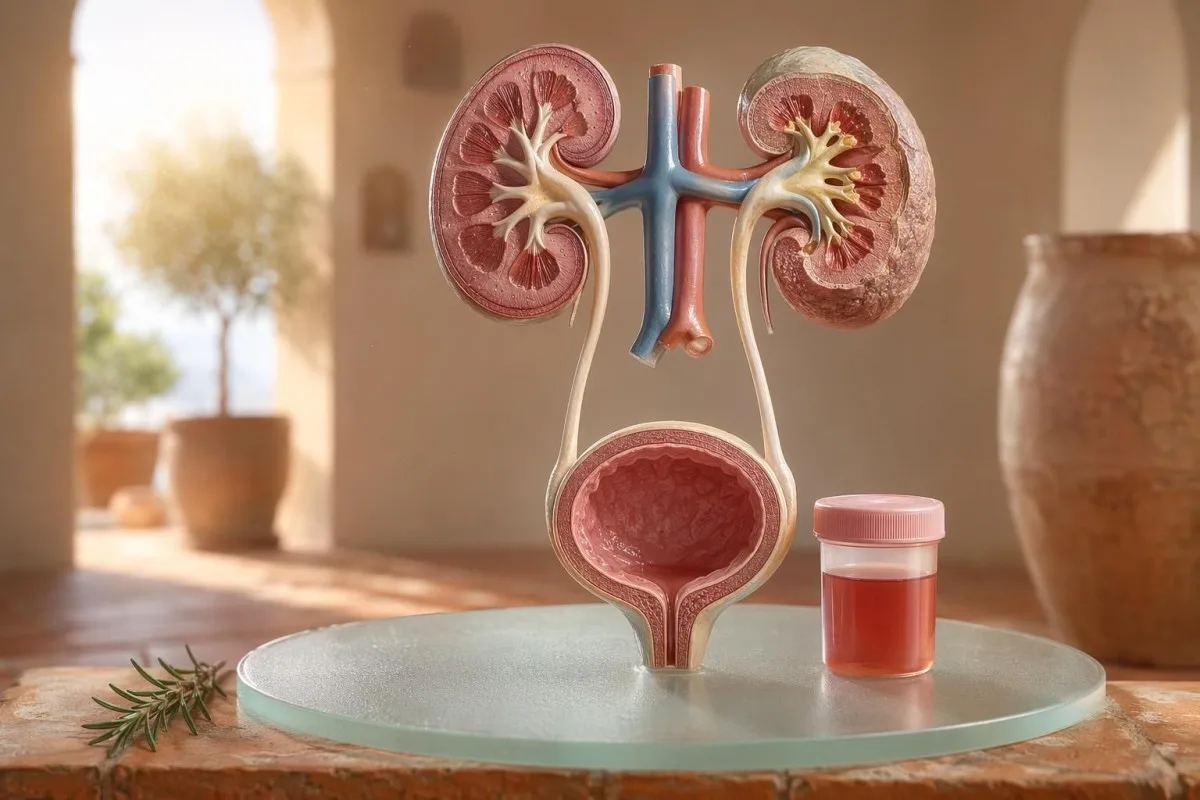
មគ្គុទេសក៍សម្រាប់អ្នកជំងឺជាមុនអំពី hematuria ដែលអាចមើលឃើញ និង hematuria ក្រោមមីក្រូទស្សន៍ រួមទាំងមូលហេតុដែលឈាមលើ dipstick មិនដូចនឹងកោសិកាឈាមក្រហម (RBC) នៅក្រោមមីក្រូទស្សន៍។.


- ឈាមក្នុងទឹកនោម មានន័យថា ទឹកនោមពណ៌ក្រហមដែលអាចមើលឃើញ ពណ៌ដូចតែ ឬពណ៌ដូចកូឡា ឬ hematuria ក្រោមមីក្រូទស្សន៍ដែលរកឃើញពីការធ្វើតេស្ត; ឈាមដែលអាចមើលឃើញតែងតែសមនឹងការតាមដានផ្នែកវេជ្ជសាស្ត្រ។.
- Microscopic hematuria ជាទូទៅត្រូវបានកំណត់ថា មាន RBC ចំនួន 3 ឬច្រើនជាងនេះក្នុងមួយ high-power field នៅលើគំរូ urine microscopy ដែលប្រមូលបានត្រឹមត្រូវ។.
- ឈាមលើ Dipstick រកឃើញសកម្មភាព heme ដូច្នេះវាអាចវិជ្ជមានពី RBC ដែលនៅដដែល hemoglobin ដោយសេរី ឬ myoglobin បន្ទាប់ពីរបួសសាច់ដុំ។.
- សញ្ញាបង្ហាញ UTI រួមមាន ការឈឺចុកចាប់ពេលបត់បង់ ការកើនប្រេកង់ leukocyte esterase nitrites និងកោសិកាស។; hematuria គួរត្រូវបានពិនិត្យឡើងវិញបន្ទាប់ពីការឆ្លងរោគបានជម្រះ។.
- កាកសំណល់ក្នុងទឹកនោម (Urine sediment) ការរកឃើញដូចជា RBCs មិនធម្មតា (dysmorphic red cells) casts នៃ RBC ឬប្រូតេអ៊ីនសំខាន់ បង្ហាញទៅរកជំងឺនៃអង្គភាពចម្រោះតម្រងនោម។.
- Urine casts ជាសម្ភារៈដែលបានរាងឡើងពីបំពង់តម្រងនោម; កំណកកោសិកាឈាម (red cell casts) គឺជាសញ្ញាព្រមានផ្នែកជំងឺតម្រងនោមដែលត្រូវពិនិត្យក្នុងសប្តាហ៍នោះ ជាពិសេសនៅពេលមានសម្ពាធឈាមខ្ពស់ ឬ eGFR កំពុងធ្លាក់ចុះ។.
- សញ្ញាព្រមានអំពីមហារីក (Cancer red flags) រួមមានឈាមក្នុងទឹកនោមដែលមិនឈឺចាប់ឃើញច្បាស់ (painless visible hematuria), ដុំឈាម, អាយុលើស 45 ឆ្នាំ, ប្រវត្តិជក់បារី, ការប៉ះពាល់សារធាតុគីមីក្នុងការងារ និងកើតឡើងវិញនូវឧប្បត្តិហេតុមិនអាចពន្យល់បាន។.
- ឈាមក្នុងទឹកនោមពេលហាត់ប្រាណ (Exercise hematuria) ជាធម្មតាធូរស្រាលក្នុងរយៈពេល 48-72 ម៉ោង; ប្រសិនបើនៅតែមានឈាមបន្ទាប់ពីសម្រាក មិនគួរទៅស្តីបន្ទោសលើការរត់ ឬជិះកង់ទេ។.
- ការវាយតម្លៃបន្ទាន់ ត្រូវការសម្រាប់ឈាមក្នុងទឹកនោមដែលមានគ្រុនក្តៅ, ឈឺចង្កេះ (flank pain), ដុំឈាមដែលបណ្តាលឲ្យស្ទះផ្លូវទឹកនោម, ការមានផ្ទៃពោះ, របួស ឬរោគសញ្ញានៃភាពស្លេកស្លាំងធ្ងន់ធ្ងរ។.
ឈាមក្នុងទឹកនោមមានន័យអ្វី មុនពេលអ្នកភ័យ
ឈាមក្នុងទឹកនោម អាចកើតពីការឆ្លង, ដុំថ្ម, ជំងឺដែលប៉ះពាល់ដល់ការតម្រងរបស់តម្រងនោម, ស្ថានភាពរបស់ក្រពេញប្រូស្តាត ឬប្លោកនោម, ថ្នាំ, ការហាត់ប្រាណ ឬការបំពុលពីពេលមករដូវ។ ច្បាប់ជាក់ស្តែងគឺសាមញ្ញ៖ ឈាមដែលឃើញច្បាស់, ឈាមក្នុងទឹកនោមតាមមីក្រូស្កុបដែលកើតឡើងវិញ, ឬឈាមក្នុងទឹកនោមដែលមានការឈឺ, គ្រុនក្តៅ, ដុំឈាម, សម្ពាធឈាមខ្ពស់, ប្រូតេអ៊ីន ឬមុខងារតម្រងនោមថយចុះ ត្រូវការការវាយតម្លៃជាបន្ទាន់ មិនមែនរង់ចាំដោយមើលស្ថានការណ៍ទេ។.
បង្គន់ពណ៌ផ្កាឈូកមួយលើកក្រោយពីប៊ីតរ៉ូត (beetroot) ឬអាហារបំប៉នថ្មី មិនមែនជាបញ្ហាគ្លីនិកដូចគ្នានឹងទឹកនោមពណ៌ដូចកូឡា (cola) បន្ទាប់ពីឈឺបំពង់កទេ។ ក្នុងការពិនិត្យរបស់ខ្ញុំ រឿងពណ៌ច្រើនតែប្រាប់ខ្ញុំថាគួរមើលកន្លែងណាមុន៖ ពណ៌ក្រហមភ្លឺជាមួយដុំឈាម ជាទូទៅទាក់ទងនឹងផ្លូវទឹកនោមផ្នែកខាងក្រោម ខណៈដែលទឹកនោមពណ៌ត្នោត ឬពណ៌ដូចតែ អាចមានន័យថាមានសារធាតុ heme pigment ចាស់ៗពីតម្រងនោម។.
Hematuria មិនមែនជាការធ្វើរោគវិនិច្ឆ័យទេ; វាជាសញ្ញា។. Hematuria ដែលឃើញច្បាស់ មានន័យថាអ្នកអាចឃើញការប្រែពណ៌ ខណៈដែល hematuria តាមមីក្រូស្កុប មានន័យថាទឹកនោមមើលទៅធម្មតា ប៉ុន្តែមីក្រូស្កុបរកឃើញកោសិកាឈាមក្រហម ជាធម្មតានៅ 3 ឬច្រើនជាងកោសិកាឈាមក្រហមក្នុងមួយវាលកម្លាំងខ្ពស់ (high-power field)។.
កាន់តេស្ទី គឺជា ឧបករណ៍វិភាគឈាម AI ដែលជួយឲ្យអ្នកជំងឺភ្ជាប់លទ្ធផលទឹកនោមជាមួយសូចនាករតម្រងនោមដូចជា creatinine, eGFR, CRP, hemoglobin និង platelets ក្នុងខ្សែពេលវេលាតែមួយ។ ប្រសិនបើពណ៌ងឿងឆ្ងល់ នោះ មគ្គុទេសក៍ពណ៌ទឹកនោម ពន្យល់ថា ហេតុអ្វីបានជាទឹកនោមពណ៌ក្រហម ត្នោត ពណ៌ទឹកក្រូច និងលឿងខ្មៅ មិនមែនសុទ្ធតែមានន័យដូចគ្នាទេ។.
ឈាមលើ Dipstick មិនដូចនឹង RBCs នៅលើការពិនិត្យក្រោមមីក្រូទស្សន៍
ការធ្វើតេស្តដំបងទឹកនោម (urine dipstick) ដែលសម្គាល់ ឈាមវិជ្ជមាន រកឃើញសកម្មភាពគីមីដូច heme មិនមែនចាំបាច់ជាកោសិកាឈាមក្រហមដែលនៅគ្រប់សភាពនោះទេ។ មីក្រូស្កុបឆ្លើយសំណួរបន្ទាប់៖ តើមាន RBC ពិតប្រាកដនៅក្នុងសំណល់ទឹកនោមដែរឬទេ ហើយបើមាន តើវាមើលទៅដូចអ្វី?
ការធ្វើតេស្តដោយ dipstick លឿន ព្រោះបន្ទះប្រតិកម្មឆ្លើយតបនឹងសកម្មភាព peroxidase របស់ heme។ នោះមានន័យថាលទ្ធផលវិជ្ជមានអាចកើតឡើងជាមួយ RBC ដែលនៅគ្រប់សភាព, hemoglobin ដោយសេរីក្រោយការបែកបាក់កោសិកាឈាមក្រហម, ឬ myoglobin ក្រោយការរងរបួសសាច់ដុំធ្ងន់ធ្ងរ។.
មីក្រូស្កុបយឺតជាង ប៉ុន្តែច្បាស់លាស់ជាង។ មន្ទីរពិសោធន៍ដោយយកចិត្តទុកដាក់នឹង សំណល់ទឹកនោម (urine sediment), ហើយរាយការណ៍ចំនួនកោសិកាឈាមក្រហមក្នុងមួយវាលកម្លាំងខ្ពស់; គ្រូពេទ្យជាច្រើនប្រើ 0-2 RBC/hpf ជាធម្មតា និង 3 ឬច្រើនជាង RBC/hpf ជាមិនធម្មតាក្នុងមនុស្សពេញវ័យ។.
ភាពមិនត្រូវគ្នាមានសារៈសំខាន់។ Dipstick ដែលមានឈាម 3+ និង 0-2 RBC/hpf ធ្វើឲ្យខ្ញុំសួរអំពី rhabdomyolysis, hemolysis, សារធាតុបំពុលដែលមានអុកស៊ីតកម្ម និងការគ្រប់គ្រងសំណាក ជាជាងទៅលោតទៅរកជំងឺប្លោកនោមភ្លាមៗ; ភាពខុសគ្នានេះស្រដៀងនឹងហេតុផលក្នុងការពន្យល់របស់យើង ការប្រៀបធៀបការវិភាគទឹកនោម និងវប្បធម៌.
ពេលណាដែលសញ្ញា UTI អាចពន្យល់ hematuria និងពេលណាមិនអាច
ការឆ្លងមេរោគផ្លូវទឹកនោម (UTI) អាចបណ្តាលឲ្យ ឈាមក្នុងទឹកនោម, ជាពិសេសនៅពេលមានការឈឺចាប់ពេលនោម (burning), ការបន្ទាន់ (urgency), ភាពញឹកញាប់ (frequency), អារម្មណ៍មិនស្រួលនៅអាងត្រគាក (pelvic discomfort), leukocyte esterase, nitrites និងកោសិកាឈាមស។ ប៉ុន្តែ hematuria គួរតែបាត់បន្ទាប់ពីការព្យាបាល; RBC នៅតែបន្តបន្ទាប់ពី UTI ត្រូវពិនិត្យមើលជាថ្មី។.
ក្នុង UTI ខាងក្រោមដែលច្បាស់លាស់ ខ្ញុំរំពឹងថានឹងមានជាក្រុម៖ dysuria, frequency, leukocyte esterase វិជ្ជមាន, pyuria ជាញឹកញាប់លើសពី 5-10 white cells/hpf និងពេលខ្លះ nitrites។ ការវិជ្ជមានតាមវប្បធម៌បែបប្រពៃណីគឺ 100,000 CFU/mL ប៉ុន្តែចំពោះស្ត្រីដែលមានរោគសញ្ញា ចំនួនទាបដូចជា 1,000-10,000 CFU/mL ក៏អាចមានន័យសំខាន់ខាងគ្លីនិកបាន។.
Leukocyte esterase គឺជាអង់ស៊ីមកោសិកាឈាមស មិនមែនជាភស្តុតាងថាមានបាក់តេរីទេ។ លទ្ធផលវិជ្ជមានជាមួយ hematuria អាចកើតឡើងពីការឆ្លង, ដុំថ្ម, ការចម្លងគំរូ (contamination), ឬការរលាកតម្រងនោម ដែលជាមូលហេតុដែល our អ្នកណែនាំ esterase របស់កោសិកាឈាមស បំបែកព័ត៌មានដែលមានប្រយោជន៍ចេញពី false positives ទូទៅ។.
Nitrites មានភាពជាក់លាក់ជាង ប៉ុន្តែមានភាពប្រែប្រួលតិច ព្រោះបាក់តេរីមិនមែនទាំងអស់បម្លែង nitrate ទៅជា nitrite ទេ។ ប្រសិនបើរោគសញ្ញាជាលក្ខណៈធម្មតា ប៉ុន្តែ nitrites អវិជ្ជមាន ការវប្បធម៌នៅតែអាចវិជ្ជមានបាន; ព័ត៌មានលម្អិតជាក់ស្តែងត្រូវបានគ្របដណ្តប់ក្នុង our អ្នកពន្យល់លទ្ធផលនីត្រាត.
Hematuria ដែលអាចមើលឃើញ៖ ពណ៌ កំណកឈាម និងសញ្ញាពេលវេលា
Hematuria ដែលមើលឃើញដោយភ្នែក គឺមិនមែនជារឿងដែលខ្ញុំមិនអើពើទេ ទោះបីវាកើតឡើងតែម្តងក៏ដោយ។ ទឹកនោមពណ៌ក្រហមភ្លឺ, ដុំឈាម (clots), ឬការហូរឈាមដោយគ្មានការឈឺចាប់ ត្រូវការការវាយតម្លៃឲ្យទាន់ពេលវេលា ព្រោះមូលហេតុនៃការហូរឈាមពីផ្លូវទឹកនោមខាងក្រោមកាន់តែមានលទ្ធភាព នៅពេលអាយុ និងការប៉ះពាល់នឹងការជក់បារីកើនឡើង។.
ពេលវេលាអាចមានប្រយោជន៍គួរឱ្យភ្ញាក់ផ្អើល។ ឈាមមានតែនៅដើមពេលនោម អាចបង្ហាញពីបំពង់នោម (urethra) ខណៈឈាមពេញទឹកនោម អាចមកពីប្រភពនៅប្លោកនោម ឬតម្រងនោម (kidney) ហើយឈាមនៅចុងបញ្ចប់ជួនកាលចង្អុលទៅតំបន់កញ្ចឹងកប្លោកនោម។.
ដុំឈាមជាទូទៅមានន័យថា ការហូរឈាមកើតឡើងនៅខាងក្រោមតម្រងតម្រងនោម ព្រោះដុំឈាមមិនងាយកើតឡើងបន្ទាប់ពីឆ្លងកាត់ glomeruli និង tubules។ ដុំឈាមដែលរារាំងលំហូរនោម គឺបន្ទាន់៖ ការហើមពេញប្លោកនោមអាចក្លាយជាការឈឺចាប់ក្នុងរយៈពេលប៉ុន្មានម៉ោង ហើយអាចត្រូវការការថែទាំ catheter។.
បុរសខ្លះសន្មតថា hematuria ដែលអាចមើលឃើញបន្ទាប់ពីរោគសញ្ញាផ្លូវទឹកនោម គ្រាន់តែជាការរលាកក្រពេញប្រូស្តាត។ នោះអាចជាការពិត ប៉ុន្តែបើ PSA ត្រូវបានពិនិត្យក្នុងអំឡុងពេល ឬភ្លាមៗបន្ទាប់ពីការឆ្លងមេរោគ ការបកស្រាយអាចមានភាពស្មុគស្មាញ; our ការណែនាំ PSA បន្ទាប់ពី UTI ពន្យល់ពីមូលហេតុដែលគ្រូពេទ្យជាច្រើនរង់ចាំ 6-8 សប្តាហ៍ មុនពិនិត្យ PSA ឡើងវិញ។.
កម្រិត hematuria ក្រោមមីក្រូទស្សន៍ ដែលវេជ្ជបណ្ឌិតប្រើ
Microscopic hematuria ជាទូទៅត្រូវបានកំណត់ថាជា 3 ឬច្រើនជាង RBCs ក្នុងមួយ high-power field លើគំរូទឹកនោមដែលប្រមូលបានត្រឹមត្រូវ។ សេចក្តីណែនាំ AUA/SUFU ប្រើកម្រិតនេះ ហើយណែនាំឱ្យធ្វើការវាយតម្លៃតាមកម្រិតហានិភ័យ ជាជាងព្យាបាលអ្នកជំងឺគ្រប់រូបដូចគ្នា (Barocas et al., 2020)។.
ការធ្វើ dipstick តែមួយមុខ មិនគួរប្រើដើម្បីធ្វើរោគវិនិច្ឆ័យ microscopic hematuria ទេ។ នៅក្នុងសេចក្តីណែនាំ AUA/SUFU របស់ Barocas et al. 2020 និយមន័យពឹងផ្អែកលើ microscopy ព្រោះ dipsticks អាចវិជ្ជមានពី heme pigments ដោយគ្មានកោសិកាឈាមក្រហមដែលនៅគ្រប់សភាព។.
ការធ្វើតេស្តឡើងវិញ មិនមែនជាភាពទន់ខ្សោយទេ; វាជាវេជ្ជសាស្ត្រល្អ។ ប្រសិនបើមាន 3-10 RBC/hpf បន្ទាប់ពីការហាត់ប្រាណខ្លាំង ក្តៅខ្លួន ការរួមភេទ ការមករដូវ ឬការប្រមូលគំរូដែលមានការចម្លងកខ្វក់ ខ្ញុំជាធម្មតាចង់បានគំរូស្អាតសម្រាប់ធ្វើឡើងវិញ មុនបញ្ជាឱ្យធ្វើការស្កេន ដូចដែលយើងណែនាំសម្រាប់ ណែនាំការធ្វើតេស្តឡើងវិញលទ្ធផលមិនប្រក្រតី.
ហានិភ័យផ្លាស់ប្តូរផ្លូវ។ មនុស្សអាយុ 24 ឆ្នាំដែលមាន 4 RBC/hpf បន្ទាប់ពីរត់ពាក់កណ្តាលម៉ារ៉ាតុង មិនដូចគ្នានឹងបុរសអាយុ 68 ឆ្នាំដែលជាអតីតអ្នកជក់បារី ហើយមាន 25 RBC/hpf ក្នុងគំរូពីរដាច់ពីគ្នា ទោះបីជាទាំងពីរនាក់បច្ចេកទេសសុទ្ធតែបំពេញតាមនិយមន័យ hematuria ក៏ដោយ។.
កាកសំណល់ក្នុងទឹកនោម ប្រូតេអ៊ីន និង urine casts ដែលផ្លាស់ប្តូរការត្រួតពិនិត្យបន្ត
កាកសំណល់ក្នុងទឹកនោម (Urine sediment) អាចបង្ហាញថា hematuria ទំនងជាមកពីតម្រងតម្រងនោមដែរឬទេ។ កោសិកាឈាមក្រហមដែលមានរូបរាងខុសធម្មតា (dysmorphic red cells) casts កោសិកាឈាមក្រហម (red cell casts) និងការផ្លាស់ប្តូរប្រូតេអ៊ីនយ៉ាងសំខាន់ ធ្វើឱ្យមានការព្រួយបារម្ភទៅរកជំងឺ glomerular ជាជាងការឆ្លងមេរោគប្លោកនោមសាមញ្ញ។.
កោសិកាឈាមក្រហមដែលមើលទៅមិនស្មើគ្នា មានស្នាមបែក (blebbed) ឬរាងជារង្វង់ ត្រូវបានហៅថា dysmorphic RBCs ហើយវាបង្ហាញពីការឆ្លងកាត់តម្រង glomerular ដែលកំពុងរលាក។ Acanthocytes លើសពីប្រហែល 5% នៃកោសិកាឈាមក្រហមក្នុងទឹកនោម ជាញឹកញាប់ត្រូវបានព្យាបាលជាសញ្ញា glomerular ដ៏ខ្លាំង ទោះបីជាមន្ទីរពិសោធន៍ខុសគ្នាអំពីកម្រិតដែលពួកគេរាយការណ៍វាដោយទំនុកចិត្តក៏ដោយ។.
ប្រូតេអ៊ីនជាសញ្ញាទីពីរដែលខ្ញុំមិនអើពើ។ សមាមាត្រ albumin-creatinine ក្នុងទឹកនោមលើសពី 30 mg/g ឬប្រហែល 3 mg/mmol គឺមិនប្រក្រតី ហើយ hematuria ដែលអាចមើលឃើញរួមជាមួយប្រូតេអ៊ីន សមនឹងការពិនិត្យផ្តោតលើតម្រងនោមឱ្យលឿនជាងមុន; our មគ្គុទេសក៍ protein ក្នុងទឹកនោម ផ្តល់កម្រិតកាត់សម្រាប់អ្នកជំងឺជាក់លាក់។.
Urine casts គឺជាផ្សិតតូចៗ (tiny molds) ដែលបង្កើតនៅក្នុងបំពង់តម្រងនោម។ Hyaline casts អាចលេចឡើងបន្ទាប់ពីខ្វះជាតិទឹក ឬការហាត់ប្រាណ ប៉ុន្តែ red cell casts គឺមិនប្រក្រតី ហើយជាទូទៅចង្អុលទៅ glomerulonephritis; សម្រាប់ឯកសារយោង urinalysis ទូលំទូលាយ សូមមើល our ការណែនាំពេញលេញអំពីការវិភាគទឹកនោម.
ថ្ម (stones) ការហាត់ប្រាណ និងមូលហេតុបណ្តោះអាសន្ននៃ hematuria
ថ្ម (Stones) និងការហាត់ប្រាណខ្លាំង អាចបណ្តាលឱ្យ hematuria ប៉ុន្តែវាមានពេលវេលាខុសគ្នា។ hematuria ពាក់ព័ន្ធនឹងថ្ម ជាញឹកញាប់ភ្ជាប់ជាមួយការឈឺចង្កេះ (flank pain) ឬគ្រីស្តាល់ ខណៈ hematuria ពីការហាត់ប្រាណ ជាទូទៅគួរតែធូរស្រាលក្នុងរយៈពេល 48-72 ម៉ោងបន្ទាប់ពីសម្រាក។.
ថ្មតម្រងនោម ជាទូទៅបណ្តាលឱ្យមានរលកនៃការឈឺចង្កេះ ចង្អោរ និង microscopic ឬ visible hematuria។ គ្រីស្តាល់ calcium oxalate មិនបញ្ជាក់ថាមានថ្មទេ ប៉ុន្តែវាបង្កើនការសង្ស័យនៅពេលភ្ជាប់ជាមួយការឈឺចាប់ប្រកាច់ (colicky pain); our អ្នកណែនាំគ្រីស្តាល់អុកសាឡាតកាល់ស្យូម ពន្យល់ពីសញ្ញា microscopy ក្នុងទឹកនោម។.
Exercise hematuria គឺពិត។ ខ្ញុំបានឃើញអ្នករត់ម៉ារ៉ាតុងដែលមាន 10-20 RBC/hpf នៅព្រឹកបន្ទាប់ពីការប្រណាំង មានវប្បធម៌ធម្មតា គ្មានប្រូតេអ៊ីន និង urinalysis ឡើងវិញទាំងស្រុងធម្មតា 3 ថ្ងៃក្រោយ។.
គ្រោះថ្នាក់គឺការសន្មតថាអ្វីៗទាំងអស់មកពីការហ្វឹកហាត់។ ប្រសិនបើ hematuria នៅតែបន្តលើសពី 72 ម៉ោង លេចឡើងជាមួយទឹកនោមពណ៌ដូច cola ងងឹត ឬភ្ជាប់ជាមួយ CK ខ្ពស់ creatinine កំពុងកើនឡើង ឬឈឺសាច់ដុំ សូមមើលលើសពីប្លោកនោម; our មគ្គុទេសក៍ការធ្វើលំហាត់ប្រាណ និង creatinine គ្របដណ្តប់លើការត្រួតស៊ីគ្នារវាងតម្រងនោម និងសាច់ដុំ។.
សញ្ញាគ្រោះថ្នាក់នៅតម្រងនោម ដែលវេជ្ជបណ្ឌិតមិនអើពើ
Hematuria ជាមួយនឹងប្រូតេអ៊ីន សម្ពាធឈាមខ្ពស់ ការហើម eGFR ថយចុះ red cell casts ឬ creatinine កើនឡើងយ៉ាងលឿន គឺជាសញ្ញាព្រមានខ្លាំងសម្រាប់តម្រងនោម។ ការរកឃើញទាំងនេះបង្ហាញថា ឯកតាចម្រោះអាចកំពុងរលាក ហើយការរង់ចាំប៉ុន្មានខែអាចធ្វើឲ្យបាត់បង់មុខងារតម្រងនោម។.
KDIGO 2024 កំណត់ជំងឺតម្រងនោមរ៉ាំរ៉ៃ ដោយសញ្ញាសម្គាល់នៃការខូចខាតតម្រងនោម ដូចជា albuminuria ឬ eGFR ទាបជាង 60 mL/min/1.73 m² យ៉ាងហោចណាស់ 3 ខែ។ ទោះជាយ៉ាងណា ក្នុងស្ថានភាពស្រួចស្រាវ hematuria រួមជាមួយនឹងការកើនឡើង creatinine ក្នុងរយៈពេលប៉ុន្មានថ្ងៃ ឬសប្តាហ៍ គឺគ្រប់គ្រាន់ដើម្បីបង្កើនកម្រិតការពិនិត្យ មុនពេលបំពេញលក្ខណៈកំណត់រយៈពេល 3 ខែ។.
Albuminuria ជាញឹកញាប់ជាសញ្ញាព្រមានដំបូង។ ACR 30-300 mg/g គឺកើនឡើងមធ្យម ខ្ពស់ជាង 300 mg/g គឺកើនឡើងខ្លាំង ហើយការភ្ជាប់មួយក្នុងចំណោមនោះជាមួយ hematuria ធ្វើឲ្យការពិភាក្សាប្រែប្រួល; our មគ្គុទេសក៍ ACR សម្រាប់តម្រងនោម បង្ហាញពីមូលហេតុដែល albumin ក្នុងទឹកនោមរកឃើញការខូចខាតមុនពេលមានរោគសញ្ញា។.
សម្ពាធឈាមមានសារៈសំខាន់នៅទីនេះ។ BP ថ្មី 160/100 mmHg ជាមួយនឹងទឹកនោមពណ៌ដូចកូឡា និងការហើមកជើង បន្ទាប់ពីការឆ្លងមេរោគបំពង់ក ធ្វើឲ្យខ្ញុំគិតពី glomerulonephritis មិនមែន cystitis ទេ; ប្រសិនបើ eGFR មិនច្បាស់ our មគ្គុទេសក៍ពិនិត្យឡើងវិញ GFR ពន្យល់ថាពេលណាដែល cystatin C អាចជួយបញ្ជាក់មុខងារតម្រងនោម។.
សញ្ញាហានិភ័យមហារីក ដោយមិនបង្កការភ័យខ្លាច
Hematuria ដែលមើលឃើញដោយគ្មានការឈឺចាប់ ជាពិសេសបន្ទាប់ពីអាយុ 45 ឆ្នាំ សមនឹងការវាយតម្លៃបន្ទាន់ ព្រោះមហារីកប្លោកនោម និងផ្នែកខាងលើនៃប្រព័ន្ធទឹកនោមអាចបង្ហាញរូបរាងបែបនេះ។ NICE NG12 ណែនាំការបញ្ជូនទៅកាន់ការពិនិត្យសង្ស័យមហារីក សម្រាប់ hematuria ដែលមើលឃើញមិនអាចពន្យល់បានក្នុងមនុស្សពេញវ័យអាយុ 45 ឆ្នាំឡើងទៅ ឬ hematuria ដែលមើលឃើញនៅតែបន្ត បន្ទាប់ពីការព្យាបាល UTI។.
Hematuria ភាគច្រើនមិនមែនជាមហារីកទេ។ ប៉ុន្តែ ករណីដែលខ្ញុំចាំ គឺករណីស្ងៀមៗ៖ គ្មានការឈឺចាប់ គ្មានគ្រុនក្តៅ គ្មានអារម្មណ៍ឆេះ គ្រាន់តែមានវគ្គ 2 ដងនៃទឹកនោមពណ៌ក្រហម ក្នុងបុរសអាយុ 56 ឆ្នាំដែលជាអ្នកជក់បារី ហើយស្ទើរតែមិនអើពើនឹងវា។.
ហានិភ័យកើនជាបន្តបន្ទាប់ មិនមែនជាទ្វេ (binary) ទេ។ អាយុលើស 45-50 ប្រវត្តិជក់បារី ការប៉ះពាល់ cyclophosphamide ការព្យាបាលដោយកាំរស្មីលើអាងត្រគាក hematuria ដែលមើលឃើញកើតឡើងវិញ ការប៉ះពាល់សារធាតុ aromatic amine ក្នុងការងារ និងដុំឈាម ទាំងអស់បង្កើនតម្រូវការសម្រាប់ cystoscopy ឬការថតរូបភាព។.
សញ្ញាសម្គាល់របស់ប្រូស្តាត មិនអាចបដិសេធមូលហេតុពីប្លោកនោមបានទេ។ ប្រសិនបើមានរោគសញ្ញាទឹកនោម ការផ្លាស់ប្តូរ PSA និង hematuria ត្រួតគ្នា សូមពិនិត្យពេលវេលា និងស្ថានភាពឆ្លងមេរោគឲ្យបានយកចិត្តទុកដាក់; our មគ្គុទេសក៍សញ្ញាសម្គាល់ប្រូស្តាត ពន្យល់ពីមូលហេតុដែល PSA គ្រាន់តែជាផ្នែកមួយនៃការវាយតម្លៃការបត់បង់ទឹកនោមរបស់បុរសប៉ុណ្ណោះ.
ថ្នាំ Anticoagulants និងការជូនដំណឹងខុស
ថ្នាំបញ្ចុះឈាមអាចបង្ហាញការហូរឈាម ប៉ុន្តែមិនគួរចោទប្រកាន់ដោយស្វ័យប្រវត្តិថាជាមូលហេតុនៃទឹកនោមមានឈាម (hematuria) នោះទេ។ ទឹកនោមមានឈាមដែលទាក់ទងនឹងថ្នាំប្រឆាំងកកឈាម (anticoagulant-associated hematuria) នៅតែត្រូវការការវាយតម្លៃ ព្រោះថ្នាំអាចបង្ហាញដុំថ្ម ការឆ្លងរោគ ជំងឺតម្រងនោម ឬការលូតលាស់នៅផ្លូវទឹកនោម.
Warfarin, apixaban, rivaroxaban, aspirin និង clopidogrel អាចធ្វើឲ្យការហូរឈាមកាន់តែច្បាស់។ ប្រសិនបើ INR ខ្ពស់ជាងកម្រិតគោលដៅ ការកែតម្រូវវាមានសារៈសំខាន់ ប៉ុន្តែ INR ធម្មតាមិនធ្វើឲ្យ hematuria ដែលមើលឃើញមានសុវត្ថិភាពនោះទេ.
មូលហេតុមួយចំនួនដែលមិនបណ្តាលឲ្យហូរឈាម អាចធ្វើឲ្យមើលទៅដូចជា hematuria។ Rifampicin អាចធ្វើឲ្យទឹកនោមប្រែពណ៌ទឹកក្រូច-ក្រហម, phenazopyridine អាចធ្វើឲ្យវាក្លាយជាពណ៌ទឹកក្រូចភ្លឺ, beetroot អាចធ្វើឲ្យទឹកនោមមានស្នាមពណ៌ចំពោះអ្នកដែលងាយរងផលប៉ះពាល់ និងការខ្វះជាតិទឹកអាចធ្វើឲ្យពណ៌សារធាតុចម្រុះងងឹតគ្រប់គ្រាន់ដើម្បីធ្វើឲ្យអ្នកជំងឺភ័យ; ការតាមដានថ្នាំបញ្ចុះឈាមត្រូវបានគ្របដណ្តប់ក្នុង ការធ្វើតេស្តសុវត្ថិភាពថ្នាំបញ្ចុះឈាម.
ការធ្វើ dipstick ក៏អាចធ្វើឲ្យយល់ច្រឡំក្រោយការហាត់ប្រាណខ្លាំង ឬការរបួសសាច់ដុំ។ បើ heme pad វិជ្ជមាន ប៉ុន្តែមាន RBC តិច ឬគ្មាន RBC នោះ នាំឲ្យខ្ញុំគិតទៅ myoglobin ជាពិសេស ប្រសិនបើ CK ខ្ពស់ជាង 1,000 IU/L និងទឹកនោមងងឹត.
កុមារ ការមានផ្ទៃពោះ និងការចម្លងពីរដូវ
Hematuria ក្នុងកុមារ ពេលមានផ្ទៃពោះ ឬនៅជុំវិញពេលមករដូវ ត្រូវការការយកគំរូឲ្យស្អាតជាង និងកម្រិតសម្រេចពិនិត្យឡើងវិញទាបជាង។ លទ្ធផលទឹកនោមដូចគ្នាអាចមានអត្ថន័យខុសគ្នា អាស្រ័យលើអាយុ ស្ថានភាពមានផ្ទៃពោះ ក្តៅខ្លួន សម្ពាធឈាម និងគុណភាពនៃការប្រមូលគំរូ.
ការចម្លងពីឈាមរដូវកើតឡើងជាញឹកញាប់ ហើយគ្មាននរណាម្នាក់គួរមានអារម្មណ៍អៀនខ្មាស់ចំពោះវាទេ។ ប្រសិនបើលទ្ធផលមិនស្របនឹងការរំពឹងទុក សូមធ្វើការយកគំរូ midstream clean-catch ឡើងវិញ យ៉ាងហោចណាស់ 48 ម៉ោងបន្ទាប់ពីការហូរឈាមឈប់; ប្រសិនបើនៅតែមាន RBC 3 ឬច្រើនជាងនេះក្នុងមួយ hpf នោះនឹងមានអត្ថន័យកាន់តែច្រើន.
កុមារជាញឹកញាប់ត្រូវការមើលតាមទស្សនៈផ្សេង។ Hematuria បន្ទាប់ពីជំងឺឆ្លងវីរុសអាចជាបណ្តោះអាសន្ន ប៉ុន្តែ hematuria ដែលមានការហើម សម្ពាធឈាមខ្ពស់ ប្រូតេអ៊ីន ឬបរិមាណទឹកនោមថយចុះ ត្រូវការការពិនិត្យជាបន្ទាន់ដោយគ្រូពេទ្យកុមារ.
ការមានផ្ទៃពោះបង្កើនហានិភ័យ ព្រោះ UTI ដុំថ្ម ភាពតានតឹងតម្រងនោមដែលទាក់ទងនឹង preeclampsia និងការចម្លងអាចត្រួតគ្នា។ សម្រាប់លំនាំការព្រមានអំពីការមានផ្ទៃពោះក្នុងថ្ងៃតែមួយ ដែលពាក់ព័ន្ធនឹងការរកឃើញតម្រងនោម ថ្លើម ប្លាកែត និងទឹកនោម សូមមើល สัญญาณเตือนทางห้องปฏิบัติการในการตั้งครรภ์.
ការធ្វើតេស្តដែលវេជ្ជបណ្ឌិតជាធម្មតាបញ្ជាទិញបន្ទាប់ពី hematuria
ការធ្វើតេស្តបន្ទាប់ជាទូទៅបន្ទាប់ពី hematuria គឺ urinalysis ឡើងវិញជាមួយ microscopy, urine culture ប្រសិនបើអាចមានការឆ្លងរោគ, ការធ្វើតេស្តឈាមមុខងារតម្រងនោម, ការវាស់ប្រូតេអ៊ីនក្នុងទឹកនោម ឬ ACR, ការវាស់សម្ពាធឈាម និងការថតរូបភាព ឬ cystoscopy តាមកម្រិតហានិភ័យ។ លំដាប់ពិតប្រាកដអាស្រ័យលើរោគសញ្ញា និងហានិភ័យ.
សម្រាប់រោគសញ្ញា cystitis មិនស្មុគស្មាញ ការធ្វើ culture និងការព្យាបាលអាចធ្វើមុន បន្ទាប់មកធ្វើ urine microscopy ឡើងវិញបន្ទាប់ពីរោគសញ្ញាបានធូរស្រាល។ ចំពោះ hematuria ដែលមើលឃើញដោយគ្មានការឈឺចាប់ គ្រូពេទ្យជាច្រើនធ្វើការថតរូបភាព និង cystoscopy ឆាប់ជាងនេះ ព្រោះការរង់ចាំវគ្គកើតឡើងឡើងវិញអាចធ្វើឲ្យការធ្វើរោគវិនិច្ឆ័យយឺត.
ការធ្វើតេស្តឈាមបន្ថែមបរិបទដែលទឹកនោមតែមួយមុខមិនអាចផ្តល់បាន។ Creatinine, eGFR, urea ឬ BUN, electrolytes, CBC, CRP, complement C3/C4 និងសញ្ញាសម្គាល់អូតូអ៊ុយមីន (autoimmune markers) អាចសមស្រប នៅពេលសង្ស័យថាមានការរលាកតម្រងនោម; our មគ្គុទេសក៍សមាមាត្រ BUN/creatinine ពន្យល់ពីលំនាំតម្រងនោមមួយដែលកើតឡើងជាញឹកញាប់.
AI Kantesti អាចរៀបចំលទ្ធផលទាំងនោះឲ្យទៅជាទិដ្ឋភាពនិន្នាការ (trend view) ប៉ុន្តែការពិនិត្យដោយវេជ្ជបណ្ឌិតនៅតែចាំបាច់ នៅពេលមានសញ្ញាព្រមាន។ វិធីសាស្ត្រសុវត្ថិភាពផ្នែកព្យាបាលរបស់យើងត្រូវបានពិពណ៌នានៅក្នុង សុពលភាពវេជ្ជសាស្រ្ត, រួមទាំងរបៀបដែលដោះស្រាយក្រុមលទ្ធផលមិនប្រក្រតី ជាជាងគ្រាន់តែលេខឯករាជ្យ.
របៀបដែល Kantesti ជួយរៀបចំបរិបទនៃ hematuria
កាន់តេស្ទី គឺជា សេវាកម្មបកស្រាយការធ្វើតេស្តរបស់ AI lab ដែលអានសញ្ញាសម្គាល់ឈាមដែលនៅជិតទឹកនោមក្នុងបរិបទ ដូចជា creatinine, eGFR, CRP, hemoglobin, platelets និង glucose។ វាមិនធ្វើរោគវិនិច្ឆ័យ hematuria ទេ ប៉ុន្តែវាជួយអ្នកជំងឺរៀបចំសេចក្តីសង្ខេបដែលស្អាត និងមានប្រយោជន៍ជាងមុនសម្រាប់វេជ្ជបណ្ឌិតរបស់ពួកគេ.
ពេលខ្ញុំពិនិត្យ hematuria ខ្ញុំមិនសូវមើលលទ្ធផលតែមួយដោយខ្លួនឯងទេ។ Creatinine ធម្មតា 0.82 mg/dL, eGFR មានស្ថិរភាពខ្ពស់ជាង 90, គ្មានប្រូតេអ៊ីន និង culture អវិជ្ជមាន មានអារម្មណ៍ខុសគ្នាខ្លាំងពី hematuria ដែលមាន eGFR 48 និង CRP 65 mg/L.
កាន់តេស្ទី គឺជា វេទិកាបកស្រាយសូចនាករជីវសាស្ត្រដោយ AI ដែលអាចដំណើរការឯកសារ PDF តេស្តឈាមដែលបានផ្ទុកឡើង ឬរូបថត និងភ្ជាប់សញ្ញាសម្គាល់តម្រងនោម ការរលាក និងភាពស្លេកស្លាំងដែលលើស/ក្រោមកម្រិតជាមួយនឹងពេលវេលានៃរោគសញ្ញា។ វិធីសាស្ត្រនៅពីក្រោយការអានលំនាំនោះត្រូវបានពិពណ៌នានៅក្នុង our មគ្គុទេសក៍បច្ចេកវិទ្យា, ហើយការគ្របដណ្តប់លើសញ្ញាសម្គាល់ទូលំទូលាយត្រូវបានរៀបរាប់នៅក្នុង ការណែនាំអំពីសញ្ញាសម្គាល់ជីវសាស្រ្ត.
គន្លឹះជាក់ស្តែងមួយ៖ មុននឹងផ្ទុកឡើង សូមពិនិត្យថា ឯកតា កាលបរិច្ឆេទ និងអត្តសញ្ញាណអ្នកជំងឺត្រូវបានចាប់យកត្រឹមត្រូវ។ កំហុស OCR អាចបម្លែង 0.9 ទៅជា 9.0 ដែលជារឿងតម្រងនោមខុសគ្នាទាំងស្រុង; our បញ្ជីត្រួតពិនិត្យការផ្ទុកឡើង PDF បង្ហាញពីកំហុសដែលខ្ញុំសុំឲ្យអ្នកជំងឺចាប់បានជាមុន។.
កំណត់ត្រាស្រាវជ្រាវ អភិបាលកិច្ច និងបញ្ជីត្រួតពិនិត្យចុងក្រោយ
គិតត្រឹមថ្ងៃទី 11 ខែកក្កដា ឆ្នាំ 2026 ផែនការដែលមានសុវត្ថិភាពបំផុតសម្រាប់អ្នកជំងឺ គឺត្រូវបញ្ជាក់ថា hematuria ពិតឬអត់ ស្វែងរកសញ្ញានៃការឆ្លង រកមើលសញ្ញាព្រមានអំពីតម្រងនោម និងបន្តទៅកាន់ការពិនិត្យបន្ទាន់ចំពោះ hematuria ដែលអាចមើលឃើញ ឬបន្តកើតមាន។ កុំផ្អែកតែលើពណ៌ទឹកនោមតែប៉ុណ្ណោះ។.
ខ្ញុំ Thomas Klein, MD ហើយដំបូន្មានរបស់ខ្ញុំមានភាពប្រុងប្រយ័ត្នដោយចេតនា ព្រោះ hematuria ដែលខកខានអាចមានសារៈសំខាន់។ ខ្លឹមសារផ្នែកវេជ្ជសាស្ត្ររបស់ Kantesti ត្រូវបានពិនិត្យជាមួយការត្រួតពិនិត្យផ្នែកគ្លីនិកពី ក្រុមប្រឹក្សាប្រឹក្សាវេជ្ជសាស្ត្រ, ហើយតួនាទីរបស់យើងគឺជួយឲ្យអ្នកជំងឺសួរសំណួរបានល្អជាងមុន មិនមែនជំនួសវេជ្ជបណ្ឌិតរបស់អ្នកទេ។.
កាន់តេស្ទី គឺជា ឧបករណ៍វិភាគតេស្តឈាមដោយ AI ត្រូវបានប្រើប្រាស់ដោយមនុស្សរាប់លាននាក់នៅទូទាំង 127 ប្រទេស ដូច្នេះ ភាពឯកជន និងភាពច្បាស់លាស់ផ្នែកគ្លីនិក ត្រូវតែបង្កើតក្នុងដំណើរការការងារ មិនមែនបន្ថែមនៅពេលក្រោយទេ។ អ្នកអាចអានបន្ថែមអំពី Kantesti LTD រចនាសម្ព័ន្ធក្រុមហ៊ុននៅចក្រភពអង់គ្លេស និងបេសកកម្មផ្នែកគ្លីនិករបស់យើងនៅ អំពីយើង.
ក្រុមស្រាវជ្រាវ AI Clinical Research Group របស់ Kantesti។ (2026)៖ កម្មវិធីវិភាគឈាមដោយ AI៖ តេស្ត 2.5M ត្រូវបានវិភាគ | របាយការណ៍សុខភាពសកល 2026។ Zenodo។ DOI: https://doi.org/10.5281/zenodo.18175532។ ResearchGate: https://www.researchgate.net/។ Academia.edu: https://www.academia.edu/.
ក្រុមស្រាវជ្រាវ AI Clinical Research Group របស់ Kantesti។ (2026)៖ តេស្តឈាម RDW៖ មគ្គុទេសក៍ពេញលេញសម្រាប់ RDW-CV, MCV និង MCHC។ Zenodo។ DOI: https://doi.org/10.5281/zenodo.18202598។ ResearchGate: https://www.researchgate.net/។ Academia.edu: https://www.academia.edu/.
សំណួរដែលសួរញឹកញាប់
Máu trong nước tiểu có phải lúc nào cũng nghiêm trọng không?
ឈាមក្នុងទឹកនោមមិនតែងតែធ្ងន់ធ្ងរទេ ប៉ុន្តែវាគួរតែត្រូវបានបញ្ជាក់ និងពន្យល់។ លទ្ធផលមីក្រូស្កូបតែម្តងនៃ 3-5 RBC/hpf បន្ទាប់ពីហាត់ប្រាណ ឬពេលមករដូវ អាចស្រកចេញនៅពេលធ្វើតេស្តឡើងវិញ ខណៈដែលឈាមដែលមើលឃើញ ដុំឈាម គ្រុនក្តៅ ឈឺចង្កេះ (flank pain) ប្រូតេអ៊ីន សម្ពាធឈាមខ្ពស់ ឬ GFR ឬ eGFR ថយចុះ ត្រូវការការពិនិត្យភ្លាមៗ។ ចំពោះមនុស្សពេញវ័យអាយុលើស 45 ឆ្នាំ ការមានឈាមក្នុងទឹកនោមដែលមើលឃើញដោយមិនដឹងមូលហេតុ សមនឹងការវាយតម្លៃបន្ទាន់ ទោះបីជាមិនមានការឈឺចាប់ក៏ដោយ។.
តើអ្វីជាភាពខុសគ្នារវាងឈាមលើឧបករណ៍ជ្រលក់ (dipstick blood) និង RBCs ក្នុងទឹកនោម?
ការធ្វើតេស្តដោយឌីបស្ទិករកឃើញសកម្មភាពគីមីស្រដៀងនឹង heme ខណៈពេលដែលមីក្រូស្កុបនៃទឹកនោមរាប់ចំនួន RBC ពិតប្រាកដ។ ឌីបស្ទិកអាចវិជ្ជមានពី RBC ដែលនៅដដែល, hemoglobin ដែលនៅដោយខ្លួនឯង, ឬ myoglobin បន្ទាប់ពីរបួសសាច់ដុំ ដូច្នេះវាមិនដូចគ្នាទៅនឹង hematuria ដែលបានបញ្ជាក់នោះទេ។ Hematuria មីក្រូស្កុប ជាទូទៅត្រូវបានកំណត់ថាជា RBC ចំនួន 3 ឬច្រើនជាងនេះក្នុងមួយ high-power field លើសំណាកដែលប្រមូលបានត្រឹមត្រូវ។.
Nhiễm trùng đường tiểu (UTI) có thể gây ra máu trong nước tiểu không?
បាទ/ចាស ការឆ្លងមេរោគផ្លូវទឹកនោម (UTI) អាចបណ្តាលឲ្យមានឈាមក្នុងទឹកនោម ជាពិសេសនៅពេលមានអារម្មណ៍ឆេះ កើតមានញឹកញាប់ និងបន្ទាន់។ ក៏មានវត្តមាននៃ leukocyte esterase, nitrites និងកោសិកាឈាមស (white cells) ផងដែរ។ ការធ្វើតេស្តវប្បធម៌ទឹកនោម (urine culture) ដែលវិជ្ជមានតាមបែបប្រពៃណីជាញឹកញាប់មានចំនួន 100,000 CFU/mL ប៉ុន្តែចំនួនទាបអាចមានសារៈសំខាន់ចំពោះអ្នកជំងឺដែលមានរោគសញ្ញា។ ជាទូទៅ គួរតែពិនិត្យឡើងវិញនូវ hematuria បន្ទាប់ពីការព្យាបាល ព្រោះឈាមដែលនៅតែបន្តអាចបង្ហាញពីគ្រួស ការខូចខាតតម្រងនោម ឬមូលហេតុផ្សេងទៀតនៃបញ្ហាផ្លូវទឹកនោម។.
តើខ្ញុំគួរទៅពិនិត្យបន្ទាន់នៅពេលណា សម្រាប់ឈាមក្នុងទឹកនោម?
ស្វែងរកការថែទាំបន្ទាន់ ប្រសិនបើមានឈាមក្នុងទឹកនោម រួមជាមួយគ្រុនក្តៅ ឈឺចង្កេះខ្លាំង ក្អួត ការមានផ្ទៃពោះ ការរងរបួសថ្មីៗ ដុំឈាម មិនអាចបត់បង់ទឹកនោម វិលមុខដួលសន្លប់ ឬរោគសញ្ញានៃភាពស្លេកស្លាំងធ្ងន់ធ្ងរ។ ការពិនិត្យវេជ្ជសាស្ត្រក្នុងសប្តាហ៍ដូចគ្នាក៏សមស្របផងដែរ សម្រាប់ hematuria ដែលមានប្រូតេអ៊ីន casts នៃកោសិកាឈាមក្រហម (red cell casts) សម្ពាធឈាមខ្ពស់ថ្មី ឬ creatinine កើនឡើង។ ចរន្តទឹកនោមដែលត្រូវបានស្ទះដោយដុំឈាម អាចក្លាយទៅជាឈឺចាប់ និងមិនមានសុវត្ថិភាពក្នុងរយៈពេលប៉ុន្មានម៉ោង។.
Việc tập thể dục có thể gây tiểu máu vi thể không?
ការធ្វើលំហាត់ប្រាណខ្លាំងអាចបណ្តាលឲ្យមានឈាមក្នុងទឹកនោមកម្រិតមីក្រូស្កូប (microscopic hematuria) ជាពិសេសបន្ទាប់ពីការរត់ចម្ងាយឆ្ងាយ ការជិះកង់ខ្លាំង ឬកីឡាទាក់ទងរាងកាយ។ ឈាមក្នុងទឹកនោមដែលទាក់ទងនឹងការធ្វើលំហាត់ប្រាណជាធម្មតានឹងបាត់ទៅវិញក្នុងរយៈពេល 48-72 ម៉ោងបន្ទាប់ពីសម្រាក ហើយមិនគួរមានជាប់ជានិច្ចនូវប្រូតេអ៊ីន (protein) ធ្លាក់ចុះ eGFR ឬឈឺសាច់ដុំធ្ងន់ធ្ងរឡើយ។ ប្រសិនបើការធ្វើតេស្ត dipstick មានលទ្ធផលវិជ្ជមាន ប៉ុន្តែ microscopy បង្ហាញ RBC តិចតួច គ្រូពេទ្យអាចពិនិត្យ CK ព្រោះ myoglobin ពីការរងរបួសសាច់ដុំអាចធ្វើឲ្យ dipstick មានប្រតិកម្ម។.
តើស្នាមកកក្នុងទឹកនោមមានន័យដូចម្តេច នៅពេលរកឃើញឈាម?
Casts nước tiểu là vật liệu dạng khuôn được hình thành bên trong các ống thận, và loại của chúng thay đổi ý nghĩa của tiểu máu. Casts hyaline có thể xuất hiện khi mất nước hoặc khi vận động, nhưng casts hồng cầu là bất thường và gợi ý viêm thận do cầu thận. Máu trong nước tiểu kèm casts hồng cầu, protein và huyết áp cao nên được điều trị như một dấu hiệu cảnh báo về thận (kidney red flag) hơn là một nhiễm trùng tiểu đơn giản.
ድርቀት ሽንት ከደም ጋር እንዲመስል ሊያደርግ ይችላል?
ድርቀት ሽንት ብዙውን ጊዜ ጥቁር ቢጫ ወይም አምበር ያደርገዋል, ነገር ግን ብዙውን ጊዜ በሽንት ውስጥ ቀይ የደም ሴሎችን አይፈጥርም. ውሃ ይጠጡ እና ቀለሙ ሮዝ, ቀይ, ቡናማ, ወይም የኮላ-like ከሆነ የሽንት ምርመራ ያድርጉ, በተለይም ከተሟጠጠ በኋላ ከቀጠለ ወይም ከህመም ጋር ከመጣ.
ទទួលការវិភាគឈាមដោយ AI ដែលមានថាមពលថ្ងៃនេះ
ចូលរួមជាមួយអ្នកប្រើប្រាស់ជាង 2 លាននាក់នៅទូទាំងពិភពលោក ដែលទុកចិត្ត Kantesti សម្រាប់ការវិភាគលទ្ធផលតេស្តមន្ទីរពិសោធន៍ភ្លាមៗ និងត្រឹមត្រូវ។ ផ្ទុកឡើងលទ្ធផលពិនិត្យឈាមរបស់អ្នក ហើយទទួលការបកស្រាយយ៉ាងទូលំទូលាយនៃសញ្ញាសម្គាល់ (biomarkers) 15,000+ ក្នុងរយៈពេលប៉ុន្មានវិនាទី។.
📚 ឯកសារស្រាវជ្រាវដែលបានយោង
Klein, T., Mitchell, S., & Weber, H. (2026). កម្មវិធីវិភាគឈាមដោយ AI៖ ការវិភាគលើការធ្វើតេស្តចំនួន 2.5M | របាយការណ៍សុខភាពសកល 2026.។ ការស្រាវជ្រាវវេជ្ជសាស្ត្រដោយ AI របស់ Kantesti។.
Klein, T., Mitchell, S., & Weber, H. (2026). ការពិនិត្យឈាម RDW៖ មគ្គុទេសក៍ពេញលេញសម្រាប់ RDW-CV, MCV និង MCHC.។ ការស្រាវជ្រាវវេជ្ជសាស្ត្រដោយ AI របស់ Kantesti។.
📖 ឯកសារយោងវេជ្ជសាស្ត្រខាងក្រៅ
Barocas DA និងគ. (2020)។. Microhematuria: គោលការណ៍ណែនាំ AUA/SUFU.។ Journal of Urology។.
វិទ្យាស្ថានជាតិដើម្បីសុខភាព និងការថែទាំឧត្តមភាព (National Institute for Health and Care Excellence) (2015)។. សង្ស័យថាមានមហារីក៖ ការទទួលស្គាល់ និងការបញ្ជូនទៅព្យាបាល។ NICE guideline NG12.។ NICE Guideline។.
ក្រុមការងារជំងឺតម្រងនោម៖ ការកែលម្អលទ្ធផលសកល (KDIGO) (2024)។. ប្រការណែនាំអនុវត្តគ្លីនិក KDIGO ឆ្នាំ 2024 ស្ដីពីការវាយតម្លៃ និងការគ្រប់គ្រងជំងឺតម្រងនោមរ៉ាំរ៉ៃ.។ Kidney International.
⚕️ ការបដិសេធផ្នែកវេជ្ជសាស្ត្រ
អត្ថបទនេះមានគោលបំណងសម្រាប់ការអប់រំប៉ុណ្ណោះ ហើយមិនមែនជាការផ្តល់ដំបូន្មានផ្នែកវេជ្ជសាស្ត្រទេ។ សូមពិគ្រោះជាមួយអ្នកផ្តល់សេវាសុខភាពដែលមានសមត្ថភាពជានិច្ច សម្រាប់ការសម្រេចចិត្តអំពីការធ្វើរោគវិនិច្ឆ័យ និងការព្យាបាល។.
សញ្ញាទុកចិត្ត E-E-A-T
បទពិសោធន៍
ការពិនិត្យព្យាបាលដោយវេជ្ជបណ្ឌិតលើដំណើរការការបកស្រាយលទ្ធផលមន្ទីរពិសោធន៍។.
ជំនាញ
ផ្តោតលើវិទ្យាសាស្ត្រមន្ទីរពិសោធន៍ថា សញ្ញាសម្គាល់ (biomarkers) មានឥរិយាបថយ៉ាងដូចម្តេចក្នុងបរិបទព្យាបាល។.
ភាពមានសិទ្ធិអំណាច
សរសេរដោយវេជ្ជបណ្ឌិត Thomas Klein ជាមួយការពិនិត្យឡើងវិញដោយវេជ្ជបណ្ឌិត Sarah Mitchell និងសាស្ត្រាចារ្យវេជ្ជបណ្ឌិត Hans Weber។.
ភាពគួរឱ្យទុកចិត្ត
ការបកស្រាយដោយផ្អែកលើភស្តុតាង ជាមួយផ្លូវបន្តតាមដានច្បាស់លាស់ ដើម្បីកាត់បន្ថយការភ័យខ្លាច។.