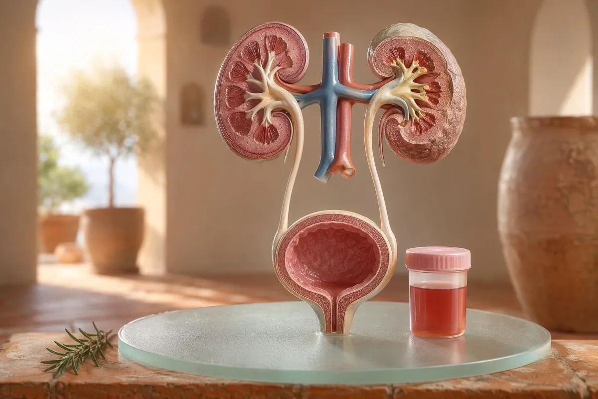
Panduan yang mengutamakan pasien untuk hematuria yang terlihat dan mikroskopis, termasuk mengapa darah pada dipstick tidak sama dengan sel darah merah di bawah mikroskop.


- Darah dalam urine berarti urine berwarna merah yang terlihat, seperti teh, atau seperti cola, atau hematuria mikroskopis yang ditemukan pada pemeriksaan; darah yang terlihat selalu memerlukan tindak lanjut medis.
- Hematuria mikroskopik biasanya didefinisikan sebagai 3 atau lebih sel darah merah per high-power field pada sampel mikroskopi urine yang dikumpulkan dengan benar.
- Darah pada dipstick mendeteksi aktivitas heme, sehingga dapat positif dari RBC yang masih utuh, hemoglobin bebas, atau mioglobin setelah cedera otot.
- Petunjuk ISK meliputi rasa perih saat berkemih, frekuensi, leukocyte esterase, nitrit, dan sel darah putih; hematuria harus diperiksa ulang setelah infeksi reda.
- Sedimen urine temuan seperti RBC dismorfik, silinder RBC, atau protein yang bermakna mengarah pada penyakit unit penyaring ginjal.
- Silinder urine adalah material yang terbentuk dari tubulus ginjal; silinder eritrosit adalah tanda bahaya nefrologi dalam minggu yang sama, terutama bila disertai tekanan darah tinggi atau eGFR yang menurun.
- Tanda bahaya kanker meliputi hematuria makroskopik yang tidak nyeri, bekuan, usia lebih dari 45 tahun, riwayat merokok, paparan bahan kimia di tempat kerja, dan episode berulang yang tidak dapat dijelaskan.
- Hematuria setelah olahraga biasanya membaik dalam 48–72 jam; darah yang menetap setelah istirahat tidak boleh disalahkan pada lari atau bersepeda.
- Evaluasi segera diperlukan untuk darah dalam urin bila disertai demam, nyeri pinggang, bekuan yang menyebabkan sumbatan saluran kemih, kehamilan, trauma, atau gejala anemia berat.
Apa arti darah dalam urine sebelum Anda panik
Darah dalam urine dapat berasal dari infeksi, batu, penyakit penyaring ginjal, kondisi prostat atau kandung kemih, obat-obatan, olahraga, atau kontaminasi saat menstruasi. Aturan praktisnya sederhana: darah yang terlihat, hematuria mikroskopik berulang, atau hematuria disertai nyeri, demam, bekuan, tekanan darah tinggi, protein, atau fungsi ginjal yang menurun memerlukan evaluasi segera, bukan menunggu dengan waspada.
Warna merah muda tunggal di mangkuk toilet setelah bit (beetroot) atau suplemen baru bukan masalah klinis yang sama dengan urin berwarna seperti cola setelah sakit tenggorokan. Dalam praktik saya, cerita warna sering memberi tahu saya ke mana harus melihat terlebih dahulu: merah terang dengan bekuan cenderung berasal dari saluran kemih bagian bawah, sedangkan urin cokelat atau seperti teh dapat berarti pigmen heme yang lebih lama dari penyaring ginjal.
Hematuria bukan diagnosis; itu adalah tanda. Hematuria makroskopik berarti Anda dapat melihat perubahan warna, sedangkan hematuria mikroskopik berarti urin tampak normal tetapi mikroskopi menemukan sel darah merah, biasanya pada 3 atau lebih sel darah merah per lapang pandang pembesaran tinggi.
Kantesti adalah seorang Analisa tes darah AI yang membantu pasien menghubungkan temuan pada urin dengan penanda ginjal seperti kreatinin, eGFR, CRP, hemoglobin, dan trombosit dalam satu timeline. Jika warnanya membingungkan, panduan warna urin menjelaskan mengapa urin merah, cokelat, oranye, dan kuning tua tidak semuanya berarti hal yang sama.
Darah pada dipstick tidak sama dengan RBC pada pemeriksaan mikroskopi
Dipstik urin yang bertanda darah positif mendeteksi aktivitas kimia yang menyerupai heme, bukan berarti sel darah merah utuh pasti ada. Mikroskopi menjawab pertanyaan berikutnya: apakah benar ada RBC di sedimen urin, dan jika ya, seperti apa bentuknya?
Pemeriksaan dipstik cepat karena bantalan reagen bereaksi dengan aktivitas peroksidase dari heme. Artinya, hasil positif dapat terjadi dengan RBC yang masih utuh, hemoglobin bebas setelah pecahnya sel darah merah, atau mioglobin setelah cedera otot yang signifikan.
Mikroskopi lebih lambat tetapi lebih spesifik. Laboratorium yang teliti akan memusingkan urin, memeriksa sedimen urin, dan melaporkan sel darah merah per lapang pandang pembesaran tinggi; banyak klinisi menggunakan 0–2 RBC/hpf sebagai normal dan 3 atau lebih RBC/hpf sebagai abnormal pada orang dewasa.
Ketidaksesuaian itu penting. Dipstik dengan darah 3+ dan 0–2 RBC/hpf membuat saya menanyakan tentang rabdomiolisis, hemolisis, kontaminan pengoksidasi, dan penanganan sampel, bukan langsung melompat ke penyakit kandung kemih; perbedaannya mirip dengan penalaran dalam panduan kami perbandingan urinalisis dan kultur.
Kapan petunjuk ISK menjelaskan hematuria, dan kapan tidak
Infeksi saluran kemih dapat menyebabkan darah dalam urin, terutama bila disertai rasa terbakar, urgensi, frekuensi, ketidaknyamanan di daerah pelvis, leukocyte esterase, nitrites, dan sel darah putih. Namun hematuria harus membaik setelah pengobatan; RBC yang menetap setelah UTI perlu ditinjau ulang.
Pada UTI bawah yang jelas, saya mengharapkan pola: disuria, frekuensi, leukocyte esterase positif, piuria sering di atas 5-10 sel darah putih/hpf, dan kadang nitrites. Kultur tradisional yang positif adalah 100.000 CFU/mL, tetapi pada wanita simtomatik, hitungan yang lebih rendah seperti 1.000-10.000 CFU/mL tetap dapat bermakna secara klinis.
Leukocyte esterase adalah enzim sel darah putih, bukan bukti adanya bakteri. Hasil positif dengan hematuria dapat terjadi akibat infeksi, batu, kontaminasi, atau peradangan ginjal, itulah sebabnya panduan leukocyte esterase memisahkan petunjuk yang berguna dari positif palsu yang umum.
Nitrites lebih spesifik tetapi kurang sensitif karena tidak semua bakteri mengubah nitrat menjadi nitrit. Jika gejalanya khas tetapi nitrites negatif, kultur tetap bisa positif; rincian praktis dibahas dalam penjelas nitrites.
Hematuria yang terlihat: petunjuk warna, bekuan, dan waktu
Hematuria yang terlihat tidak pernah sesuatu yang saya abaikan, bahkan jika hanya terjadi sekali. Urin merah terang, bekuan, atau perdarahan tanpa nyeri memerlukan penilaian yang tepat waktu karena penyebab dari saluran kemih bagian bawah menjadi lebih mungkin seiring bertambahnya usia dan paparan merokok.
Waktu bisa sangat berguna. Darah hanya pada awal saat berkemih dapat mengarah ke uretra, darah di seluruh aliran bisa berasal dari sumber kandung kemih atau ginjal, dan darah terminal di dekat akhir kadang mengarah ke area leher kandung kemih.
Gumpalan biasanya berarti sumber perdarahan berada di bawah penyaring ginjal karena gumpalan tidak mudah terbentuk setelah melewati glomerulus dan tubulus. Gumpalan yang menghambat aliran urin bersifat gawat: distensi kandung kemih dapat menjadi nyeri dalam hitungan jam, dan perawatan kateter mungkin diperlukan.
Pria kadang mengira hematuria yang terlihat setelah gejala saluran kemih hanyalah iritasi prostat. Itu mungkin benar, tetapi jika PSA diperiksa selama atau segera setelah infeksi, interpretasinya bisa menjadi rumit; kami Panduan PSA setelah UTI menjelaskan mengapa banyak klinisi menunggu 6–8 minggu sebelum mengulang PSA.
Ambang hematuria mikroskopis yang digunakan dokter
Hematuria mikroskopis umumnya didefinisikan sebagai 3 atau lebih RBC per high-power field pada spesimen urin yang dikumpulkan dengan benar. Panduan AUA/SUFU menggunakan ambang ini dan merekomendasikan evaluasi berbasis risiko, bukan mengobati setiap pasien dengan cara yang sama (Barocas et al., 2020).
Tes dipstick saja tidak boleh digunakan untuk mendiagnosis hematuria mikroskopis. Dalam panduan AUA/SUFU Barocas et al. 2020, definisinya bergantung pada mikroskopi karena dipstick bisa positif dari pigmen heme tanpa sel darah merah yang utuh.
Mengulang pemeriksaan bukanlah kelemahan; itu pengobatan yang baik. Jika seseorang memiliki 3–10 RBC/hpf setelah olahraga berat, demam, hubungan seksual, menstruasi, atau pengumpulan yang terkontaminasi, biasanya saya ingin sampel ulang yang bersih sebelum memesan pemindaian, sama seperti yang kami sarankan untuk untuk hasil lab yang abnormal berulang.
Risiko mengubah jalurnya. Seorang usia 24 tahun dengan 4 RBC/hpf setelah setengah maraton tidak sama dengan pria usia 68 tahun mantan perokok dengan 25 RBC/hpf pada dua spesimen, meskipun keduanya secara teknis memenuhi definisi hematuria.
Sedimen urine, protein, dan silinder urine yang mengubah penilaian lanjutan
Sedimen urine dapat mengungkap apakah hematuria kemungkinan berasal dari penyaring ginjal. Sel darah merah dismorfik, silinder sel darah merah, dan pergeseran protein yang bermakna mengarahkan kekhawatiran ke penyakit glomerulus, bukan infeksi kandung kemih yang sederhana.
Sel darah merah yang tampak tidak rata, bergelembung (blebbed), atau berbentuk cincin disebut RBC dismorfik, dan itu menunjukkan adanya perjalanan melalui penyaring glomerulus yang meradang. Acanthocytes di atas kira-kira 5% sel darah merah urin sering diperlakukan sebagai petunjuk glomerulus yang kuat, meskipun laboratorium berbeda dalam seberapa yakin mereka melaporkannya.
Protein adalah petunjuk kedua yang tidak boleh saya abaikan. Rasio albumin-kreatinin urin di atas 30 mg/g, atau sekitar 3 mg/mmol, bersifat abnormal, dan hematuria yang terlihat plus protein layak mendapat peninjauan ginjal yang lebih cepat; kami panduan protein dalam urin memberikan ambang batas tingkat pasien.
Silinder urine adalah cetakan kecil yang terbentuk di dalam tubulus ginjal. Silinder hialin dapat muncul setelah dehidrasi atau olahraga, tetapi silinder sel darah merah bersifat abnormal dan biasanya mengarah ke glomerulonefritis; untuk rujukan urinalisis yang lebih luas, lihat kami panduan lengkap analisis urin.
Batu, olahraga, dan penyebab sementara hematuria
Batu dan olahraga berat dapat menyebabkan hematuria, tetapi keduanya memiliki waktu yang berbeda. Hematuria terkait batu sering disertai nyeri pinggang atau kristal, sedangkan hematuria akibat olahraga biasanya akan membaik dalam 48–72 jam setelah istirahat.
Batu ginjal secara klasik menyebabkan gelombang nyeri pinggang, mual, dan hematuria mikroskopis atau yang terlihat. Kristal kalsium oksalat tidak membuktikan adanya batu, tetapi meningkatkan kecurigaan bila dipasangkan dengan nyeri kolik; kami panduan kristal kalsium oksalat menjelaskan petunjuk mikroskopi urin.
Hematuria akibat olahraga itu nyata. Saya telah melihat pelari maraton dengan 10–20 RBC/hpf pada pagi hari setelah lomba, kultur normal, tanpa protein, dan urinalisis ulang yang benar-benar normal 3 hari kemudian.
Bahayanya adalah menyalahkan semuanya pada latihan. Jika hematuria menetap di luar 72 jam, muncul dengan urin berwarna seperti cola gelap, atau disertai CK tinggi, kreatinin yang meningkat, atau nyeri otot, lihat lebih jauh dari kandung kemih; kami panduan kreatinin saat berolahraga mencakup tumpang tindih ginjal dan otot.
Tanda bahaya ginjal yang tidak diabaikan dokter
Hematuria dengan protein, tekanan darah tinggi, pembengkakan, eGFR yang menurun, silinder sel darah merah, atau kreatinin yang meningkat cepat adalah tanda bahaya ginjal. Temuan ini menunjukkan unit penyaring mungkin sedang meradang, dan menunggu berbulan-bulan dapat mengorbankan fungsi ginjal.
KDIGO 2024 mendefinisikan penyakit ginjal kronis berdasarkan penanda kerusakan ginjal, seperti albuminuria, atau eGFR di bawah 60 mL/menit/1,73 m² selama setidaknya 3 bulan. Namun, pada kondisi akut, hematuria disertai kenaikan kreatinin dalam hitungan hari atau minggu sudah cukup untuk meningkatkan prioritas sebelum definisi 3 bulan terpenuhi.
Albuminuria sering menjadi sinyal peringatan dini. ACR 30-300 mg/g meningkat sedang, di atas 300 mg/g meningkat berat, dan menggabungkannya dengan hematuria mengubah pembahasan; kami panduan ACR ginjal menjelaskan mengapa albumin urin menemukan kerusakan sebelum gejala.
Tekanan darah penting di sini. BP baru 160/100 mmHg dengan urin seperti cola dan pembengkakan pergelangan kaki setelah infeksi tenggorokan membuat saya memikirkan glomerulonefritis, bukan sistitis; jika eGFR tidak pasti, panduan kami panduan pemeriksaan ulang GFR menjelaskan kapan sistatin C dapat memperjelas fungsi ginjal.
Petunjuk risiko kanker tanpa panik
Hematuria terlihat tanpa nyeri, terutama setelah usia 45 tahun, layak dievaluasi secara mendesak karena kanker kandung kemih dan saluran kemih bagian atas dapat muncul seperti ini. NICE NG12 merekomendasikan rujukan dugaan-kanker untuk hematuria terlihat yang tidak dapat dijelaskan pada orang dewasa usia 45 tahun ke atas, atau hematuria terlihat yang menetap setelah pengobatan ISK.
Kebanyakan hematuria bukan kanker. Namun, kasus yang saya ingat adalah yang tenang: tidak ada nyeri, tidak ada demam, tidak ada rasa terbakar, hanya dua episode urin merah pada perokok usia 56 tahun yang hampir mengabaikannya.
Risikonya kumulatif, bukan biner. Usia di atas 45-50, riwayat merokok, paparan siklofosfamid, radioterapi panggul, hematuria terlihat berulang, paparan amina aromatik terkait pekerjaan, dan bekuan darah semuanya meningkatkan kebutuhan untuk sistoskopi atau pencitraan.
Penanda prostat tidak menyingkirkan penyebab dari kandung kemih. Jika gejala saluran kemih, perubahan PSA, dan hematuria saling tumpang tindih, tinjau waktu dan status infeksi dengan saksama; kami panduan penanda prostat menjelaskan mengapa PSA hanya satu bagian dari penilaian saluran kemih pada pria.
Obat-obatan, antikoagulan, dan alarm palsu
Pengencer darah dapat mengungkap adanya perdarahan, tetapi tidak boleh langsung disalahkan untuk hematuria. Hematuria terkait antikoagulan tetap perlu evaluasi karena obat tersebut dapat mengungkap batu, infeksi, penyakit ginjal, atau pertumbuhan pada saluran kemih.
Warfarin, apixaban, rivaroxaban, aspirin, dan clopidogrel dapat membuat perdarahan menjadi lebih jelas. Jika INR berada di atas kisaran target, koreksi itu penting, tetapi INR normal tidak membuat hematuria yang tampak menjadi tidak berbahaya.
Beberapa penyebab yang tidak menyebabkan perdarahan dapat meniru hematuria. Rifampisin dapat mengubah urin menjadi oranye-merah, phenazopyridine dapat membuatnya menjadi oranye terang, bit dapat mewarnai urin pada orang yang rentan, dan dehidrasi dapat menggelapkan pigmen hingga membuat pasien takut; pemantauan antikoagulan dibahas dalam pemeriksaan keselamatan pengencer darah.
Dipstick juga dapat menyesatkan setelah olahraga berat atau cedera otot. Hasil positif pada bantalan heme dengan sedikit atau tanpa RBC mendorong pemikiran saya ke arah mioglobin, terutama jika CK di atas 1.000 IU/L dan urin tampak gelap.
Anak-anak, kehamilan, dan kontaminasi dari periode menstruasi
Hematuria pada anak-anak, kehamilan, atau sekitar menstruasi memerlukan pengambilan sampel yang lebih bersih dan ambang batas yang lebih rendah untuk ditinjau. Hasil urin yang sama dapat berarti hal yang berbeda tergantung usia, status kehamilan, demam, tekanan darah, dan kualitas pengambilan.
Kontaminasi menstruasi adalah hal yang umum dan tidak ada alasan untuk merasa malu karenanya. Jika hasilnya tidak sesuai dugaan, ulangi sampel midstream clean-catch setidaknya 48 jam setelah perdarahan berhenti; hematuria yang menetap dengan 3 atau lebih RBC/hpf menjadi lebih bermakna.
Anak-anak sering memerlukan sudut pandang yang berbeda. Hematuria setelah penyakit virus dapat bersifat sementara, tetapi hematuria disertai pembengkakan, tekanan darah tinggi, protein, atau penurunan keluaran urin memerlukan peninjauan pediatrik segera.
Kehamilan meningkatkan taruhannya karena UTI, batu, stres ginjal terkait preeklamsia, dan kontaminasi dapat saling tumpang tindih. Untuk pola peringatan kehamilan pada hari yang sama yang melibatkan temuan ginjal, hati, trombosit, dan urin, lihat tanda bahaya pemeriksaan laboratorium kehamilan.
Pemeriksaan yang biasanya dipesan dokter setelah hematuria
Pemeriksaan berikut yang biasanya dilakukan setelah hematuria adalah urinalisis ulang dengan mikroskopi, kultur urin jika infeksi mungkin, tes darah fungsi ginjal, protein urin atau ACR, pengukuran tekanan darah, serta pencitraan atau sistoskopi berdasarkan risiko. Urutan yang tepat bergantung pada gejala dan risiko.
Untuk gejala sistitis yang tidak rumit, kultur dan pengobatan mungkin didahulukan, lalu mikroskopi urin ulang setelah gejala mereda. Untuk hematuria tampak yang terlihat dan tidak nyeri, banyak klinisi bergerak lebih cepat ke pencitraan dan sistoskopi karena menunggu episode berulang dapat menunda diagnosis.
Tes darah menambah konteks yang tidak dapat diberikan oleh urin saja. Kreatinin, eGFR, urea atau BUN, elektrolit, CBC, CRP, komplemen C3/C4, dan penanda autoimun mungkin sesuai bila dicurigai adanya peradangan ginjal; panduan rasio BUN/kreatinin menjelaskan satu pola ginjal yang umum.
AI Kantesti dapat mengorganisasi hasil tersebut menjadi tampilan tren, tetapi peninjauan dokter tetap penting bila ada tanda bahaya. Pendekatan keselamatan klinis kami dijelaskan dalam Validasi Medis, termasuk bagaimana klaster yang tidak normal ditangani, bukan hanya angka yang terisolasi.
Cara Kantesti membantu mengatur konteks hematuria
Kantesti adalah seorang layanan interpretasi tes lab AI yang membaca penanda darah yang berdekatan dengan urin dalam konteks, seperti kreatinin, eGFR, CRP, hemoglobin, trombosit, dan glukosa. Ini tidak mendiagnosis hematuria, tetapi membantu pasien menyiapkan ringkasan yang lebih bersih dan lebih berguna untuk dokter mereka.
Saat saya meninjau hematuria, saya jarang melihat satu hasil saja. Kreatinin normal 0,82 mg/dL, eGFR stabil di atas 90, tanpa protein, dan kultur negatif terasa sangat berbeda dibanding hematuria dengan eGFR 48 dan CRP 65 mg/L.
Kantesti adalah seorang platform interpretasi biomarker AI yang dapat memproses PDF hasil tes darah yang diunggah atau foto serta mengaitkan penanda ginjal, peradangan, dan anemia di luar batas dengan waktu munculnya gejala. Metode di balik pembacaan pola tersebut dijelaskan dalam Panduan Teknologi, dan cakupan penanda yang lebih luas diuraikan dalam panduan biomarker.
Satu tips praktis: sebelum mengunggah, periksa bahwa satuan, tanggal, dan identitas pasien tertangkap dengan benar. Kesalahan OCR dapat mengubah 0,9 menjadi 9,0, yang merupakan cerita ginjal yang sama sekali berbeda; daftar periksa unggah PDF menunjukkan kesalahan yang saya minta pasien untuk diperiksa terlebih dahulu.
Catatan penelitian, tata kelola, dan daftar periksa akhir
Per 11 Juli 2026, rencana pasien yang paling aman adalah memastikan apakah hematuria itu nyata, mengidentifikasi petunjuk infeksi, mencari tanda bahaya ginjal, dan melakukan eskalasi untuk hematuria yang terlihat atau menetap. Jangan hanya mengandalkan warna urin.
Saya Thomas Klein, MD, dan nasihat saya sengaja bersifat konservatif karena hematuria yang terlewat bisa menjadi penting. Konten medis Kantesti ditinjau dengan pengawasan klinis dari kami Dewan Penasehat Medis, dan peran kami adalah membantu pasien mengajukan pertanyaan yang lebih baik, bukan menggantikan dokter mereka.
Kantesti adalah seorang Alat analisis pemeriksaan darah berbasis AI digunakan oleh jutaan orang di 127 negara, jadi privasi dan kejelasan klinis harus dibangun ke dalam alur kerja, bukan ditambahkan kemudian. Anda dapat membaca lebih lanjut tentang Kantesti LTD, struktur perusahaan kami di Inggris, dan misi klinis kami di Tentang Kami.
Kantesti AI Clinical Research Group. (2026). AI Blood Test Analyzer: 2,5 Juta Tes Dianalisis | Laporan Kesehatan Global 2026. Zenodo. DOI: https://doi.org/10.5281/zenodo.18175532. ResearchGate: https://www.researchgate.net/. Academia.edu: https://www.academia.edu/.
Kantesti AI Clinical Research Group. (2026). RDW Blood Test: Complete Guide to RDW-CV, MCV & MCHC. Zenodo. DOI: https://doi.org/10.5281/zenodo.18202598. ResearchGate: https://www.researchgate.net/. Academia.edu: https://www.academia.edu/.
Pertanyaan yang Sering Diajukan
Apakah darah dalam urin selalu serius?
Darah dalam urin tidak selalu serius, tetapi perlu dikonfirmasi dan dijelaskan. Hasil mikroskopis satu kali sebesar 3–5 RBC/hpf setelah olahraga atau menstruasi mungkin hilang pada pemeriksaan ulang, sedangkan darah yang terlihat, gumpalan, demam, nyeri pinggang, protein, tekanan darah tinggi, atau eGFR yang menurun memerlukan peninjauan segera. Pada orang dewasa usia lebih dari 45 tahun, hematuria terlihat tanpa sebab yang jelas memerlukan penilaian segera meskipun tidak ada nyeri.
Apa perbedaan antara darah pada dipstick dan RBC dalam urin?
Dipstick darah mendeteksi aktivitas kimia yang menyerupai heme, sedangkan mikroskopi urin menghitung sel darah merah yang benar-benar ada. Dipstick dapat menjadi positif dari RBC yang masih utuh, hemoglobin bebas, atau mioglobin setelah cedera otot, sehingga tidak identik dengan hematuria yang terkonfirmasi. Hematuria mikroskopik umumnya didefinisikan sebagai 3 atau lebih RBC per lapang pandang berdaya tinggi pada sampel yang dikumpulkan dengan benar.
Dapatkah infeksi saluran kemih (ISK) menyebabkan darah dalam urin?
Ya, infeksi saluran kemih (ISK) dapat menyebabkan darah dalam urin, terutama saat disertai rasa terbakar, frekuensi, urgensi, serta adanya leukocyte esterase, nitrit, dan sel darah putih. Hasil kultur urin yang positif secara tradisional sering kali 100.000 CFU/mL, tetapi hitungan yang lebih rendah dapat berarti pada pasien yang memiliki gejala. Hematuria biasanya perlu diperiksa ulang setelah pengobatan karena darah yang menetap dapat mengarah pada batu, penyakit ginjal, atau penyebab lain dari saluran kemih.
Kapan saya harus pergi ke layanan perawatan segera untuk hematuria?
Carilah perawatan segera jika terdapat darah dalam urin disertai demam, nyeri pinggang yang hebat, muntah, kehamilan, trauma baru-baru ini, gumpalan, tidak dapat mengeluarkan urin, pingsan, atau gejala anemia berat. Pemeriksaan medis dalam minggu yang sama juga sesuai untuk hematuria dengan protein, silinder sel darah merah, tekanan darah tinggi baru, atau kreatinin yang meningkat. Aliran urin yang tersumbat akibat gumpalan dapat menjadi nyeri dan tidak aman dalam hitungan jam.
Apakah olahraga dapat menyebabkan hematuria mikroskopik?
Olahraga berat dapat menyebabkan hematuria mikroskopik, terutama setelah lari jarak jauh, bersepeda intens, atau olahraga kontak. Hematuria terkait olahraga biasanya membaik dalam 48–72 jam setelah istirahat dan tidak boleh disertai protein menetap, penurunan eGFR, atau nyeri otot berat. Jika darah pada dipstick positif tetapi mikroskopi menunjukkan sedikit RBC, klinisi dapat memeriksa CK karena mioglobin akibat cedera otot dapat memicu hasil dipstick.
Apa arti silinder urin ketika ditemukan darah?
Silinder urin adalah material berbentuk yang terbentuk di dalam tubulus ginjal, dan jenisnya mengubah makna hematuria. Silinder hialin dapat muncul dengan dehidrasi atau olahraga, tetapi silinder sel darah merah adalah hal yang tidak normal dan mengindikasikan peradangan ginjal glomerulus. Darah dalam urin dengan silinder sel darah merah, protein, dan tekanan darah tinggi harus diperlakukan sebagai tanda bahaya ginjal, bukan sebagai UTI sederhana.
Bisakah dehidrasi membuat urin terlihat seperti darah?
Dehidrasi sering membuat urin berwarna kuning tua atau kuning keemasan, tetapi biasanya tidak menimbulkan sel darah merah dalam urin. Minumlah air dan jadwalkan tes urin jika warnanya merah muda, merah, coklat, atau seperti cola, terutama jika menetap setelah terhidrasi atau disertai rasa sakit.
Dapatkan Analisis Tes Darah Berbasis AI Hari Ini
Bergabunglah dengan lebih dari 2 juta pengguna di seluruh dunia yang mempercayai Kantesti untuk analisis instan dan akurat terhadap tes lab. Unggah hasil tes darah Anda dan terima interpretasi komprehensif biomarker 15,000+ dalam hitungan detik.
📚 Publikasi Riset yang Dirujuk
Klein, T., Mitchell, S., & Weber, H. (2026). Penganalisis Tes Darah AI: 2,5M Tes Dianalisis | Laporan Kesehatan Global 2026. Kantesti Penelitian Medis AI.
Klein, T., Mitchell, S., & Weber, H. (2026). Tes Darah RDW: Panduan Lengkap untuk RDW-CV, MCV & MCHC. Kantesti Penelitian Medis AI.
📖 Referensi Medis Eksternal
National Institute for Health and Care Excellence (2015). Dugaan kanker: pengenalan dan rujukan. Panduan NICE NG12. Panduan NICE.
Kidney Disease: Improving Global Outcomes CKD Work Group (2024). Pedoman Praktik Klinis KDIGO 2024 untuk Evaluasi dan Tatalaksana Penyakit Ginjal Kronis. Kidney International.
⚕️ Penafian Medis
Artikel ini hanya untuk tujuan edukasi dan tidak merupakan nasihat medis. Selalu konsultasikan dengan penyedia layanan kesehatan yang berkualifikasi untuk keputusan diagnosis dan perawatan.
Sinyal Kepercayaan E-E-A-T
Pengalaman
Tinjauan klinis yang dipimpin dokter terhadap alur kerja interpretasi hasil lab.
Keahlian
Fokus pada kedokteran laboratorium tentang bagaimana biomarker berperilaku dalam konteks klinis.
Kewenangan
Ditulis oleh Dr. Thomas Klein dengan peninjauan oleh Dr. Sarah Mitchell dan Prof. Dr. Hans Weber.
Kepercayaan
Interpretasi berbasis bukti dengan jalur tindak lanjut yang jelas untuk mengurangi kepanikan.