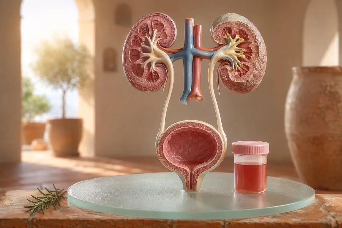
Πρώτης προτεραιότητας οδηγός για ορατή και μικροσκοπική αιματουρία, συμπεριλαμβανομένου του γιατί το αίμα από τη δοκιμασία ταινίας δεν είναι το ίδιο με τα ερυθρά αιμοσφαίρια κάτω από το μικροσκόπιο.


- Αίμα στα ούρα σημαίνει ορατά κόκκινα, ούρα χρώματος τσαγιού ή κόλα, ή μικροσκοπική αιματουρία που ανιχνεύεται σε εξετάσεις· το ορατό αίμα πάντα αξίζει ιατρική παρακολούθηση.
- Μικροσκοπική αιματουρία συνήθως ορίζεται ως 3 ή περισσότερα ερυθρά αιμοσφαίρια ανά οπτικό πεδίο υψηλής μεγέθυνσης σε δείγμα μικροσκοπικής εξέτασης ούρων που έχει συλλεχθεί σωστά.
- Το αίμα από τη δοκιμασία ταινίας ανιχνεύει δραστηριότητα αιμής, οπότε μπορεί να είναι θετικό από άθικτα ερυθρά αιμοσφαίρια, ελεύθερη αιμοσφαιρίνη ή μυοσφαιρίνη μετά από τραυματισμό μυών.
- Ενδείξεις ουρολοίμωξης (UTI) περιλαμβάνουν αίσθημα καύσου, συχνουρία, λευκοκυτταρική εστεράση, νιτρώδη και λευκά κύτταρα· η αιματουρία πρέπει να επανελέγχεται αφού υποχωρήσει η λοίμωξη.
- Το ουρικό ίζημα ευρήματα όπως δυσμορφικά ερυθρά αιμοσφαίρια, κυλινδρικά στοιχεία ερυθρών αιμοσφαιρίων ή σημαντική πρωτεΐνη παραπέμπουν σε νόσο της μονάδας διήθησης των νεφρών.
- Κύλινδροι στα ούρα είναι μορφοποιημένο υλικό από τα νεφρικά σωληνάρια· οι κύλινδροι ερυθρών αιμοσφαιρίων είναι προειδοποιητικό σημάδι νεφρολογίας της ίδιας εβδομάδας, ειδικά με υψηλή αρτηριακή πίεση ή μειούμενο GFR.
- Σημεία κινδύνου για καρκίνο περιλαμβάνουν ανώδυνη ορατή αιματουρία, θρόμβους, ηλικία άνω των 45, ιστορικό καπνίσματος, έκθεση σε χημικές ουσίες στην εργασία και επαναλαμβανόμενα ανεξήγητα επεισόδια.
- Αιματουρία μετά από άσκηση συνήθως υποχωρεί μέσα σε 48-72 ώρες· το επίμονο αίμα μετά την ανάπαυση δεν πρέπει να αποδίδεται στο τρέξιμο ή το ποδήλατο.
- Απαιτείται άμεση αξιολόγηση απαιτείται για αίμα στα ούρα με πυρετό, πλευρικό άλγος, θρόμβους που προκαλούν απόφραξη της ούρησης, κύηση, τραύμα ή συμπτώματα σοβαρής αναιμίας.
Τι σημαίνει αίμα στα ούρα πριν πανικοβληθείτε
Αίμα στα ούρα μπορεί να προέρχεται από λοίμωξη, λίθους, νόσο που επηρεάζει το φιλτράρισμα των νεφρών, καταστάσεις του προστάτη ή της ουροδόχου κύστης, φάρμακα, άσκηση ή επιμόλυνση από την περίοδο. Ο πρακτικός κανόνας είναι απλός: το ορατό αίμα, η επαναλαμβανόμενη μικροσκοπική αιματουρία ή η αιματουρία με πόνο, πυρετό, θρόμβους, υψηλή αρτηριακή πίεση, πρωτεΐνη ή μειωμένη νεφρική λειτουργία χρειάζονται άμεση αξιολόγηση και όχι παρακολούθηση χωρίς δράση.
Ένα μεμονωμένο ροζ χρώμα στην τουαλέτα μετά από παντζάρι ή ένα νέο συμπλήρωμα δεν είναι το ίδιο κλινικό πρόβλημα με τα ούρα χρώματος κόλα μετά από πονόλαιμο. Στην πρακτική μου, η ιστορία του χρώματος συχνά μου λέει πού να κοιτάξω πρώτα: το έντονο κόκκινο με θρόμβους τείνει να αφορά το κατώτερο ουροποιητικό, ενώ τα καφέ ή χρώματος τσαγιού ούρα μπορεί να σημαίνουν παλαιότερες χρωστικές αίματος (heme) από τα φίλτρα των νεφρών.
Η αιματουρία δεν είναι διάγνωση· είναι σημάδι. Ορατή αιματουρία σημαίνει ότι βλέπετε αλλαγή στο χρώμα, ενώ μικροσκοπική αιματουρία σημαίνει ότι τα ούρα φαίνονται φυσιολογικά αλλά η μικροσκόπηση βρίσκει ερυθρά αιμοσφαίρια, συνήθως 3 ή περισσότερα ερυθρά αιμοσφαίρια ανά οπτικό πεδίο υψηλής μεγέθυνσης.
Το Καντέστι είναι ένα Αναλυτής εξέτασης αίματος AI που βοηθά τους ασθενείς να συνδέσουν τα ευρήματα από τα ούρα με δείκτες νεφρών όπως κρεατινίνη, eGFR, CRP, αιμοσφαιρίνη και αιμοπετάλια σε μία χρονική γραμμή. Αν το χρώμα μπερδεύει, ο οδηγός χρώματος ούρων εξηγεί γιατί τα κόκκινα, καφέ, πορτοκαλί και σκούρα κίτρινα ούρα δεν σημαίνουν όλα το ίδιο πράγμα.
Το αίμα από τη δοκιμασία ταινίας δεν είναι το ίδιο με τα RBC στη μικροσκόπηση
Ένα τεστ ταινίας ούρων με ένδειξη θετικό αίμα ανιχνεύει χημική δραστηριότητα τύπου heme, όχι απαραίτητα ακέραια ερυθρά αιμοσφαίρια. Η μικροσκόπηση απαντά στην επόμενη ερώτηση: υπάρχουν πραγματικά RBCs στο ίζημα των ούρων και, αν ναι, πώς φαίνονται;
Η εξέταση με ταινία είναι γρήγορη επειδή το αντιδραστικό επίθεμα αντιδρά με τη δραστηριότητα υπεροξειδάσης του heme. Αυτό σημαίνει ότι ένα θετικό αποτέλεσμα μπορεί να εμφανιστεί με ακέραια RBCs, ελεύθερη αιμοσφαιρίνη μετά από διάσπαση των ερυθρών αιμοσφαιρίων ή μυοσφαιρίνη μετά από σημαντική μυϊκή κάκωση.
Η μικροσκόπηση είναι πιο αργή αλλά πιο ειδική. Ένα προσεκτικό εργαστήριο θα φυγοκεντρίσει τα ούρα, θα εξετάσει το ίζημα ούρων, και θα αναφέρει ερυθρά αιμοσφαίρια ανά οπτικό πεδίο υψηλής μεγέθυνσης· πολλοί κλινικοί χρησιμοποιούν 0-2 RBC/hpf ως φυσιολογικό και 3 ή περισσότερα RBC/hpf ως μη φυσιολογικό στους ενήλικες.
Η ασυμφωνία έχει σημασία. Μια δοκιμαστική ταινία (dipstick) με 3+ αίμα και 0-2 RBC/hpf με κάνει να αναρωτιέμαι για ραβδομυόλυση, αιμόλυση, οξειδωτικούς ρυπαντές και χειρισμό του δείγματος, αντί να πηδάω κατευθείαν στη νόσο της ουροδόχου κύστης· η διαφορά είναι παρόμοια με τη συλλογιστική στο δικό μας σύγκριση ανάλυσης ούρων και καλλιέργειας.
Πότε οι ενδείξεις για λοίμωξη ουροποιητικού εξηγούν την αιματουρία και πότε όχι
Μια λοίμωξη του ουροποιητικού συστήματος μπορεί να προκαλέσει αίμα στα ούρα, ειδικά με αίσθημα καύσου, επιτακτικότητα, συχνουρία, δυσφορία στην πύελο, εστεράση λευκοκυττάρων, νιτρώδη και λευκά αιμοσφαίρια. Όμως η αιματουρία πρέπει να υποχωρήσει μετά τη θεραπεία· τα επίμονα ερυθρά μετά από UTI χρειάζονται δεύτερη ματιά.
Σε μια απλή κατώτερη UTI, αναμένω ένα σύμπλεγμα: δυσουρία, συχνουρία, θετική εστεράση λευκοκυττάρων, πυουρία συχνά πάνω από 5-10 λευκά κύτταρα/hpf, και μερικές φορές νιτρώδη. Η παραδοσιακή θετικότητα της καλλιέργειας είναι 100.000 CFU/mL, αλλά σε συμπτωματικές γυναίκες, χαμηλότεροι αριθμοί όπως 1.000-10.000 CFU/mL μπορεί να είναι ακόμη κλινικά σημαντικοί.
Η εστεράση λευκοκυττάρων είναι ένα ένζυμο των λευκών αιμοσφαιρίων, όχι απόδειξη βακτηρίων. Ένα θετικό αποτέλεσμα με αιματουρία μπορεί να συμβεί από λοίμωξη, λίθους, επιμόλυνση ή φλεγμονή του νεφρού, γι’ αυτό το δικό μας οδηγός εστεράσης λευκοκυττάρων διαχωρίζει χρήσιμες ενδείξεις από συχνούς ψευδώς θετικούς.
Τα νιτρώδη είναι πιο ειδικά αλλά λιγότερο ευαίσθητα, επειδή δεν μετατρέπουν όλα τα βακτήρια το νιτρικό σε νιτρώδες. Αν τα συμπτώματα είναι τυπικά αλλά τα νιτρώδη είναι αρνητικά, η καλλιέργεια μπορεί ακόμη να είναι θετική· οι πρακτικές λεπτομέρειες καλύπτονται στο δικό μας επεξηγητής αποτελέσματος νιτρωδών.
Ορατή αιματουρία: χρώμα, θρόμβοι και ενδείξεις χρονισμού
Η ορατή αιματουρία δεν είναι ποτέ κάτι που το απορρίπτω, ακόμη κι αν συμβεί μία φορά. Τα έντονα κόκκινα ούρα, οι θρόμβοι ή η ανώδυνη αιμορραγία χρειάζονται έγκαιρη αξιολόγηση, επειδή τα αίτια από το κατώτερο ουροποιητικό σύστημα γίνονται πιο πιθανό καθώς αυξάνονται η ηλικία και η έκθεση στο κάπνισμα.
Ο χρόνος μπορεί να είναι εκπληκτικά χρήσιμος. Αίμα μόνο στην αρχή της ούρησης μπορεί να υποδηλώνει την ουρήθρα, αίμα σε όλη τη ροή μπορεί να προέρχεται από την ουροδόχο κύστη ή από νεφρικές πηγές, και τελικό αίμα κοντά στο τέλος μερικές φορές δείχνει προς την περιοχή του αυχένα της κύστης.
Οι θρόμβοι συνήθως σημαίνουν ότι η αιμορραγία προέρχεται κάτω από τα φίλτρα του νεφρού, επειδή οι θρόμβοι δεν σχηματίζονται εύκολα αφού περάσουν από σπειράματα και σωληνάρια. Θρόμβος που αποφράσσει τη ροή των ούρων είναι επείγον: η διάταση της κύστης μπορεί να γίνει επώδυνη μέσα σε ώρες και μπορεί να χρειαστεί φροντίδα με καθετήρα.
Οι άνδρες μερικές φορές υποθέτουν ότι η ορατή αιματουρία μετά από ουρολογικά συμπτώματα είναι απλώς ερεθισμός του προστάτη. Αυτό μπορεί να ισχύει, αλλά αν έγινε έλεγχος PSA κατά τη διάρκεια ή λίγο μετά από λοίμωξη, η ερμηνεία μπορεί να είναι μπερδεμένη· το δικό μας PSA μετά από λοίμωξη ουροποιητικού καθοδηγεί εξηγεί γιατί πολλοί κλινικοί περιμένουν 6-8 εβδομάδες πριν επαναλάβουν το PSA.
Τα όρια μικροσκοπικής αιματουρίας που χρησιμοποιούν οι γιατροί
Η μικροσκοπική αιματουρία συνήθως ορίζεται ως 3 ή περισσότερα RBC ανά υψηλού μεγέθους οπτικό πεδίο σε σωστά συλλεγμένο δείγμα ούρων. Η κατευθυντήρια οδηγία AUA/SUFU χρησιμοποιεί αυτό το όριο και συνιστά αξιολόγηση βάσει κινδύνου αντί για αντιμετώπιση κάθε ασθενούς με τον ίδιο τρόπο (Barocas et al., 2020).
Ένα μόνο dipstick δεν πρέπει να χρησιμοποιείται για τη διάγνωση της μικροσκοπικής αιματουρίας. Στην κατευθυντήρια οδηγία AUA/SUFU του Barocas et al. 2020, ο ορισμός εξαρτάται από τη μικροσκόπηση, επειδή τα dipsticks μπορεί να είναι θετικά από χρωστικές αίμης χωρίς ακέραιους ερυθρούς αιμοσφαίρους.
Η επανάληψη των εξετάσεων δεν είναι αδυναμία· είναι καλή ιατρική. Αν κάποιος έχει 3-10 RBC/hpf μετά από έντονη άσκηση, πυρετό, σεξ, εμμηνόρροια ή επιμολυσμένη συλλογή, συνήθως θέλω ένα καθαρό επαναληπτικό δείγμα πριν ζητήσω απεικονιστικές εξετάσεις, όπως ακριβώς συμβουλεύουμε για οδηγός για επαναληπτικές μη φυσιολογικές εξετάσεις.
Ο κίνδυνος αλλάζει την πορεία. Ένας 24χρονος με 4 RBC/hpf μετά από μισό μαραθώνιο δεν είναι το ίδιο με έναν 68χρονο πρώην καπνιστή με 25 RBC/hpf σε δύο δείγματα, παρότι και οι δύο πληρούν τεχνικά τον ορισμό της αιματουρίας.
Ουρικό ίζημα, πρωτεΐνη και κυλινδρικά στοιχεία στα ούρα που αλλάζουν τον έλεγχο
Το ουρικό ίζημα μπορεί να αποκαλύψει αν η αιματουρία είναι πιθανό να προέρχεται από τα φίλτρα του νεφρού. Δυσμορφικά ερυθρά αιμοσφαίρια, κυλινδρικά ερυθρών αιμοσφαιρίων και σημαντική μετατόπιση πρωτεΐνης στρέφουν την ανησυχία προς σπειραματική νόσο και όχι προς μια απλή λοίμωξη της ουροδόχου κύστης.
Τα ερυθρά αιμοσφαίρια που φαίνονται άνισα, με φυσαλίδες (blebbed) ή σε σχήμα δακτυλίου ονομάζονται δυσμορφικά RBC, και υποδηλώνουν διέλευση μέσα από ένα φλεγμαίνον σπειραματικό φίλτρο. Ακανθοκύτταρα πάνω από περίπου 5% των ερυθρών αιμοσφαιρίων στα ούρα αντιμετωπίζονται συχνά ως ισχυρή ένδειξη για σπειραματοπάθεια, αν και τα εργαστήρια διαφέρουν ως προς το πόσο με βεβαιότητα τα αναφέρουν.
Η πρωτεΐνη είναι η δεύτερη ένδειξη που δεν αγνοώ. Ο λόγος λευκωματίνης-κρεατινίνης στα ούρα πάνω από 30 mg/g, ή περίπου 3 mg/mmol, είναι μη φυσιολογικός, και η ορατή αιματουρία μαζί με πρωτεΐνη αξίζει ταχύτερη επανεξέταση με εστίαση στους νεφρούς· το δικό μας οδηγός πρωτεΐνης στα ούρα δίνει όρια ανά επίπεδο ασθενούς.
Κύλινδροι στα ούρα είναι μικροί μύκητες που σχηματίζονται μέσα στα νεφρικά σωληνάρια. Οι υαλώδεις κύλινδροι μπορεί να εμφανιστούν μετά από αφυδάτωση ή άσκηση, αλλά οι κύλινδροι ερυθρών αιμοσφαιρίων είναι μη φυσιολογικοί και συνήθως δείχνουν προς σπειραματονεφρίτιδα· για μια πιο ευρεία αναφορά στη γενική εξέταση ούρων, δείτε το δικό μας πλήρης οδηγός ανάλυσης ούρων.
Πέτρες, άσκηση και προσωρινές αιτίες αιματουρίας
Οι πέτρες και η έντονη άσκηση μπορούν να προκαλέσουν αιματουρία, αλλά έχουν διαφορετικά χρονικά πλαίσια. Η αιματουρία από πέτρα συχνά συνοδεύεται από πόνο στο πλάι ή κρυστάλλους, ενώ η αιματουρία από άσκηση συνήθως θα πρέπει να υποχωρεί μέσα σε 48-72 ώρες από την ανάπαυση.
Οι νεφρικές πέτρες κλασικά προκαλούν κύματα πόνου στο πλάι, ναυτία και μικροσκοπική ή ορατή αιματουρία. Οι κρύσταλλοι οξαλικού ασβεστίου δεν αποδεικνύουν πέτρα, αλλά αυξάνουν την υποψία όταν συνδυάζονται με κολικοειδή πόνο· το δικό μας κρύσταλλοι οξαλικού ασβεστίου καθοδηγεί εξηγεί την ένδειξη από τη μικροσκόπηση των ούρων.
Η αιματουρία από άσκηση είναι πραγματική. Έχω δει δρομείς μαραθωνίου με 10-20 RBC/hpf το πρωί μετά από έναν αγώνα, φυσιολογική καλλιέργεια, χωρίς πρωτεΐνη, και εντελώς φυσιολογική επαναληπτική γενική εξέταση ούρων 3 ημέρες αργότερα.
Ο κίνδυνος είναι να τα αποδίδουμε όλα στην εκπαίδευση. Αν η αιματουρία επιμένει πέρα από 72 ώρες, εμφανίζεται με σκούρο χρώμα ούρων τύπου κόλα ή συνοδεύεται από υψηλή CK, αυξανόμενη κρεατινίνη ή μυϊκό πόνο, κοιτάξτε πέρα από την ουροδόχο κύστη· ο οδηγός κρεατινίνης από την άσκηση καλύπτει την επικάλυψη νεφρών και μυών.
Οι «κόκκινες σημαίες» από τους νεφρούς που οι γιατροί δεν αγνοούν
Αιματουρία με πρωτεΐνη, υψηλή αρτηριακή πίεση, οίδημα, μειωμένο eGFR, κυλίνδρους ερυθρών αιμοσφαιρίων ή ταχέως αυξανόμενη κρεατινίνη είναι «κόκκινη σημαία» για τους νεφρούς. Τα ευρήματα αυτά υποδηλώνουν ότι οι μονάδες διήθησης μπορεί να είναι φλεγμαίνουσες και η αναμονή μηνών μπορεί να κοστίσει σε λειτουργία των νεφρών.
Το KDIGO 2024 ορίζει τη χρόνια νεφρική νόσο από δείκτες νεφρικής βλάβης, όπως η αλβουμινουρία, ή eGFR κάτω από 60 mL/min/1.73 m² για τουλάχιστον 3 μήνες. Σε οξείες καταστάσεις, ωστόσο, η αιματουρία μαζί με αύξηση της κρεατινίνης μέσα σε ημέρες ή εβδομάδες αρκεί για κλιμάκωση πριν επιτευχθεί ο ορισμός των 3 μηνών.
Η αλβουμινουρία είναι συχνά το πρώιμο προειδοποιητικό σήμα. ACR 30-300 mg/g είναι μέτρια αυξημένο, πάνω από 300 mg/g είναι σοβαρά αυξημένο, και ο συνδυασμός οποιουδήποτε από τα δύο με αιματουρία αλλάζει τη συζήτηση· ο οδηγός ACR για τους νεφρούς εξηγεί γιατί η αλβουμίνη στα ούρα βρίσκει βλάβη πριν από τα συμπτώματα.
Η αρτηριακή πίεση μετρά εδώ. Μια νέα ΑΠ 160/100 mmHg με ούρα τύπου κόλα και οίδημα αστραγάλων μετά από λοίμωξη του φάρυγγα με κάνει να σκέφτομαι σπειραματονεφρίτιδα, όχι κυστίτιδα· αν το eGFR είναι αβέβαιο, ο οδηγός επανελέγχου GFR εξηγεί πότε η κυστατίνη C μπορεί να διευκρινίσει τη νεφρική λειτουργία.
Ενδείξεις κινδύνου για καρκίνο χωρίς πανικό
Ανώδυνη ορατή αιματουρία, ειδικά μετά την ηλικία των 45, αξίζει επείγουσα αξιολόγηση επειδή οι καρκίνοι της ουροδόχου κύστης και του ανώτερου ουροποιητικού συστήματος μπορούν να παρουσιαστούν έτσι. Το NICE NG12 συνιστά παραπομπή για ύποπτο καρκίνο σε ανεξήγητη ορατή αιματουρία σε ενήλικες 45 ετών και άνω, ή σε ορατή αιματουρία που επιμένει μετά από θεραπεία για ουρολοίμωξη.
Η περισσότερη αιματουρία δεν είναι καρκίνος. Παρ’ όλα αυτά, αυτά που θυμάμαι είναι τα «ήσυχα»: χωρίς πόνο, χωρίς πυρετό, χωρίς κάψιμο—μόνο δύο επεισόδια κόκκινων ούρων σε έναν 56χρονο καπνιστή που σχεδόν το αγνόησε.
Ο κίνδυνος είναι σωρευτικός, όχι δυαδικός. Η ηλικία πάνω από 45-50, το ιστορικό καπνίσματος, η έκθεση σε κυκλοφωσφαμίδη, η πυελική ακτινοθεραπεία, η υποτροπιάζουσα ορατή αιματουρία, η επαγγελματική έκθεση σε αρωματικές αμίνες και οι θρόμβοι αυξάνουν την ανάγκη για κυστεοσκόπηση ή απεικόνιση.
Οι δείκτες του προστάτη δεν αποκλείουν αιτίες από την ουροδόχο κύστη. Αν υπάρχουν ουρολογικά συμπτώματα, αλλαγές στο PSA και αιματουρία αλληλεπικαλύπτονται, εξετάστε προσεκτικά το χρονικό διάστημα και την κατάσταση λοίμωξης· το δικό μας οδηγός για δείκτες προστάτη εξηγεί γιατί το PSA είναι μόνο ένα μέρος της αξιολόγησης του ανδρικού ουροποιητικού.
Φάρμακα, αντιπηκτικά και ψευδείς συναγερμοί
Τα αντιπηκτικά μπορούν να αποκαλύψουν αιμορραγία, αλλά δεν πρέπει να κατηγορούνται αυτόματα για την αιματουρία. Η αιματουρία που σχετίζεται με αντιπηκτικά εξακολουθεί να χρειάζεται αξιολόγηση, επειδή το φάρμακο μπορεί να αναδείξει λίθους, λοίμωξη, νεφρική νόσο ή ανάπτυξη στο ουροποιητικό σύστημα.
Η βαρφαρίνη, η απιξαμπάνη, η ριβαροξαμπάνη, η ασπιρίνη και η κλοπιδογρέλη μπορούν να κάνουν την αιμορραγία πιο εμφανή. Αν το INR είναι πάνω από το επιθυμητό εύρος, η διόρθωσή του έχει σημασία, αλλά ένα φυσιολογικό INR δεν καθιστά την ορατή αιματουρία ακίνδυνη.
Πολλές αιτίες χωρίς αιμορραγία μιμούνται την αιματουρία. Η ριφαμπικίνη μπορεί να κάνει τα ούρα πορτοκαλί-κόκκινα, η φαιναζοπυριδίνη μπορεί να τα κάνει έντονα πορτοκαλί, το παντζάρι μπορεί να χρωματίσει τα ούρα σε ευαίσθητα άτομα και η αφυδάτωση μπορεί να σκουρύνει τη χρωστική τόσο ώστε να τρομάξει τους ασθενείς· η παρακολούθηση αντιπηκτικών καλύπτεται στα εργαστηριακά ασφάλειας για αντιπηκτικά.
Το δοκιμαστικό ταινίας μπορεί επίσης να παραπλανήσει μετά από έντονη άσκηση ή μυϊκό τραυματισμό. Ένα θετικό επίθεμα αιμής με λίγα ή καθόλου RBC ωθεί τη σκέψη μου προς τη μυοσφαιρίνη, ειδικά αν η CK είναι πάνω από 1.000 IU/L και τα ούρα είναι σκούρα.
Παιδιά, εγκυμοσύνη και επιμόλυνση από περίοδο
Η αιματουρία σε παιδιά, στην εγκυμοσύνη ή γύρω από την περίοδο χρειάζεται καθαρότερη δειγματοληψία και χαμηλότερο όριο για επανεξέταση. Το ίδιο αποτέλεσμα ούρων μπορεί να σημαίνει διαφορετικά πράγματα ανάλογα με την ηλικία, την κατάσταση εγκυμοσύνης, τον πυρετό, την αρτηριακή πίεση και την ποιότητα συλλογής.
Η επιμόλυνση από την έμμηνο ρύση είναι συχνή και κανείς δεν πρέπει να ντρέπεται γι’ αυτό. Αν το αποτέλεσμα είναι απροσδόκητο, επαναλάβετε ένα δείγμα μέσης ροής καθαρού αλιεύματος τουλάχιστον 48 ώρες μετά τη διακοπή της αιμορραγίας· η επίμονη παρουσία 3 ή περισσότερων RBC/hpf γίνεται τότε πιο ουσιαστική.
Τα παιδιά συχνά χρειάζονται διαφορετικό φακό. Η αιματουρία μετά από ιογενή νόσο μπορεί να είναι παροδική, αλλά η αιματουρία με οίδημα, υψηλή αρτηριακή πίεση, πρωτεϊνουρία ή μειωμένη παραγωγή ούρων χρειάζεται άμεση παιδιατρική αξιολόγηση.
Η εγκυμοσύνη αυξάνει το διακύβευμα, επειδή η ουρολοίμωξη, οι λίθοι, η νεφρική επιβάρυνση που σχετίζεται με προεκλαμψία και η επιμόλυνση μπορεί να αλληλεπικαλύπτονται. Για μοτίβα προειδοποίησης εγκυμοσύνης την ίδια ημέρα που αφορούν ευρήματα νεφρών, ήπατος, αιμοπεταλίων και ούρων, δείτε το δικό μας εγκυμοσύνη: εργαστηριακές «κόκκινες σημαίες».
Εξετάσεις που συνήθως ζητούν οι γιατροί μετά από αιματουρία
Οι συνήθεις επόμενες εξετάσεις μετά την αιματουρία είναι επαναληπτική γενική εξέταση ούρων με μικροσκόπηση, καλλιέργεια ούρων αν είναι πιθανή λοίμωξη, αιματολογικές εξετάσεις νεφρικής λειτουργίας, πρωτεΐνη στα ούρα ή ACR, μέτρηση αρτηριακής πίεσης και απεικόνιση ή κυστεοσκόπηση με βάση τον κίνδυνο. Η ακριβής σειρά εξαρτάται από τα συμπτώματα και τον κίνδυνο.
Για συμπτώματα μη επιπλεγμένης κυστίτιδας, μπορεί πρώτα να προηγηθούν καλλιέργεια και θεραπεία, ακολουθούμενα από επαναληπτική μικροσκόπηση ούρων αφού υποχωρήσουν τα συμπτώματα. Για ανώδυνη ορατή αιματουρία, πολλοί κλινικοί προχωρούν νωρίτερα σε απεικόνιση και κυστεοσκόπηση, επειδή η αναμονή για επαναλαμβανόμενα επεισόδια μπορεί να καθυστερήσει τη διάγνωση.
Οι αιματολογικές εξετάσεις προσθέτουν πλαίσιο που δεν μπορεί να δώσει μόνο το δείγμα ούρων. Η κρεατινίνη, το eGFR, η ουρία ή BUN, οι ηλεκτρολύτες, το CBC, το CRP, το συμπλήρωμα C3/C4 και οι αυτοάνοσοι δείκτες μπορεί να είναι κατάλληλοι όταν υπάρχει υποψία φλεγμονής των νεφρών· το δικό μας οδηγό λόγου BUN/κρεατινίνης εξηγεί ένα κοινό νεφρικό μοτίβο.
Το Kantesti AI μπορεί να οργανώσει αυτά τα αποτελέσματα σε προβολή τάσης, αλλά η κλινική επανεξέταση από ιατρό παραμένει απαραίτητη όταν υπάρχουν ενδείξεις κινδύνου. Η προσέγγιση κλινικής ασφάλειας περιγράφεται στο Ιατρική Επικύρωση, συμπεριλαμβανομένου του πώς αντιμετωπίζονται μη φυσιολογικά συσσωρευμένα ευρήματα αντί για μεμονωμένοι αριθμοί.
Πώς το Kantesti βοηθά στην οργάνωση του πλαισίου της αιματουρίας
Το Καντέστι είναι ένα υπηρεσία ερμηνείας δοκιμών AI που διαβάζει δείκτες αίματος κοντά στα ούρα σε πλαίσιο, όπως κρεατινίνη, eGFR, CRP, αιμοσφαιρίνη, αιμοπετάλια και γλυκόζη. Δεν διαγιγνώσκει αιματουρία, αλλά βοηθά τους ασθενείς να ετοιμάσουν μια καθαρότερη, πιο χρήσιμη περίληψη για τον κλινικό τους.
Όταν αξιολογώ την αιματουρία, σπάνια κοιτάζω μόνο ένα αποτέλεσμα. Μια φυσιολογική κρεατινίνη 0,82 mg/dL, σταθερό eGFR πάνω από 90, χωρίς πρωτεΐνη και αρνητική καλλιέργεια μοιάζει πολύ διαφορετική από αιματουρία με eGFR 48 και CRP 65 mg/L.
Το Καντέστι είναι ένα Πλατφόρμα ερμηνείας βιοδεικτών AI που μπορεί να επεξεργαστεί PDF εξετάσεων αίματος ή φωτογραφίες που έχουν ανέβει και να συνδέσει δείκτες νεφρικής δυσλειτουργίας, φλεγμονής και αναιμίας εκτός ορίων με το χρονικό διάστημα των συμπτωμάτων. Οι μέθοδοι πίσω από αυτή την ανάγνωση μοτίβου περιγράφονται στο δικό μας Οδηγός Τεχνολογίας, και η ευρύτερη κάλυψη δεικτών περιγράφεται στο οδηγός βιοδεικτών.
Μια πρακτική συμβουλή: πριν από την ανάρτηση, ελέγξτε ότι οι μονάδες, οι ημερομηνίες και οι αναγνωριστικοί κωδικοί ασθενούς καταγράφονται σωστά. Τα σφάλματα OCR μπορούν να μετατρέψουν το 0,9 σε 9,0, κάτι που είναι μια εντελώς διαφορετική ιστορία για τα νεφρά· το δικό μας checklist ανεβάσματος PDF δείχνει τα λάθη που ζητάω από τους ασθενείς να εντοπίζουν πρώτοι.
Σημειώσεις έρευνας, διακυβέρνηση και τελικός έλεγχος
Από τις 11 Ιουλίου 2026, το ασφαλέστερο πλάνο για τον ασθενή είναι να επιβεβαιωθεί αν η αιματουρία είναι πραγματική, να εντοπιστούν ενδείξεις λοίμωξης, να αναζητηθούν «κόκκινες σημαίες» για τα νεφρά και να γίνει κλιμάκωση για ορατή ή επίμονη αιματουρία. Μην βασίζεστε μόνο στο χρώμα των ούρων.
Είμαι ο Thomas Klein, MD, και η συμβουλή μου είναι σκόπιμα συντηρητική, επειδή η αιματουρία που διαφεύγει μπορεί να έχει σημασία. Το Kantesti's ιατρικό περιεχόμενο ελέγχεται με κλινική εποπτεία από το δικό μας Ιατρική Συμβουλευτική Επιτροπή, και ο ρόλος μας είναι να βοηθάμε τους ασθενείς να κάνουν καλύτερες ερωτήσεις, όχι να αντικαθιστούμε τον γιατρό τους.
Το Καντέστι είναι ένα Εργαλείο ανάλυσης αιματολογικών εξετάσεων με AI-powered χρησιμοποιείται από εκατομμύρια ανθρώπους σε 127 χώρες, οπότε η προστασία της ιδιωτικότητας και η κλινική σαφήνεια πρέπει να ενσωματωθούν στη ροή εργασίας και όχι να προστεθούν αργότερα. Μπορείτε να διαβάσετε περισσότερα για το Kantesti LTD, τη δομή της εταιρείας μας στο Ηνωμένο Βασίλειο και την κλινική μας αποστολή στο Σχετικά με εμάς.
Kantesti AI Clinical Research Group. (2026). AI Blood Test Analyzer: 2.5M Tests Analyzed | Global Health Report 2026. Zenodo. DOI: https://doi.org/10.5281/zenodo.18175532. ResearchGate: https://www.researchgate.net/. Academia.edu: https://www.academia.edu/.
Kantesti AI Clinical Research Group. (2026). RDW Blood Test: Complete Guide to RDW-CV, MCV & MCHC. Zenodo. DOI: https://doi.org/10.5281/zenodo.18202598. ResearchGate: https://www.researchgate.net/. Academia.edu: https://www.academia.edu/.
Συχνές Ερωτήσεις
Είναι πάντα σοβαρό το αίμα στα ούρα;
Η παρουσία αίματος στα ούρα δεν είναι πάντα σοβαρή, αλλά πρέπει να επιβεβαιωθεί και να εξηγηθεί. Ένα μεμονωμένο μικροσκοπικό αποτέλεσμα 3-5 RBC/hpf μετά από άσκηση ή εμμηνόρροια μπορεί να υποχωρήσει σε επαναληπτικό έλεγχο, ενώ το ορατό αίμα, θρόμβοι, πυρετός, πόνος στο πλάι, πρωτεΐνη, υψηλή αρτηριακή πίεση ή μειωμένο GFR απαιτούν άμεση αξιολόγηση. Σε ενήλικες άνω των 45 ετών, η ανεξήγητη ορατή αιματουρία αξίζει επείγουσα εκτίμηση ακόμη κι αν δεν υπάρχει πόνος.
Ποια είναι η διαφορά ανάμεσα στο αίμα με ταινία (dipstick) και τα RBCs στα ούρα;
Η δοκιμασία με ταινία (dipstick) ανιχνεύει αιμοειδή χημική δραστηριότητα, ενώ η μικροσκοπική εξέταση των ούρων μετρά πραγματικά ερυθρά αιμοσφαίρια. Μια ταινία μπορεί να είναι θετική από άθικτα RBC, ελεύθερη αιμοσφαιρίνη ή μυοσφαιρίνη μετά από τραυματισμό του μυός, επομένως δεν είναι ταυτόσημη με επιβεβαιωμένη αιματουρία. Η μικροσκοπική αιματουρία συνήθως ορίζεται ως 3 ή περισσότερα RBC ανά πεδίο υψηλής μεγέθυνσης σε κατάλληλα συλλεγμένο δείγμα.
Μπορεί μια ουρολοίμωξη (UTI) να προκαλέσει αίμα στα ούρα;
Ναι, μια UTI μπορεί να προκαλέσει αίμα στα ούρα, ειδικά όταν υπάρχει αίσθημα καύσου, συχνουρία, επιτακτικότητα, λευκοκυτταρική εστεράση, νιτρώδη και λευκά αιμοσφαίρια. Η παραδοσιακή θετικότητα καλλιέργειας ούρων είναι συχνά 100.000 CFU/mL, αλλά οι χαμηλότεροι αριθμοί μπορεί να έχουν σημασία σε συμπτωματικούς ασθενείς. Η αιματουρία θα πρέπει συνήθως να επανελέγχεται μετά τη θεραπεία, επειδή το επίμονο αίμα μπορεί να υποδεικνύει λίθους, νεφρική νόσο ή άλλη αιτία από το ουροποιητικό σύστημα.
Πότε πρέπει να πάω στα επείγοντα/επείγουσα φροντίδα για αιματουρία;
Ζητήστε άμεση ιατρική φροντίδα για αίμα στα ούρα με πυρετό, έντονο πόνο στη νεφρική χώρα, εμετούς, εγκυμοσύνη, πρόσφατο τραυματισμό, θρόμβους, αδυναμία αποβολής ούρων, λιποθυμία ή συμπτώματα σοβαρής αναιμίας. Εντός της ίδιας εβδομάδας ιατρική επανεξέταση είναι επίσης κατάλληλη για αιματουρία με πρωτεΐνη, κυλίνδρους ερυθρών αιμοσφαιρίων, νέα υψηλή αρτηριακή πίεση ή αυξανόμενη κρεατινίνη. Ένα αποφραγμένο ρεύμα ούρων από θρόμβους μπορεί να γίνει επώδυνο και μη ασφαλές μέσα σε λίγες ώρες.
Μπορεί η άσκηση να προκαλέσει μικροσκοπική αιματουρία;
Η έντονη άσκηση μπορεί να προκαλέσει μικροσκοπική αιματουρία, ιδιαίτερα μετά από τρέξιμο μεγάλων αποστάσεων, έντονη ποδηλασία ή αθλήματα επαφής. Η αιματουρία που σχετίζεται με την άσκηση συνήθως υποχωρεί εντός 48-72 ωρών από την ανάπαυση και δεν θα πρέπει να συνοδεύεται από επίμονη πρωτεϊνουρία, πτώση eGFR ή σοβαρό μυϊκό πόνο. Αν το αίμα στο δοκιμαστικό χαρτί είναι θετικό αλλά η μικροσκόπηση δείχνει λίγα RBC, οι κλινικοί ιατροί μπορεί να ελέγξουν την CK επειδή η μυοσφαιρίνη από μυϊκή κάκωση μπορεί να ενεργοποιήσει το δοκιμαστικό χαρτί.
Τι σημαίνουν τα κυλινδρικά στοιχεία ούρων (urine casts) όταν ανευρίσκεται αίμα;
Τα κυλινδρικά στοιχεία στα ούρα είναι σχηματισμένο υλικό που δημιουργείται εντός των νεφρικών σωληναρίων, και ο τύπος τους αλλάζει το νόημα της αιματουρίας. Οι υαλώδεις κύλινδροι μπορεί να εμφανιστούν με αφυδάτωση ή άσκηση, αλλά οι ερυθροκυτταρικοί κύλινδροι είναι παθολογικοί και υποδηλώνουν φλεγμονή του σπειράματος. Αίμα στα ούρα με ερυθροκυτταρικούς κυλίνδρους, πρωτεϊνουρία και υψηλή αρτηριακή πίεση θα πρέπει να αντιμετωπίζονται ως «κόκκινη σημαία» για τον νεφρό και όχι ως απλή ουρολοίμωξη (UTI).
Η αφυδάτωση μπορεί να κάνει τα ούρα να μοιάζουν με αίμα;
Η αφυδάτωση συχνά κάνει τα ούρα σκούρο κίτρινο ή κεχριμπαρένιο, αλλά συνήθως δεν δημιουργεί ερυθρά αιμοσφαίρια στα ούρα. Πιείτε νερό και κανονίστε μια εξέταση ούρων εάν το χρώμα είναι ροζ, κόκκινο, καφέ ή σαν κόκα-κόλα, ειδικά εάν επιμένει μετά την ενυδάτωση ή συνοδεύεται από πόνο.
Λάβετε σήμερα ανάλυση εξετάσεων αίματος με AI
Εγγραφείτε σε πάνω από 2 εκατομμύρια χρήστες παγκοσμίως που εμπιστεύονται το Kantesti για άμεση, ακριβή ανάλυση εργαστηριακών εξετάσεων. Ανεβάστε τα αποτελέσματα εξετάσεων αίματος και λάβετε ολοκληρωμένη ερμηνεία βιοδεικτών του 15,000+ μέσα σε δευτερόλεπτα.
📚 Παραπομπές σε δημοσιεύσεις έρευνας
Klein, T., Mitchell, S., & Weber, H. (2026). Αναλυτής AI για εξετάσεις αίματος: 2,5M εξετάσεις που αναλύθηκαν | Παγκόσμια Έκθεση Υγείας 2026. Ιατρική έρευνα του Kantesti με AI.
Klein, T., Mitchell, S., & Weber, H. (2026). Εξέταση αίματος RDW: Πλήρης οδηγός για RDW-CV, MCV & MCHC. Ιατρική έρευνα του Kantesti με AI.
📖 Εξωτερικές ιατρικές αναφορές
Εθνικό Ινστιτούτο για την Υγεία και την Αριστεία στη Φροντίδα (NICE) (2015). Υποψία καρκίνου: αναγνώριση και παραπομπή. Κατευθυντήρια οδηγία NICE NG12. NICE Guideline.
Νεφρική νόσος: Βελτιώνοντας τα παγκόσμια αποτελέσματα (KDIGO) Ομάδα Εργασίας για τη ΧΝΝ (2024). Οδηγός Κλινικής Πρακτικής KDIGO 2024 για την Αξιολόγηση και Διαχείριση της Χρόνιας Νεφρικής Νόσου. Kidney International.
⚕️ Ιατρική αποποίηση ευθύνης
Αυτό το άρθρο προορίζεται μόνο για εκπαιδευτικούς σκοπούς και δεν αποτελεί ιατρική συμβουλή. Συμβουλεύεστε πάντα έναν κατάλληλο επαγγελματία υγείας για αποφάσεις σχετικά με τη διάγνωση και τη θεραπεία.
Σήματα εμπιστοσύνης E-E-A-T
Εμπειρία
Κλινική ανασκόπηση από ιατρό για τις ροές εργασίας ερμηνείας εργαστηριακών αποτελεσμάτων.
Πραγματογνωμοσύνη
Εστίαση στην εργαστηριακή ιατρική στο πώς συμπεριφέρονται οι βιοδείκτες στο κλινικό πλαίσιο.
Αυθεντικότητα
Γραμμένο από τον Δρ. Thomas Klein με ανασκόπηση από την Δρ. Sarah Mitchell και τον Καθ. Dr. Hans Weber.
Αξιοπιστία
Ερμηνεία βασισμένη σε τεκμηριωμένα δεδομένα με σαφείς οδούς παρακολούθησης για τη μείωση του συναγερμού.