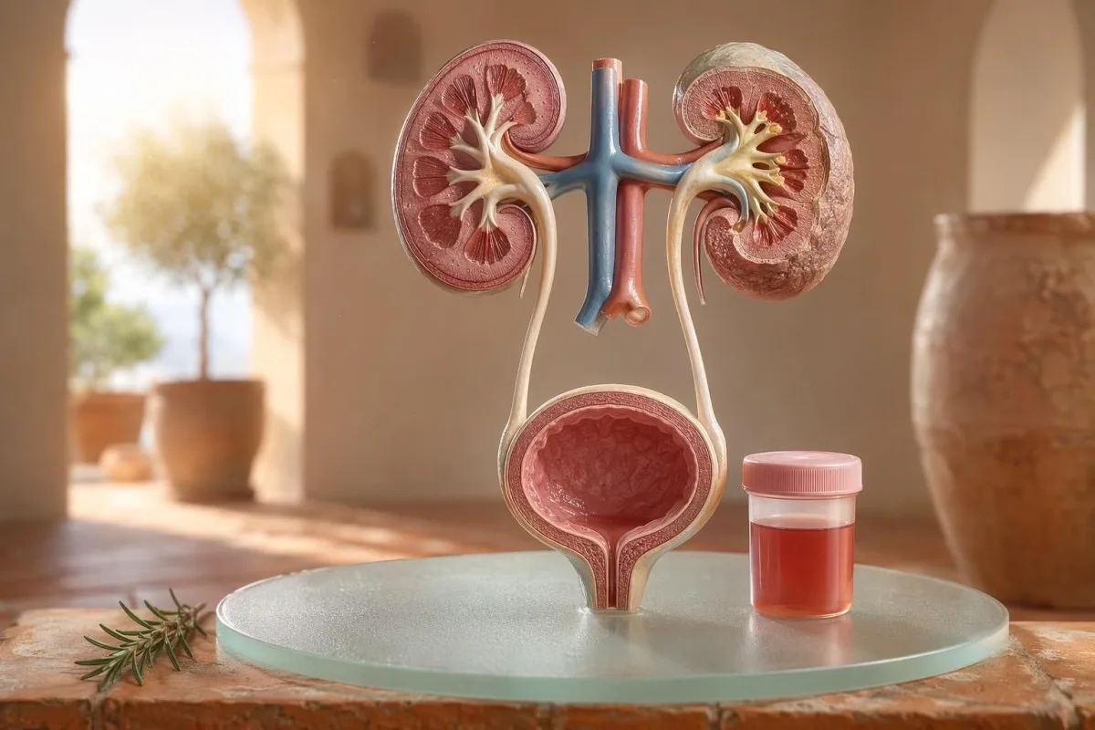
Një udhëzues i orientuar drejt pacientit për hematurinë e dukshme dhe atë mikroskopike, duke përfshirë pse gjaku në shirit (dipstick) nuk është i njëjtë me qelizat e kuqe nën mikroskop.


- Gjaku në urinë do të thotë urinë e kuqe e dukshme, me ngjyrë çaji ose me ngjyrë kola, ose hematuri mikroskopike e gjetur gjatë testimit; gjaku i dukshëm gjithmonë meriton ndjekje mjekësore.
- Hematuri mikroskopike zakonisht përkufizohet si 3 ose më shumë RBC për fushë me fuqi të lartë në një mostër të mbledhur siç duhet për mikroskopinë e urinës.
- Gjaku në shirit (dipstick) zbulon aktivitetin e hemit, ndaj mund të dalë pozitiv nga RBC të paprekura, hemoglobina e lirë ose mioglobina pas dëmtimit të muskujve.
- Të dhëna për UTI përfshijnë djegie, shpeshtësi, esterazë leukocitare, nitrite dhe qeliza të bardha; hematuria duhet të kontrollohet sërish pasi infeksioni të jetë pastruar.
- Sedimenti i urinës gjetje të tilla si RBC dismorfike, cilindrat e RBC ose proteinë domethënëse tregojnë sëmundje të njësisë filtruese të veshkave.
- Cilindrat e urinës janë materiale të formuara nga tubulat renale; cilindrat eritrocitarë janë një shenjë alarmi e nefrologjisë brenda të njëjtës javë, sidomos me hipertension të lartë ose eGFR në rënie.
- Shenja alarmi për kancer përfshijnë hematurinë e dukshme pa dhimbje, koagulat, moshë mbi 45 vjeç, histori duhanpirjeje, ekspozim profesional ndaj kimikateve dhe episode të përsëritura pa shpjegim.
- Hematuria pas ushtrimit zakonisht qetësohet brenda 48-72 orësh; gjaku që vazhdon pas pushimit nuk duhet t’i atribuohet vrapimit apo çiklizmit.
- Vlerësim urgjent nevojitet për gjakun në urinë me temperaturë, dhimbje në anë (flank pain), koagula që shkaktojnë bllokim urinar, shtatzëni, traumë ose simptoma të anemisë së rëndë.
Çfarë do të thotë gjaku në urinë para se të panikoni
Gjaku në urinë mund të vijë nga infeksioni, gurët, sëmundja e filtrimit të veshkave, gjendrat e prostatës ose gjendje të fshikëzës, medikamente, ushtrimi ose kontaminimi nga menstruacionet. Rregulli praktik është i thjeshtë: gjaku i dukshëm, hematuria mikroskopike e përsëritur, ose hematuria me dhimbje, temperaturë, koagula, hipertension, proteinë, ose funksion i reduktuar i veshkave kërkon vlerësim të shpejtë, jo pritje vigjilente.
Një ngjyrë rozë e vetme në tualet pas panxharit të kuq (beetroot) ose një suplement i ri nuk është i njëjti problem klinik si urina me ngjyrë “cola” pas një dhimbje fyti. Në praktikën time, historia e ngjyrës më tregon shpesh ku të shikoj i pari: e kuqe e ndezur me koagula tenton të lidhet me traktin e poshtëm urinar, ndërsa urina kafe ose me ngjyrë çaji mund të nënkuptojë pigmente më të vjetra të hemit nga filtrat e veshkës.
Hematuria nuk është një diagnozë; është një shenjë. Hematuria e dukshme do të thotë që e shihni ndryshimin e ngjyrës, ndërsa hematuria mikroskopike do të thotë që urina duket normale, por mikroskopia gjen qeliza të kuqe, zakonisht në 3 ose më shumë qeliza të kuqe të gjakut për fushë me zmadhim të lartë.
Kantesti është një Analizues i testeve të gjakut AI që i ndihmon pacientët t’i lidhin gjetjet në urinë me shënuesit e veshkave si kreatinina, eGFR, CRP, hemoglobina dhe trombocitet në një afat kohor të vetëm. Nëse ngjyra është konfuze, udhëzuesi ynë për ngjyrën e urinës shpjegon pse urina e kuqe, kafe, portokalli dhe e verdhë e errët nuk nënkuptojnë të gjitha të njëjtën gjë.
Gjaku në shirit (dipstick) nuk është i njëjtë me RBC-të në mikroskopi
Një test me shirit urinar i shënuar me gjak pozitiv zbulon aktivitet kimik të ngjashëm me hemin, jo domosdoshmërisht qeliza të kuqe të gjakut të plota. Mikroskopia i përgjigjet pyetjes tjetër: a ka qeliza reale RBC në sedimentin urinar dhe, nëse po, si duken ato?
Testimi me shirit është i shpejtë sepse pad-i i reagentit reagon me aktivitetin e peroksidazës të hemit. Kjo do të thotë që një rezultat pozitiv mund të ndodhë me RBC të plota, hemoglobinë të lirë pas prishjes së qelizave të kuqe, ose mioglobinë pas një dëmtimi të konsiderueshëm të muskujve.
Mikroskopia është më e ngadaltë, por më specifike. Një laborator i kujdesshëm e centrifugon urinën, ekzaminon sedimentin urinar, dhe raporton qelizat e kuqe për fushë me zmadhim të lartë; shumë klinicistë përdorin 0-2 RBC/hpf si normale dhe 3 ose më shumë RBC/hpf si jonormale te të rriturit.
Mospërputhja ka rëndësi. Një shirit me 3+ gjak dhe 0-2 RBC/hpf më bën të pyes për rabdomiolizë, hemolizë, ndotës oksidues dhe trajtimin e mostrës, në vend që të hidhem menjëherë te sëmundja e fshikëzës; diferenca është e ngjashme me arsyetimin në udhëzuesin tonë të krahasimi i analizës së urinës dhe kulturës.
Kur shenjat e UTI-së shpjegojnë hematurinë dhe kur jo
Një infeksion i traktit urinar mund të shkaktojë gjak në urinë, veçanërisht kur ka djegie, urgjencë, shpeshtësi, parehati në legen, esterazë leukocitare, nitrite dhe qeliza të bardha. Por hematuria duhet të pastrohet pas trajtimit; RBC-të e vazhdueshme pas një UTI kërkojnë një vlerësim të dytë.
Në një UTI të thjeshtë të poshtme, pres një grupim: disuri, shpeshtësi, esterazë leukocitare pozitive, piuri shpesh mbi 5-10 qeliza të bardha/hpf, dhe ndonjëherë nitrite. Pozitiviteti tradicional i kulturës është 100,000 CFU/mL, por te gratë simptomatike, numra më të ulët si 1,000-10,000 CFU/mL mund të kenë ende kuptim klinik.
Esteraza leukocitare është një enzimë e qelizave të bardha, jo provë e baktereve. Një rezultat pozitiv me hematuri mund të ndodhë nga infeksioni, gurët, kontaminimi ose inflamacioni i veshkave, prandaj udhëzuesi ynë për esterazën leukocitare ndan të dhënat e dobishme nga pozitivet e zakonshme të rreme.
Nitritet janë më specifike por më pak të ndjeshme, sepse jo të gjitha bakteret e shndërrojnë nitratin në nitrit. Nëse simptomat janë tipike por nitritet janë negative, kultura prapë mund të jetë pozitive; detajet praktike mbulohen në shpjeguesin e rezultatit të nitriteve.
Hematuria e dukshme: të dhëna për ngjyrën, koagulat dhe kohën
Hematuria e dukshme nuk është kurrë diçka që e anashkaloj, edhe kur ndodh vetëm një herë. Urina me ngjyrë të kuqe të ndezur, mpiksjet ose gjakderdhja pa dhimbje kërkojnë vlerësim në kohë, sepse shkaqet e traktit urinar të poshtëm bëhen më të mundshme ndërsa rriten mosha dhe ekspozimi ndaj duhanit.
Koha mund të jetë befasuese e dobishme. Gjaku vetëm në fillim të urinimit mund të sugjerojë uretrën, gjaku gjatë gjithë rrjedhës mund të vijë nga burime në fshikëz ose në veshkë, dhe gjaku në fund afër fundit ndonjëherë tregon drejt zonës së qafës së fshikëzës.
Koagulat zakonisht do të thonë se burimi i gjakderdhjes është poshtë filtrave të veshkave, sepse koagulat nuk formohen lehtë pasi të kalojnë përmes glomerulëve dhe tubulave. Një koagulat që bllokon rrjedhën e urinës është urgjente: distensioni i fshikëzës mund të bëhet i dhimbshëm brenda disa orësh dhe mund të nevojitet kujdes me kateter.
Burrat ndonjëherë supozojnë se hematuria e dukshme pas simptomave urinare është thjesht irritim i prostatës. Kjo mund të jetë e vërtetë, por nëse PSA u kontrollua gjatë ose menjëherë pas infeksionit, interpretimi mund të bëhet i ndërlikuar; ne Udhëzuesi PSA pas UTI shpjegon pse shumë klinicistë presin 6-8 javë përpara se të përsërisin PSA-n.
Pragjet e hematurisë mikroskopike që përdorin mjekët
Hematuria mikroskopike zakonisht përkufizohet si 3 ose më shumë RBC për fushë me fuqi të lartë në një mostër urine të mbledhur siç duhet. Udhëzimi AUA/SUFU përdor këtë prag dhe rekomandon vlerësim të bazuar në rrezik, në vend të trajtimit të çdo pacienti njësoj (Barocas et al., 2020).
Vetëm një dipstick nuk duhet të përdoret për të diagnostikuar hematurinë mikroskopike. Në udhëzimin AUA/SUFU të Barocas et al. 2020, përkufizimi varet nga mikroskopia, sepse dipstickët mund të dalin pozitivë nga pigmentet e hemit pa qeliza të kuqe të plota.
Përsëritja e testimit nuk është dobësi; është mjekësi e mirë. Nëse dikush ka 3-10 RBC/hpf pas ushtrimit të rëndë, temperaturës, seksit, menstruacioneve ose një mostre të kontaminuar, zakonisht dua një mostër të pastër të përsëritur përpara se të porosis skanime, njësoj siç këshillojmë për përsëritja e analizave jonormale.
Rreziku e ndryshon rrugën. Një 24-vjeçar me 4 RBC/hpf pas një gjysmë-maratonë nuk është i njëjtë me një 68-vjeçar ish-duhanpirës me 25 RBC/hpf në dy mostra, edhe pse të dyja teknikisht plotësojnë përkufizimin e hematurisë.
Sedimenti i urinës, proteina dhe cilindrat e urinës që ndryshojnë vlerësimin
Sedimenti i urinës mund të zbulojë nëse hematuria ka gjasa të vijë nga filtrat e veshkave. Eritrocite dismorfike, cilindër eritrocitesh dhe një zhvendosje domethënëse e proteinave e zhvendosin shqetësimin drejt sëmundjes glomerulare, jo drejt një infeksioni të thjeshtë të fshikëzës.
Eritrocitet që duken të pabarabarta, me “bleb” ose në formë unaze quhen RBC dismorfike dhe sugjerojnë kalim përmes një filtri glomerular të inflamuar. Acanthocytes mbi afërsisht 5% të eritrociteve urinare shpesh trajtohen si një shenjë e fortë glomerulare, megjithëse laboratorët ndryshojnë në sa me besueshmëri i raportojnë ato.
Proteina është e dyta; nuk e injoroj. Një raport albuminë-kreatininë në urinë mbi 30 mg/g, ose rreth 3 mg/mmol, është jonormal dhe hematuria e dukshme plus proteina meriton një rishikim më të shpejtë të fokusuar te veshkat; ne proteinë në urinë japin pragje në nivel pacienti.
Cilindrat e urinës janë myqe të vogla të formuara brenda tubulave të veshkave. Cilindrat hialinë mund të shfaqen pas dehidratimit ose ushtrimit, por cilindrat e eritrociteve janë jonormalë dhe zakonisht tregojnë drejt glomerulonefritit; për një referencë më të gjerë të analizës së urinës, shihni tonë udhëzues i plotë për analizën e urinës.
Gurët, ushtrimet dhe shkaqe të përkohshme të hematurisë
Gurët dhe ushtrimi i fortë mund të shkaktojnë hematuri, por kanë afate kohore të ndryshme. Hematuria e lidhur me gurin shpesh shoqërohet me dhimbje në anë (flank) ose me kristale, ndërsa hematuria nga ushtrimi zakonisht duhet të qetësohet brenda 48-72 orësh pushimi.
Gurët e veshkave klasike shkaktojnë valë dhimbjeje në anë, nauze dhe hematuri mikroskopike ose të dukshme. Kristalet e oksalatit të kalciumit nuk e provojnë një gur, por e rrisin dyshimin kur shoqërohen me dhimbje kolikative; udhëzuesi ynë për kristalet e oksalatit të kalciumit shpjegon shenjën e mikroskopisë së urinës.
Hematuria nga ushtrimi është reale. Kam parë vrapues maratonë me 10-20 RBC/hpf në mëngjesin pas një gare, me kulturë normale, pa proteina dhe me një analizë urine të përsëritur plotësisht normale 3 ditë më vonë.
Rreziku është t’i fajësosh të gjitha te trajnimi. Nëse hematuria vazhdon përtej 72 orësh, shfaqet me urinë të errët si “cola”, ose shoqërohet me CK të lartë, kreatininë në rritje, ose dhimbje muskulore, shiko përtej fshikëzës; ne udhëzuesi i kreatininës gjatë ushtrimeve mbulon mbivendosjen midis veshkave dhe muskujve.
“Flamujt e kuq” të veshkave që mjekët nuk i injorojnë
Hematuria me proteinë, presion i lartë i gjakut, ënjtje, eGFR i ulur, cilindrat e eritrociteve ose kreatinina që rritet me shpejtësi janë një flamur i kuq për veshkat. Këto gjetje sugjerojnë se njësitë filtruese mund të jenë të inflamuara, dhe pritja me muaj mund të kushtojë funksionin e veshkave.
KDIGO 2024 e përcakton sëmundjen kronike të veshkave nga shënues të dëmtimit të veshkave, si albuminuria, ose eGFR nën 60 mL/min/1.73 m² për të paktën 3 muaj. Megjithatë, në situata akute, hematuria plus një rritje e kreatininës brenda ditëve ose javëve mjafton për ta përshkallëzuar përpara se të plotësohet përkufizimi 3-mujor.
Albuminuria shpesh është sinjali i hershëm paralajmërues. Një ACR prej 30-300 mg/g është e rritur mesatarisht, mbi 300 mg/g është e rritur rëndë, dhe çiftëzimi i secilës me hematurinë e ndryshon bisedën; our udhëzuesi ACR për veshkat shpjegon pse albumina në urinë gjen dëmtim përpara simptomave.
Presioni i gjakut ka rëndësi këtu. Një BP e re prej 160/100 mmHg me urinë si “cola” dhe ënjtje të kyçit të këmbës pas një infeksioni të fytit më bën të mendoj për glomerulonefrit, jo për cistit; nëse eGFR është e pasigurt, our udhëzuesi i rivlerësimit të GFR shpjegon kur cistatina C mund të sqarojë funksionin e veshkave.
Shenja të rrezikut për kancer pa panik
Hematuria e dukshme pa dhimbje, veçanërisht pas moshës 45 vjeç, meriton vlerësim urgjent sepse kanceret e fshikëzës dhe të traktit të sipërm urinar mund të paraqiten në këtë mënyrë. NICE NG12 rekomandon referim për dyshim kanceri për hematurinë e dukshme të pashpjeguar te të rriturit 45 vjeç e lart, ose hematurinë e dukshme që vazhdon pas trajtimit të UTI.
Shumica e hematurisë nuk është kancer. Megjithatë, rastet që mbaj mend janë ato të qeta: pa dhimbje, pa temperaturë, pa djegie—vetëm dy episode të urinës së kuqe te një duhanpirës 56-vjeçar që pothuajse e injoroi.
Rreziku është kumulativ, jo binar. Mosha mbi 45-50, historia e duhanpirjes, ekspozimi ndaj ciklofosfamidit, radioterapia pelvike, hematuria e dukshme e përsëritur, ekspozimi profesional ndaj aminave aromatike dhe koagulat të gjitha rrisin nevojën për cistoskopi ose imazheri.
Markerët e prostatës nuk e përjashtojnë shkakun nga fshikëza. Nëse simptomat urinare, ndryshimet në PSA dhe hematuria mbivendosen, rishiko me kujdes kohën dhe statusin e infeksionit; our udhëzues për markerin e prostatës shpjegon pse PSA është vetëm një pjesë e vlerësimit urinar te meshkujt.
Medikamente, antikoagulantë dhe alarme të rreme
Holluesit e gjakut mund të zbulojnë gjakderdhje, por nuk duhet të fajësohen automatikisht për hematurinë. Hematuria e lidhur me antikoagulantët ende kërkon vlerësim, sepse ilaçi mund të zbulojë gurë, infeksion, sëmundje të veshkave ose një rritje në traktin urinar.
Warfarina, apixabani, rivaroxabani, aspirina dhe clopidogeli mund ta bëjnë gjakderdhjen më të dukshme. Nëse INR është mbi intervalin e synuar, korrigjimi i saj ka rëndësi, por një INR normale nuk e bën hematurinë e dukshme të padëmshme.
Disa shkaqe jo-hemorragjike imitojnë hematurinë. Rifampicina mund ta kthejë urinën portokalli-kuq, fenazopiridina mund ta bëjë atë portokalli të ndezur, panxhari i kuq mund ta ngjyrosë urinën te personat e ndjeshëm dhe dehidratimi mund ta errësojë pigmentin aq sa t’i frikësojë pacientët; monitorimi i antikoagulantëve mbulohet në tonë analizat e sigurisë për holluesit e gjakut.
Shiritëza (dipstick) mund të mashtrojë edhe pas ushtrimeve të forta ose dëmtimit të muskujve. Një rezultat pozitiv në heme pad me pak ose pa RBC e shtyn mendimin tim drejt mioglobinës, sidomos nëse CK është mbi 1,000 IU/L dhe urina është e errët.
Fëmijët, shtatzënia dhe kontaminimi nga periudha
Hematuria te fëmijët, gjatë shtatzënisë ose rreth menstruacioneve kërkon marrje më të pastër të mostrës dhe një prag më të ulët për rishikim. E njëjta analizë e urinës mund të nënkuptojë gjëra të ndryshme, në varësi të moshës, statusit të shtatzënisë, temperaturës, presionit të gjakut dhe cilësisë së grumbullimit.
Kontaminimi menstrual është i zakonshëm dhe askush nuk duhet të ndihet në siklet për këtë. Nëse rezultati është i papritur, përsëritni një mostër të pastër “midstream clean-catch” të paktën 48 orë pasi të ketë ndalur gjakderdhja; hematuria e vazhdueshme me 3 ose më shumë RBC/hpf bëhet më domethënëse.
Fëmijët shpesh kanë nevojë për një qasje tjetër. Hematuria pas një sëmundjeje virale mund të jetë e përkohshme, por hematuria me ënjtje, presion të lartë të gjakut, proteinë ose ulje të prodhimit të urinës kërkon rishikim të shpejtë nga pediatri.
Shtatzënia i rrit peshat, sepse UTI, gurët, stresi i veshkave i lidhur me preeklampsinë dhe kontaminimi mund të mbivendosen. Për modelet e paralajmërimit të shtatzënisë për të njëjtën ditë që përfshijnë gjetje të veshkave, mëlçisë, trombociteve dhe urinës, shihni tonë shenjat paralajmëruese laboratorike në shtatzëni.
Testet që mjekët zakonisht urdhërojnë pas hematurisë
Testet e zakonshme të ardhshme pas hematurisë janë analiza e përsëritur e urinës me mikroskopi, kultura e urinës nëse infeksioni është i mundshëm, analizat e funksionit të veshkave në gjak, proteina në urinë ose ACR, matja e presionit të gjakut dhe imazheria ose cistoskopi sipas rrezikut. Rendi i saktë varet nga simptomat dhe rreziku.
Për simptomat e cistitit të pakomplikuar, kultura dhe trajtimi mund të vijnë të parat, të ndjekura nga mikroskopia e përsëritur e urinës pasi simptomat të zgjidhen. Për hematurinë e dukshme pa dhimbje, shumë klinicistë kalojnë më herët te imazheria dhe cistoskopi, sepse pritja për episode të përsëritura mund ta vonojë diagnozën.
Analizat e gjakut shtojnë kontekst që urina vetëm nuk mund ta japë. Kreatinina, eGFR, ure ose BUN, elektrolitet, CBC, CRP, komplementi C3/C4 dhe markerët autoimunë mund të jenë të përshtatshëm kur dyshohet inflamacion i veshkave; tonë për raportin BUN/ kreatininë shpjegon një model të zakonshëm të veshkave.
AI Kantesti mund t’i organizojë këto rezultate në një pamje trendi, por rishikimi nga mjeku mbetet thelbësor kur ka flamuj të kuq. Qasja jonë e sigurisë klinike përshkruhet në Validimi Mjekësor, duke përfshirë si trajtohen grupimet jonormale, jo numrat e izoluar.
Si Kantesti ndihmon të organizohet konteksti i hematurisë
Kantesti është një shërbimin e interpretimit të analizave të AI që lexon markerët e gjakut afër urinës në kontekst, si kreatinina, eGFR, CRP, hemoglobina, trombocitet dhe glukoza. Nuk diagnostikon hematurinë, por i ndihmon pacientët të përgatitin një përmbledhje më të pastër dhe më të dobishme për mjekun e tyre.
Kur unë rishikoj hematurinë, rrallë shoh vetëm një rezultat. Një kreatininë normale prej 0.82 mg/dL, eGFR e qëndrueshme mbi 90, pa proteinë dhe kulturë negative duket shumë ndryshe nga hematuria me eGFR 48 dhe CRP 65 mg/L.
Kantesti është një platformë për interpretimin e biomarkerëve nga AI që mund të përpunojë PDF-të e analizave të gjakut ose fotot e ngarkuara dhe të lidhë markerët e veshkave, inflamacionit dhe anemisë jashtë intervalit me kohën e simptomave. Metodat pas leximit të atij modeli përshkruhen në tonë Udhëzues Teknologjik, dhe mbulimi më i gjerë i markerëve përshkruhet në udhëzues për bioshënuesit.
Një këshillë praktike: përpara se të ngarkoni, kontrolloni që njësitë, datat dhe identifikuesit e pacientit të jenë kapur saktë. Gabimet OCR mund ta kthejnë 0.9 në 9.0, që është një histori krejtësisht tjetër e veshkave; tonë lista kontrolluese për ngarkimin e PDF tregon gabimet që u kërkoj pacientëve t’i kapin së pari.
Shënime kërkimore, qeverisje dhe lista përfundimtare e kontrollit
Që nga 11 korriku 2026, plani më i sigurt për pacientin është të konfirmohet nëse hematuria është reale, të identifikohen shenja infeksioni, të kërkohen shenja alarmi për veshkat dhe të përshkallëzohet hematuria e dukshme ose e vazhdueshme. Mos u mbështet vetëm te ngjyra e urinës.
Unë jam Thomas Klein, MD, dhe këshilla ime është qëllimisht konservatore sepse hematuria e humbur mund të ketë rëndësi. Përmbajtja mjekësore e Kantesti rishikohet me mbikëqyrje klinike nga Bordi Këshillimor Mjekësor, dhe roli ynë është t’i ndihmojmë pacientët të bëjnë pyetje më të mira, jo t’ua zëvendësojmë mjekun.
Kantesti është një Mjet për analizën e testeve të gjakut me AI përdoret nga miliona njerëz në 127 vende, ndaj privatësia dhe qartësia klinike duhet të ndërtohen në procesin e punës, jo të shtohen më vonë. Mund të lexoni më shumë rreth Kantesti LTD, strukturës sonë të kompanisë në Mbretërinë e Bashkuar dhe misionit tonë klinik në Rreth Nesh.
Grupi i Kërkimit Klinik i AI Kantesti. (2026). Analizues i Testit të Gjakut me AI: 2.5M Teste të Analizuara | Raporti Global i Shëndetit 2026. Zenodo. DOI: https://doi.org/10.5281/zenodo.18175532. ResearchGate: https://www.researchgate.net/. Academia.edu: https://www.academia.edu/.
Grupi i Kërkimit Klinik i AI Kantesti. (2026). Testi i Gjakut RDW: Udhëzues i Plotë për RDW-CV, MCV & MCHC. Zenodo. DOI: https://doi.org/10.5281/zenodo.18202598. ResearchGate: https://www.researchgate.net/. Academia.edu: https://www.academia.edu/.
Pyetje të Shpeshta
A është gjithmonë serioze prania e gjakut në urinë?
Prezența sângelui în urină nu este întotdeauna gravă, dar trebuie confirmată și explicată. Un rezultat microscopic unic de 3-5 RBC/hpf după exercițiu sau menstruație poate să se clarifice la testarea repetată, în timp ce sângele vizibil, cheagurile, febra, durerea în regiunea lombară, proteinele, tensiunea arterială crescută sau eGFR redus necesită evaluare promptă. La adulții peste 45 de ani, hematuria vizibilă neexplicată merită o evaluare urgentă chiar dacă nu există durere.
Quali është ndryshimi midis gjakut në shiritin reaktiv (dipstick) dhe RBC-ve në urinë?
Dipstick-u zbulon aktivitet kimik të ngjashëm me hemin, ndërsa mikroskopia e urinës numëron qelizat e vërteta të kuqe të gjakut. Një dipstick mund të dalë pozitiv nga RBC-të të paprekura, hemoglobina e lirë ose mioglobina pas dëmtimit të muskujve, prandaj nuk është identik me hematurinë e konfirmuar. Hematuria mikroskopike zakonisht përkufizohet si 3 ose më shumë RBC për fushë me fuqi të lartë në një mostër të mbledhur siç duhet.
A mund të shkaktojë një infeksion urinar (ITU) gjak në urinë?
Po, një infeksion urinar (UTI) mund të shkaktojë gjak në urinë, veçanërisht kur ka djegie, si dhe kur janë të pranishme edhe frekuenca, urgjenca, esteraza e leukociteve, nitritet dhe qelizat e bardha. Pozitiviteti tradicional i kulturës së urinës shpesh është 100.000 CFU/mL, por numrat më të ulët mund të kenë rëndësi te pacientët simptomatikë. Hematuria zakonisht duhet të kontrollohet sërish pas trajtimit, sepse gjaku i vazhdueshëm mund të tregojë gurë, sëmundje të veshkave ose një tjetër shkak të traktit urinar.
Kur duhet të shkoj në urgjencë për hematuri?
Kërkoni kujdes urgjent nëse ka gjak në urinë me temperaturë, dhimbje të forta në anë (flank), të vjella, shtatzëni, traumë të fundit, mpiksje, pamundësi për të kaluar urinën, të fikët, ose simptoma të anemisë së rëndë. Rishikimi mjekësor brenda të njëjtës javë është gjithashtu i përshtatshëm për hematuri me proteinë, cilindër qelizash të kuqe (red cell casts), rritje të re të presionit të gjakut ose kreatininë në rritje. Një rrjedhje e bllokuar e urinës nga mpiksjet mund të bëhet e dhimbshme dhe e pasigurt brenda disa orësh.
A mund të shkaktojë ushtrimi hematuri mikroskopike?
Ushtrimi i vështirë mund të shkaktojë hematuri mikroskopike, veçanërisht pas vrapimit në distanca të gjata, çiklizmit intensiv ose sporteve të kontaktit. Hematuria e lidhur me ushtrimin zakonisht zgjidhet brenda 48–72 orëve pas pushimit dhe nuk duhet të shoqërohet me proteinuri të vazhdueshme, rënie të eGFR ose dhimbje të rënda muskulore. Nëse gjaku në shiritin testues është pozitiv, por mikroskopia tregon pak RBC, klinicistët mund të kontrollojnë CK, sepse mioglobina nga dëmtimi i muskujve mund të aktivizojë shiritin testues.
Çfarë nënkuptojnë cilindrat e urinës kur gjendet gjak?
Cilindrat urinarë janë materiale të formuara në formë të derdhur brenda tubulave renale dhe lloji i tyre ndryshon kuptimin e hematurisë. Cilindrat hialinë mund të shfaqen me dehidratim ose ushtrim, por cilindrat me qeliza të kuqe janë jonormale dhe sugjerojnë inflamacion të veshkave glomerulare. Prania e gjakut në urinë me cilindër të qelizave të kuqe, proteinë dhe presion të lartë të gjakut duhet të trajtohet si një shenjë alarmi renale, jo si një UTI e thjeshtë.
A mund të bëjë dehidrimi që urina të duket si gjak?
Dehidrimi shpesh e bën urinën të verdhë të errët ose ngjyrë qelibari, por zakonisht nuk krijon qeliza të kuqe në urinë. Pini ujë dhe caktoni një analizë urinarie nëse ngjyra është rozë, e kuqe, kafe ose si Coca-Cola, veçanërisht nëse vazhdon pas hidratimit ose shoqërohet me dhimbje.
Merrni sot analizë të analizave të gjakut me AI
Bashkohuni me mbi 2 milionë përdorues në mbarë botën që i besojnë Kantesti për analizë të menjëhershme dhe të saktë të analizave laboratorike. Ngarkoni rezultatet e analizave të gjakut dhe merrni interpretim gjithëpërfshirës të biomarkerëve të 15,000+ brenda sekondash.
📚 Publikime kërkimore të cituara
Klein, T., Mitchell, S., & Weber, H. (2026). Analizues i analizave të gjakut me AI: 2.5M analiza të shqyrtuara | Raporti Global i Shëndetit 2026. Kantesti Kërkim Mjekësor me AI.
Klein, T., Mitchell, S., & Weber, H. (2026). Analiza e gjakut për RDW: Udhëzues i plotë për RDW-CV, MCV & MCHC. Kantesti Kërkim Mjekësor me AI.
📖 Referenca të jashtme mjekësore
Instituti Kombëtar për Shëndetin dhe Kujdesin e Përsosur (NICE) (2015). Kancer i dyshuar: njohja dhe referimi. Udhëzimi NICE NG12. Udhëzimi NICE.
Sëmundja e veshkave: Përmirësimi i rezultateve globale (CKD) Grupi i punës (2024). Udhëzimi i Praktikës Klinike KDIGO 2024 për Vlerësimin dhe Menaxhimin e Sëmundjes Kronike të Veshkave. Kidney International.
⚕️ Mohim përgjegjësie mjekësore
Ky artikull është vetëm për qëllime edukative dhe nuk përbën këshilla mjekësore. Gjithmonë konsultohuni me një ofrues të kualifikuar të kujdesit shëndetësor për vendimet e diagnozës dhe trajtimit.
Sinjalet e Besimit E-E-A-T
Përvoja
Rishikim klinik i udhëhequr nga mjeku i flukseve të punës për interpretimin e analizave.
Ekspertizë
Fokus i mjekësisë laboratorike mbi mënyrën se si sillen biomarkerët në kontekstin klinik.
Autoritariteti
Shkruar nga Dr. Thomas Klein me rishikim nga Dr. Sarah Mitchell dhe Prof. Dr. Hans Weber.
Besueshmëria
Interpretim i bazuar në prova, me rrugë të qarta për ndjekje për të reduktuar alarmin.